Experimental and clinical analysis of fractures around the hip joint
PhD thesis István Flóris MD
Semmelweis University, Medical School Clinical Medicine, Doctoral School
Supervisor: Dr. Péter Cserháti MD, Ph.D Official reviewers: Dr. Norbert Wiegand MD, Ph.D
Dr. Lajos Bartha MD, Ph.D Head of the Final Examination Committee:
Dr. Péter Lakatos MD, DSc.
Members of the Final Examination Committee:
Dr. Zoltán Bejek MD, Ph.D Dr. Gergely Pánics MD, Ph.D
Budapest, 2015
1 1. Introduction
Recently developed novel implants applied in musculoskeletal surgery allow of efficient treatment of fractures and provide sufficient stability for functional aftercare, exercises and weight bearing. All these make possible to quickly restore the pre-injury functions and status of the musculoskeletal system and ability to move and walk.
As little as possible damage to the soft tissues adjacent to the fracture site plays a very important role in up-to-date trauma surgery in addition to modern ways of fracture fixation. Conditions of physiological bone healing are provided by the means of prevalent minimally invasive surgical techniques and fracture fixation.
However, stability of implants applied for fracture fixation does not depend exclusively only on localization, pattern and type of fractures, but also on bone quality.
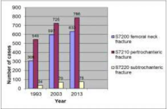
Osteoporosis, the most frequent metabolic bone disease became already endemic. Parallel to the increasing average age, also the number of osteoporotic fractures is rapidly increasing in the last two decades (Table 1).
The number of hip fractures per 100.000 inhabitants above the age of 50 years in the medical attendance region of the National Institute of Traumatology was as follows: 279 (0.279% incidence) in the year 1993, 440 (0.440%) in the year 2003 and 472 (0.472%) in the year 2013. That means, there is still an upward trend.
Table 1: The tendency of the number of hip fractures in people above the age of 50 years at the National Institute of Traumatology – distribution of fracture localization
2
In my dissertation, I provide an overview of the prevention, diagnostics and ways of treatment of osteoporosis, the most frequent bone metabolism disease;
it may be a useful practical guideline for orthopaedic and trauma surgeons.
The aim of my investigation is to determine, whether or not up-to-date intra- and extramedullary implants inserted into bones containing normal amounts of minerals are suitable to provide sufficient stability even in osteoporotic bones, with special respect to hip fractures, causing most surgical difficulties and requiring careful rehabilitation.
Thus, we have to face up to a complex problem: we have to apply a biomechanically appropriate implant, which is suitable for early weight bearing of the lower extremities, and furthermore, is also suitable for reliable anchorage even is osteoporotic bones with reduced mineral content. This elderly population is usually in poor general condition, has several concomitant diseases and requires minimal possible surgical intervention and burden with the lowest possible complication ratio.
Based on experimental and clinical investigations, I provide a guideline for the algorithm of surgical treatment of hip injuries, selection of the appropriate implants based on fracture pattern, the patients’ general condition and bone quality.
2. Aims
In my dissertation, which is summary of my more than 20 years long scientific work performed at the National Institute of Traumatology and its successors, I investigated and evaluated four main issues.
1. I evaluated the diagnostics problems related to hip fractures and worked out a diagnostic protocol. I analyzed the effects of fracture reduction and implant position on mechanical complications. I performed a finite element model in order to determine the optimal position of the screws.
2. I performed mechanical and clinical investigation of the implants developed for increased stability and amended the technique of the double canulated screw fixation (Manninger), which has been developed at the National Institute of Traumatology.
My aim was to develop a new implant in order to increase the stability of the conventional screw fixation of femoral neck fractures; this implant is suitable for the osteosynthesis of unstable femoral neck fractures with low ratio of complications.
3
3. I evaluated the outcome and complications of 720 patients operated on for femoral neck fractures and 1214 patients for trochanteric fractures with respect to fracture reduction and screw position.
I draw my conclusions based on our own results and that of the international literature and provide a guideline for hip fracture treatment and set up a treatment algorithm.
4. I developed the selection of the proper hip prosthesis type implanted in case of posttraumatic damage to the hip joint. My aim was to provide a guideline for the selection of the proper hip prosthesis type implanted in case of posttraumatic damage to the hip joint.
3. Methods
3.1. Diagnostic problems of hip fractures
In order to evaluate the diagnostic problems, I investigated the occurrence of occult hip fractures. I reviewed the data of 1295 patients with femoral neck and trochanteric fractures treated at Péterfy Hospital Trauma Centre.
I enrolled into the investigation those cases, when hip injury caused by low energy trauma has been examined radiologically during the primary care and the report written by the radiologist or trauma surgeon was negative or ambiguous regarding a fracture, and when the fracture was diagnosed within 6 weeks from primary injury, without repeated trauma. After that, an experienced radiologist and trauma surgeon re-analyzed the primary X-rays.
3.2. Evaluation of fracture reduction and screw position
I applied a finite element model for the investigation of impaired stability caused implant position and screw malposition.
For finite element modelling we used the integrated SolidWorks Simulation software of SolidWorks 2010 CAD appliance.
During the course of biomechanical modelling, we positioned the femoral neck screws in the same manner as in a real surgery. The connecting subchondral bone layer was simulated with a 20 x 20 mm large and 4 mm thick rectangular prism containing the built in 3.5 mm deep companion piece of the screw thread. We examined also the case, when the bone layer was simulated by a cap following the real geometry of the femoral head and we found that both models are suitable to reveal the effects of wrong subchondral screw positioning. We always applied the load to the model onto the lower surface of the implant; in case of modelling straight traction forces
4
perpendicular to the surface and when simulating oblique forces, parallel to the sideward surface of the bone layer. We performed the load examinations also when the screw was inserted obliquely, in an angle of 5 degrees (Figure 1).
Figure 1: Load and fixations points. Malposition: the screw is inserted into the bone in an angle of 5 degrees
3.3. Improvement of the double canulated (Manninger) screw technique developed at the National Institute of Traumatology, mechanical and clinical examination of implants providing increased stability
The standard screw has a core diameter of 7 mm, a thread diameter of 8 mm and a thread length of 24 mm.
As soon as after the evaluation of the initial results and mechanical complications, we developed the reinforcement of the lateral supporting point of the caudal screw: we applied a 2 mm thick small plate providing tension loop effect. The ratio of redisplacements could be reduced with this method.
The standard implants and those of the methods providing increased stability were tested on cadaver bones. We performed the biomechanical investigations on the TIRE test 2300 appliance of the Department of Material Sciences and Engineering, at the Technical University, Budapest.
We performed an osteotomy in an angle of 45 degrees in the central third of the femoral neck taken from 4-4 pairs of cadaver femora. We performed screw fixation only with two screws on one side; we fixed the other side in addition to the two screws with an implant providing increased stability and examination under load was performed.
3.4. Evaluation of early mechanical complications (early
redisplacements) of 720 patients operated on for femoral neck fracture
Patients older than 50 years of age with surgically treated type Garden III- IV femoral neck fractures were involved into the study; all of them underwent
5
surgery within 24 hours after injury. I investigated their ability to walk before and after injury, their general condition according to ASA and the mechanical complications as well.
I compared the results of 92 patients, who underwent primary prosthesis implantation for femoral neck fracture with those 92 patients’ outcome, who underwent secondary prosthesis implantation for a complication of the osteosynthesis.
3.5. Comparative clinical examination of 1214 patients treated for trochanteric fractures
I examined the results and complications of 1214 patients. I compared the outcomes of extra- and intramedullary fixation methods in order to set up a treatment algorithm for trochanteric fractures, as mentioned among the aimes of my study.
I addition, I examined also the outcomes and complications of prosthesis implantations performed for complications of trochanteric fractures.
3.6. Analysis of arthroplasties performed for complications of acetabular fractures
I selected from acetabular fractures caused by high energy trauma those ones, which resulted in posttraumatic complication, osteoarthritis of the hip or posttraumatic necrosis of the femoral head has developed and prosthesis implantation was necessary; all enrolled patients were treated at our institution during a 5-year period from 01.01.2002 to 31.12.2006.
I analyzed the primary treatment and complications of the cases requiring prosthesis implantation and evaluated the type of the prosthesis, with special respect to cup selection and necessity of acetabular reconstruction (replacement of the bony defect, removal of metal implants).
4. Results
4.1. Examination of occult fractures
The incidence of occult femoral neck fractures was 2.89% and in case of trochanteric fractures 1.07%.
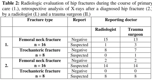
We analyzed the fractures also retrospectively, at the time of setting the final diagnosis of a fracture; we found that radiologic symptoms of a suspected fracture were present also at the time of primary examination (Table 2).
6
Table 2: Radiologic evaluation of hip fractures during the course of primary care (1.), retrospective analysis of X-rays after a diagnosed hip fracture (2.) by a radiologist (I.) and a trauma surgeon (II.)
Fracture type Report Reporting doctor
Radiologist Trauma surgeon 1.
Femoral neck fracture n = 16
Negative 15 13
Suspected 1 3
Trochanteric fracture n = 8
Negative 8 7
Suspected 0 1
2.
Femoral neck fracture n = 16
Negative 2 2
Suspected 14 14
Trochanteric fracture n = 8
Negative 0 0
Suspected 8 8
4.2. The role of implant positioning
Loading stress arising in case of straight pulling of the femoral neck screw producing a force greater than the limiting stress in the bone is 400 N. In case of oblique traction, the loading stress producing a force greater than the limiting stress in the bone is 350 N (Figure 2).
Figure 2: Tension forces on the screws in case of straight (perpendicular) and oblique (alignment of 5 degrees) pull directions
4.3. Development of implants providing increased stability
We tested the ultimate tensile strength of the screws 9.5 mm in diameter versus those of 8 mm: the previous one was 50% higher. In order to increase the rotational stability of the Manninger screws, we developed the so called blade-screw. In this case, we made a slash into the caudal screw along its axis longitudinally and inserted a 5 mm wide blade into that slot.
7
We tested the stability of three 8 mm in diameter screws versus the classical double canulated screw fixation: the stability was 30% stronger compared to that of classical double canulated screw fixation.
Development of the angular stable Manninger screw: there are threads in the satellite plate and sleeve providing angular stability and keeping the collo- diaphyseal angle in a stable manner. We insert conventional, 8 mm in diameter Manninger screws into the sleeves.
Characteristics of the implant system: the 1.5-2 cm long flatted lateral end of the conventional femoral neck screw fits into the sleeves; it allows of 1.5 cm sliding, preventing the screws from migrating to medial direction and, as a consequence, also perforation of the femoral head is avoidable.
4.4. Results of the investigation of patients with femoral neck fractures Investigating and comparing the ability to walk before and after injury and considering the ASA classification, I concluded that the half of ASA II-III patients were able to walk (at least in a limited manner), but the majority of ASA IV patients were unable to walk 4 months after surgery, or they did not return for a follow-up or died.
The ratio of early redisplacements (within 4 months) was 7.6% in group Graden III and 25.5% in group Graden IV. According to the AO classification, we observed the highest ratio of redisplacement in group 31-B3, i. e. in case of subcapital fractures. In case of mid-cervical fractures with slight varus displacement (group AO B2.2) and appropriate fracture reduction and implant position, the ratio of redisplacement was about 5% (Table 3).
Table 3: distribution of redisplacements according to AO classification Cases (n=489) Redisplacement (n=73)
31-B2.1 0 0
31-B2.2 257 14 (5.4%)
31-B2.3 8 2 (25.0%)
31-B3.1 51 12 (23.5%)
31-B3.2 37 10 (24.3%)
31-B3.3 136 35 (26.5%)
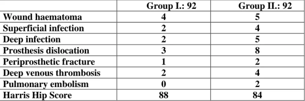
When comparing the complications after primary (I.) and secondary (II.) arthroplasties, I concluded that the ratio of complications is higher in group II, and the functional outcome is better in group I. (Table 4).
8
Table 4: The ratio of general and surgical complications and the functional outcome (HHS score) after primary (I) and secondary (II) arthroplasties
Group I.: 92 Group II.: 92
Wound haematoma 4 5
Superficial infection 2 4
Deep infection 2 5
Prosthesis dislocation 3 8
Periprosthetic fracture 1 2
Deep venous thrombosis 2 4
Pulmonary embolism 0 2
Harris Hip Score 88 84
4.5. Results of the investigation of patients with trochanteric fractures I analyzed the results based on the AO classification.
In group A1, after DHS synthesis, the average duration of surgery was slightly longer; wound haematomas and infections occurred more frequently after intramedullary fixation. There was no redisplacement after the use of a two-hole plate; this proves that in case of a stable, two-part fracture also a two-hole plate provides sufficient stability.
Among surgical complications after intramedullary fixation, the ratio of intraoperative fracture displacement (secondary redisplacement of an undisplaced or only slightly displaced fracture, caused by nail insertion) was higher (3 cases, 3.1%) No redisplacement could be observed during postoperative investigations, neither after intramedullary, nor after extramedullary fixation. Also the ratio of wound haematomas and infections was higher after intramedullary fixation (10.6–6.3% and 6.3–3.6%, respectively).
Duration of surgery and hospital care was longer in case of A2 fractures;
more blood transfusion was necessary after DHS synthesis, wound haematomas and infections occurred more frequently (8.3%–8.3% after extramedullary, and 6.7%–7.4% after intramedullary fixation.
9
Out of 22 redisplacements 12 occurred after the use of a two-hole plate, 8 after the use of a four-hole plate and 2 the use of a five-hole plate. In this group, the ratio of redisplacements after extramedullary fixation is almost twice as high as after intramedullary fixation (13.1% and 6.7%, respectively).
In case of A3 fractures, duration of intramedullary nailing was longer, but the ratio of complications was the highest after DHS synthesis among the three methods. The ratio of general surgical complications is almost equally high after DHS and DCS synthesis, the ratio of redisplacements is slightly lower after DCS synthesis (15.8% and 9.1%, respectively).
Postoperative redisplacement is less frequent after intramedullary fixation (10.2%) as after DHS synthesis; its ratio is similar to that after DCS synthesis, but intraoperative displacement of the lateral wall occurs relatively frequently (6 cases), which impairs the stability of the osteosynthesis.
Cut-out or Z-effect is twice as frequent after intramedullary fixation when compared to DCS (8.8% and 4.5%) and also to DHS synthesis. Thus, in case of A3 fractures, general surgical complications are more frequent after extramedullary fixation, but the ratio of redisplacements is high (10.2%) also after intramedullary fixation.
In case of A3 fractures, when also the lateral wall or the greater trochanter is broken, the ratio of redisplacements after 95 degrees DCS synthesis is similar to that after intramedullary fixation, but, in turn, there was no intraoperative fracture redisplacement.
There were complications requiring repeated surgery in 7 cases out of the examined 27 after prosthesis implantation for complications of trochanteric fractures. These were 3 prosthesis dislocations, all of them occurred after implantation of short stem prosthesis.
There were 3 periprosthetic fractures, two of them after implantation of cemented, short stem prosthesis and one after implantation of cemented, long stem prosthesis (Table 5).
Table 5: distribution of complications requiring repeated surgery Short stem
prosthesis (14)
Long stem prosthesis (13)
Periprosthetis fracture 2 1
Dislocation 3 0
Deep infection 1 0
10
4.6. Outcome of prosthesis implantations performed for complications after acetabular fractures
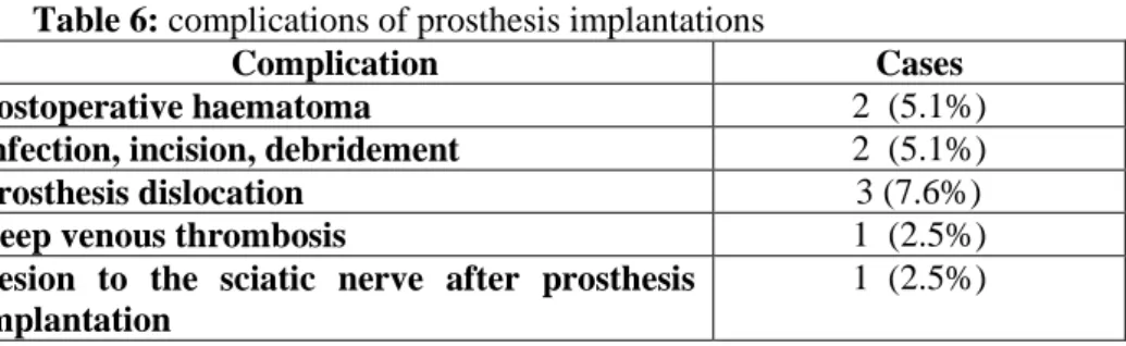
Table 6 demonstrates the complications of 39 patients, who underwent prosthesis implantation after complications of acetabular fractures.
Table 6: complications of prosthesis implantations
Complication Cases
Postoperative haematoma 2 (5.1%)
Infection, incision, debridement 2 (5.1%)
Prosthesis dislocation 3 (7.6%)
Deep venous thrombosis 1 (2.5%)
Lesion to the sciatic nerve after prosthesis implantation
1 (2.5%)
Functional outcome was evaluated according to Harris Hip Score; there was an increase of 39 points on the average (preoperatively 42, postoperatively 81).
5. Conclusions
5.1. Diagnostic algorithm of occult hip fractures
We developed the diagnostic algorithm of occult hip fractures based on our investigations and results:
1. In case of clinically suspected hip fracture, X-rays of the injured hip are performed in two planes, focused on the hip.
2. If the X-rays are negative and the patient is able to walk, may be discharged, but has to return for a follow-up on the third week and X-rays need to be repeated. If the patient is unable to walk, hospitalization is necessary and the following imaging diagnostics are suggested, in the order of their sensitivity: MRI, radioisotope scan, CT scan and ultrasonography.
4. As an alternative of specific imaging diagnostics, the patient undergoing analgesic therapy is gradually mobilized at the hospital, and, after physiologic load of the hip joint, X-rays are repeated 48-72 hours after primary injury and signs of a fracture are searched repeatedly.
5. If hip fracture can be excluded with repeated imaging diagnostics as well,
11
the patient may be discharged and has to return for a follow-up within 6 weeks and imaging diagnostics have to be repeated.
5.2. Effect of implant malposition
In case of double canulated screw fixation, if one of the screws is inserted in a malposition of 5 degrees, the pull-out force decreases by 12.5% as verified with the simplest biomechanical model. The finite element model verified that position or malposition of the screws plays an important role in the stability of the osteosynthesis.
5.3. Treatment algorithm of femoral neck fractures
I developed the treatment algorithm of femoral neck fractures based on biomechanical and clinical investigations.
Treatment of fractures type B1.1: Double canulated screw fixation (Manninger) after closed reduction of the hypervalgus position. If the axial malalignment on the lateral view is greater than 10°, it needs reduction.
Treatment of fractures type B1.2: In situ screw fixation is advisable for the fixation of stable fractures with less than 15° valgus malalignment in order to prevent secondary redisplacement and to start functional treatment as soon as possible.
Treatment of fractures type B1.3: In situ canulated screw fixation is advisable for the fixation of stable, undisplaced fractures in order to prevent secondary redisplacement and to start functional treatment as soon as possible.
Treatment of fractures type B2.1: Use of angular stable implants is advisable in case of biomechanically unstable, extracapsular fractures.
Treatment of fractures type B2.2: Simple fractures with slight varus displacement in the central third of the femoral neck usually heal after appropriate reduction and good positioning of the implants independently from the patients’ age, thus double canulated screw fixation is advisable.
Implants providing increased stability (e.g. angular stable implants or femoral neck screws with DCD attachment) are preferable in case of comminuted fractures. Primary prosthesis implantation is advisable only if good reduction cannot be achieved; total arthroplasty at the age of 65-75 years and bipolar hemiarthrosplasty above the age of 75 years.
Treatment of fractures type B2.3: Osteosynthesis is indicated for the treatment of steep, partially extracapsular (type Pauwels III.) fractures, but because of the instability implants providing increased stability (femoral neck
12
screws with DCD attachment, angular stable femoral neck screw, DHS) or angular stable implants are required.
Treatment of fractures type B3.1: Subcapital fractures with slight varus displacement but without vertical shift. Closed reduction and osteosynthesis with double canulated screws and a plate providing tension loop effect is advisable for patients younger than 65 years of age. In case of unsuccessful reduction and good bone quality, uncemented total hip arthroplasty is advisable. Arthroplasty is advisable for patients above the age of 65 years;
total, cemented arthroplasty at the age of 65-75 years and bipolar hemiarthrosplasty above the age of 75 years.
In order to minimize the burden, closed reduction and fixation with canulated screws is advisable for patients in poor general condition, provided, the reduction is appropriate.
Treatment of fractures type B3.2: There is a varus displacement and a vertical shift as well. If good reduction is achieved, preservation of the femoral head is advisable under the age of 65 years. Total hip arthroplasty is preferable above that age. If the reduction is poor, total hip arthroplasty is preferable also for patients under the age of 65 years.
Treatment of fractures type B3.3: Because of extremely high ratio of mechanical complications, hip replacement is principally preferable in case of fractures with considerable varus displacement, external rotation and vertical shift.
Indications and conditions of hip arthroplasty:
1. More than 24 hours elapsed after injury
2. Type Garden III, AO B2.2 mid-cervical fractures, if good reduction cannot be achieved
3. Type Garden IV, AO 31-B3 subcapital fractures above the age of 65 years
4. Type Garden IV, AO 31-B3 subcapital fractures under the age of 65 years, if good reduction cannot be achieved
5. Failure of the primary osteosynthesis, secondary hip replacement because of redisplacement
6. Secondary hip replacement because of necrosis of the femoral head 7. Pathologic fracture of the femoral neck
8. Stage ASA I-III patients
9. Ability to move and walk prior to injury 10. Appropriate mental status and compliance
13
5.4. Treatment algorithm of trochanteric fractures
I developed the treatment algorithm of trochanteric fractures based on biomechanical and clinical investigations.
Treatment of fractures type A1.1: Extramedullary implants are recommended for the treatment of undisplaced, simple, two-part trochanteric fractures in the intertrochanteric line; a two-hole DHS provides sufficient stability.
Treatment of fractures type A1.2: Extramedullary implants are recommended also in this case; two-hole DHS in case of good bone quality and four-hole DHS in case of poor bone quality.
Treatment of fractures type A1.3: Two-part fractures medially exceeding the level of the lesser trochanter. A longer attachment (four-hole plate) is required in case of extramedullary fixation. If the fracture involves also more distal segments, intramedullary fixation is preferable in order to avoid a large exposure required in case of extramedullary fixation. However, biomechanical stability of these two methods is similar in case of stable fractures.
Treatment of fractures type A2.1: This type of fracture is still stable, only the lesser trochanter is broken out and its displacement is not considerable. From biomechanical point of view, intramedullary fixation is more advantageous. In case of extramedullary fixation, a longer attachment (at least a four-hole plate) and reconstruction of the medial wall is required.
Treatment of fractures type A2.2: Unstable, comminuted fractures, also the lesser trochanter is broken and displaced. Intramedullary fixation is recommended.
Treatment of fractures type A2.3: Comminuted, unstable fracture, both the greater and lesser trochanters are broken. From biomechanical point of view, intramedullary fixation is recommended.
Treatment of fractures type A3.1: Reverse, oblique, intertrochanteric fracture pattern, with or without fracture of the greater trochanter and/or lateral wall. If the lateral wall is intact, intramedullary fixation is recommended. If the lateral wall is broken, but the tip of the greater trochanter is not involved, intramedullary fixation is preferable in addition to reconstruction of the lateral wall with a cerclage. If the greater trochanter is also involved, extramedullary fixation is recommended laterally with a proximal angular stable femoral plate or DCS.
Treatment of fractures type A3.2: Transverse, intertrochanteric fracture pattern. Intramedullary fixation is preferable, similarly to fractures of type
14
A3.1. If the lateral wall is unstable, reconstruction is needed with a cerclage or extramedullary fixation is recommended.
Treatment of fractures type A3.3: Unstable, comminuted, transverse or oblique fracture pattern. Intramedullary fixation is recommended, if the lateral wall or the greater trochanter is intact. If the greater trochanter is also involved, extramedullary fixation is recommended; if the fracture of the lateral wall does not involve the greater trochanter, intramedullary fixation is preferable supplemented with a cerclage.
If in case of A3 fractures appropriate reduction is not feasible, open reduction is necessary by the means of a bone adaptor forceps and cerclage.
Nail insertion should only be performed after fracture reduction; otherwise the fragments will be fixed in incorrect position, resulting in mechanical complications.
Long intramedullary implant is required, if the fracture exceeds the level of the lesser trochanter at least 4 cm distally.
Indications for hip replacement after trochanteric fractures:
1. pathologic trochanteric fracture caused by bone tumour 2. pre-existing osteoarthritis of the hip joint prior to injury
3. inveterate traochanteric fracture (open reduction and internal fixation will be probably unsuccessful)
4. failure of a previous osteosynthesis or pseudoarthrosis, if the patient’s age and quality of the proximal fragment preclude repeated osteosynthesis
5. pseudoarthrosis secondary to a trochenteric fracture or (infrequently) posttraumatic necrosis of the femoral head and/or osteoarthritis of the hip joint
5.5. Hip replacement after acetabular fractures
Selection of the cup is the primary issue. It depends on the severity of the deformity, extent of the defect, pseudoarthrosis requiring osteosynthesis or eventually, in case of non-union, bone substitution. Based on my investigations, I provide a guideline for the selection of the cup and necessity of bone substitution during the course hip replacement after acetabular fractures (Table 7).
15
Table 7: Guidelines for the selection of the cup and bone substitution
Type of the defect Treatment
Central, cavital defect (< 10 mm Ø) Substitution with cancellous bone chips harvested from the femoral head, uncemented, threaded or cemented cup Segmental, peripheral defect (< 10 mm
Ø)
Substitution with cancellous bone chips harvested from the femoral head, uncemented, press-fit or cemented cup Central, medial defect (10-25 mm Ø) Substitution with cancellous bone
chips harvested from the femoral head, titanium mesh, uncemented, threaded or cemented cup
Central, medial defect (> 25 mm Ø) Bone substitution with cortico- cancellous graft or cancellous bone chips, Müller’s socket, cemented cup Dorsal wall or column defect > 25 mm
Ø
Bone substitution with cortico- cancellous graft, Müller’s socket, cemented cup or dorsal plate osteosynthesis, cemented cup
Large defect involving two columns, pseudoarthrosis after transverse or T- shaped fractures
Bone substitution with cortico- cancellous graft or cancellous bone chips, Burch-Schneider’s socket, cemented cup
Type of the pseudoarthrosis Treatment Anterior coloumn; the pseudoarthrosis
does not involve the weight bearing surface
Bone substitution with cancellous bone chips, uncemented press-fit or cemented cup
Dorsal wall and coloumn, the pseudoarthrosis involves the weight bearing surface, <10 mm
Bone substitution with cancellous bone chips, uncemented threaded or cemented cup
Dorsal wall and coloumn, the Substitution with cancellous bone
16 pseudoarthrosis involves the weight bearing surface, 10-25 mm
chips, titanium mesh, cemented cup Dorsal wall and coloumn, the
pseudoarthrosis involves the weight bearing surface, >25mmØ
Substitution with cancellous bone chips, cortico-cancellous graft, Müller’s socket, cemented cup, or dorsal approach, reduction of the fragments around the pseudoarthrosis, filling up the pseudoarthrosis with cancellous bone chips, plate osteosynthesis, cemented cup
The most relevant publications related to the topic of the dissertation 1. Fekete K, Laczko T, Floris I, Cserhati P, Tasnadi L. (2002)
Treatment of femoral neck fractures in Hungary with the Manninger screw
Injury. Dec;33 Suppl 3:C19-23 IF: 0,408
2. Flóris I, Cserháti P, Baktai J, Gál T, Gloviczki B, and Vendégh Zs.
(2011) Treatment of the displaced femoral neck fractures: indications and limits of osteosynthesis
Eur J Trauma Emerg Surg 37:277-285 IF: 0,328
3. Bodzay T, Flóris I, Váradi K (2011) Comparison of stability in the operative treatment of pelvic injuries in a finite element model Arch Orthop Trauma Surg. 131(10):1427-33
IF: 1,310
4. Flóris I, Bodzay T, Vendégh Z, Gloviczki B, Balázs P. (2013) Short- term results of total hip replacement due to acetabular fractures.
Eklem Hastalik Cerrahisi. Joint Dis Rel Surg 24(2):64-71.
IF: 0,634
Other publications related to the topic of the dissertation 5. Flóris I, Dóczi J, Füles P, Martsa B. (1995) Osteoporotikus törések
kezelése Lege Artis Medicinae 6: 508-514
6. Flóris I. (1996) Ritka másodlagos dislocatiójú combnyaktörés diagnosztikai nehézségei
17
Magyar Traumatológia, Ortopédia, Kézsebészet, Plasztikai Sebészet, XXXIX. évfolyam. 2. 175-178
7. Flóris I, Dóczi J, Martsa B. (1996) Osteoporosisban létrejött törések kezelése
Kórház III. Évf. 10. Szám 6-13. oldal
8. Flóris I, Farkas T, Martsa B, Melly A. (1999) Probleme bei der Versorgung osteoporotischer Mehrfachfrakturen im hohen Lebensalter
Ostosynthese International 2/99, 70-71
9. Flóris I, Martsa B. (2001) Osteoporosis (összefoglaló közlemény)
Orvosi értesítő III. évfolyam április 10. Flóris I, Laczkó T, Baktai J, Fekete K, Manninger J, Gloviczky B.
(2004) Increasing the stability under the operative treatment of
femoral neck fracture in case of serious osteoporosis European Journal of Trauma 184 Supplement
11. Flóris I, Martsa B, Szita J, Kecskeméti Á, Vendégh Z. (2005)
Tomportáji törések összehasonlító klinikai vizsgálata: extra- vagy
intramedullaris rögzítés?
Magyar Traumatológia, Ortopédia, Kézsebészet, Plasztikai sebészet 48:127-138
12. Bodzay T, Burján T, Bagdi C, Flóris I, Vendégh Zs, Várady K.
(2007)
Evaluation of stabilization methods of pelvic ring injuries by finite element modeling
Eklem Hastalik Cerrahisi. Joint Dis Rel Surg 18(3):108-115 13. Kricsfalusy M, Flóris I, Cserháti P. (2009) A csípőtáji törések
ellátási gyakorlata, eredményei és problémái az elmúlt évtizedben Ca és Csont 12. évf. 1. sz.
14. Flóris I, Baktai J, Vendégh Zs, Bodzay T, Cserháti P. (2004) Treatment of the intracapsular Femoral Neck Fracture:
Osteosynthesis or Arthroplasty?
Eur J Trauma Emerg Surg 34 (Suppl I):1–143
15. Floris I, Baktai I, Bodzay T, Laczko T, Cserhati P. (2009) Percutaneous Osteosynthesis of Femoral Neck Fracture with Cannulated Screws – a New Modified Instrumentarium Eur J Trauma Emerg Surg 171
18
16. Flóris I, Cserháti P, Laczkó T, Baktai J, Kádas I, Manninger Jenő.
(2010) Diszlokált combnyaktörések ellátása: osteosynthesis vagy arthroplastica?
Magyar Traumatológia Ortopédia Kézsebészet Plasztikai Sebészet 53:197-207
17. Tóth F, Flóris I, Melly A, Tasnádi L, Kárpáti. (2010) A csípõtáji törések ellátásában történt szemléletváltozás
Orvosképzés 2:97-184.
18. Cserháti P, Laczkó T, Flóris I, Somogyi P. (2010) A csípőtáji törések kezelésének és rehabilitációjának értékelése a SAHFE
európai projekt révén Rehabilitáció 20(1): 101–106.
19. Flóris I, Bodzay T, Balázs P, Gál T, Karsay P. (2011) Acetabulum törések késői szövődményei miatt végzett csípőízületi
arthroplasticák eredményei Magyar Traumatológia Ortopédia Kézsebészet Plasztikai Sebészet 54.
1. 13-26
20. Flóris I, Kricsfalusy M, Udvardy C, Somogyi P. (2011) A csípőtáji combcsonttörést szenvedett betegek kezelése és rehabilitációja napjainkban
LAM KID 1(2) 41-45
21. Bagi I. Flóris I (2011) A combnyaktörést rögzítő csavarok subchondrális pozícionálási hibája következtében létrejövő stabilitáscsökkentő hatás vizsgálata végeselemes modellen.
Magyar Traumatológia Ortopédia Kézsebészet Plasztikai Sebészet 54(3):265-271
22. Floris I, Vendegh Z, Baktai J, Gloviczky B, Balázs P. (2012) Treatment of the displaced femoral neck fractures: indications and limit of osteosynthesis Eur J Trauma Emerg Surg 38 (Suppl 1):S1–S217
23. Manninger J, Bagi I, Flóris I, Laczkó T, Soltay P, Cserháti P, Vámos G. (2002)
A kanülált csavarozás biomechanikai vonatkozásai: Kísérletek, fejlesztések
In: Manninger J, Kazár Gy, Fekete K, Cserháti P (szerk.) A combnyaktörés kezelése osteosynthesissel. Budapest: Medicina Könyvkiadó,. 99-128.
19 24. Flóris I. (2003) Osteoporosis
In: Renner Antal (szerk.) Traumatológia 2. Kiadás. Budapest:
Medicina Könyvkiadó, 79-82.
25. Manninger J, Bagi I, Flóris I, Laczkó T, Soltay P, Cserháti P, Vámos G, Kádas I. (2005) Biomechanische Aspekte der kanülierten Verschraubung: Experimente und Entwiklung
In: Manninger J, Bosch U, Cserháti P, Fekete K, Kazár Gy (szerk.) Osteosynthese der Schenkelhalfstruktur: Ein Bildatlas. Springer Verlag 115-158.
26. Manninger J, Bagi I,Flóris I. (2007) Biomechanical aspects of cannulated screw fixation. Experimental investigations and developments
In: Manninger et al: Internal fixation of femoral neck fractures An atlas. Springer Verlag Chapter 5, 105-147.
27. Flóris I. (2007) Flanged screw
In: Manninger et al: Internal fixation of femoral neck fractures Springer Verlag Chapter 8, pp. 210-211.
28. Kazár G, Cserháti P, Bosch U, Baktai J, Fekete K,, Flóris I. (2007) Results of treatment
In: Manninger et al: Internal fixation of femoral neck fractures Springer Verlag Chapter 11 259-278. oldal
29. Melly A, Farkas T, Kecskeméti A, Flóris I, Kádas I. (1995) Die Behandlung von Humerusschaftfrakturen mit einem Fixateur externe und Methodenwechsel.
Osteosynthese International. Leuven University Press. 289-291.
30. Flóris I, Martsa B, Füles P, Hargitai E. (1995) Tillaux törések Aktuális kérdések a gyermektraumatológiában, Szerk.: Ács G.65-67.
31. Flóris I, Hargitai E, Martsa B, Renner A. (1996) Tillaux törések kezelése
Magyar Traumatológia, Ortopédia, Kézsebészet, Plasztikai Sebészet, XXXIX. évfolyam, 4:229-307
32. Flóris I, Detre Z, Martsa B, Hargitai E. (1998) A triplane törésről Megjelent: Magyar Traumatológia, Ortopédia, Kézsebészet, Plasztikai sebészet, XLI. évfolyam 1:9-14
20
33. Flóris I, Farkas I, Melly A, Füles P. (1998) Secundaere Operation zur Wiederherstellung des oberen Sprunggelenks nach schweren Verletzungen
Acta Chirurgica Austriaca 140:146-148
34. Melly A, Salacz T, Magyari Z, Flóris I (1998) Varusosteotomy of pylon fractures after primary treatment with fixateur externe Acta Chirurgica Austriaca. Jahrgang 30. Suppl.:1998. P.: 160.
35. Flóris I, Farkas T. (1998) Secondary operation for correction of impairments around the ankle joint after severe injury
Supplementum of 6th European Regional Conference of Rehabilitation International, Rehabilitation Following Accidents, 56- 57. o.
36. Flóris I, Martsa B, Farkas T, Sashegyi M, Soltay P. (1999) Versorgunk von gelenknahen und intrarticularen Radius Trümmerfrakturen mit Fixateur Externe
TraumaLinc (München) 4:12-16
37. Eiben A. Süveges E, Flóris I, Genti György. (2000) Két különböző mesenchymalis daganat egy beteg esetében
Magyar Reumatológia 169.o.
38. Melly A, Farkas T, Flóris I, Végh G, Szántay C. (2000)
Artroskopische Versorgung bei Impressions-Tibiakopffrakturen European Surgery-Acta Chirurgica Austriaca - EUR SURG 32:18-19.
39. Bodzay T, Flóris I, Vendégh Zs, Szita J. (2006) Treatment of unstable pelvic injuries- a rewiew of 10 years
Eur J Trauma 32. S 1 31-32 . 2006.
40. Bodzay T, Szita J, Flóris I. (2010) Medencetörések ellátásának modern szemlélete. Minimálinvazív lehetőségek és kiterjesztett rekonstruktív ellátás
Orvosképzés 85:(3) pp. 251-259.
41. Bodzay T, Papp E, Nardai G, Gal T, Floris I, Szita J. (2011) Whole- body MSCT versus conventional radiography +organspecific CT:
one years’s prospective study Eur J Trauma Emerg Surg 37 (Suppl 1):S1–S205
42. Füles P, Flóris I, Farkas T. (1994) Autolog szalagpótlások, augmentatiók
In: Farkas Tamás (szerk.) Szalagsérülések: Traumatológiai témakörök.
Budapest: &, 1994. pp. 47-49.
43. Flóris I. (2010) Autolog szalagpótlások, augmentatiok
21
In: Renner Antal (szerk.) Szalagsérülések. 369 p. Budapest: Kadix Press, 87-95. (Traumatológia Témakörök)
44. Flóris I. (2011) Csontanyagcsere betegségek, osteoporosis
In: Renner Antal (szerk.) Traumatológia. 1108 p.
Budapest: Medicina Könyvkiadó, 188-193.
45. Flóris I. (2011) Térdtáji törések
In: Renner Antal (szerk.) Traumatológia. 1108 p.
Budapest: Medicina Könyvkiadó, 753-771.