Transduction
A L L A N C A M P B E L L
I. D i s c o v e r y a n d G e n e r a l F e a t u r e s 49 I I . N a t u r e of t h e T r a n s d u c i n g P a r t i c l e 50 I I I . T r a n s d u c t i o n of t h e G a l a c t o s e G e n e s b y B a c t e r i o p h a g e λ 51
A . P r o p e r t i e s of B a c t e r i o p h a g e λ 51 Β . D i s c o v e r y of T r a n s d u c t i o n b y λ 55 C. I d e n t i f i c a t i o n of D e f e c t i v e P h a g e as t h e T r a n s d u c t i o n V e c t o r 57
D . N a t u r e of t h e A s s o c i a t i o n b e t w e e n P h a g e a n d B a c t e r i a l G e n e s 60
E . Effect of N o n t r a n s d u c i n g P h a g e in T r a n s d u c t i o n 62 F . G e n e t i c a n d P h y s i c a l V a r i a b i l i t y of t h e T r a n s d u c i n g P h a g e 64
G. M e c h a n i s m of t h e R e c o m b i n a t i o n a l E v e n t 70 H . M e c h a n i s m of L o w F r e q u e n c y T r a n s d u c t i o n 72
I V . G e n e r a l T r a n s d u c t i o n 73 A. R e l a t i o n t o S p e c i a l T r a n s d u c t i o n 73
B . T r a n s d u c t i o n of t h e L a c t o s e G e n e s b y P h a g e P I 75 C. P r o p o r t i o n of T r a n s d u c i n g P a r t i c l e s i n O r d i n a r y P h a g e L y s a t e s 76
D . A b o r t i v e T r a n s d u c t i o n 77 E . U n i f o r m i t y of t h e T r a n s d u c e d F r a g m e n t s 79
F. M a t e r i a l O r i g i n of T r a n s d u c e d G e n e s 80 V . Effect of I r r a d i a t i o n o n T r a n s d u c t i o n 80
V I . U s e s of T r a n s d u c t i o n 81 A . M a p p i n g of M u t a t i o n s 81 B . C o m p l e m e n t a t i o n T e s t s 82 C. B i o c h e m i c a l G e n e t i c s 83 V I I . E v o l u t i o n a r y I m p l i c a t i o n s 83 V I I I . R e v i e w A r t i c l e s on T r a n s d u c t i o n 84
R e f e r e n c e s 84 I. Discovery a n d G e n e r a l Features
I n 1952, Zinder and Lederberg1 reported t h a t certain temperate bacterial viruses could act as vectors in the transfer of bacterial genes from one cell line to another. The phenomenon was first observed with phage PLT22 in Salmonella. I t has now been extensively studied with several phages of the Enterobacteriaceae but has also been observed in such diverse genera as Pseudomonas,2 Bacillus* and Micrococcus,4 and probably is of as general occurrence among bacteria as is phage itself.
Zinder and Lederberg introduced at t h a t time the term "genetic transduc
tion" to denote not only the virus-mediated transfer t h a t they had observed, but any recombinational process involving intercellular transmission of a genetic fragment, rather t h a n of a complete genome. Their discovery con
stituted a new example of such fragmentary recombination. Their very
49
logical terminology has not gained wide currency, however. The other well- documented example of fragmentary recombination—transformation by free D N A (deoxyribonucleic acid)—already had a name of its own, and the word "transduction" has therefore frequently come t o be used to refer only to the phage-mediated transduction which Zinder and Lederberg dis
covered.
We have no strong preference for one usage or the other. As the subject matter of this chapter will be solely transduction by phage, it will be more convenient for our purposes to use the t e r m in the restricted sense popular usage has given it.
Like transformation with free D N A , transduction involves the entrance into a cell of a small piece of genetic material which is homologous to some portion of the genome already present and which can in some fraction of the recipient cells replace its homolog totally or partially. Transduction pro
vides, therefore, a method of high resolution for recombination analysis of small regions of the bacterial genome. This property has been extensively exploited, and much of our knowledge of fine structure genetics of bacteria is derived from transduction d a t a .5 I t is also possible to do transductions in many genera in which a conjugational mating system has not yet been dem
onstrated. I n some genera, transduction represents the only method for studying genetic recombination. Until recently, this was the case for Sal
monella.
A survey of the literature on transduction reveals immediately t h a t the bulk of experimental work has been limited to such uses of transduction as a tool for finding out something about the genetics of the bacterial host. The concepts employed have been reasonably simple and obvious, and we do not intend to dedicate an amount of space here commensurate with the volume of this work. We shall rather concern ourselves primarily with a limited number of investigations which have specifically clarified certain aspects of the mechanism of transduction itself.
II. N a t u r e o f the Transducing Particle
The basic experimental result of Zinder and Lederberg was t h a t a lysate which contains bacteriophage has also some transducing activity for each of the genes of the host on which it was grown. The frequency is usually low, and surprisingly variable from one lysate to another. For a typical gene, one finds on the order of 1 transduction per 105 plaque-forming particles. One of the first points which had to be understood was whether the transducing activity was associated with the phage particles themselves rather t h a n wTith something else in the lysate. Zinder6 found t h a t the transducing ability and the plaque-producing ability fractionated in the same way on centrifugation and ultrafiltration, had the same specificity of attachment to particular bac-
terial strains, and showed identical kinetics of inactivation by antiserum, heat, and ultrasonic irradiation. Therefore t h e two activities must be lo
cated on particles of similar size and surface properties.
The most reasonable picture to draw was t h a t the transducing activity resides in particles which contain a small piece of bacterial D N A surrounded by a n ordinary phage coat with a tail. Later chemical and microscopic work, which we will describe presently, has verified this interpretation in certain cases. T h e question which remained unanswered for a long time was how different these transducing particles might be from the average phage parti
cle in the lysate. I n particular, does the transducing particle contain an ordinary phage genome in addition to some pieces of bacterial D N A ? If so, does every phage particle contain some such extra D N A , or is this restricted to a rare abnormal class of particles? Finally, if both phage and bacterial D N A occur within the same particle, is there some enduring intimate ge
netic association between t h e two, or are they merely accidental bedfellows who later can go their separate ways?
The direct study of transduction by phage P L T 2 2 has not provided a definitive answer to these questions. W h a t evidence we do have for this phage has come subsequent to t h e study of some specialized systems of transduction by other phages which happened to be technically well suited for investigations of the mechanism of transduction. I n these cases, it is clear t h a t transduction is performed by a special class of particles within the lysate which differ both physically and genetically from the phage parti
cles themselves and whose genetic behavior indicates t h a t phage and bacte
rial D N A have become permanently connected into a new genetic struc
ture.
The system most thoroughly studied in this respect is the transduction of the galactose genes of Escherichia coli K12 by bacteriophage λ. We will therefore describe in some detail the properties of this system and the his
tory of the development of our knowledge of it.
III. Transduction o f the G a l a c t o s e G e n e s b y B a c t e r i o p h a g e λ
A . P R O P E R T I E S OF BACTERIOPHAGE λ
I n 1953, Lederberg and Lederberg7 described some of the properties of a new bacteriophage (λ), for which the wild-type strain of Escherichia coli K12 was lysogenic. This phage had escaped attention for m a n y years simply because no one h a d tested the strain for lysogenicity on a n y indicator which was sensitive to it. Phage λ is generally now propagated on derivatives of E. coli K12 which have been cured of the λ prophage by exposure to heavy doses of ultraviolet irradiation. I t s utility in transduction studies derives partly from the special character of t h e transduction it performs b u t
equally to the large background of information which has been accumulated both on the characteristics of the phage and on the genetics of the host.
The genetic m a p of bacteriophage λ has been carefully worked out by Kaiser.8 Most of the known m u t a n t s either affect plaque size, host range,9 or ability to lysogenize. I n addition, Jacob et al.10 have identified numerous defective m u t a n t s of λ. These are lethal m u t a n t s which have lost the ability to carry out properly some step in the normal lytic cycle.
As with any temperate phage, infection of a sensitive cell by bacterio
phage λ has at least two possible outcomes. (1) The cell lyses and liberates
ade his
F I G . 1. G e n e t i c m a p of Escherichia coli K 1 2 . (After T a y l o r and A d e l b e r g .5 5) T>
t h r e o n i n e - r e q u i r i n g ; lac, u n a b l e t o f e r m e n t l a c t o s e ; gal, u n a b l e t o f e r m e n t g a l a c t o s e ; try, t r y p t o p h a n - r e q u i r i n g ; his, h i s t i d i n e - r e q u i r i n g ; ade, a d e n i n e - r e q u i r i n g ; mal, u n a b l e t o f e r m e n t m a l t o s e ; xyl, u n a b l e t o f e r m e n t x y l o s e ; arg, arginine requiring. T h e p h a g e s λ and 434 are l o c a t e d a c c o r d i n g t o t h e work of J a c o b and W o l l m a n .5 6
phage (lytic pathway). I n this pathway, the synthetic capacities of the cell are diverted from the synthesis of cellular material to t h a t of phage material.
The blueprints for the phage material, as well as for the catalysts affecting the diversion and those causing the ultimate destruction of the cell, seem to be carried in the phage genome. A genetic defect in any of these functions will cause a lethal mutation, the bearer of which would quickly perish in the following cycles of lytic growth. (2) T h e cell does not lyse, but survives as a lysogenic complex. The phage is converted into a latent form called prophage and reproduces as such. I n the case of bacteriophage λ, the pro
phage is located a t a particular point on the bacterial chromosome, as shown in Fig. 1. As long as the lysogenic complex is multiplying as such, the pro
phage is reproduced like any other component of the bacterial genome. As
far as we know, none of the specific phage-directed syntheses of the lytic cycle occurs in the lysogenic cell. The prophage is replicated by the same machinery which replicates the rest of the host material. A m u t a t i o n alter
ing any of these phage-specific syntheses is therefore not deleterious t o t h e prophage. The prophage state resembles diploidy in higher organisms in t h a t it creates a shelter under which genetic variability can accumulate.
The appearance of defective m u t a n t s in the lytic cycle and in t h e prophage state is diagrammed in Fig. 2.
A phage m u t a n t which is unable to carry out some function or functions of the lytic cycle is called a defective phage. A nondefective phage is called
F I G . 2. D i a g r a m m a t i c r e p r e s e n t a t i o n of t h e origin a n d f a t e of d e l e t e r i o u s m u t a n t s in l y t i c and l y s o g e n i c c y c l e s . P , p h a g e ; B , b a c t e r i u m ; P ' , d e l e t e r i o u s p h a g e m u t a n t , B ( P ) , b a c t e r i u m l y s o g e n i c for p r o p h a g e P .
active. A defective phage in the prophage state is a defective prophage. A bacterial strain carrying a defective prophage is called a defective lysogen.
Active lysogens are recognized by the production of phage. This occurs spontaneously by the rare sporadic lysis of occasional cells in the culture.
With some phages, including λ, lysis can be induced in almost all the cells of the population by a variety of treatments, the simplest of which is expo
sure to small doses of ultraviolet (UV) irradiation. Defective lysogens are distinguished from active lysogens by their failure to produce phage, either spontaneously or following ultraviolet induction. They are distinguishable from nonlysogenic cells by two means. (1) They retain the specific immunity to superinfection by the homologous phage which characterizes nonlyso
genic strains. (2) If they are induced with UV (to destroy the immunity) and then superinfected with genetically marked active phage, genetic re-
combinants between the defective prophage and the superinfecting phage can be isolated.
For example, suppose we start with an active lysogen carrying wild-type λ and isolate a defective lysogen from it. We superinfect the defective lyso
gen with the double m u t a n t \hc, where h and c are two mutations (host range and clear plaque, respectively), each of which leads, under proper conditions, to a type of plaque visibly different from t h a t of wild type.
Qualitatively, this is equivalent to making the cross \h+ c+ i X \h c i+, where i stands for the lethal mutation causing the defect. All eight possible recombinant types will be produced in such a cross, but those carrying the i gene will be lethal and unable to form plaques. The fact t h a t , among the active phages, one finds individuals of the genotypes h+ c+ i+, h+ c i+, and h c+ i+, proves t h a t the h and c genes were present in the defective phage, although they cannot be directly detected there. The defect is therefore lo
calized. Other genetic markers can recombine away from it. The frequency of such recombination depends on the distance between the marker in ques
tion and the i gene studied. F r o m a study of such frequencies one can arrive at a consistent approximate location of each defect.
Since m a n y different steps are necessary for the proper functioning of the lytic cycle, one expects the existence of m a n y different types of defective phage in which the mutation has blocked different steps. This is indeed found, and various defective m u t a n t s are also located at different points on the linkage m a p of the phage.
Analogous to the defective m u t a n t s are the conditionally lethal or sensi
tive mutants, which manifest some specific block in the lytic cycle only if grown under certain conditions.1 1 Some of these, for example, are blocked at high temperatures or extreme p H ' s at which the wild-type phage can grow. Others respond to the action of particular suppressor genes in the bacterial host and are able to grow only on those hosts carrying the appro
priate suppressor gene. These have been called host-dependent (hd) or sup
pressor-sensitive (sus) m u t a n t s . The bacterial strain carrying the suppressor gene t h a t allows the m u t a n t s to grow is called a permissive (pm+) strain;
strains lacking the suppressor are nonpermissive (pm~). As with the true defectives, sensitive m u t a n t s occur at numerous places on the phage genetic m a p and apparently cause blocks in m a n y distinct functions.
The host-dependent m u t a n t s especially have been handy tools for study
ing transduction, for reasons which we shall discuss later. For m a n y pur
poses, they are more convenient to use t h a n the " t r u e " defectives, because it is possible to prepare lysates, perform phage crosses, etc., on the strain on which they grow. Thus, the localization of these m u t a n t s on the genetic m a p of λ can be based on ordinary phage crosses and does not require super
infection of defective lysogens.
The superinfection immunity of lysogenic strains is itself under the ge
netic control of the phage. This is clearly demonstrated by crosses between λ and other related phages, independently isolated from nature, which can recombine genetically with λ, but whose immunity specificity is different.
The specificity has been localized in t h a t part of the chromosome in which are found clear plaque m u t a n t s , which are unable to lysogenize. According to current theory, the immunity region should contain both a regulator gene, which synthesizes a specific repressor, and an operator gene, which responds to the same repressor by shutting off, directly or indirectly, all the synthetic activities of the phage particle of which it comprises a part.1 2 A stock in which the immunity determinant from such a related phage has been introduced into an otherwise λ genome1 3 has been very useful in trans
duction studies.
B . DISCOVERY OF TRANSDUCTION BY λ
Whereas bacteriophages such as P L T 2 2 will transduce any marker of their host, this is not true for all temperate phages. Indeed, out of 13 differ
ent temperate coliphages independently isolated from nature, only one was shown to have this property.1 4 I n particular, such generalized transduction is not shown by λ. Bacteriophage λ can transduce a cluster of loci which determine the synthesis of some enzymes concerned with the metabolism of galactose, but no other genes of E. coli are known to be transduced by it.1 5
Morse found t h a t this ''specialized" transduction has m a n y features which distinguish it from the generalized transduction carried out by phage PLT22. I n the first place, if λ is grown lytically on gal+ bacteria (the stand
ard procedure in generalized transduction), no transducing activity is ob
tained. Transducing activity appears only if one starts with a culture of gal+ bacteria lysogenic for λ. Active lysates can then be prepared by induc
ing this culture to lyse with ultraviolet irradiation. When a gal~ E. coli cul
ture is infected with such a lysate, about one gal+ colony is obtained per 106 phage particles added.
When m a n y such gal+ colonies are isolated and purified by restreaking, one finds two classes. About one third have been stably converted from gal~ to gal+. If one induces these strains (which are almost always lysogen- ized by the infection), they give rise again to lysates whose activity in trans
duction is similar to t h a t of the lysates derived from the original parent strain. Everything happens as though the gal+ genes transferred by the phage had substituted and replaced their homologs in the chromosome of the recipient. This is the type of result one would expect in generalized transduction.
The other two thirds of the transductants, however, have much different
and more interesting properties. They are persistently unstable and segre
gate gal~ progeny at a rate of about 10~3 per bacterium per division. The gal+ genes, instead of replacing their homologs, have been added to the chromosomal complement so t h a t the resulting cell is diploid for this small region of genetic material. The hypothesis of diploidy was confirmed by the use of different gal~ m u t a n t s which are recombinationally a n d / o r physio
logically distinct from each other.1 6 One always finds among the segregants of such unstable strains only those gal~ types t h a t one used in synthesizing the stock in the first place.
For example, suppose we use the m u t a n t s gak and gal2. These two were each isolated as gal~ m u t a n t s of E. coli K12. Further examination showed t h a t they were recombinationally distinct, i.e., a cross between a gak and a gal2 strain produced some gal+ recombinants. They are also physiologically distinct, and have been shown each to lack a different enzyme of galactose metabolism. If a strain of K12 gak (λ) is induced, the resulting lysate will convert recipient cells of K12 gal2 to gal+ at the usual rate of 10~6. It is de
void of transducing activity on a gak recipient. We can isolate the unstable transductants produced from a gak donor and a gal2 recipient and study the gal~ segregants which they produce. W h a t is found is t h a t most of these are gal2. The added fragment of genetic material which came from the donor has been lost, and we are back with the original recipient strain. Less fre
quently, somatic recombination between the added fragment and the gal region of the recipient results in the production of segregants which are gal\
or the double m u t a n t gak gal2.
There is therefore no doubt t h a t partial diploidy is indeed involved.
Morse et al.1B called such strains which are diploid for a very limited region of genetic material syngenotes. If the two gal regions of an individual are nonidentical genetically, it is a heterogenote ; if identical, a homogenote.
They also called the added fragment of genetic material the exogenote, and its homolog in the recipient the endogenote. They found t h a t the heteroge- notes produce, on induction, lysates which can give, under optimal condi
tions, almost one transduction per plaque-forming particle present. This was called high frequency transduction (Hft) in contrast to the low fre
quency transduction (Lft) seen with the original donor strain.
If we start with a heterogenote such as the one described above, in which the exogenote is gak+ gal2 and the endogenote is gak gal2+, a variety of seg- regational and recombinational events is possible. For example, the gal~
segregants found in cultures of such a strain might in principle be one of two things: haploid segregants, which have lost the exogenote, or gal~ ho- mogenotes produced by somatic recombination between exo- and endoge
note. I n fact, both types are found, but haploid segregants are about ten times more frequent t h a n homogenotes. They are most easily distinguish-
able by the fact t h a t homogenotes and not haploids produce lysates which are Hft when tested on recipients carrying other gal~ alleles. To equate this property with diploidy requires a more complete understanding of the sys
tem t h a n was possible at the time, but this interpretation was verified in some cases by the behavior of gal+ reverse m u t a n t s isolated from these strains. Revertants from a haploid should be stable gal+ and those from a homogenote should (in the absence of further segregational or recombina
tional events) be unstable, since the gene is perpetuated in two independent lines of descent and the mutation will have occurred in either one or the other. This is a very direct test for diploidy and correlates completely with the ability to produce Hft lysates.
C . IDENTIFICATION OF D E F E C T I V E P H A G E AS T H E TRANSDUCTION V E C T O R
It seemed obvious, whatever these complex findings might mean, t h a t high frequency transduction was a very good system for studying the prop
erties of transducing particles, as they must constitute a fairly high propor
tion of the total number of visible particles in the lysate. Convincing evi
dence was soon obtained t h a t the transductions are carried out by a special class of particles which contain some genetic material derived from the phage but do not have a complete phage genome and are unable to form p l a q u e s .1 7 - 1 9
If one performs transduction with an Hft lysate at a phage/bacterium ratio sufficiently low so t h a t one is looking at the result of infection of cells by a single transducing particle, one finds t h a t almost all of the transduc
tions are heterogenotes. Unlike those produced at higher phage/bacterium ratios, however, these heterogenotes are defective rather t h a n active lyso
gens. They are immune to superinfection by λ, they lyse beautifully after UV induction, but no infectious particles are liberated during such lysate.
T h e rather inexact t e r m ' 'defective heterogenote" is used to denote these strains which, as a consequence of being lysogenic for the transducing phage, are both heterogenotic and defectively lysogenic.
If one superinfects these defective heterogenotes with genetically marked active phage, the results are somewhat different from t h a t obtained with an ordinary defective lysogen. For example, suppose a defective heterogenote made from Xh c+ is superinfected with Xh+ c. Among the active phage lib
erated, one finds some Xh+ c and some Xh+ c+ but no Xh c or Xh c+. Since the h gene is a selective marker, if one particle in 108 were Xh it could easily be detected. It is as though the h gene had disappeared completely from the phage. The c gene, on the other hand, is obviously still there. If one performs superinfection experiments with various other m u t a n t s of λ, one finds the expected recombinant types for most markers, except for those in one region
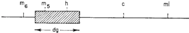
of the m a p (dg region) which includes the h locus and which comprises about 25 % of the known genetic m a p of λ.1 7
Figure 3 shows the result of such superinfection experiments for a number of markers. I t is seen t h a t a continuous section of genetic material, taken from the middle of the genetic map, is missing. The acquisition of the galac
tose genes seems to require a concomitant loss of phage genes. T h e c genes, which control superinfection immunity, are present, which explains t h e immunity of the defective heterogenotes and probably accounts also for the ability of t h e defective phage to lysogenize at all.
An Hft lysate would then be a mixture of two types of particles: (a) ordinary phage particles, which do not transduce, and (b) transducing parti
cles, which do not form plaques. A cell infected with a transducing particle in single infection might be lysogenized, giving rise to a transduction, or it might lyse without liberating anything, like a defective heterogenote in
duced by ultraviolet. If a population of cells is infected with an Hft lysate at a multiplicity of about 0.4 phage per cell and individual lysing cells local-
mf i m R h c mi
J
d g ^ -
F I G . 3. G e n e t i c m a p of λ, s h o w i n g t h e dg region. (After Arber.1 7)
ized on a fixed grid and observed in the electron microscope, one sees t h a t roughly half of the cells which lyse fail to liberate any visible particle similar to phage.1 7 Arber has called the transducing phage Xdg (for défectif, galactose- transducteur). T h a t region of the phage chromosome which is missing from t h e transducing phage is referred t o as the dg region.
Since Xdg cannot multiply in single infection, it follows t h a t the phage yield from a population of cells infected with an Hft lysate at very low multiplicity will contain very little transducing activity, because most of the Xdg enter cells which are not simultaneously infected with active λ phage. If the infection is done at high multiplicity, however, the transduc
ing activity is multiplied about to the same extent as the active phage.1 7 Whereas one can infer from these observations t h a t the lysate is a mixture of two types of particles, they do not rigorously exclude the possibility t h a t all particles in the lysate are really identical and t h a t the defect arises in a fraction of the individuals after the time of infection. Additional experi
ments somewhat more elaborate t h a n those described here made this possi
bility unlikely, but a complete proof required the physical separation of the two types of particles from the Hft lysate.
Such a separation was simply and beautifully achieved by the use of
density gradient centrifugation.2 0 Whereas an ordinary λ lysate gives only one sharp band in the ultracentrifuge, an Hft lysate gives two, as shown in Fig. 4. T h e plaque-forming particles are concentrated in one band and the
llllllll mm llf:
III
F I G . 4. B a n d i n g of n o r m a l p h a g e a n d t r a n s d u c i n g p a r t i c l e s f r o m s e v e r a l i n d e p e n d e n t H f t l y s a t e s . T h e dark b a n d t o t h e right is N1 5- l a b e l e d λ w h i c h w a s a d d e d t o all l y s a t e s as a d e n s i t y m a r k e r a n d w a s u s e d t o a l i g n t h e p h o t o g r a p h s . T h e c e n t r a l b a n d is n o r m a l λ. T h e t r a n s d u c i n g p a r t i c l e s b a n d e i t h e r t o i t s left or t o i t s right, d e p e n d i n g o n t h e d e n s i t y . R e p r o d u c e d from t h e J. Mol. Biol.20 w i t h p e r m i s s i o n of t h e p u b l i s h e r s .
transducing particles in another. Whereas the transducing particles from any one Hft lysate have a uniform density, those from different lysates m a y band at quite different densities. We shall return to this point later.
The absence of a connected region of genetic material which characterizes
the transducing phage has not yet been found among any of the defective m u t a n t s of λ which have been isolated as such. Among bacterial and phage m u t a n t s in general, multisite mutations, which cover a region rather t h a n a point, are common, although less frequent t h a n point mutations. Such mutations have been attributed to small deletions or other structural aber
rations, and, in one case, the genetic distance across such a "deletion" has been shown to be much less t h a n t h a t across the material normally present there.2 1 From the results of λ transduction, however, we can say t h a t some unknown fraction of all multisite m u t a n t s in phage must represent not sim
ple deletions but rather replacements of phage genes by genes from the host.
As we shall discuss later (see Section I I I , G ) , it is not necessary to suppose t h a t the host genes have actually gone where the phage genes were, but at least the loss of the phage genes was accompanied by the gain of some ex
traneous genetic material. The important point is that, by all the operations of phage genetics, the dg region behaves as a deletion. If Xdg had first turned up in a stock unmarked at the gal locus, it would have been classified simply as a deletion defective. The methods used for studying Xdg genetically have borrowed heavily from the techniques and concepts developed from study
ing deletions in other systems.2 2
D . N A T U R E OF T H E ASSOCIATION B E T W E E N P H A G E AND BACTERIAL G E N E S
The transducing particles thus contain some bacterial genes and some phage genes. W h a t is the connection between the two? Is there a genuine physical association, such as we suppose occurs between any two genes of the phage? Or do the defective phage just leave a convenient empty space within their shells into which a galactose gene can be neatly accommodated?
This question has been answered first from biological evidence, which is highly indicative, and more recently by chemical evidence, which is quite conclusive. The biological evidence is worth recounting first because it shows us some interesting features of the system.
In the first place, if the association between defective phage and trans
duced genes were a loose one, they should be easily separated from each other. Thus, a defective heterogenote might be expected to lose only the gal genes but retain the defective prophage or vice versa. W h a t is observed, instead, is t h a t , when the gal genes are lost, the defective prophage is usually lost, too. If one picks a number of independent gal~ segregants from a defec
tive heterogenote, one finds t h a t about 90 % have lost the immunity which indicates the presence of the defective phage.
W h a t about the other 10%? Do these show t h a t the genes can really be
come detached from the defective phage? As we mentioned earlier, in the discussion of lysogenic heterogenotes, a gal~ segregant can in principle arise
from a heterogenote in two ways: either through loss, to give a haploid strain, or through internal recombination, to give a still diploid homogenote.
One expects t h a t the haploids should have lost the immunity and the ho- mogenotes should have retained it. The homogenotes can be independently distinguished by the study of reverse m u t a n t s , or by their ability to give Hft lysates following induction and superinfection. T h u s far, all the indi
viduals examined which were gal~ but still immune have proven to be ho
mogenotes.
All the results on segregation from defective heterogenotes are compatible with the formation of a permanent stable association between the genes from the bacterium and those of the defective phage. Those types which would constitute obvious exceptions (e.g., a cell which was still segregating gal~ but had lost the immunity) have never been observed. One might, how
ever, still maintain t h a t a cell can carry a variety of such loose fragments and shed t h e m all at once because of some special state t h a t the cell occa
sionally enters rather t h a n because of a physical connection between the various fragments.
T h e segregation patterns of lysogenic heterogenotes are also in accord with the idea of an indissoluble union of phage and bacterial genes in the transducing particle. If lysogenic gal* cells are infected with an Hft lysate, most of the transductants are doubly lysogenic, carrying both Xdg and λ prophage. If both phages are genetically marked, such cells on induction liberate transducing particles and a mixture of active phages with genetic markers from the two parents, although never from the dg region of the Xdg parent. Gal~ segregants from such strains are usually singly lysogenic, and have lost most frequently the phage alleles with which the gal+ genes entered the cell.2 3 This was taken to indicate linkage between the gal genes and the transducing parent; b u t alternatively one could say it means merely t h a t both the Xdg prophage and the gal genes remain separate from, and more easily dislodgeable than, the prophage of active λ.
The real proof has come from transformation studies. Kaiser and Hog- ness2 4 have shown t h a t D N A prepared from free transducing particles can transform gal~ cells into gal+. This transformation is really a lysogenization of the bacterial recipient by the D N A of t h e transducing phage and results mainly in the formation of heterogenotic rather t h a n haploid transformants.
The number of transformants is directly proportional to the amount of D N A added. The transformation requires the presence of added "helper"
phage, but, even at multiplicities of 0.1 phage D N A equivalents per bac
terium, most of the transformants have acquired some genetic markers from the transducing phage.
This implies t h a t the phage genes and the galactose genes remain associ
ated on a single physical structure through the extraction and isolation of
the D N A . Thus, the Xdg particle, like active λ, consists of a single D N A molecule covered by a protein coat. T h e biological experiments, further
more, indicate strongly t h a t this D N A molecule retains its unity during intracellular growth, both in the vegetative and in the prophage state. T h e problem to which attention must next be addressed is the origin of this new t y p e of D N A molecule which contains components from two diverse sources.
E . E F F E C T OF NONTRANSDUCING P H A G E IN TRANSDUCTION
Before discussing the origin of Xdg, we shall complete our description of the experiments mentioned thus far by returning to a point we have deliber
ately glossed over. When we recounted the characterization of the transduc
ing phage as defective, we specified t h a t the multiplicity of infection (i.e., the phage/bacterium ratio in the transduction) must be sufficiently low t h a t we are sure to be looking at the results of single infection by the transducing particle. This seems such an obvious requirement t h a t the reader unac
quainted with this work m a y wonder why the defectivity was not discovered immediately, or even in low frequency transduction.
The reason is purely technical. I n single infection, Xdg lysogenizes very poorly. If the same cell is simultaneously infected with an active phage particle, the lysogenization is greatly increased. We say t h a t the active phage helps its defective relative to lysogenize, and therefore to transduce.
An Hft lysate is a mixture of active λ and Xdg. When a population of cells is infected with such a lysate, a certain fraction of those cells receiving Xdg phage will receive an active λ as well. At multiplicities of infection much less t h a n one, this fraction is approximately equal to the multiplicity.
However, because of the helping effect, this fraction will account for the majority of transductions down to quite low multiplicities.
For example, suppose t h a t a cell infected with one active phage and one transducing phage has a probability of giving rise to a recoverable trans- ductant colony which is 40 times t h a t of a cell infected with a transducing phage alone. Consider a population of' gal~ cells infected with an Hft lysate in the proportion of 1 active phage: 10 bacterial cells (multiplicity of in
fection, 0.1). Of every 100 cells which receive a transducing particle at all, approximately 90 will receive only the Xdg, whereas 10 will be simultane
ously infected with an active λ phage. However, each of these 10 cells has 40 times the probability of giving rise t o a transduction which the other 90 have. Among the recovered transductions, then, a fraction 400/490, or 82 %, will arise by multiple infection, even at a multiplicity where only 10 % of the cells were multiply infected.
Figure 5 shows the type of data from which such conclusions are drawn.1 8 On the ordinate is plotted the number of transductions per active phage
particle added ( T / P ) as the multiplicity of infection is varied. A t very low multiplicities this quantity equals the product of the number of transducing particles per active phage in the lysate by the probability of transduction in single infection. As multiplicities are raised from 0.01 to 1, the T / P value rises some 20- to 40-fold. T h a t this is due to a helping effect is verified by a control experiment in which all the cells are infected at a multiplicity of 2 with a λ lysate obtained from a gal~ bacterium which has no transduc
ing activity whatsoever, and only the multiplicity of transducing phage is varied. The T / P is then independent of multiplicity, and some 20- to 40- fold greater t h a n t h a t obtained in single infection.
ι Η
Multiplicity of infection
F I G . 5. Efficiency of t r a n s d u c t i o n b y a n H f t l y s a t e as a f u n c t i o n of t h e m u l t i p l i c i t y of i n f e c t i o n . C u r v e Β : h i g h f r e q u e n c y t r a n s d u c i n g l y s a t e alone. C u r v e A : s a m e l y s a t e w i t h c o n s t a n t b a c k g r o u n d m u l t i p l i c i t y (1.7) of n o n t r a n s d u c i n g p h a g e . R e p r o d u c e d f r o m Virology18 w i t h t h e p e r m i s s i o n of t h e p u b l i s h e r s .
All of the transductants arising from mixedly infected cells seem t o become lysogenized by the active phage as well as by the transducing phage. They are t h u s double lysogens, which is easily verified by the use of a genetically marked helping phage. The lysogenic heterogenotes originat
ing from Lft have properties identical with these double lysogens and presumably arise in the same way. I t is technically difficult to perform low frequency transduction a t very low multiplicities, but it is possible to demonstrate the presence of defective phage in Lft lysates by density gradient centrifugation.2 5
T h e mechanism of the helping effect is not understood. I t has been suggested t h a t the main effect is t o permit vegetative multiplication of the transducing phage, thereby increasing the number of copies available to
lysogenize and thus augmenting the frequency of lysogenization.2 6 We know t h a t help is blocked whenever the recipient cell carries a prophage of the same immunity specificity as the helping phage.2 3 We know also t h a t some of those cells singly infected with a transducing phage are lost through lysis,1 7 but it is not clear whether the fraction of cells lysing is changed by the helping phage. The main interest of the whole phenomenon at the moment is t h a t it creates a technical difficulty which has confused the interpretation of results in the past and might do so in other systems in the future.
F . G E N E T I C AND PHYSICAL VARIABILITY OF T H E TRANSDUCING P H A G E
The model of transduction arrived at by the experiments described thus far can now be recapitulated briefly. When one prepares a lysate of λ by inducing a lysogenic strain, there are produced, in addition to ordinary λ phage particles, very rare abnormal phage (about 1 in 105) which have picked u p the galactose genes from nearby on the host chromosome and have lost a block of their own phage genetic material. These are ordinarily detected by transduction and recovered in the form of lysogenic strains which carry a normal λ prophage in addition to the prophage form of the abnormal (Xdg) phage. The normal phage is present in these strains for the technical reason t h a t low frequency transduction cannot be performed at very low multiplicities with the expectation of any recovery at all, and at higher multiplicities the helping effect occurs. Among the phage liberated by such strains, the abnormal type is no longer very rare. I t is produced in an amount about equal to t h a t of the normal phage. This information is summarized in Table I. Table I I shows the relationship between the sym
bolic and verbal descriptions of the various types of strains involved.
Such an Hft lysate contains two discrete classes of particles—normal phages which produce plaques, and abnormal phages which mediate trans
duction. If we consider t h a t an Hft lysate can be prepared from a culture of a lysogenic heterogenote which has been grown from a single cell, we see t h a t the two classes of particles are lineally derived from the two pro
phages carried by this heterogenote. We therefore expect that, except for the intervention of additional rare events, all the transducing particles in this lysate will be identical.
On the other hand, in the case of low frequency transduction, the lysate is prepared from a culture ultimately descended from a single cell which carried one λ prophage in the normal, active form. The transductions one sees have resulted from rare events which have occurred sometime after this single cell was isolated, and different isolates may have resulted from different events. There is therefore no a priori expectation as to whether
or not the transducing phage derived from different events in low frequency transduction should be identical.
The problem has been attacked by physical as well as genetic methods.
We have mentioned the result of the physical studies already. Whereas
T A B L E I
C O M P A R I S O N O F L O W F R E Q U E N C Y T R A N S D U C T I O N W I T H H T G H F R E Q U E N C Y T R A N S D U C T I O N
Type of
trans- Donor duction
Approximate composition
of lysate
Transductants
High m.o.i. Low m.o.i.
Lft gal+(X) 1 05λ : 1 Xdg M gal+(X) (Xdg) N o t f e a s i b l e0 H gai+(x)
H f t gal- (X) (Xdg) 1 X:Xdg gal~ (X) (Xdg) gal~ (Xdg)
gal+(X) (rare) gal+ (rare)
a L o w f r e q u e n c y t r a n s d u c t i o n is n o t feasible a t r e a l l y l o w m u l t i p l i c i t i e s , a l t h o u g h b y special m e t h o d s o n e c a n s h o w t h a t s o m e of t h e t r a n s d u c t a n t s are gal~(Xdg)?b
T A B L E I I
R E L A T I O N B E T W E E N T E R M S A N D S Y M B O L S I N λ T R A N S D U C T I O N
Symbol Term
gal+
gal~
S e n s i t i v e b a c t e r i a
gal+(X) gah(X)
A c t i v e l y s o g e n s or l y s o g e n i c s t r a i n s
gal+(\dg gal+) D e f e c t i v e gal+ h o m o g e n o t e gah(Xdg gal+)
gal+(Xdg gah) gah(Xdg gah)
D e f e c t i v e h e t e r o g e n o t e s
gah(Xdg gah) D e f e c t i v e gah h o m o g e n o t e gah(X)(Xdg gal+) L y s o g e n i c h e t e r o g e n o t e
there is no detectable density heterogeneity among the transducing particles within any one Hft lysate, there is almost invariably some difference be
tween the density seen in one Hft lysate and t h a t in any other which was derived from a different Lft event.2 0 Some kinds of Xdg particles are denser t h a n ordinary λ. Others are lighter. T h e density of a particle probably
depends on the ratio of D N A to protein. If the protein content of all the particles is the same, the density depends directly on the D N A content.
I t would thus seem t h a t some Xdg phages have more D N A t h a n active λ and t h a t others have less. Of 10 independent lysogenic heterogenotes studied, a different density was found for each; therefore the total number of possible density classes must be very large.
Genetic methods have yielded concordant results.2 7 One transducing particle might in principle differ from another either in the extent of bac
terial genetic material picked up, or in t h a t of the phage genetic material lost. Differences of the former type have not yet been found, but the latter have been studied rather extensively with the aid of the sus m u t a n t s of λ.
As explained earlier, these are m u t a n t s which can form plaques on some strains of E. coli K12 (pm+) and not on others (pmr) (see Table I I I ) . The
T A B L E I I I
P L A T I N G P R O P E R T I E S O F A C T I V E A N D D E F E C T I V E λ
Phage types Bacterial strain"
N a m e Symbol pm+ pmr
W i l d - t y p e λ λ+ + +
H o s t - d e p e n d e n t ( s u p p r e s - Xhdn or \susn + —
s o r - s e n s i t i v e ) m u t a n t
D e f e c t i v e m u t a n t of λ \in — —
T r a n s d u c i n g λ \dg — —
° K e y : + , forms p l a q u e s ; — , d o e s n o t form p l a q u e s .
pm~ strains provide an absolute selection for any wild-type recombinants which m a y be produced in a cross between two sus m u t a n t s , or between one sus m u t a n t and one true defective. If a defective heterogenote carrying Xdg is induced and then superinfected with a sus m u t a n t , wild-type recom
binants will appear if the sus mutation lies outside of the dg region. None will appear if it lies inside. By making such crosses between a particular heterogenote and a collection of sus mutants, one can define the extent of the dg region in t h a t heterogenote.
I n practice, m a n y such crosses can be performed on the surface of a single Petri plate (Fig. 6). On a background of pmr cells, drops of various defective heterogenotes, various sus mutants, and paired combinations of the two are placed. After drying, the plates are irradiated briefly to induce the heterogenotes. The appearance of plaques in the spot in excess of those produced by reverse m u t a n t s in the phage lysate itself shows t h a t wild- type recombinants are formed. Since we are asking only whether or not a given genetic site is contained in the transducing phage, we are not con-
F I G . 6. C r o s s e s b e t w e e n s u p p r e s s o r s e n s i t i v e m u t a n t s and Xdg. T h e p l a t e w a s first s e e d e d w i t h a b a c k g r o u n d of pmr c e l l s , and t h e n in t h e c e n t e r of e a c h s q u a r e w a s p l a c e d a loopful f r o m a c u l t u r e of a d e f e c t i v e h e t e r o g e n o t e ( t h e s a m e o n e for all t h e s q u a r e s of a row) and from a l y s a t e of an sus m u t a n t ( t h e s a m e o n e for all t h e s q u a r e s of a c o l u m n ) . A f t e r an i n d u c i n g d o s e of u l t r a v i o l e t l i g h t , t h e y w e r e i n c u b a t e d o v e r n i g h t . F o r m a t i o n of p l a q u e s o n t h e pmr b a c k g r o u n d s h o w s t h e a b i l i t y of t h e Xdg t o p r o d u c e w i l d - t y p e r e c o m b i n a n t s w h e n crossed w i t h t h e sus m u t a n t in q u e s t i o n .
derived from the same Lft event are genetically as well as physically identical. Second, particles derived from separate events are frequently different. The differences thus far revealed have all involved the position cerned with how m a n y such recombinants are produced, but only with their presence or absence.
Such studies have shown, first, t h a t all of the transducing particles
of the left end point, which generally falls within a region of the chromo
some especially rich in sus m u t a n t s . Figure 7 shows the different classes of transducing particles which have been revealed by this technique. Figure 8 is a diagrammatic representation of the origin of the various types of defective phages shown in Table I I I .
One sees, first of all, t h a t there is a "common core" to all the dg regions—
a group of genes which are missing from every transducing λ thus far examined. This core region covers a group of sus m u t a n t s and also the h locus. T h e dg regions of various Xdg's penetrate to different extents the section of the genome to the left of the common core.
sus,, I sûs,e| sûs,o| sus, I sus6 6| sûs8 l| sus,12| sus7e| sus3 6| sus,2 3| sûs4 |sus9 6 B| SUS55| SUS,oi|sus44| SUS3 6 SUS3 2 S U SI 07 S U S4 8 SUS9 3 SUS2 2 SUS4 2 SUS20 SUS15 SUS2 5 SUS28 SUS26 SUS9 susl 2 sus2
A B C D E F G H I
F I G . 7. D i a g r a m m a t i c r e p r e s e n t a t i o n of t h e v a r i a b l e p e n e t r a t i o n of t h e dg region i n t o a s e c t i o n of t h e p h a g e c h r o m o s o m e . T h e e n t i r e s e c t i o n s h o w n is t o t h e left of t h e h g e n e in F i g . 3. E a c h solid line r e p r e s e n t s t h a t p o r t i o n of t h e p h a g e c h r o m o s o m e w h i c h is p r e s e n t in a p a r t i c u l a r t r a n s d u c i n g p h a g e . T h e m u t a n t s are all of t h e s u p - pressor s e n s i t i v e (sus) t y p e . F o r s i m p l i c i t y , o n l y o n e m u t a n t h a s b e e n s h o w n in e a c h region, and t h e regions h a v e b e e n e q u a l l y s p a c e d . T h e c a p i t a l l e t t e r s r e p r e s e n t c o m - p l e m e n t a t i o n c l a s s e s i n t o w h i c h t h e m u t a n t s c a n b e g r o u p e d .
The results of the crosses between all of the m u t a n t s and all of the trans- ducing phages constitute, by themselves, an adequate system for ordering along a linear linkage m a p both the sus mutations and the end points of the dg regions of the transducing phages. The validity of the method is shown by the fact t h a t one can make a diagram such as t h a t of Fig. 7 where every dg region is represented as an uninterrupted segment of the linear structure.
The order of some of the sus m u t a n t s has also been corroborated by standard two- or three-factor crosses. The results shown in Fig. 7 indicate t h a t the end point of the dg region can occur at a great many points, perhaps any point, within a certain part of the phage chromosome. They indicate definitely t h a t it can fall within a genetic locus as well as between loci.
For some transducing phages, both physical and genetic measurements have been made. The results show a general correlation between D N A
content a n d number of phage genes present. T h e longer t h e dg region, t h e less dense t h e transducing phage. T h e correlation is not perfect, however, which means t h a t there are other variables which can change independently of this end point of t h e dg region.
F r o m all of these results, it is obvious t h a t a n Lft lysate differs from a n Hft lysate not only in t h e ratio of transducing particles to active phages, but also in t h e spectrum of types of transducing particles formed. An Hft lysate contains only t h e one type which is a copy of t h e particular Xdg prophage carried b y t h e heterogenote from which t h e lysate was made.
Other types presumably can occur a t t h e same level as in a n Lft lysate, b u t
Mutation
Prophage of lysogenic strain
Prophages of different defective lysogens
Recombination with galactose region following UV induction
Prophages of defective heterogenotes F I G . 8. O r i g i n of d e f e c t i v e l y s o g e n i c s t r a i n s .
they are hidden b y t h e 105 times greater amount of identical transducing particles present.
One might therefore wonder whether a n Lft lysate contains a n even wider range of transducing particles t h a n are recovered among t h e prophages of heterogenotes. I t is quite possible t h a t this is so. Lft lysates give a higher proportion of stable nonheterogenotic transductions t h a n do Hft lysates.1 5 This probably reflects t h e presence of a class of transducing particles which lysogenize poorly, if a t all, and therefore rarely, if ever, form heterogenotes.
Special methods will be required t o obtain a n y information as to t h e nature of these particles. Most people have preferred t o study t h e heterogenotes because it is easier. A n y generalizations about transduction a t t h e present moment must be tempered with a n understanding of the bias introduced b y studying a limited class of objects which happen t o be convenient t o work with.
G . M E C H A N I S M OF T H E RECOMBINATIONAL E V E N T
The transducing phage can be considered as a genetic recombinant between the host bacterium and the bacteriophage. We are using ''recombin
ation" in a very broad sense to cover any process in which two or more (parent) individuals with different genetic characters interact to produce a new individual, some of whose genetic specificity is derived from each of the parents.
Recombination as thus defined can be of two basically different types, which we can call equal and unequal, respectively. I n equal recombination, the genetic elements of the two parents can be put in a one-to-one cor
respondence with each other, and the rule is t h a t each offspring must receive a complete set of genetic elements. Recombination which is not equal is unequal.
I n organisms where a sexual process constitutes an essential part of the reproductive cycle, equal recombination is far more common t h a n unequal recombination. This is necessary because the products of unequal recom
bination are likely to be lethal due to duplications or deficiencies of genetic material. Unequal recombination certainly does occur, though, giving rise to chromosomal aberrations. Equal recombination is probably the rule also in bacteria and phages. Otherwise, multisite mutations would occur with a far higher frequency t h a n is observed.2 2
The physical bases of equal recombination are, first, the mitotic ap
paratus, and, second, the precise pairing of homologous regions within the chromosome. Such an exact matching of homologous parts of the parent structure is essential for equal recombination within the chromosome, regardless of whether the mechanism is by chromosome breakage or by copy choice, and irrespective of whether the individual recombinational event is reciprocal or nonreciprocal. Unequal recombination, on the other hand, does not require any genetic homology between the two parent structures.
I n transduction, the λ phage loses a specific region of its own genome concomitantly with picking up a specific portion of the bacterial genome.
This could be t h e result of equal recombination. Certain regions of the phage D N A might match corresponding regions in the bacterial D N A , and within these regions crossing over or miscopying might occur. The facts t h a t the same ''common core" of the dg region is absent from all the transducing phages, and t h a t the deletion of phage genes is interstitial rather t h a n terminal gives superficial plausibility to such a scheme. The simplest model for an origin by equal recombination is shown in Fig. 9.
However, equal recombination of this type should give rise to recom
binants containing the same amount of D N A as the parents in the region of homology. According to Fig. 9, the transducing phage might contain
more or less D N A t h a n the λ phage itself, but all transducing phages should have the same D N A content. Insofar as D N A content can be deduced from density, this is contrary to experimental fact.
The argument is supported also by genetic evidence. Suppose t h a t there were a region (such as ALAR of Fig. 9) in which the λ chromosome matched perfectly the bacterial chromosome, and t h a t the transducing phage orig
inated by equal recombination in this region. Then the active λ phage and the transducing phage would likewise match each other in this region.
Since the transducing phage can only mature in the presence of active phage, every Xdg particle has had some opportunity to recombine with active λ, and one product of such recombination should be a transducing phage which now contains a larger amount of phage material t h a n it had
gal
ARh d6 h BL BR c p4
4A AR gal
Host chromosome
Phage chromosome
c P4 Chromosome of transducing phage
F I G . 9. S i m p l e s t m o d e l for origin of t h e t r a n s d u c i n g p h a g e b y e q u a l r e c o m b i n a t i o n . I t is a s s u m e d t h a t t h e c h r o m o s o m e s of t h e p h a g e and t h e b a c t e r i u m are h o m o l o g o u s b e t w e e n A L a n d A R a n d b e t w e e n B L a n d B R and t h a t a t r a n s d u c i n g p h a g e c a n be p r o d u c e d b y r e c o m b i n a t i o n at a n y p o i n t s A and Β w i t h i n t h e s e r e g i o n s . T h e s u p p r e s s o r - s e n s i t i v e m u t a n t s are d e n o t e d b y t h e i r old n a m e of h o s t - d e p e n d e n t (hdi and hde). T h e m u t a t i o n p4 is at t h e s a m e p l a c e as mi of F i g . 3 . R e p r o d u c e d from Virology28 w i t h t h e p e r m i s s i o n of t h e p u b l i s h e r s .
previously. We can recognize the point at which recombination has taken place as the end point of the dg region, determined genetically by crosses with the sus m u t a n t s .
Now, the fact is t h a t the end point of the dg region has never been ob
served to change after primary isolation among m a n y hundreds of indi
viduals examined. More important, if we select among transducing phages those which have recombined with an active phage and have acquired from it a genetic marker at the extreme left-hand end of the linkage group, we still do not detect any change in the end point of the dg region.2 8 We conclude t h a t such recombinants arise by equal recombination within the region of Xdg which was derived from λ and never in t h a t derived from the bacterium.
Likewise, if one selects for individual Xdg prophages in which the gal~
allele from the bacterial chromosome has substituted for the gal+ allele of Xdg (giving a defective homogenote), we find no change in the end point
of the dg region. The transducing phage thus contains some genetic material derived from the phage and able to undergo equal recombination with the homologous regions of the phage, and other genes derived from the bac
terium which can recombine with the bacterial chromosome. W h a t is absent in the picture is any common ground, any portion of the structure which falls into both categories at once.
Both physical and genetic methods therefore agree in ruling out equal recombination between two perfectly matched regions. Unfortunately, they cannot eliminate the possibility of matching which, though imperfect, is sufficient to serve as a basis for precise pairing. We do not expect the match
ing to be completely perfect anyway. Perfect matching occurs only between truly identical structures. I n equal recombination between identical parents, no record is left of where or whether the recombinational event has occurred. One can study this only by introducing genetic markers, thereby destroying identity. The success of classical recombinational genetics depends on the fact t h a t the individual markers generally have negligible effects on the mechanics of the recombination process. This assumption is not necessarily valid in molecular genetics at the level studied here. If the end point of the dg region does represent a point of equal recombination, the matching regions of phage and bacterial chromosome must differ at least enough so t h a t the wild-type allele of the sus m u t a n t s studied cannot be recovered by recombination from Xdg at a detectable frequency.
We conclude t h a t there is no direct evidence favoring equal recombina
tion, and t h a t , if genetic homologies between phage and host are involved, these homologies are very imperfect. I t must be mentioned also t h a t , whereas we know t h a t an interstitial region of the phage chromosome is missing, there is no real evidence t h a t the galactose genes have replaced them rather t h a n adding to some other part of the structure. I t is thus quite feasible to entertain models for the origin of transducing λ in which the gal genes add to the end of the phage chromosome rather t h a n to the middle.
H . M E C H A N I S M OF L O W F R E Q U E N C Y TRANSDUCTION
All of this work says t h a t , once the primary transductional event has occurred, a new stable genetic structure is formed which rarely, if ever, changes its properties. The fixed character of this primary change through
out the introduction and exchange of genes into various parts of the struc
ture by ordinary recombination is reminiscent of inversions or translocations in higher organisms and suggests t h a t some alteration more far reaching t h a n equal recombination between homologous regions has transpired. At any rate, since nothing very interesting happens after the primary event, one is forced to delve backwards and t r y to study the event itself.