"Abnormal Metabolites" of Amino Acid Origin
HERBERT SPRINCE
Department of Research Biochemistry Veterans Administration Hospital Coatesville, Pennsylvania and Department of Biochemistry Graduate School of Medicine University of Pennsylvania Philadelphia, Pennsylvania
I. Introduction: General Aspects 162 A. Definition of Abnormal Metabolites 162 B. Origin of Abnormal Metabolites: Metabolic Aspects 163
C. Factors Involved in Formation of Abnormal Metabolites . . . . 167 D. Methods Used in Determination of Relative Magnitudes of Metabolic
Pathways 169 E. Methods for Expressing Values of Urinary Metabolites . . . . 171
F. Chemical Nature of Abnormal Urinary Metabolites Originating from
Amino Acids 172 II. Amino Acids as Abnormal Metabolites 172
A. Normal Urinary Excretion of Amino Acids 173 B. Classification of Pathological Aminoacidurias 176 C. Abnormal Amino Acid Excretion in Pathological Aminoacidurias . . 177
D. Methodology 185 III. Keto Acids as Abnormal Metabolites 194
A. Normal Urinary Keto Acids 194 B. Abnormal Urinary Keto Acids 194
C. Methodology 195 IV. Phenolic Compounds as Abnormal Metabolites 197
A. Normal Urinary Excretion of Phenolic Compounds 197 B. Abnormal Urinary Excretion of Phenolic Compounds . . . . 201
C. Methodology 204 V. Indolic Compounds as Abnormal Metabolites 210
A. Normal Urinary Excretion of Indolic Compounds 210 B. Abnormal Urinary Excretion of Indolic Compounds 214
C. Methodology 218 VI. Imidazolic Compounds as Abnormal Metabolites 225
A. Normal Urinary Excretion of Imidazolic Compounds 227 B. Abnormal Urinary Excretion of Imidazolic Compounds . . . . 229
C. Methodology 232 References 237
161
I . INTRODUCTION: GENERAL ASPECTS
This chapter will be devoted to a discussion of abnormal urinary metabolites originating from amino acids and newer methods concerned with their measurement. The presentation will be restricted to those metabolites most frequently regarded as "abnormal metabolites" of amino acid metabolism as well as certain newcomers now beginning to appear in the current literature. It is obvious that to some extent the selection of metabolites for review and the emphases placed thereon are bound to be arbitrary. An attempt, however, will be made to deal with metabolites not only of special interest to the author, but also of general interest and importance in the field of amino acid metabolism.
A. Definition of Abnormal Metabolites
The term "abnormal metabolites" in its most complete sense may be defined as those metabolites (in blood and/or urine) which first appear in, or are increased (or decreased) above (or below) their normal range as a result of, certain metabolic disturbances. As most generally used, however, the term refers primarily to increases above normal range rather than decreases. The metabolic disturbances may be of genetic, pathologic, pharmacologic, toxicologic, homeostatic, or nutritional origin.
The biochemical geneticist may use the term to refer primarily to those metabolites which relate to a congenital abnormality of molecular struc- ture [e.g., the abnormality of the hemoglobin molecule in sickle-cel]
anemia (1, 2)] or of metabolic function [e.g., the abnormal accumulation of homogentisic acid in alkaptonuria or phenylpyruvic acid in phenyl- ketonuria (3, 4 ) ] . Such a definition supposes that abnormal metabolites arise from a genetic mutation or defect in an enzyme system. This represents perhaps, the most restricted use of the term. The clinical bio- chemist, on the other hand, has extended the use of this term to certain metabolites normally found in small amounts, but markedly increased in pathological states. This is especially true where a defect may exist in renal tubular reabsorption [e.g., cystinuria (5)] or where an alternate metabolic pathway may be involved [e.g., the serotonin—5-hydroxyin- doleacetic acid pathway from tryptophan which is known to be increased in carcinoid disease (6)]. Similarly, the pharmacologist often speaks of abnormal metabolites arising from the administration of drugs [e.g., the increase in urinary tryptamine or serotonin after administration of monoamine oxidase inhibitors (7)]. The toxicologist is inclined to think of detoxicants as abnormal metabolites which serve to indicate the in- gestion of toxic chemical compounds [e.g., the excretion of mercapturic acid after intake of chlorobenzene (8)]. The stress physiologist talks
about abnormal increases of metabolites resulting from disturbances in homeostatic mechanisms due to a variety of stress stimuli (9) [e.g., increase of catecholamines in emotional disturbance (10-12)]. And finally, even the nutritional biochemist can lay claim to the importance of studying abnormal metabolites which may arise as a result of diet
[e.g., increased 5-hydroxyindoleacetic acid excretion as a result of in- gestion of bananas and certain other fruits (13-15)], vitamin deficiency states [e.g., xanthurenic acid excretion in vitamin B6 deficiency (16)], and a host of other factors.
It is apparent, therefore, that the term "abnormal metabolites" is a loose term which has come to assume a shade of meaning peculiar to the field of study involved. As currently used in the literature, it applies not only to metabolites which first appear only under abnormal conditions, but also to certain metabolites which normally occur in relatively small amounts, yet are markedly increased under abnormal conditions. In this review, we shall use the term "abnormal metabolites" as set forth in the first two sentences of the preceding paragraph. Moreover, we shall limit ourselves to a discussion of abnormal metabolites of natural origin, as distinguished from those resulting from the intake of a chemical compound foreign to the body (e.g., detoxicants).
B. Origin of Abnormal Metabolites: Metabolic Aspects
At the biochemical level, abnormal metabolites arise as a result of disturbances in the metabolic pathways of intermediary metabolism.
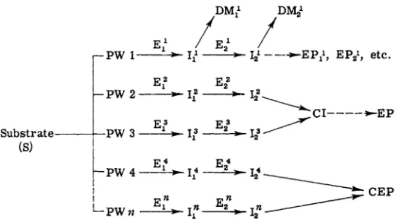
Hence, a brief discussion of the general aspects of intermediary metab- olism is in order for a better understanding as to how abnormal metabo- lites develop. A simplified diagram is presented in Fig. 1. For a recent review, see Kopin (17).
In Fig. 1 any given substrate (S) may be metabolized over a number of different (alternate) metabolic pathways (PW 1, PW 2, etc.). Each pathway may involve a number of enzyme systems (E1} E2, etc.) oper- ating in sequence and giving rise to a series of intermediates I2, etc.) which eventually are transformed into one or more end products (EPi, EP2, etc.). During the course of metabolism, any given intermediate of a single pathway may be channeled into "divergent reactions" (17) to form deviate metabolites (DM1 ? DM2, etc.) leading, perhaps, to new pathways. Conversely, intermediates from different pathways may be channeled into "convergent reactions" (17) to form common inter- mediates (CI) or common end products (CEP). In normal metabolism, certain alternate pathways assume major or minor priority, depending on a number of factors to be discussed in the next section (Section I,C).
In acquired metabolic disease states, the priority of such pathways may
be altered by the pathological process. In an inborn metabolic disorder, the normal pathway may be blocked by the congenital absence of an enzyme system or altered by excessive channeling into a particular path
way (e.g., as in gout). In any of these conditions, an abnormal increase (or decrease) of substrate, enzyme, intermediary metabolite, or end product may result in the blood or urine. These substances can then be regarded as abnormal metabolites.
In terms of intermediary metabolism, therefore, abnormal metabolites are known to develop in at least six different ways. A discussion of each follows.
FIG. 1. Schematic diagram of metabolic pathways. S = substrate; PW 1, 2, etc. = pathway 1, 2, etc.; Ε = enzyme; I = intermediate; D M = deviate metab
olite ; CI = common intermediate; EP = end product; CEP = common end prod
uct. Exponents refer to pathway number. Subscripts refer to sequence number within a given pathway.
1. Abnormal Increase in the Blood or Urine of the Substrate Itself This occurs in a number of different types of aminoacidurias which will be discussed in Section II,B. At this point, it may suffice to give some examples of a few of the more well-defined aminoacidurias wherein an abnormal excretion of the substrate (amino acid) itself takes place.
The probable cause of the aminoaciduria in the following instances is noted in parentheses. Thus, for example, in phenylketonuria (congenital enzymatic defect), there may be an increase in urinary phenylalanine (18); in cystinuria (congenital renal tubular defect), an increase in urinary cystine, lysine, arginine, and ornithine (19); in Wilson's disease
(acquired renal tubular defect), an increase in urinary threonine and cystine (20) and the appearance of urinary proline and citrulline, the
latter two not found in normal urine (21); and in chronic protein de
ficiency of the kwashiorkor type ("no threshold" aminoaciduria), an increase in β-aminoisobutyric acid and ethanolamine (22, 23).
2. Abnormal Appearance or Increase in the Blood or Urine of an Intermediary Metabolite
Such cases generally result from an enzyme defect which may be congenital or acquired. Examples of intermediary metabolites excreted abnormally in the urine due to a congenital enzymatic defect are: homo- gentisic acid in alkaptonuria (3), phenylpyruvic acid in phenylketonuria
(4), and monoiodotyrosine and diiodotyrosine in cretinism related to a defect of "dehalogenating enzyme" also known as iodotyrosine de- iodinase (24). Among the intermediary metabolites which arise by virtue of an acquired enzymatic defect and appear abnormally in the urine are pyruvic acid in thiamine deficiency (25) and the p- hydroxyphenyl compounds (p-hydroxyphenylpyruvic acid and p-hydroxy- phenyllactic acid) in ascorbic acid deficiency (26, 27).
3. Abnormal Appearance or Increase in Blood or Urine of a uDivergent Reaction" Metabolite
Perhaps, the most outstanding example of this type is the urinary excretion of xanthurenic acid in pyridoxine deficiency (16). Another ex
ample which fits into this category is the recently reported excretion of 5-methoxytryptamine, a metabolic deviate of serotonin metabolism, in rheumatic fever (28). Both of these cases may be regarded as acquired enzymatic defects. As an example of a congenital defect of this type, there may be considered the increased urinary oxalate excretion of primary hyperoxaluria (29). In this disease, oxalate is thought to be a metabolic deviate of glyoxylic acid metabolism. The precise nature of the biochemical defect in primary hyperoxaluria, however, is not yet known.
4. Abnormal Alteration of Metabolic Pathways Resulting in the Diversion of Substrate from a Normally Preferred Pathway
into One of Lesser Priority
A number of examples of this type of metabolic abnormality readily come to mind. As in preceding instances, the aberrant alteration may originate from a congenital disorder or an acquired disease state. Ex
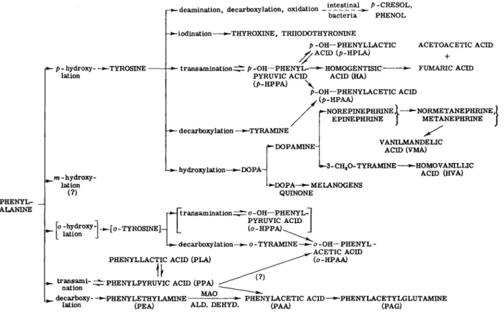
amples of congenital disorders of this type are to be found, again, in phenylketonuria and also in albinism. In phenylketonuria, phenylalanine is diverted from its usual conversion into tyrosine and its metabolites (p-hydroxylation pathway) to the formation of phenylpyruvic acid and its metabolites (keto acid pathway). This is due to the congenital
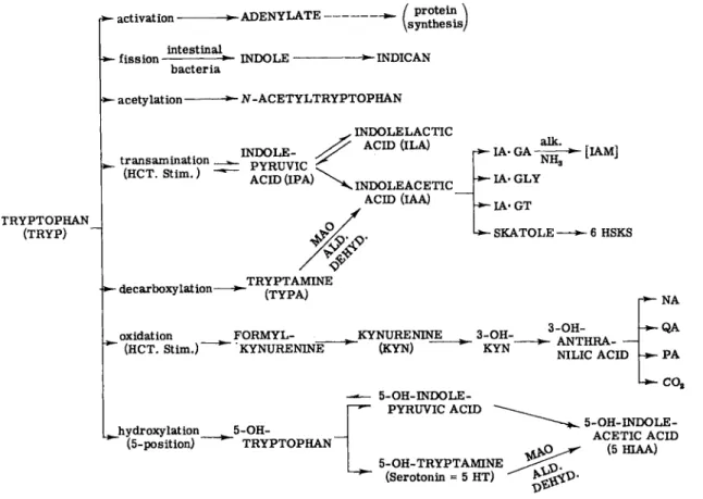
absence of phenylalanine hydroxylase (4). A similar situation may occur in albinism, wherein tyrosine cannot form melanin due to the congenital absence of the enzyme tyrosinase (30). Tyrosine may thus be channeled into other pathways, but, as yet, this aspect has not been sufficiently investigated (30). Examples of alterations of metabolic pathways ac- quired in a disease state are to be found in carcinoid disease and in melanogenuria. In carcinoid tumors, tryptophan is diverted from its nor- mal oxidative pathway (kynurenine, nicotinic acid, etc.) into the 5- hydroxylation pathway (5-hydroxytryptophan, serotonin, etc.). It has been estimated that, in normal subjects, only about 1% of dietary tryptophan is converted to 5-hydroxyindole compounds; in carcinoid dis- ease, this may rise to 60% (6). In melanogenuria, tyrosine, which nor- mally is metabolized by the transamination pathway (p-hydroxyphenyl- pyruvic acid, homogentisic acid, etc.), is channeled in increased amounts into a pathway of lesser priority, the oxidative pathway (dopa, dopa quinone, and melanogens) (31). The excreted melanogens undergo auto- oxidation in air to form melanin, the dark brown pigment found in the urine of melanoma.
5. Enzymes in Relation to Abnormal Metabolites
In certain pathological situations (e.g., myocardial infarction, liver disease, etc.), enzyme activity levels (e.g., serum transaminases) may be elevated. In such cases, however, the enzymes involved cannot be re- garded as truly abnormal metabolites of intermediary metabolism since their increased levels are due primarily to liberation from damaged tissue rather than an inherent metabolic disturbance at the biochemical level. Most cases of enzymes dealing with abnormal intermediary metabolism are characterized by the congenital absence or acquired malfunction of a normally occurring tissue enzyme, as in phenyl- ketonuria (absence of phenylalanine hydroxylase) or in pyridoxine de- ficiency (lack of coenzyme for kynureninase), respectively. Recently, two excellent monographs have appeared dealing with metabolic dis- turbances of enzymes in congenital and acquired disease states. See Abderhalden's book on "Clinical Enzymology" (32) and Wilkinson's publication "An Introduction to Diagnostic Enzymology" (33).
6. Abnormal Increase in Blood or Unne of a Metabolic End Product Metabolic end products may also be increased in abnormal amounts as a result of congenital disorders or acquired disease states. Such in- creases may be the basis for diagnostic tests in clinical chemistry. Thus, o-hydroxyphenylacetic acid is excreted normally in urine in amounts of 1 to 2 mg per day in nonphenylketonurics. In phenylketonuria this range
may be increased from 100 to 400 mg per day (34, 35). At such elevated levels, this urinary metabolite is practically diagnostic for this disorder.
o-Hydroxyphenylacetic acid is an end product chiefly of phenylalanine metabolism via (?) the intermediary metabolite, phenylpyruvic acid (35). It is, therefore, a metabolic end product directly related to the congenital disorder, phenylketonuria. Another case in point is 5-hydroxy- indoleacetic acid. Normally, it is excreted in a range of 3 to 10 mg per day; in carcinoid disease, this range is elevated from 25 to 1000 mg per day. Values of 5-hydroxyindoleacetic acid greater than 25 mg per day are considered diagnostic for carcinoid (36). 5-Hydroxyindoleacetic acid is an end product of tryptophan metabolism via the serotonin pathway. It is, therefore, a metabolic end product directly related to an acquired disease state, carcinoid disease.
C. Factors Involved in Formation of Abnormal Metabolites
From the preceding sections, over-all intermediary metabolism may be pictured as a biochemical milieu resulting from a vast network of different substrate-enzyme systems, capable of operating over a number of alternative metabolic pathways, and organized into patterns of steady state activity for the maintenance of the organism. When metabolic pathways are made to operate at steady state levels by factors best suited for the maintenance of the organism in health, they are said to be functioning normally, and they give rise to normal metabolites. When these same pathways are altered by interfering factors (e.g., congenital absence or acquired malfunction of an enzyme system), they are said to be functioning abnormally, and they give rise to abnormal metabolites.
The factors which affect steady state patterns of metabolic pathways are of two general types: (1) cellular factors, acting at the cellular level, and (2) organismal factors, occurring within the organism, but affecting the cellular factors. Cellular factors are those dealing with concentrations of substrates, enzymes, coenzymes, cations, anions, and activators or inhibitors. Also involved are the effects of cytoarchitecture, permeability, active transport, pH, oxidation-reduction potentials, negative feedback, repression, induction (enzyme adaptation), and mutation. A discussion of these factors is beyond the scope of this chapter. For this, the reader is referred to the excellent reviews of Conn and Stumpf (37), Pardee (38), Cold Spring Harbor Symposia in Quantitative Biology of 1962 (39), and Drabkin (40). Organismal factors are those involving (1) genetic dis- orders, (2) acquired disease states, (3) stress and hormonal influences,
(4) diet, and (5) miscellaneous factors such as sex, age, environmental influences, and drug intake. Examples of some of these factors have already been noted in preceding paragraphs. These, together with ex- amples of additional factors, are now summarized in more concise form.
1. Genetic Disorders
Abnormal metabolites are known to result from genetic defects in (a) molecular structure (e.g., abnormal hemoglobin of sickle-cell anemia), (b) enzymatic function due to lack of an enzyme (e.g., phenylketonuria), (c) abnormal channeling of a metabolic pathway (e.g., gout), (d) renal tubular absorption (e.g., cystinuria), or (e) intestinal absorption, as well as renal tubular absorption [e.g., Hartnup disease (41)]. References to the first four types of genetic defects are to be found in Sections I,A and I,B. In some instances a renal defect may be coupled with a specific enzyme deficiency, as suggested by Dickinson for xanthinuria (42) and by Jepson for Hartnup disease (41).
2. Acquired Disease States
Abnormal metabolites can arise from metabolic disturbances in
duced by acquired disease states. Acquired disturbances may be in the nature of: (a) enzymatic malfunctions due to lack of necessary coen
zymes (e.g., vitamin deficiency states, pyridoxine, thiamine, etc.), (b) alterations in metabolic pathways due to acquired disease states (e.g., carcinoid disease), (c) acquired renal tubular defects (e.g., Wilson's disease), or (d) excessive tissue destruction [e.g., excretion of β-amino- isobutyric acid and ethanolamine in kwashiorkor (22)].
3. Stress and Hormonal Factors
Various types of stress increase normal metabolites to abnormal levels. Physiological stress (exercise) increases the blood content of pyruvate and lactate. Psychic stress increases the blood content of hydrocortisone (43) and the urinary level of catecholamines (10-12).
Both of these stress hormones are known to increase the levels of certain tissue enzymes. For example, hydrocortisone raises the level of tryptophan pyrrolase (44, 44a) and tyrosine transaminase (45) in rat liver. The catecholamine epinephrine also elevates tryptophan pyrrolase to some extent (44, 46) and has been shown to stimulate phosphorylase activity (47, 47a). These and many other examples which could be cited suggest that hormones may alter metabolic pathways to produce ab
normal metabolites. However, it should be emphasized that, despite numerous observations, no conclusive demonstration has yet been made of such a direct relationship. For a recent review, see Karlson (48).
4. Dietary Factors
Dietary factors can give rise to abnormal metabolites. Space permits only a few examples. Thus, coffee is known to increase urinary phenols
(49), bananas increase urinary 5-hydroxyindoleacetic acid (13-15), and rhubarb increases urinary oxalic acid (29). Care must be taken to avoid ingestion of these foods prior to clinical testing of urine for these metabolites. Another case in point is sprue (malabsorption syndrome), wherein an abnormal excretion of urinary indoles has been observed (50, 51). The increased indole excretion is considered to be a nonspecific secondary effect reflecting a disturbance in intestinal transport and metabolism of proteins and peptides (51). Symptoms of this disease can be traced to the ingestion of wheat, rye, oats, and barley. The offending substances have been found to be glutamine peptides which are believed to be harmful to the small intestine. The defect may be due to the genetic maldevelopment of intestinal proteolytic enzymes. A disturbance in folic acid metabolism has also been implicated (52, 52a,b).
5. Miscellaneous Factors
A number of other factors may influence the formation of abnormal metabolites. For example, sex and age are factors involved in the metabolic abnormality of gout. Thus, it has been found that gout occurs twenty times more frequently in men than in women. Moreover, male relatives of gout patients show high values of plasma urate after puberty;
female relatives show such values generally only after menopause (53).
Environmental factors such as cold are known to alter protein metab- olism to the extent of increasing the urinary excretion of certain amino acids and other nitrogenous compounds (54). Urinary catecholamine excretion is also elevated (55). Finally, the administration of drugs may raise the urinary levels of naturally occurring metabolites to abnormally high values [e.g., the increase in urinary tryptamine or serotonin after administration of monoamine oxidase inhibitors (7)].
D. Methods Used in Determination of Relative Magnitudes of Metabolic Pathways
The existence of alternate pathways of metabolism for a given com- pound immediately raises the question of how can the relative magnitudes of the degree of channeling of the various pathways be determined. Once this could be achieved, a measurable comparison of the activity of any given pathway under normal and abnormal conditions of metabolism (i.e., in health and disease) would then become possible. This would extend our insight into the origin of abnormal metabolites and, perhaps, point the way to their metabolic control.
In recent years, the problem of relative magnitudes of metabolic path- ways has been approached by the use of radioactive isotopes. Estima- tion by isotopic analysis becomes necessary (a) when one or more
key metabolites or end products of a given pathway can be formed from several precursors (e.g., expired C02) or (b) when one or more key metabolites or end products originating from a single precursor can be formed by more than one pathway (e.g., metabolism of epinephrine). A single isotope (single-labeling technique) or two different isotopes ad- ministered simultaneously (double-labeling technique) may be used, depending on the nature of the study involved. At this point, it may be in order to discuss briefly three examples of metabolites which have been studied in considerable detail by such methods, namely glucose, epinephrine, and tryptophan.
1. Glucose Metabolism Studies
The relative magnitudes of the alternate pathways of glucose have been investigated with a single isotope, e.g., radioactive carbon C1 4 (56-58). The approach has been to use glucose labeled with C1 4 in various positions (e.g., glucose-l-C1 4, glucose-6-C1 4) and to determine the relative utilization of these labeled compounds by the different pathways of glucose metabolism, namely glycolysis, the tricarboxylic acid cycle, the hexose monophosphate shunt system, etc. This is accomplished by comparing the relative yields of C1 402 formation, disappearance and randomization of C1 4 from glucose, and the appearance of C1 4 in certain key metabolites, such as serum lipids, from the specifically labeled glucose-C1 4 precursors.
2. Epinephrine Metabolism Studies
More recently, the relative magnitudes of the alternate pathways of epinephrine in man have been studied by utilizing two isotopes simul- taneously, e.g., carbon C1 4 and hydrogen (tritium) H3. The method as described by Kopin (59) for general application involves the estimation of the relative magnitudes of alternate pathways in the formation of a urinary metabolite from a single precursor. Data can be obtained by a single experiment in vivo in health or disease states. This is accom- plished by the simultaneous administration of the precursor and an intermediate labeled with different isotopes (in this case, epinephrine-7- H3 and metanephrine-methoxy-C1 4, respectively). The ratios of the iso- topes occurring in each of the excreted metabolites (epinephrine, free and conjugated; metanephrine, free and conjugated; 3-methoxy-4-hydroxy- mandelic acid; and 3-methoxy-4-hydroxyphenylglycol) is then obtained.
After a determination of the total radioactivity in the metabolites in- volved, the magnitude of each pathway can be calculated in terms of a percentage of the precursor substance. Details of the procedure and calculations involved in this method have been presented by Kopin (17, 59).
3. Tryptophan Metabolism Studies
The metabolic pathways of tryptophan have been, for some time, the subject of considerable research by means of radioactive isotopes (60).
Recent studies of this nature in the rat have been concerned primarily with the major intermediary pathway in vivo by which tryptophan is converted into C 02 (60, 61). For such studies both radioactive carbon C1 4 and hydrogen H3 have been used. The problem has been approached by three different techniques. These have involved the determination of:
(1) the C1 4-labeling pattern of amino acids in the protein of rats receiv
ing C1 4-labeled tryptophan, (2) the C1 4-labeling pattern of acetate trapped by cyclohexylalanine in the urine of rats receiving C1 4-labeled tryptophan or C1 4-labeled 3-hydroxyanthranilate, and (3) the labeling of a postulated intermediate, glutaric acid, by "metabolite overloading"
with OMabeled tryptophan plus unlabeled glutaric acid or HMabeled 3-hydroxyanthranilic acid plus unlabeled glutaric acid. In this way, evidence has accrued that the major route for the complete oxidation of the benzene ring of tryptophan in vivo is by way of the kynurenine—
3-hydroxyanthranilate pathway. Glutarate was found, indeed, to be an intermediate in the eventual formation of acetate and C 02 (60-62).
E. Methods for Expressing Values of Urinary Metabolites
Data obtained with both normal and abnormal urinary metabolites derived from amino acids may be expressed in several different ways.
The three most commonly used expressions are: (1) the amount of the metabolite excreted in the urine as a function of time, e.g., mg of metabolite/24-hr urine collection, (2) the amount of metabolite excreted in relation to urinary creatinine output, e.g., mg of metabolite/mg urinary creatinine, and (3) the amount of metabolite excreted in the urine per unit time in relation to its concentration in the blood (clear
ance of metabolite). Actually, clearance is generally defined by the formula C = UV/B, where C = volume of blood containing the amount of metabolite excreted in the urine per minute (ml/min), U — concen
tration of metabolite in urine (mg/ml), V = volume (ml) of urine formed per minute, and Β = concentration of metabolite in blood (mg/ml).
Some pertinent comments have been made by Bigwood et al. (63) with respect to all three of these expressions. To permit better comparison of results of different studies, it is often desirable to include with the first expression (amount of metabolite/24 hr) such related data as:
total urine volume/24 hr, total nitrogen excretion/24 hr, creatinine out
put/24 hr, sex, age, body weight, body height, and dietary intake and medication immediately preceding and during the 24-hour test period.
To compare data obtained with infants and children with data of adults,
the amount excreted per 24 hours may be calculated in terms of kilogram body weight, e.g., mg/24 hr/kilogram body weight. Reliance upon data expressed solely in terms of creatinine output as a basis of comparison is debatable. At least two recent reports have raised anew the question of the constancy of creatinine excretion, especially during short collection periods (64, 65). Clearance tests also present certain limitations. Thus, they may be of dubious value in "load tests" (e.g., with amino acids) where overloading results in large variations in blood concentration even within a short observation time. On the other hand, such tests are useful in detecting metabolic abnormalities relating to renal tubular defects, either genetic or acquired [e.g., in cystinuria (66, 67)].
Recently, an approach has been made to the development of survey methods for the detection of abnormal urinary metabolites at the clinical level by Karlsson (68). These studies have involved screening families with recurrent mental retardation for excessive urinary excretion of nonurea organic carbon and nitrogen. Values for nonurea organic nitro- gen consistently above 60 mg/kg/day and for nonurea organic carbon consistently exceeding 120 mg/kg/day were considered abnormal under the conditions of this study. On the basis of such tests, Karlsson claims to have found families with recurrent mental retardation wherein the mental defect may have an as yet unknown metabolic basis.
F. Chemical Nature of Abnormal Urinary Metabolites Originating from Amino Acids
The number of metabolites which can be regarded as originating either directly or indirectly from the various amino acids in abnormal metabolic states is obviously multitudinous. From the practical standpoint of dis- cussing an approach to the methodological analyses of such metabolites, it is desirable to classify them on the basis of the nature of their chemical groupings. Based on our current knowledge of such abnormal metabolites, it is now possible to discern at least five major groups: (1) amino acids per se, (2) keto acid metabolites, (3) phenolic metabolites, (4) indolic metabolites, and (5) imidazolic metabolites. The remainder of this chapter will be devoted to a discussion of these five major groups of metabolites, the nature of the abnormalities involved in the genesis of these metabolites, and newer methods currently available for their meas- urement.
II. AMINO ACIDS AS ABNORMAL METABOLITES
Amino acids are known to be excreted in the urine in both health and disease. In this context, the term "aminoaciduria" has occasionally been used in its broadest sense. Care must be taken, however, with the defini-
tion of this term since it is currently employed primarily to refer to certain disease states. Before passing onto a consideration of amino acids as abnormal metabolites and newer methods of their measurement as such, it would be well to review briefly our present knowledge of the normal excretion of amino acids in urine.
A. Normal Urinary Excretion of Amino Acids
It is only with the advent of paper chromatographic and ion-exchange column chromatographic techniques that a reliable appraisal of the pat- tern of normal excretion of urinary amino acids has begun to develop
(63, 69). Paper chromatographic techniques are most useful in studying the pattern of excretion from the standpoint of qualitative identification of urinary constituents. At best, they are semiquantitative. Ion-exchange methods, on the other hand, offer the most reliable means of obtaining quantitative data of a high degree of precision. The two techniques often complement each other in studies involving identification and quantita- tive estimation of amino acids in urine.
Paper chromatographic methods (utilizing a urine volume containing 250 /xg of total nitrogen) have revealed a fairly uniform excretion pat- tern of amino acids in normal adult urine (22, 70, 71). Generally, gly- cine is the dominant spot, followed by alanine, glutamine, serine, taurine, histidine, and methylhistidine. In some instances, two equally dominant spots may occur involving either glycine and taurine or glycine and /?-aminoisobutyric acid. The latter combination occurs in about 5-10%
of the adult population and is genetic in character. A number of other amino acids (excreted in amounts less than 15 mg/day) are present, but cannot be detected when the above amount of urine is used for chromatography. For the detection and estimation of these, a variety of methods involving special modifications of paper and ion-exchange chromatographic procedures, as well as other techniques, have been em- ployed. In blood plasma, the most prominent amino acids identified by paper chromatography of a desalted plasma ultrafiltrate are glutamine, glycine, and alanine; small amounts of a number of other amino acids can also be detected (22).
Ion-exchange column chromatography (72-75) has been used exten- sively to determine the amino acids present in normal urine in both the free and bound states. By such means, the normal excretion of total free amino acids in the adult human male was found by Stein (72) to be approximately 1.1 gm/24 hr. This value is equivalent to about 180 mg of total amino acid nitrogen or about 1.2% of the total nitrogen excreted.
It also corresponds to about 120 mg of «-amino nitrogen. The normal total bound urinary amino acids liberated from acid hydrolysis was
estimated by Stein (72) to be about 2.0 gm (twice the value of the free amino acids). The bound amino acids consist primarily of conjugates of glycine, glutamic acid, and aspartic acid. It should be emphasized that these figures are only average values, and that considerable variation in normal individuals is known to occur.
Latest figures for the normal urinary excretion of the individual free amino acids, obtained primarily by ion-exchange methods, are given in Table I which is based on data from Westall (71). Plasma values also from Westall (71) are presented by way of comparison. A number of amino acids not listed in Table I are also known to be excreted in trace amounts (71).
Table I is a consolidation of two tables presented by Westall (71) in a recent review wherein he compiled the recently acquired quantitative data of several different investigators separately. In Table I, the indi
vidual values for a given amino acid have been consolidated into a com
posite mean wherever possible from WestalPs tables. The over-all pre
cision of the newer ion-exchange chromatographic methods based on recovery assays has been stated to be on the order of magnitude of
± 5 % to ± 3 % (69, 75). Larger variations, however, are known to occur with at least three amino acids (methionine, tryptophan, and glutamine) determined by these methods (69). In the case of tryptophan, the loss may be as high as 40-60%. It is for this reason that the figure for tryptophan inserted in Table I is a value determined by a microbio
logical method. Finally, it must be stressed that the normal range of excretion of a given amino acid can vary markedly in different indi
viduals and even in the same individual from day to day (70). The reasons for such fluctuations are not entirely understood.
A number of factors have been considered in relation to their pos
sible effect on the normal urinary excretion pattern of amino acids.
These are diet, age, sex, hormonal factors, and genetic factors. For detailed discussion and references, the reader is referred to the recent reviews of Bigwood et al. (63), Jagenburg (76), Ivor Smith (22), Wes- tal (71), Soupart (69), and Schreier (77). Diet within the limits of normal variation has relatively little significance. Even a marked in
crease in dietary protein results only in a relatively small rise in free amino acid excretion. Some exceptions, however, do occur. Histidine and methylhistidine levels in the urine can be closely correlated with dietary meat intake. Fasting subjects (36 hours) have been shown to excrete increased amounts of taurine and β-aminoisobutyric acid. A gross aminoaciduria resembling the Fanconi syndrome occurs after the admin
istration of DL-amino acids or protein hydrolyzates. D-Amino acids are rapidly excreted as such because they are poorly metabolized. Age is
TABLE I
FREE AMINO ACIDS IN BLOOD AND U R I N E OF A NORMAL HUMAN ADULT, ADAPTED FROM WESTALL (71)
Urine6
Blood plasma0
Range Composite mean Composite
Amino acids Range mean Male Female Male Female Alanine 2 . 4 - 7 . 6 3 .12 5-71 9-44 32 24
β- Alanine — 3-10 2-9 6 3
α-Aminoadipic acid — 5-13 0-13 8 4
α-Aminobutyric acid 0 . 1 - 0 . 3 0 .22 — — — —
j8-Aminoisobutyric — 6-37 10-52 17.5 29
Asparagine — 0 .58 — — — —
Aspartic acid 0.01-0.3 0 .16 3-29 2-11 9 4
Arginine 1.2-3.0 1 .94 0-14 0-11 8 4
Citrulline — 0 .5 — — — —
Cystine 0 . 8 - 5 . 0 1 .80 3-33 0-13 11 6 Glutamic acid 0 . 5 - 1 . 2 0. .84 7-40 — 11.5 — Glutamine 4 . 6 - 9 . 7 7. .52 40-103 43-88 73 62 Glycine 0 . 8 - 5 . 4 1, .94 53-200 67-312 115 142 Histidine 0 . 8 - 3 . 8 1. .39 20-320 79-208 150.3 128 Isoleucine 0 . 7 - 4 . 2 1. 23 5-30 5-20 15.3 10
Leucine 1.0-5.2 1. 75 5-25 2-16 11 9
Lysine 1.4r-5.8 2. 64 0-48 0-16 12.6 8
Methionine 0 . 2 - 1 . 0 0. ,45 5-11 3-12 10.3 5 1-Methylhistidine — 0. .11 9-210 26-155 91.7 65 3-Methylhistidine — 0. .08 33-87 30-69 52.5 48
Ornithine 0 . 6 - 0 . 8 0. ,74 0-4 0-11 1 2 Phenylalanine 0 . 7 - 4 . 0 1. ,25 8-31 6-41 15 13
Proline 1.5-5.7 2. .05 — — <10 —
Serine 0 . 3 - 2 . 0 1 .34 25-75 22-61 42.5 37 Taurine 0 . 2 - 0 . 8 0 .48 35-300 27-161 112.7 87 Threonine 0 . 9 - 3 . 6 1. .68 2-50 5-33 22.5 23 Tryptophan 0.4^3.0 1. .12 free, rangec = 8.5-56.0 Tyrosine 0 . 8 - 2 . 5 1. ,17 7-50 9-26 25.7 15
Valine 1.9-4.2 2. 71 4-17 0-30 8.3 6
° Values in mg/100 ml.
6 Values in mg/24 hr.
c As determined by a microbiological method (261).
believed to be a factor affecting amino acid excretion, but there is yet much discussion as to the nature of the change (22, 69, 71, 77, 78).
Perhaps the most striking feature is the increased excretion of proline and hydroxyproline in newborn infants. This begins at about the fifth or sixth day of life and lasts for about 5 to 6 months. After this age, these two amino acids are rarely detected in normal individuals (79).
Sex, despite conflicting claims, is not generally regarded to be an ap
preciable factor causing differences in amino acid excretion. However, Soupart (69) has claimed that histidine output is influenced by the men
strual cycle and is definitely increased in pregnancy. Hormones are known to affect urinary amino acid output. Adrenocorticotropic hormone
(ACTH) and the glucocorticoids produce a marked increase. On the other hand, the somatotropic hormone (STH), anabolic testosterone derivatives, and insulin effect a definite decrease in amino acid excretion.
Finally, genetic factors may play a role, as in the case of β-aminoiso- butyric acid alluded to previously. β-Aminoisobutyric acid excretion is normally independent of age, sex, and diet. Race is a factor. The inci
dence of "high excretors" is lowest in Caucasians, intermediate in Negroes, and highest in Mongolians and American Indians (80). It is believed to arise from thymine or valine metabolism. Increased excretion has also been correlated with excessive tissue destruction, as in fasting or certain disease states.
B. Classification of Pathological Aminoacidurias
Since the pioneer work of Dent in 1946 and 1947 (81, 82), an in
creasing number of diseases has been found to be associated with ab
normal patterns of amino acid excretion in the urine (83). Based on renal clearance studies over a period of years, Dent (81, 82, 84, 85) has classified pathological aminoacidurias into three major categories: (1)
"overflow" aminoacidurias, (2) "renal" aminoacidurias, and (3) "no- threshold" aminoacidurias.
The overflow type is characterized by increased excretion of amino acids due to an abnormal increase in plasma concentration of amino acids. Kidney function is normal. The aminoaciduria may be generalized
(i.e., involve a number of amino acids due to tissue damage, as in liver disease states) or specific (i.e., involve only a specific amino acid due to an enzyme defect, as for example phenylalanine in phenylketo
nuria). The distinction between generalized vs. specific aminoacidurias has been stressed by Chisolm (83). Moreover, the aminoaciduria may result from an acquired disease state (again, as in liver disease) or from an inborn metabolic disorder (again, as in phenylketonuria).
The renal type of aminoaciduria is characterized by increased excre
tion of amino acids due to a defect in renal tubular reabsorption. This defect may be specific for certain amino acids only, or it may be general
ized when widespread kidney damage has occurred. The plasma concen
tration of amino acids is generally normal. Here again, a combination of factors may be involved. Thus, the aminoaciduria may be generalized and acquired (as in lead poisoning or in Wilson's disease), generalized and inborn (as in the Fanconi syndrome), specific and acquired (as in
ascorbic acid deficiency), or specific and inborn (as in cystinuria). See Chisolm (83) for further examples.
The no-threshold aminoaciduria occurs primarily with amino acids or closely related compounds having a high renal clearance and not usually found in plasma. In certain abnormal conditions, the plasma concentration of these substances may be raised sufficiently to result in a high urinary output, despite relatively low plasma levels. This is be
cause no renal reabsorption mechanism for these compounds exists, and overflow readily occurs. This form of aminoaciduria, thus, may be re
garded as a special case of the overflow type. The following compounds have been found to be excreted in this manner: argininosuccinic acid, phosphoethanolamine, and β-aminoisobutyric acid (85).
Dent's classification of aminoacidurias has not gone unchallenged.
Both Bigwood (63) and Soupart (69) have questioned whether present knowledge of plasma amino acid levels and renal clearance techniques was sufficiently advanced to permit such a classification. Soupart (69) has proposed his own classification involving only two major categories:
generalized aminoacidurias (congenital and acquired) and specific amino
acidurias (congenital and acquired). Ivor Smith (22) has extended Dent's classification to include another type of aminoaciduria characterized by having a raised plasma amino acid level along with a renal tubular de
fect. Celiac disease, adult idiopathic steatorrhea, and phosphorus poison
ing are listed in this category. Paine (86) has suggested a different type of classification based on the paper chromatographic excretion pattern of urinary amino acids. This classification sets forth three categories:
(1) "single aminoacidurias" characterized by the abnormal occurrence or greatly increased excretion of a single amino acid, as in argininosuc
cinic aciduria or glycinuria, respectively; (2) "multiple but patterned aminoacidurias" as in cystinuria or Hartnup disease; and (3) "general
ized aminoacidurias" as used in the usual sense of the term (e.g., Fan- coni syndrome or Wilson's disease). Division of all three categories into genetic or acquired subgroups may be possible. All of these classifica
tions have their own merits in elucidating the nature of aminoacidurias.
At best, however, they are provisional and subject to revision as new findings are uncovered.
C. Abnormal Amino Acid Excretion in Pathological Aminoacidurias The number of pathological aminoacidurias now known is many and varied and is constantly increasing. In many instances, however, the exact relationship of the aminoaciduria to the disease state is still obscure. Table II, based on Paine's classification (86), presents an up
dated list of known pathological aminoacidurias.
In this section, we shall examine only the more recently reported
TABLE II
THE PATHOLOGICAL AMINOACIDURIAS, ADAPTED FROM PAINE (86)
Aminoaciduria0 Amino acids involved I. Single aminoacidurias
Argininosuccinic aciduria (1) Citrullinuria (2)
Cystathioninuria (3) Glycinuria (4)
a. with nephrolithiasis b. with retardation Hydroxy prolinuria (7) Phenylketonuria Isovalthinuria (9) in
hypercholesterolemia (?)
Argininosuccinic acid Citrulline
Cystathionine Glycine Glycine Hydroxyproline Phenylalanine
S- (Isopropy lcarboxy methyl) cysteine
II. Multiple but patterned aminoacidurias Cystinuria
Hartnup disease
Histidinuria (5) Homocystinuria (6) Maple-syrup urine disease Oasthouse disease Prolinuria (8)
III. Generalized aminoacidurias
Cystine, lysine, arginine, and ornithine Tryptophan and indole metabolites: indican, in-
doleacetylglutamine, and indoleacetic acid; cer
tain other amino acids
Histidine, α-alanine, and threonine Homocystine and methionine
Leucine, isoleucine, and valine and corresponding keto acids; methionine in plasma
Phenylalanine, tyrosine, and methionine Proline, hydroxyproline, and glycine
Characterized by urinary excretion of a large number of different amino acids in excessive amounts. This is the most numerous category. Generalized aminoacid
urias occur in liver disease, renal disease (nephrosis), intestinal disease (celiac disease), Wilson's disease, the de Toni-Fanconi syndrome, the Lowe syndrome, Vitamin D deficiency, galactosemia, heavy-metal poisoning (lead, mercury, uranium), lysol poisoning, protein catabolism, and other conditions.
a Numbers in parentheses indicate the division in Section II,C where a full discussion of the nature of the aminoaciduria may be found.
(from 1957 onwards) pathological aminoacidurias, primarily to survey the methods by which they have been detected and evaluated. These are chiefly of the single or multiple but patterned type of aminoacidurias as set forth in Table II. It will not be possible to consider these in detail, nor the generalized aminoacidurias at all, since that would be beyond the scope of this review. For over-all detailed discussion and references pertaining to the pathological aminoacidurias, the reader is referred to the recent reviews of Chisolm (83), Jagenburg (76), Paine (86), Menkes
et al. (87), Bigwood et al. (63), Soupart (69), Westall (71), Harris (70), and the books by Stanbury et al. (88), Hsia (89), and Harris (53).
1. Argininosuccinic Aciduria
In 1958, Allan et al. (90) described a hereditary disorder in two sibs characterized by mental deficiency, abnormal electroencephalographic patterns, friable hair, and the marked excretion of an abnormal amino acid as revealed by paper chromatography. The disorder was shown to be a single aminoaciduria of the no-threshold type. The clearance of the abnormal metabolite was 105 ml/min/1.73 m2. Westall (91) found the abnormal substance to be argininosuccinic acid (ASA), a known inter
mediate in the biosynthesis of urea. It is not found in normal urine. It was isolated by paper chromatography with a two-solvent system (luti- dine/water and phenol/NH3) and obtained as an insoluble barium salt from 75% alcohol. It exists in three forms: as the open-chain acid and as two cyclic anhydrides (B and C forms). The three forms can be separated rapidly and convincingly by high-voltage paper electro
phoresis. In the body, ASA exists primarily in open-chain form. It has been estimated by ion-exchange chromatography with Dowex 50. Levin et al. (92) claim to have assayed ASA semiquantitatively with one- dimensional paper chromatography using as solvent systems butanol/ace- tic acid/water or phenol/NH3. The staining reagent was a cadmium acetate-ninhydrin mixture (93). The spot was eluted with methanol and read colorimetrically against an ASA standard at 509 τημ. Levin's data indicate the level of ASA to be 4.4 mg/100 ml in plasma and 9.5 mg/100 ml in cerebrospinal fluid. The latter value suggests a disturbance of ASA metabolism in the brain. Urine values vary from 1.5-3.0 gm/24 hr, depending on dietary intake of protein.
2. Citrullinuria
Citrullinuria, a new aminoaciduria associated with mental retarda
tion, was first reported in 1962 by McMurray et al. (94). It was found during a chromatographic screening program in the urine of an 18- month-old child whose parents were first cousins. Citrulline levels were abnormally high in both plasma and urine. No other urinary amino acids were present in abnormal amounts. Thus, it would appear to be a single aminoaciduria of the overflow type, possibly (?) congenital in nature. Plasma values of citrulline ranged from 20-30 mg/100 ml in citrullinuria, compared with normal values of 0.38-0.57 mg/100 ml.
Citrulline was markedly increased in the cerebrospinal fluid (6 mg/100 ml) compared to values of <0.1 mg/100 ml in other mental defectives.
This suggests an abnormality of citrulline metabolism in the brain as in
the case of argininosuccinic acid. Urine values varied from 0.48-2.15 gm/day in citrullinuria, compared to Stein's (72) value for normal urine of <0.01 gm/day. Citrulline has been detected in the urine of Hartnup's disease, the Fanconi syndrome, and cystinuria (95). However, the above case was believed to be a distinct clinical entity (95, 96) for reasons which are beyond the scope of this discussion.
Urinary citrulline in this disease was demonstrated by paper chroma
tography with a two-solvent system (butanol/acetic acid/water and phenol/ethanol/ammonia/water). Isolation of citrulline was effected by chromatography on an ion-exchange resin (Dowex 50) followed by pre
cipitation as the copper salt. Identification was achieved by color reac
tions with Ehrlich's reagent and diacetylmonoxime, melting point, specific optical rotation, elemental analyses, and infrared spectrum.
Quantitative analyses were made by the colorimetric method of Archi
bald (97), which is based on the presence of an ureido group.
3. Cystathioninuria
Cystathioninuria was first reported in an elderly female imbecile in 1959 by Harris et al. (98), and more recently in 1963 in a male acro
megalic with congenital defects and mental aberrations by Frimpter et al. (99). Frimpter found a plasma value of 0.45 mg/100 ml, cerebro
spinal fluid value of 0.021 mg/100 ml, and a urine range of 960-1300 mg/24 hr. This is a single aminoaciduria, hereditary in nature, and of the overflow type; it should not be confused with cystinuria. Feeding methionine increases urinary cystathionine; pyridoxine decreases cysta
thionine excretion. Metabolically, cystathionine is an intermediate in the conversion of methionine to cysteine. Harris et al. (98) have postulated a metabolic block in the cleavage of cystathionine to cysteine and homo- serine which normally involves pyridoxal phosphate. There is, however, no evidence of vitamin deficiency in cystathioninuria.
Cystathioninuria has been demonstrated by two-dimensional paper chromatography. Harris et al. (98) first used as solvent systems phenol/NH3 followed by lutidine/H20. Later, a particularly useful solvent system was the combination methanol/H2O/10 Ν HCl/pyridine
(32/7/1/4). Frimpter et al. (99) employed as solvent systems: bu
tanol/acetic acid/H20 vs. butanol/pyridine/H20. In all cases, the spray was ninhydrin. Ion-exchange column chromatography was employed by both investigators for more precise quantitation.
4. Glycinurias
Several forms of glycinuria are known. They would appear to be dis
tinct clinical entities.
The first form, reported in 1957 by deVries et al. (100), was found to be a congenital disorder characterized by a hyperglycinuria without an accompanying hyperglycinemia and associated with nephrolithiasis.
Excretion of other amino acids was normal. The disturbance thus appears to be due to a specific renal tubular defect in the reabsorption of glycine.
The discovery of this aminoaciduria resulted from a paper chromato- graphic study of the urine from a patient (20-year-old female) with recurrent bilateral nephrolithiasis. Two-dimensional paper chromatog- raphy was used with two solvent systems (phenol first, followed by lutidine/collidine/diethylamine). Color was developed with 0.1% nin- hydrin in isopropanol. The glycine spot was abnormally large, but otherwise the chromatogram was normal. Identity of glycine was estab- lished by cochromatography of the urine with glycine and by the o-phthalylaldehyde reaction of Curzon (101). Spot intensity was not decreased by acid hydrolysis. Fasting serum gave a glycine spot of normal intensity estimated to be 1.5 mg/100 ml of serum. This indicated that the glycinuria was probably due to a renal defect. Semiquantitative determinations of urinary glycine by paper chromatography gave a value of about 650 mg/24 hr. By a microbiological method using an Escherichia coli mutant, the urinary output of glycine was found to average 991 mg/24 hr, compared to a normal average of 156 mg/24 hr obtained with eight normal adults. Glycine:creatinine clearance ratios showed that 70% of the glycine filtered in the glomeruli was excreted in the urine. In normal controls only 7% is excreted.
A second form of glycinuria was reported in 1961 by Childs et al.
(102) in a 3-year-old male child. This disorder was characterized by both hyperglycinemia and hyperglycinuria; recurrent episodes of vom- iting, acidosis and ketosis; and marked osteoporosis and developmental retardation. The severity of the recurrent attacks could be related to the amount of protein or amino acids in the diet. Leucine in combination with other amino acids induced symptoms. The most intense response occurred with leucine, isoleucine, and threonine in doses of 2.25, 1.5 and 1.13 gm, respectively. Paper chromatography of the urine revealed ab- normally large and intense spots for glycine and acetoacetic acid. Plasma and urine levels of glycine were determined by the method of Alexander et al. (103). This method is based on the conversion of glycine to for- maldehyde which is then measured by its reaction with chromotropic acid (l,8-dihydroxynaphthalene-3,6-disulfonic acid). The mean plasma level of glycine in the patient was found to be 11.2 mg/100 ml, compared to 0.65-1.08 mg/100 ml in the controls. The mean urine level of glycine in the patient was found to be 418 mg/24 hr, compared to 12.9-68 mg/24 hr in the controls. Further study of this patient with column
chromatography using Dowex 50-X4 and Amberlite IR-120 (104) showed that marked increases in the concentrations not only of glycine, but also of serine, alanine, isoleucine, and valine occurred in the plasma.
However, only glycine excretion was increased in the urine. Isotopic studies with labeled glycine suggest an abnormality in the conversion of glycine to serine (105), but this point is not yet established. A second case of this type was reported in 1963 (106), indicating that the disease may not be as rare as originally supposed. It has yet to be demonstrated that this syndrome is an inherited metabolic disease.
Other glycinurias have been reported which according to Nyhan et al.
(106) differ from the single glycinurias discussed above. These belong to the multiple but patterned aminoacidurias (Paine). Thus, Kaser et al.
(107) have described a congenital aminoaciduria with abnormally high levels of both glucose and glycine in the urine. This was discovered in a 9-year-old boy with cystic fibrosis of the pancreas and type Β renal glucosuria. Plasma glycine was normal, but urinary glycine was in
creased. Other amino acids were normal in both plasma and urine. Plasma amino acids including glycine were measured by column chromatography.
Glycine excretion in the urine was determined by paper chromatography, high-voltage paper electrophoresis, and microbiological methods. An
other congenital aminoaciduria, characterized by increased proline, hydroxyproline, and glycine in association with cerebral dysfunction, hereditary deafness, and nephropathy, has been reported by Schafer et al. (108, 109). This will be discussed in detail in Section II,C,8.
5. Histidinuria (Histidinemia)
In 1961, Ghadimi et al. (110) described a new congenital disorder in two sibs characterized by speech retardation and abnormally large amounts of histidine in the urine. A moderate increase of α-alanine and threonine was also observed. The findings suggested an enzymatic de
ficiency, either of histidine α-deaminase or urocanase. Plasma histidine varied from 7-9 mg/100 ml in the sibs, compared to normal child con
trols of 0.4-1.8 mg/100 ml. Urine values in the sibs ranged from 616- 819 mg/24 hr, compared to 10-81 mg/24 hr in normal child controls.
The histidinuria was established by two-dimensional paper chroma
tography, first with pyridine/acetone/NH4OH (5/3/2), and followed by isopropyl alcohol/formic acid/H20 (8/1/1). Ninhydrin was used as the spray. No overlapping of other amino acids with histidine occurs. Con
firmation of the histidine spot was established by unidimensional chro
matography and staining with Pauly's reagent (diazobenzenesulfonic acid). Quantitative determinations of histidine were made by Hunter's method (111, 111a), which involves adsorption of the base on an ion-
exchange resin, elution with acid, and subjection of the eluate to a modified Knoop reaction with bromine.
6. Homocystinuria
Homocystinuria associated with mental retardation was first re- ported in 1962 by Gerritsen et al. (112), and later in greater detail by Carson et al. (113). It was found in children. It is an aminoaciduria of the overflow type, congenital in nature, and characterized by the in- creased excretion of homocystine and in some instances of methionine and methionine sulfoxides. The disorder has been found to be due to a liver deficiency or absence of cystathionine synthetase (114). This en- zyme normally forms cystathionine, an intermediary metabolite in the conversion of homocysteine to cysteine in the metabolism of methionine.
Homocysteine accumulates and forms homocystine.
Carson et al. (113) found the plasma value of homocystine in this disorder to be 1.5 mg/100 ml (not detectable in normal plasma). Plasma methionine was also increased. The urine value of homocystine was 72 mg/24 hr (not detectable in normal urine). Homocystine clearance was 3.3 ml/min (uncorrected). Gerritsen et al. (112), however, reported a lower range of urinary homocystine in this disorder (7.2-16.0 mg/24 hr).
Urinary methionine was also increased.
Homocystine was detected by paper chromatography (112, 113). It was found to have an Rf of 0.28 in n-butanol/acetic acid/water (60/
20/20) and an Rf of 0.70 in isopropanol/formic acid/water (30/2/15).
Quantitative assays of homocystine in urine were made by the ion- exchange chromatographic method of Spackman, Stein, and Moore (75).
7. Hydroxy prolinuria (Hydroxyprolinemia)
An abnormal increase in urinary excretion of hydroxy proline (HP) has recently been reported in two different clinical conditions: (a) in Marian's syndrome, a hereditary disorder of connective tissue (115, 116), and (b) in mental deficiency associated with possible renal disorder
(117).
In Marian's syndrome, Mitoma et al. (116) have evaluated paper chromatographic and colorimetric methods for measuring urinary HP.
For paper chromatography, the method of Pasieka and Morgan (118) was satisfactory. For the colorimetric procedure, the method finally used involved a preliminary hydrolysis with Ba(OH)2 when total H P values were desired, followed by colorimetric assay by a modification of the method of Wiss (119). Total urinary H P values in Marian's syndrome showed a median value of 61.6 mg/24 hr, compared to the normal adult
median value of 25.1 mg/24 hr. Similarly, the median value of free urinary HP in Marian's syndrome was 2.9 mg/24 hr, compared to 0.65 mg/24 hr for normal adults. Some overlapping occurred with respect to range in both cases.
In hydroxyprolinuria associated with mental deficiency, the urine was studied by paper chromatography with several solvent systems: phenol, lutidine, isoamyl alcohol/pyridine/water (35/35/28), and butanol/acetic acid/water (12/3/5). Ninhydrin and isatin were used as staining reagents (22). Identification was verified by high-voltage paper electrophoresis in a formic acid buffer of pH 1.9. Quantitative measurement of urines and plasma were made with an automatic amino acid analyzer by the technic of Spackman, Stein, and Moore (75). The aminoaciduria was found to be of the overflow type. The plasma H P level was 0.34 /xmole/ml (normal, less than 0.01 /onole); the urine HP excretion was 0.12 ftmole/
min (normal, 0). The urinary excretion per day was 285.4 mg/24 hr of free H P (before hydrolysis) and 324.8 mg/24 hr of total HP (after hy- drolysis). The renal clearance was 0.35 ml/min (normal, 0). Normal adult urine shows no HP. Only HP values were affected. All other plasma and urine amino acids, including proline and glycine, gave normal values. The disorder was held to be different from prolinuria. Its hereditary nature is not yet clearly established.
8. Prolinuria (Prolinemia)
Prolinuria is a newly discovered hereditary aminoaciduria reported in 1962 by Schafer et al. (109) as occurring in association with mental retardation, defective renal development, electroencephalographic sensi- tivity to photic stimulation, and nerve deafness. Of the amino acids, only plasma proline levels were increased; fasting values above 4 mg/100 ml were considered abnormal. Normal plasma proline varies from 1.3-3.4 mg/100 ml. When plasma values rose to 8-10 mg/100 ml, proline, hy- droxyproline, and glycine appeared in the urine in increased amounts. The excretion of the latter two amino acids was regarded as a secondary effect of the large proline load on a renal tubular transport system com- mon to all three amino acids.
The biochemical methods used for both plasma and urine in this study were as follows. Two-dimensional chromatography was used for initial screening as recommended by Dent (120, 120a). Results were veri- fied by high-voltage ionophoresis followed by partition chromatography
(121). Final quantitative values were obtained with a Spinco automatic amino acid analyzer by the procedure of Spackman, Stein, and Moore (75). Use was also made of E. coli mutants to establish that the proline identified by column and paper chromatography was truly free L-proline.