Atomic Force Microscopy and Nanomechanics of Bacteriophage T7
Ph.D. Thesis
Zsuzsanna Vörös
Semmelweis University Basic Medicine Doctoral School
Supervisor: Dr. Miklós Kellermayer MD, D.Sc.
Offical Reviewers: Dr. Csaba Dobó-Nagy MD, D.Sc.
Dr. Attila Gergely Végh, Ph.D.
Head of the Final Examintaion Committee:
Dr. Emil Monos MD, D.Sc.
Memebers of the Final Examintaion Committee:
Dr. Alán Alpár MD, Ph.D.
Dr. Beáta Bugyi, Ph.D.
Budapest
2018
INTRODUCTION
Viruses are infectious obligate intracellular parasites that comprise a nanometer-sized protein shell (capsid) and an internal genome. The capsid protects the genome from exposure to harmful chemical and physical effects in the extracellular environment. In DNA viruses, it also harbors the machinery for the efficient packaging and infectious exposition of the genomic material.
Our research group has had a long-time interest in the structure and stability of bacteriophage T7. Bacteriophage T7 belongs to the Podoviridae family and infects most strains of Escherichia coli (E. coli). The T7 bacteriophage is composed of a head with a non-enveloped icosahedral capsid shell of T = 7 symmetry and a short non-contractile tail. The head comprises an inner core and a shell. The inner core consists of gp14, gp15, gp16 and gp8 proteins, and the shell is an assembly of 415 pieces of gp10A structural proteins. These structural proteins arrange into two types of capsomer subunits: 5-gp10A pentamers and 6-gp10A hexamers. The pentamers are located at 11 of the 12 icosahedral vertices, and the last vertex is occupied by the dodecameric connector protein gp8. The hexamers fill out the 60 topological locations around the pentamers. The mature capsid is filled with the 40 kbp-long genomic dsDNA. In recent years, it has become increasingly recognized that the geometry of viral capsids plays an important role in their structural and mechanical stability.
Previously, our research group had investigated the structure and stability of the T7 bacteriophage, primarily with absorption spectroscopic methods. It has been found in optical melting experiments that the T7 phage passes through three major temperature-dependent transitions. The first transition occurs at around 50-55 ˚C, and is thought to correspond to the loosening of capsid structure due to the decrease in the tight packing of the internal DNA. The second transition occurs at around 80-85 ˚C, and is associated with strand separation of the dsDNA. Finally, the third transition appears at around 90-95 ˚C, and is related to the disruption of the alpha-helical protein structure.
We sought to explore the mechanism of these structural changes by using a high-resolution method: atomic force microscopy (AFM). AFM imaging allows for the precise characterization of the capsid structure, such as the general dimensions, the arrangement of the capsomers and even the identification of underlying motifs. Furthermore, force spectroscopy methods make AFM a uniquely attractive approach to investigate the mechanical properties of viral structures.
The present thesis summarizes our efforts towards the better understanding of the structure, nanomechanics and force-induced transitions of the T7 bacteriophage.
GOALS
Although viruses are known to become inactivated by heat treatment, the mechanisms of the accompanying thermally-induced structural changes within the particles are unknown.
Furthermore, the dynamics of the capsid behavior and the mechanism of the DNA ejection process are still largely unresolved. AFM is an effective technique to explore the detailed structure of these nanometer-sized particles in various environmental conditions and also allows to investigate their mechanical properties. Therefore, we endeavored to answer the following questions by using this technique:
• What kind of changes occur in the capsid structure due to heat-treatment?
• How do these structural changes influence the mechanical properties of the particles?
• How does the internal DNA contribute to the mechanical stability of the virus capsid?
• How do the mechanical properties of T7 phages relate to the topology and dynamics of capsomeric proteins and intercapsomeric interactions?
• What is the role of mechanical force in triggering DNA ejection of the T7 phage?
METHODS
AFM Imaging of virus particles
The surface-attached virus particles were imaged in PBS buffer (137 mM NaCl, 2.7 mM KCl, 10 mM Na2HPO4, 1.8 mM KH2PO4, pH 7.4) using non-contact mode AFM.
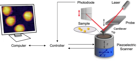
AFM operates with a sharp tip mounted at the end of a cantilever spring and brought in close proximity with the surface area to be scanned (Fig. 1.). During scanning the interaction between the sample and the tip causes the cantilever to bend. This bending is detected by a laser beam which is focused on the back part of the cantilever and reflected to a quadrant photodiode.
The x-y piezo provides the movement along the horizontal plane for scanning a selected area.
The virus particles were surface-attached by the two-step functionalization of the mica in order to cross-link the capsid protein to the surface. The first step was to place positively charged amino groups onto the mica by covalent attachment of poly-L-lysine, which was followed by the binding of zero-length cross-linker glutaraldehyde onto it. This treatment improved the stability and reliability of the AFM imaging in liquids.
Fig. 1. Schematic diagram of a typical AFM measurement setup.
Indentation experiments on virus particles
Prior to starting the indentation experiment the virus particles were scanned to verify their intactness, locate single particles and position the tip above the center of the selected particle.
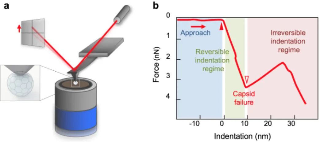
First, the viruses were manipulated by pressing the cantilever tip against the apex of the particles until reaching a predefined maximal force, then pulling it back with a constant pre-adjusted rate. As the output of the indentation experiment force-deformation curves (FDC) were recorded. The analysis of the FDC curves provided information about the mechanical properties of the capsid. Fig. 2. shows the experimental setup of an indentation, and Fig. 2.b the schematic FDC curve of the result with three main regions. As the tip approaches the surface by extending the z-piezo, the force is zero because the tip is not in contact with the sample, hence the cantilever does not bend. From the point when the tip reaches the capsid (Fig. 2.b solid triangle), force begins to rise. Initially the force curve displays a linear regime with a positive slope, which corresponds to the deformation of the virus as a Hookean particle. The slope of this linear section yields the stiffness of the particle. When the capsid is unable to withstand further loading, the force abruptly drops, which corresponds to the capsid fracture (Fig. 2.b open triangle). The regime immediately following capsid breakage corresponds to the unloaded swinging of the cantilever towards its equilibrium position. Accordingly, the slope of this region is equal to cantilever stiffness. Furthermore, the indentation depth, the distance between the contact point and the capsid failure (Fig. 2.b width of the green area), and the corresponding breaking force give further information about the capsid mechanical characteristics.
Fig. 2. Schematic diagram of AFM nanoindentation of a virus. (a) The AFM tip indents the virus particle while the cantilever bends; the exerted force on the tip is measured by the changes of laser signal detected on the quadrant photodiode. (b) The indentation force is plotted as the function of indentation depth.
RESULTS
Temperature-Dependent Topography and Nanomechanics of Bacteriophage T7
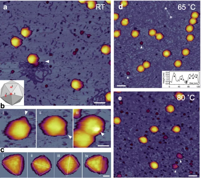
To explore the heat-induced topographical changes in T7 bacteriophages, we exposed them to a two-stage thermal treatment (65 ˚C, 80 ˚C) and studied the temperature-induced effects on the capsid using AFM. At room temperature, the surface-attached virus particles exhibited an intact, sphere-like structure in the AFM images (Fig. 3.a-c). Using a sharp tip allowed us to resolve the cogwheel shape and the central pore of the capsomers and, thereby, to identify the adsorbing symmetry of the capsids (Fig. 3.c). Around some virus particles, DNA clusters appeared on the surface due to occasional mechanically-induced DNA ejection. The conical tail complexes also became visible at different locations on most phage particles, depending on their binding orientation (Fig. 3.b). A few globular particles became apparent in the background, which may correspond to the core T7 phage proteins that got ejected simultaneously with DNA.
Fig. 3. AFM imaging of surface-immobilized untreated T7 bacteriophages. (a) Overview of a 1 μm x 1 μm non-treated sample area. White arrowhead points at the nearly instantaneous event of mechanically induced DNA ejection. Scale bar 100 nm. (b) AFM images of T7 phage particles at RT displaying their conical tail in different orientations. White arrowheads point at the tail apices. Scale bar is 30 nm. (c) High-resolution AFM images of the T7 phage surfaces with resolvable capsomers. Views along the two-fold (ii, iii) and three-fold symmetry axes (i, iv) are shown. Scale bar is 10 nm. (d) Overview of a 1 μm x 1 μm sample area treated at 65 ˚C.
White arrowheads point at large (>10 nm) globular particles. Scale bar 100 nm. Inset, topographical height map along an arbitrarily chosen line in the background (white dashed line). (e) Overview of a 1 μm x 1 μm sample area treated at 80 ˚C.
Following the topographical exploration of the capsid structure at room temperature, we heated the same sample to 65 ˚C for 15 min then cooled it back to 20 ˚C for image acquisition (Fig.
3.d). Upon 65 ˚C treatment, the topography of the sample has changed significantly (Fig. 3.d) The substrate surface became covered with a meshwork of DNA, the conical tail complex of most capsids disappeared and we found large (>10 nm) globular particles in the background which may correspond to the residues of the tail complexes that broke off. The capsid surface became more faceted as the icosahedral edges and faces got more pronounced, which could be explained by a slight shrinkage of the capsid due to the DNA release (Fig. 3.d). Upon 80 ˚C
treatment, the background was even more densely populated with DNA strands (Fig. 3.e). In addition, we observed a large number of globular particles, as well as large aggregates scattered in the background which may originate from the capsid wall. Furthermore, in high-resolution AFM images the capsomers appeared swollen with less distinct cogwheel structure.
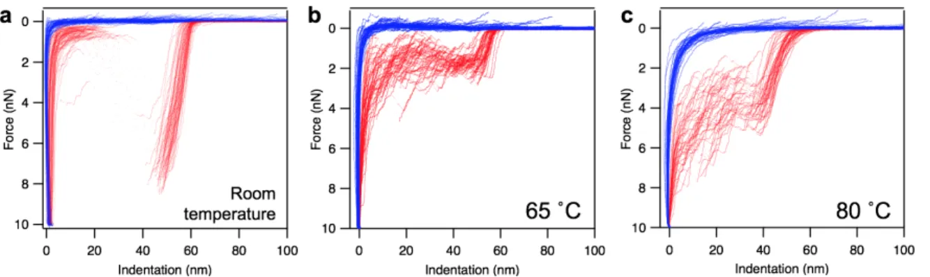
Following the topographical exploration of the heat-treated particles, we performed indentation experiments to reveal the thermally-induced changes in their nanomechanical properties (see details in Methods). The results obtained on phages at room temperature (RT), 65 ˚C and 80 ˚C are shown below (Fig. 4.).
Fig. 4. Nanomechanics of T7 phages. Overlay of force versus indentation curves collected in independent experiments on different phage particles at room temperature (a), at 65 ˚C (b) and at 80 ˚C (c).
The analysis of the force curves showed that the breaking force significantly reduced in case of the 65 ˚C-treated capsids (from 6.80 nN to 1.61 nN), indicating that the presence of packaged DNA within the phage contributes considerably to its mechanical stability. Interestingly, the breaking force increased for the 80 ˚C-treated particles. This might be explained by structural rearrangements between the capsomeric proteins that occurred between 65-80 ˚C, which stabilized their interactions and led to an increased overall mechanical stability of the phage particle. The calculated stiffness was twice as large in case of the intact T7 phages than for the empty RT capsids and the heat-treated ones. This suggest that the presence of packaged DNA contributes to the stiffness of the T7 phage. The maximal indentation value progressively increased with the heat-treatments, as a combined effect of the underlying changes in breaking forces and stiffness.
Force-Induced Structural Changes of the Bacteriophage T7
To study the mechanical stability of the phage particles, we performed fatigue experiments by repeatedly applying load with a 1.5 nN maximal force, lower than the average breaking force (7 nN). This loading force created an approximately 10 nm maximal indentation and was low enough to prevent capsid failure during the first load, but sufficiently high to induce it after
multiple cycles. For approximately one-fifth of the indented phage particles the force traces contained multiple discrete, step-like transitions (Fig. 5.).
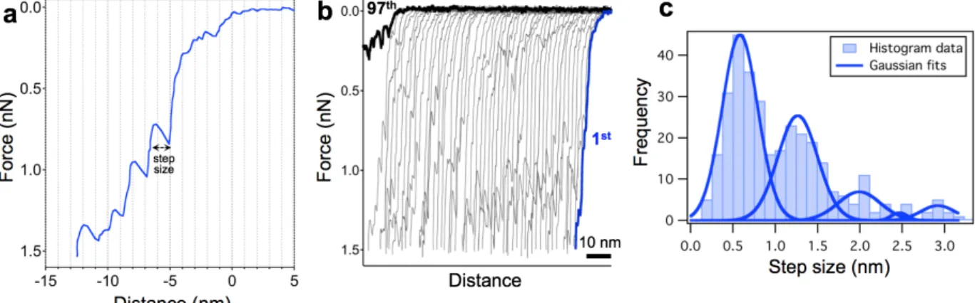
Fig. 5. Stepwise transitions and mechanical fatigue of T7 bacteriophage capsid. (a) Representative force versus distance curve obtained by indenting the capsid. Step size was measured as the distance change at the force of the sawtooth peak. (b) 97 consecutive FDCs collected on the same capsid at room temperature. The consecutive curves were shifted along the distance axis by 2 nm for better display. (c) Distribution of the transition step size with the fitted Gaussian curves. The peaks are centered at 0.58 (±0.21), 1.26 (±0.24), 1.99 (±0.25), 2.47 (±0.09) and 2.92 (±0.19) nm.
Transitions typically appeared as a sequence of sawtooth-like features, peaks followed by a sudden transient drop in force. The number of the subsequent curves often reached up to one hundred before it resulted in capsid failure and the curves systematically contained these saw- tooth like steps (Fig. 5.b). The distribution analysis of the transition step-sizes (see in Methods) showed peaks at integer multiples of approximately 0.6 nm (0.58 ± 0.21 nm, S.D). This suggests that a single transition corresponds to a ∼0.6 nm shift of the capsid structure, which might be closely related to a slight, discrete structural change closely related to the capsomers.
To explore the importance of the genomic DNA in the stepwise transitions, we carried out fatigue experiments on particles emptied by heat-treating at 65 ˚C. Here we also observed repeatable indentation force traces, however the number of the indentation prior to the capsid failure was significantly lower than at room temperature (Fig. 6.). This indicates that the DNA indeed contributes to the mechanical stability of the phage particles and increases the resilience against the mechanical fatigue of the capsids.
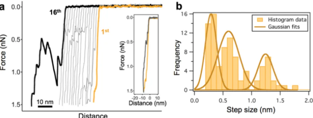
Fig. 6. Discrete nanoindentation steps in heat-treated (65 ˚C) T7 bacteriophage capsids.
(a) Force versus distance curves collected in 16 consecutive nanoindentation experiments on the same capsid. (b) Distribution of the transition step size. Gaussian were fitted on the cumulative step-size dataset. The peaks are centered at 0.29 (±0.09), 0.59 (±0.19) and 1.24 (±0.13) nm.
The distribution analysis of the transition step sizes measured on 65 ˚C treated particles showed that the peaks at 0.59 nm (± 0.37 nm) and 1.24 nm (± 0.26 nm) correspond well to the first and second peaks of the room temperature control, at integer multiples of the ~0.6 nm unit step size.
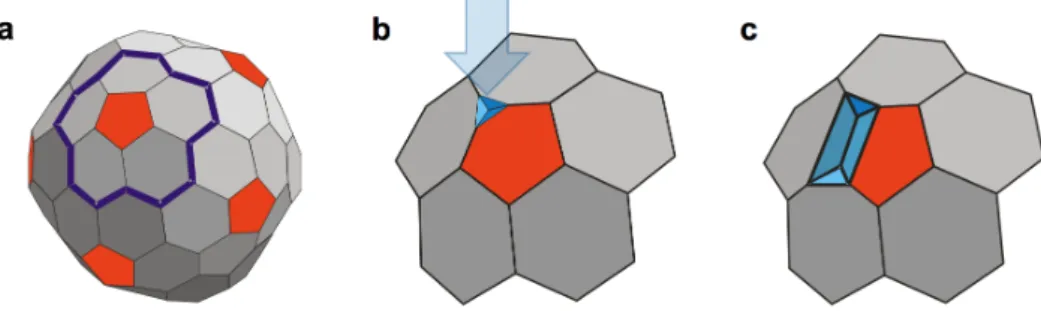
However, an additional peak appeared at 0.29 nm (±0.19 nm), which is one-half of the unit step size. We speculate that the structural failure is associated with the pentameric capsomer for three reasons: (a) the probability of observing stepwise transitions in the force traces was approximately 0.2, which compares well with the ratio of pentamers (12) to hexamers (60) covering the capsid surface; (b) the maximum number of steps leading to capsid collapse is five; and (c) because the T7 virion is faceted, the pentamers protrude from the capsid surface, making them mechanically accessible and prone to buckling. Therefore, it is likely that we observe step-like transitions when a pentamer of the T7 capsid points upward, in 5-fold symmetric orientation. Accordingly, if five structural failures are accumulated by a pentamer, it is no longer able to withstand the pressure exerted by the cantilever and allows the AFM tip to penetrate the capsid. Therefore, we speculate that the stepwise transitions are related to the buckling of the capsid pentameric unit. To correlate the unit buckling amplitude, the 0.6 nm individual step size, with the structural features of the T7 capsid we proposed a simple geometric model (Fig. 7.).
Fig. 7. Phenomenological model of the mechanically induced stepwise buckling of the T7 capsid. (a) Schematics of the T7 capsid with the involved group of capsomers (a pentamer with the surrounding hexamers) outlined (thick blue line). (b) The first buckling step induced by indentation with the AFM cantilever (blue arrow). (c) Progression of buckling along the capsid surface.
Mechanically-Driven DNA Ejection of the Bacteriophage T7
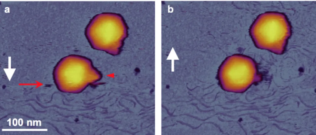
We investigated the role of mechanical force in triggering DNA injection by visualizing the topographical structure and mechanically-induced changes of T7 phages. We scanned individual surface attached capsids continuously as a function of time and recorded the DNA release from the capsids in response to the mechanical tapping of the cantilever tip (Fig. 8.).
Based on the analysis of the images we were able to explore several key features of the mechanically-driven DNA ejection process:
• The ejected and surface-adsorbed DNA was only a partial T7 genome in a kinetically trapped structural state. To validate this hypothesis, we compared the size of the area covered by ejected DNA in the AFM images with that computed from the total length of surface-equilibrated and surface-projected T7 DNA genome.
• The DNA ejection visibly occurred via the tail complex (Fig. 8.), indicating that the process took place forcibly through its natural path. To determine whether the DNA was ejected due to the cantilever tip breaking off the tail, we performed additional measurements, where the DNA ejection was triggered while tapping the body of the capsid without touching the tail.
• The DNA ejection occurred via the tail complex at a certain rate specific to T7 phage.
We estimated this rate by analyzing sets of consecutively scanned images.
• The mechanical load exerted further away from the tail complex caused perturbation that propagated through the compressible medium of the DNA-filled capsid eventually triggering the DNA ejection. We estimated the internal pressure increment that was generated due to cantilever oscillation and led to DNA ejection.
Fig. 8. In situ AFM of DNA ejection from the T7 bacteriophage. (a-b) Sequential height-contrast AFM images collected during (a) and following (b) DNA ejection. Collection of the two images was separated by 7 minutes. White arrows indicate AFM line-scanning direction. Red arrow points at the scan line in which DNA suddenly appeared, while the red arrowhead points at the tail complex, the surface of which became fuzzy after DNA ejection.
To explore the possible mechanisms behind the abrupt triggering of the DNA-ejection, we chemically fixed the surface attached particles with glutaraldehyde prior to scanning them (Fig. 9.).
Fig. 9. Chemical fixation of T7 phage particles. (a) AFM image of surface attached T7 phage particles fixated with glutaraldehyde. (b) AFM images of individual T7 particles with their tail complex pointing away from the substrate surface. Dotted lines in the bottom left image denote the fibers immobilized on the capsid surface. (c) Schematic model of T7 binding to the E. coli surface and passing through a switching step prior to genomic DNA release. Red double arrows indicate the putative conformational switch thought to be associated with a change in the fiber arrangement.
The tail fibers of the T7 phages became visible on the capsid surface and, surprisingly, the substrate had already been already covered with the ejected DNA by the time of scanning.
Conceivably, the immobilization of the tail fibers induced a conformational switching that
triggereds DNA ejection similarly as in the case of mechanical force. Although the details of the conformational switch are yet largely unknown, it might include a lever-like action of the tail fibers that results in a transition within the tail complex.
CONCLUSIONS
Thermally-Induced Structural Changes in Bacteriophage T7
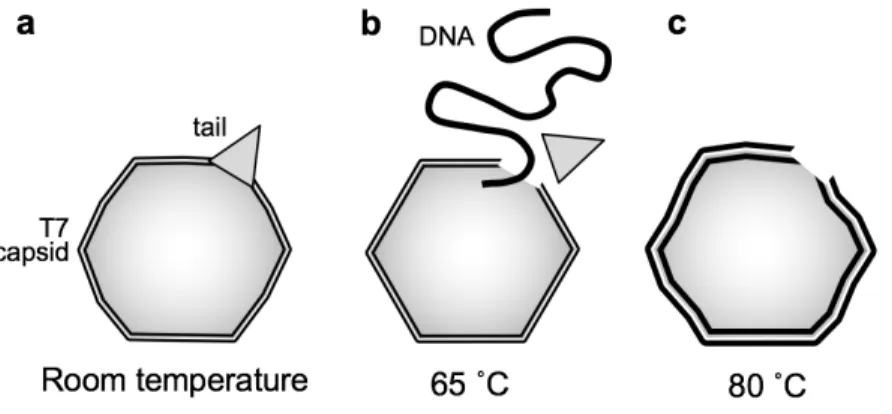
We have shown that exposing the T7 particles to 65 ˚C thermal treatment causes the release of its genomic DNA due to the abruption of the tail complex (Fig. 10.b). The loss of DNA results in a relaxation of the capsid structure accompanied by a reduced stiffness and breaking force.
Further heating to 80 ˚C leads to rearrangements within the capsid wall, caused most likely by partial denaturation of the component gp10A proteins (Fig. 10.c). Even though the loss of DNA destabilizes the capsids, they are remarkably capable of withstand high temperatures with a fairly intact global topographical structure. Thus, partial denaturation may have a stabilizing effect on the global structural constraints of the viral capsid.
Fig. 10. Schematic model of thermally-induced changes in the T7 bacteriophage. (a) At room temperature, the capsid is slightly swollen because of the DNA pressure inside. (b) Upon heating to and incubating at a temperature of 65 ˚C, the tail complex breaks off, resulting in the release of the genomic DNA. The capsid becomes more faceted due to the decrease of the capsid pressure. (c) Finally, at 80 ˚C the capsid becomes swollen and its surface irregular.
Stepwise Reversible Buckling of Bacteriophage T7
The fatigue experiments revealed that the elastic regime of the nanoindentation force traces contain discrete, stepwise transitions that cause buckling of the T7 capsid with magnitudes that are integer multiples of ~0.6 nm. These transitions are reversible and contribute to the rapid consolidation of the capsid structure against force during cantilever retraction. The stepwise transitions were present even following the removal of the genomic DNA by heat treatment, indicating that they are related to the structure and dynamics of the capsomeric proteins.
Dynamic force spectroscopy experiments showed that the thermally activated consolidation
step is ~104 times faster than spontaneous buckling, suggesting that the capsid stability is under strong dynamic control. Therefore, capsid structural dynamics may play an important role in protecting the genomic material from harsh environmental impacts.
DNA Ejection Triggered by a Sensitive Mechanical Switch
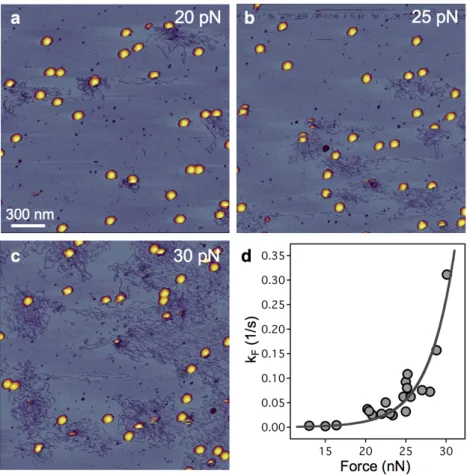
We directly visualized that tapping the capsid wall with an oscillating AFM cantilever triggers rapid DNA ejection via the tail complex. The triggering rate increased exponentially as a function of force (Fig. 11.d), following transition-state theory, across an activation barrier of 23 kcal/mol at 1.2 nm along the reaction coordinate.
Fig. 11. The effect of increasing mechanical load on the DNA ejection triggering rate. 2 x 2 μm AFM images of the same sample exposed to an average (a) 20 pN, (b) 25 pN and (c) 30 pN load. (d) Rate of DNA ejection as a function of mechanical load.
The conformation of the ejected DNA molecules revealed that it had been exposed to a propulsive force. The force arising from intra-capsid pressure assists in initiating the ejection process and transferring the DNA to outside the capsid wall. The chemical immobilization of the tail fibers also resulted in enhanced DNA ejection, suggesting that the triggering process involves a conformational switch that can be mechanically activated either by external forces or via the tail-fiber complex.
NOVEL SCIENTIFIC RESULTS
I. Exposing the T7 capsids to thermal treatment at 65 ˚C causes the release of their DNA due to the abruption of the tail complex, which leads to the relaxation of the capsid structure. Further heating to 80 ˚C results in increased mechanical stability due to partial denaturation of the capsomeric proteins kept within the global capsid arrangement.
II. The elastic regime of the nanoindentation force traces contains discrete, stepwise transitions which may be related to the buckling of the pentamers. These transitions are reversible and contribute to the rapid consolidation of the capsid structure against force during cantilever retraction. The capsid stability is under dynamic control, and in thermal equilibrium the reaction is strongly biased towards the intact structural state.
III. Tapping the capsid wall with an oscillating AFM cantilever triggers rapid DNA ejection via the tail complex. The triggering rate increases exponentially as a function of force, following transition-state theory. The triggering process might involve a conformational switch that can be activated by mechanical force.
LIST OF PUBLICATIONS
Publications related to the dissertation
Vörös Z, Csík G, Herényi L, Kellermayer MSZ. (2017) Stepwise reversible nanomechanical buckling in a viral capsid. Nanoscale 9: 1136–1143.
Kellermayer MSZ, Vörös Z, Csík G, Herényi L. (2018) Forced phage uncorking: viral DNA ejection triggered by a mechanically sensitive switch. Nanoscale 10: 1898–1904.
Vörös, Z., Csík, G., Herényi, L. & Kellermayer, M. S. Z. Temperature-dependent nanomechanics and topography of bacteriophage T7. Under review
Publications unrelated to the dissertation
Vörös Z, Yan Y, Kovari DT, Finzi L, Dunlap D. (2017) Proteins mediating DNA loops effectively block transcription. Protein Sci. 26: 1427–1438.
Reinemann DN, Sturgill EG, Das DK, Degen MS, Vörös Z, Hwang W, Ohi R, Lang MJ. (2017) Collective Force Regulation in Anti-parallel Microtubule Gliding by Dimeric Kif15 Kinesin Motors. Curr. Biol. 27: 2810–2820.