Acta Microbiologica et Immunologica Hungarica
DOI:10.1556/030.66.2019.039
© 2019 Akadémiai Kiad´o, Budapest
ORIGINAL ARTICLE
* Corresponding author:
Assoc. Prof. Alexandra Sashova Alexandrova, PhD; Department of Medical Microbiology, Medical Faculty, Medical University of Sofia, 1, G.Sofiiski Boul., 1431-Sofia, Bulgaria Phone: +359 2 91 72 719 E-mail:alex.alexandrova1@gmail.com
Phenotypic and genotypic characterization of serogroup 6 Streptococcus pneumoniae isolates collected during 10-valent
pneumococcal conjugate vaccine era in Bulgaria
ALEXANDRA SASHOVA ALEXANDROVA
1* ,
LENA PETROVA SETCHANOVA
1, DANIELA ROSENOVA PENCHEVA
2and IVAN GERGOV MITOV
11Department of Medical Microbiology, Medical Faculty, Medical University of Sofia, Sofia, Bulgaria
2Molecular Medicine Center, Department of Medical Chemistry and Biochemistry, Medical Faculty, Medical University of Sofia, Sofia, Bulgaria
Received: August 02, 2019•Accepted: October 01, 2019
ABSTRACT
Serogroup 6 remains common in the pneumococcal-conjugated vaccine era in Bulgaria; therefore, we investigated its clonal and serotype dynamics. The antibiotic susceptibilities were assessed by broth microdilution. Strains identified as serogroup 6 with latex agglutination method were subjected to serotype-specific PCRs. Erythromycin-resistant strains were analyzed by PCR for presence ofermBand mefEgenes. MLST was performed to define clonal composition of the sequence types (STs). Serogroup 6 was represented by 40 (13.3%) from 301 invasive and non-invasiveStreptococcus pneumoniaeisolates.
Molecular serotyping revealed new emerging serotype 6C (6.6%), not detected in pre-vaccine era. Among unvaccinated patients, mostly we observed serotypes 6А (57.1%) and 6В (28.6%). Serotype 6C was distinctive for vaccinated children (64%), followed by 6A (24%). Penicillin and ceftriaxone non- susceptible serogroup 6 strains were 65% and 5%, respectively; erythromycin- and clindamycin-resistant were 70.0% and 52.5%, respectively. Multidrug-resistant strains were 57.5%. Prevalent genetic determi- nant for macrolide resistance wasermBgene (75%). MLST revealed 17 STs into 5 clonal complexes and 7 singletons. Predominant genetic lineage was CC386, represented by MDR-6C non-invasive strains.
Serotype 6B, principally responsible for invasive diseases in the pre-vaccine era, retreated this position to serotype 6A.
KEYWORDS
Streptococcus pneumoniae, serogroup 6, clonal composition
INTRODUCTION
The widespread use of pneumococcal-conjugated vaccines has decreased the incidence of invasive diseases in children, reduced the carriage of vaccine-type strains, and also conferred indirect herd immunity to other age groups [1, 2].
In 2010, the 10-valent pneumococcal-conjugated vaccine (PCV10, Synflorix, GSK, Brendfort, UK) was introduced for universal vaccination into Bulgarian National Pediatric Immunization Program with over 90% coverage among targeted age groups in the period 2011–2017. Prior to PCV10 implementation, PCV7 was not used in our country.
Before the introduction of PCV10, one of the most isolated serotypes in Bulgaria was 6B, following serotypes 19F and 3. Serogroup 6 is represented only by serotype 6B in PCV7 and PCV10 and an additional serotype 6A in PCV13.
-99 67 (2020) 2, 91
Serogroup 6 has been recognized worldwide as an important cause of invasive and non-invasive pneumococcal diseases. Initially, it was composed of the serotypes 6A and 6B, but in recent years, additional types as 6C and less frequent 6D, 6E, 6F, 6G, and 6H have been reported [3–6]. This confirms that vaccination is often accompanied by a rise in the frequency of non-vaccine serotypes (NVTs) [7,8].
The ancestral origin of the new serotype 6C is still unknown. Serotypes 6A and 6B are with very high similarity in capsular loci, which only differ consistently in one nucleotide of the wciP gene. Similarly, both serotypes 6A and 6C harboredwciNβgene, but serotype 6C has a glucose residue in place of a galactose residue in the 6A cps repeating unit, which results in a glucosyl transferase in 6C and galactosyl transferase in 6A [9, 10].
In the post-vaccine era, 10 years after the introduction of PCV, serogroup 6 is still represented at high rates in our country.
The aim of this study was to examine the antimicrobial susceptibility profiles, clonal composition, and changes in capsular serotypes in serogroup 6 Streptococcus pneumo- niae isolates collected after the introduction of PCV in Bulgaria.
MATERIALS AND METHODS Patients and specimen collection
In the period January 2011–March 2019, we collect 301 invasive and non-invasive pneumococcal isolates from patients at different ages (0–84 years of age) on a voluntary basis from the Department of Medical Microbiology, Medical University–Sofia and microbiological laboratories through- out Bulgaria (Sofia, Plovdiv, and Pleven).
Among this collection of 301S. pneumoniaeisolates, 121 were invasive strains isolated from cerebrospinal fluids (CSFs), blood, and pleural fluids. A case of invasive pneu- mococcal disease (IPD) was defined as the recovery of an isolate ofS. pneumoniaefrom a normally sterile site. The rest were non-invasive isolates (n=180), obtained from the nasopharynx, middle-ear fluid (MEF), the conjunctiva, and sputum. Non-invasive pneumococcal disease (NIPD) was defined asS. pneumoniaestrains causing infection detected in the ear, eye, tracheal aspirate specimens, or nasopharynx;
no invasive (sterile sites) isolates were collected from the same patient. From children diagnosed with acute otitis media (AOM), one MEF sample or one nasopharyngeal sample per child was collected. The AOM episodes were confirmed by an otorhinolaryngologist or pediatrician. In the cases of persistent or recurrent AOM, MEFs were collected.
AOM episodes diagnosed in children without perforation were also included, in which nasopharyngeal samples were taken through the nose.
Among 301 invasive and non-invasive S. pneumoniae isolates collected during the post-vaccine period, serogroup 6 constituted 13.3% (40/301) out of all pneumococcal strains.
Serogroup 6, which was the object of this study, consisted of 8 invasive strains isolated from CSF (n=6) and blood (n=2) and 32 respiratory strains isolated from the nasopharynx (n=23), MEF (n=4), sputum (n=2), and eye (n=3).
All pneumococcal strains were confirmed with both methods–optochin susceptibility test and bile solubility.
Data on demographic characteristics, source of isolate, clinical diagnosis, and vaccination status were collected for all received isolates (TableI).
PCV status was determined on the basis of patients’age at the date when the pneumococcalstrain was isolated. Children born April 1, 2010 or thereafter and those who had received
≥3 doses of PCV10 were defined as PCV10-vaccinated. The coverage rate of PCV10 was very high (>90%) among age-eligible children according to the national epidemiologi- cal data. The remaining children and adults were defined as an unvaccinated population.
Antimicrobial susceptibility testing
The antibiotic susceptibilities (MICs) were determined by the broth microdilution method on microtiter plates (Sensititre, Trek Diagnostic Systems Ltd., UK). STR6F MIC plate was inoculated forS. pneumoniaestrains. Antibiotic susceptibilities were defined according to the breakpoints of the European Committee on Antimicrobial Susceptibility Testing (EUCAST) [11]. Pneumococcal non-meningitis isolates were classified as penicillin-non-susceptible (PNSP) with minimal inhibitory concentration (MIC) values for benzylpenicillin (MIC≥0.1 mg/L) and ampicillin/ceftriaxone/cefuroxime, iv (MIC≥1.0 mg/L), while penicillin-resistant pneumococci were isolates having a MIC of (>2 mg/L) according to EUCAST 2018 breakpoints. For meningitis isolates, all strains that have shown a penicillin MIC of≥0.12 mg/L have been interpreted as resistant.S. pneumoniaeATCC 49619 was used as a control strain for the susceptibility test. Multidrug resis- tance (MDR) was defined by non-susceptibility to at least three or more classes antimicrobial agents.
PCR detection of erythromycin resistance genes
Polymerase chain reaction (PCR) method was applied to all erythromycin-resistant strains to disclose the macrolide re- sistance determinantsermBandmefE. The PCR conditions and primers have complied with the protocol described by Sutcliffe et al. [12].
Serotyping
Serotyping wasfirst performed by capsular swelling reaction using commercial serogroup 6 and serotype specific factor antisera for determination of serotypes 6A and 6B provided by the Staten Serum Institute, Copenhagen, Denmark. All isolates identified to serogroup 6 were then subjected to PCR serotyping. To confirm the phenotypically determined ser- otypes and to identify the most recent serotypes, three PCR reactions were used.
Acta Microbiologica et Immunologica Hungarica -99
92 67 (2020) 2, 91
TableI.Phenotypicandgenotypicdataofserogroup6S.pneumoniaeisolatesrecoveredinBulgariaduringpost-PCV10era(2011–2019) SerotypeLabno.Patients’ageVaccinea SpecimenDiagnosisAntibioticRpattern
MIC(mg/L) MacrolideRgenotypeSTCCb PCTX 6A173HID7yearsNoCSFBMSusceptible≤0.03≤0.12–488CC490 15QI5yearsYesNph.AOME,C,andSxt0.06≤0.12mefE490CC490 1119HID2yearsand9monthsNoNph.URTIE,C,andSxt≤0.03≤0.12mefE13460CC490 257Pl30daysNoMEFAOMP,E,C,andSxt0.12≤0.12mefE490CC490 1126Pd59yearsNoCSFBMEandSxt0.060.01mefE490CC490 134QI2years6monthsYesNph.AOME,C,andSxt0.06≤0.12ermB13460CC490 565Pd2yearsYesCSFBMP,E,C,andSxt0.250.125mefE3614CC3614 9542Pd2yearsYesNph.URTIPandSxt0.250.03–3614CC3614 1-XII1yearYesNph.AOMP,E,Cli,C,andSxt2.01.0ermB135CC3614 394Pd1yearNoCSFBMSusceptible≤0.03≤0.12–2467CC3614 182QI1yearand10monthsYesMEFAOMP,E,Cli,C,andSxt0.25≤0.12ermB395CC395 867CM8yearsYesNphCarriageE,Cli,T,C,andSxt0.0160.023ermB13569CC395 3177Pl1yearYesEyeConjunctivitisSxt≤0.03≤0.12–600CC600 936CM4yearsYesNph.URTIP,E,Cli,T0.12≤0.12ermB386CC386 3381Pl59yearsNoCSFBMPandSxt0.12≤0.12–3510Singletonc 6B14QI6yearsand10monthsNoNph.AOME,Cli,C,T,andSxt0.06≤0.12ermB273CC395 18-XI1yearand3monthsYesNph.AOMSusceptible≤0.03≤0.12–395CC395 1933Pd84yearsNoCSFBMPandSxt0.250.03–3614CC3614 1235HID2yearsNoMEFAOMP,E,Cli,T,andSxt0.50.25ermB149Singleton 146AH59yearsNoSputumCAPP,E,Cli,andT0.12≤0.12ermB2922Singleton (Continued)
Acta Microbiologica et Immunologica Hungarica 67 (2020) 2, 91-99 93
TableI.Phenotypicandgenotypicdataofserogroup6S.pneumoniaeisolatesrecoveredinBulgariaduringpost-PCV10era(2011–2019)(Continued) SerotypeLabno.Patients’ageVaccinea SpecimenDiagnosisAntibioticRpattern
MIC(mg/L) MacrolideRgenotypeSTCCb PCTX 6C30QI4yearsYesNph.AOMP,E,Cli,andT0.12≤0.12ermB386CC386 68XI5yearsand5monthsYesNph.AOMP,E,Cli,andT0.12≤0.12ermB386CC386 104XII3yearsand6monthsYesNph.AOMP,E,Cli,andT0.120.06ermB386CC386 1209CM2yearsYesNph.URTIP,E,Cli,andT0.120.06ermB386CC386 476033Pl1yearYesEyeConjunctivitisP,E,Cli,andT0.250.06ermB386CC386 304CM2yearsand7monthsYesNph.CarriageP,E,Cli,andT0.250.06ermB386CC386 179Mн2yearsand2monthsYesNph.AOMP,E,Cli,T,andSxt0.120.06ermB386CC386 180Mн4yearsand2monthsYesNph.AOMP,E,Cli,andT0.120.06ermB386CC386 182Mн3yearsYesNph.AOMP,E,Cli,andSxt0.120.03ermB386CC386 4031CM2yearsYesNph.URTIP,E,Cli,andT0.12≤0.12ermB4310CC386 4110СМ4monthsNoNph.Bronchiolitisac.P,E,Cli,andT0.250.06ermB1876CC3614 4815Vn71yearsNoBloodPneumoniaeSusceptible≤0.03≤0.12–1135CC3614 546CM6monthsYesNph.CarriageP0.250.125–3614CC3614 3254CM13yearsNoNph.CarriageE,Cli,andT0.06≤0.12ermB2924CC395 498AH13yearsNoSputumBronchiectasisP,E,Cli,andSxt2.00.75ermB8CC395 33QI3yearsYesNph.AOMSusceptible≤0.03≤0.12–1205CC600 1224Pl1yearYesMEFAOMP,T,andSxt0.12≤0.12–8630Singleton 2523Pl1yearYesEyeConjunctivitisP,E,C,andT0.12≤0.12mefE367Singleton 3967CM31yearsNoBloodBacteriemiaSusceptible0.0080.006–1804Singleton 1559Pd2yearsYesNph.CAPP,E,Cli,T,andSxt2.01.0ermB5740Singleton Note:Pneumococcalisolateswereclassifiedaspenicillin–nonsusceptible(benzylpenicillinMIC≥0.1mg/Landampicillin/ceftriaxone/cefuroxime,iv–nonsusceptible(MIC≥1.0mg/L)accordingto EUCASTbreakpoints,2018.Abbreviations:CSF:cerebrospinalfluid;Nph:nasopharynx;MEF:middleyearfluid;BM:bacterialmeningitis;AOM:acuteotitismedia;URTI:upperrespiratorytractinfection;CAP: communityacquiredpneumonia,P:penicillin;E:erythromycin;Cli:clindamycin;Sxt:trimethoprim–sulfametoxasole;T:tetracycline;C:chloramphenicol;MIC:minimalinhibitoryconcentration;ST:sequence type;CC:clonalcomplex.a ApplicationofPCV10accordingtoage.AllchildrenbornApril1,2010orthereafterwereeligibleforvaccination.Theremainingchildrenandadultsweredefinedasanunvaccinated population.b DefinedusingeBurstalgorithm.Acut-offpointof5identicallocitothepredictorfounderwasusedtodetermineaCC,namedafterthepredominantSTinthegroup.c STwithoutdefinedCC,but belongingtoaknowneBurstgroup.
Acta Microbiologica et Immunologica Hungarica
94 67 (2020) 2, 91-99
We performed PCR for simultaneously detecting 6Аand 6C, because of their very high similarity in the cps loci. The presence of 6А/6C was proven by 149-bp amplification product of wciP gene. PCR amplification of wciNβ gene (359 bp) was used to resolve 6C. The isolates of serotype 6В were tested by PCR with primers, which amplify a part of wciPgene (155-bp product). The primers used in this study are previously published by Jin et al. [13] and Park et al.
[14]. PCR buffers and DNA polymerase were supplied by GenetBio, Korea and all DNA primers were obtained from Alpha DNA, Canada. Gene amplification was performed using a GenePro-thermal cycler (Bioer, China) as follows: 94 °C for 15 s, 35 cycles of 95 °C for 30 s, 60 °C for 60 s, 72 °C for 60 s, and 72 °C for 10 min. Electropho- resis on 1.5% agarose gels was used to distinguish PCR products.
Multilocus sequence typing (MLST)
MLST was performed as previously described [15]. Seven housekeeping genes were sequenced and compared to the pneumococcal MLST database (http://pubmlst.org/
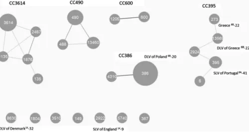
spneumoniae) to identify the alleles and respective sequence types (STs). PHYLOViZ (https://online.phyloviz.net/) was used to define clonal complexes (CCs). STs sharing five (double locus variants – DLVs) or six (single locus variants–SLVs) identical alleles were assigned to the same CC, named after the predominant ST in the group. STs not assigned to any CC were designed singletons (Figure1).
Statistical analysis
Theχ2 and/or Fisher’s exact tests were used for the analysis of categorical data, and statistical significance was set at p≤0.05.
RESULTS
Demographic and clinical data of patients
Serogroup 6 was represented by 40 (13.3%) from all 301 invasive and non-invasiveS. pneumoniaeisolates collected after PCV10 implementing in our routine immunization program. The invasive strains isolated from patients with meningitis and bacteremia were (n=8) 20%. The prevailed group of pneumococci (n=16) was from patients with AOM (40%). The rest of the patients (n=16) were with upper respiratory tract infections, bronchiolitis, bronchiec- tasis, community-acquired pneumonia and conjunctivitis (TableI). The higher proportion of the strains (n=31) was isolated from children of age 0–7 years (77.5%), among them 54.8% were from patients less than 2 years of age. Five strains were from≥59-year-old patients and the rest four patients were from middle-age group (8–31 years of age).
Out of the 31 children, 25 had received PCV10 vaccination (80.6%).
Antimicrobial non-susceptibility
Using the meningitis criteria and the MIC values for ben- zylpenicillin (MIC≥0.1 mg/L), 65% of the serogroup 6 isolates were classified as PNSP and 5% were ceftriaxone non-susceptible (Table I). Among the meningitis isolates (n=6), we observed three penicillin-resistant pneumococci having a MIC of≥0.12 mg/L, the others were susceptible to all tested antimicrobial drugs.
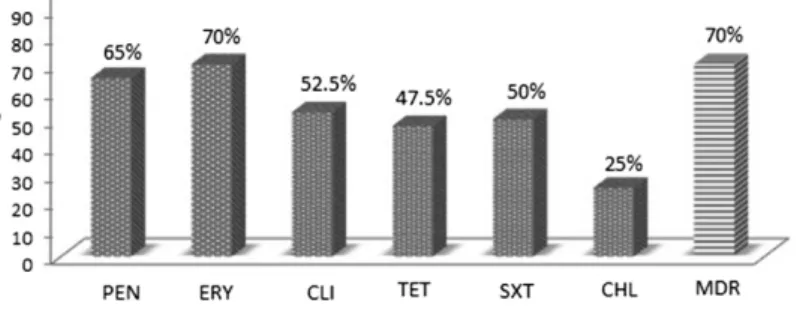
We found also high rates for erythromycin and clindamy- cin resistance of 70.0% and 52.5%, respectively. Tetracycline- resistant strains were 47.5%. Chloramphenicol resistance was observed in 25% of the strains. The resistance to trimethoprim–sulfamethoxazole reached 50% among the serogroup 6 isolates. Six isolates were susceptible to all tested classes of antimicrobial drugs. The MDR serogroup 6 isolates were 70%. The non-susceptibility rates to antimi- crobial agents of all 40 serogroup 6S. pneumoniaeisolates are shown in Figure2.
Serotyping
All of the isolates belonging to serogroup 6 were identified as serotype 6A (n=35) and 6B (n=5) by Quellung test using serotype-specific factor antisera provided by Statens Serum Institute (Copenhagen, Denmark). The results from serotype-specific PCR’s disclosed in fact three serotypes in serogroup 6: 6A (n=15), 6B (n=5), and 6C (n=20). They were 37.5%, 12.5%, and 50%, respectively.
The distribution among serotypes in children≤7 years of age was 54.8% for serotypes 6C, 35.5% for serotype 6A, and only 3 strains from serotype 6B, including two patients without an applied vaccine. In adults who are≥59 years of age, we observed heterogeneous data: equal proportions for 6A and 6B and one isolate from serotype 6C.
We determined three 6C and one 6A isolate from the rest four patients, which were non-vaccinated (13–31 years of age), born after April 1, 2010.
The results showed prevalence for serotype 6A among all invasive isolates (n=4/8), followed by 6C (n=2/8) and only one isolate from a patient with meningitis at 84 years of age from serotype 6B. All patients with meningitis were non- vaccinated and the isolated pneumococcal strains were from serotype 6A. The isolated pneumococcus from blood speci- mens was from serotype 6C from unvaccinated patients.
In the group of AOM isolates (n=16), the predominant serotype was 6C (50%), followed by serotype 6A (31.3%) and serotype 6B (18.7%). The widespread serotype isolated from patients with respiratory tract infections and conjunctivitis was serotype 6C (62.5%), which was not encountered in the pre-vaccine era in our country (1995–2010). The rest respi- ratory isolates were from serotype 6A (31.2%) and only one was from serotype 6B recovered in an old unvaccinated patient.
In the group of unvaccinated children (n=7), four strains were from serotype 6A, followed by two 6B strains and only one 6C strain. In comparison to group of vaccinated children (n=25), we discovered аn emerging serotype 6C (n=16) Acta Microbiologica et Immunologica Hungarica 67 (2020) 2, 91–99 95
with 64.0% distribution, less frequent 6A (n=6) 24%, and only one 6B isolate.
Among PNSP (n=26), we noted 61.5% serotype 6C isolates, 27% serotype 6A isolates, and 11.5% from serotype 6B. The erythromycin-resistant strains were principally from serotype 6C (53.6%) and less often from serotype 6A (35.7%) and serotype 6B (10.7%). The highest percent of MDR strains was distinctive for 6C isolates (56%), followed by 6A (28%) and 6B (16%).
PCR – Analysis of macrolide resistance genes from serogroup 6 S. pneumoniae isolates
The predominant genetic determinant for macrolide resis- tance out of all erythromycin-resistant serogroup 6 isolates wasermBgene, observed in 20 (75%) strains.MefEgene was determined in the other 25% of the strains.
The strains from serotype 6C strains possessed mostlyermB genes in their genome (75%) and two 6C strain carriedmefE gene. Efflux mechanism was prevailing for serotype 6A strains (41.7%) andermBgene was represented by 16.7% 6A pneumo- cocci. All macrolide-resistant 6B strains possessedermBgenes.
Multilocus sequence typing (MLST)
Among the isolates of serogroup 6, MLST revealed 17 STs distributed into 5 CCs named after with the predominant ST and 7 singletons (Figure1).
CC490 (n=6) was represent by 3 STs: ST490, ST488, and ST13460. All strains from CC490 were from serotype 6A, isolated from patients with meningitis and AOM. Except one strain susceptible to all antimicrobial agents, all other isolates from CC490 were erythromycin-resistant, principally har- boredmefEgene (80%).
CC3614 was the most diverse, distributed in 5 STs and 8 strains from serotype 6C (n=4), serotype 6A (n=3), and serotype 6B (n=1), isolated from patients with invasive and non-invasive diseases. The PNSP isolates in CC3614 were 75%, including 37.5% MDR strains.
CC386 was the most common among the studied pop- ulation of serogroup 6 (n=11). All of the strains, except one from ST4310, were from ST386 (n=10). These two STs were SLVs and were associated with international clone Poland6B-20/ST315 from Pneumococcal Molecular Epidemiology Network (PMEN). All pneumococci from CC386 were from serotype 6C isolated from patients less than 7 years of age with non-invasive infections, except one strain from serotype 6A. The isolates from CC386 were MDR strains. The responsible genetic determinant for macrolide resistance in CC386 was represented only by ermB gene.
CC395 was shared between PCV10 NVTs 6A and 6C and two isolates from vaccine serotype (VT) 6B. It consisted of five STs: ST395, ST273, ST13569, ST2924, and ST8, which were DLVs. ST395 was DLV of PMEN clone Portugal6A-41.
ST273 was identical to PMEN clone Greece6B-22. All repre- sentatives to this CC395 were also MDR, except one. We detectedermBgene out of all erythromycin-resistant strains from this genetic lineage.
The next clonal lineage was CC600, with ST600 and ST1205 represented by two S. pneumoniaestrains isolated from children with conjunctivitis and AOM. The strains were susceptible and resistant only to trimethoprim–sulfamethox- azole, respectively.
We disclosed seven singletons among serogroup 6:
ST8630, DLVs of PMEN clone Denmark14-32, ST1804, ST3510, ST149, ST5740, ST367, and ST2922, which are SLV of England14-9. Out of all singletons, we observe 85.7%
PNSP, including 71.4% MDR strains.
Figure 1.Clonal composition of serogroup 6 serotypes amongS. pneumoniaeisolates collected during PCV10-vaccine era in Bulgaria (2011–2019).Note:Each circle represents an ST. Circle sizes are proportional to the number of isolates within the ST. Solid, gray lines connect STs that are SLVs and thin, light lines connect STs that are DLVs according to the PHYLOVIZ tree cut-off: 2, NLV 2 rule reached (https://online.phyloviz.net/). STs that are linked belong to the same CC, named after with the predominant ST in the CC. Text in dark gray indicates PMEN-related clones. STs not assigned to any CC were designed singletons
Acta Microbiologica et Immunologica Hungarica
96 67 (2020) 2, 91-99
DISCUSSION
This study shows that in the post-PCV10-vaccine period, serogroup 6 remains common in our country. In the pre- vaccine era, serotype 6B was the most frequent, but it subsequently decreased in both adults and children in the post-vaccine era.
Compared to our previous study, serotype 6B was respon- sible for 10.5% of the invasive diseases (n=222) for the period 2006–2010 in contrast to only one case of IPD among the studied population (n=301) [16]. A significant decrease in the proportion of serotype 6B (p=0.0048) was found in this study.
In addition, we observed a significant reduction of serotype 6B among MEF isolates from 15.6% to 4.6%
(p=0.0202), when we compared results of this study with the pre-vaccine era for the same kind of specimen [17]. We concluded that PCV10 was highly successful in reducing IPD and NIPD caused by VTs in our country.
As expected, a significant increase of NVTs was found in the post-vaccine era. Compared with ourfindings from the non-vaccine period [18], we noted escalation in the propor- tions of NVT from serogroup 6 (6A and 6C) from 4.8% to 11.6% (p<0.003). Ourfindings suggest that after implemen- tation of PCV10 into our National Immunization Program, the majority of the circulating pneumococci among vaccine- eligible children was NVTs.
Like other authors, we found that serotype-specific PCR of some of our isolates previously serotyped as 6A by Quellung test was in fact 6C, due to cross-reaction of both serotypes 6C and 6A [19]. Serotype 6C was not detected before PCV10 implementation. At present, we found a rate of 6.3% for serotype 6C in the PCV10 period. Serotype 6C can be cross-reacted serologically with serotype 6A and is an example of a serotype 6A immunological variant that escaped notice for decades, due to its serological similarity to serotype 6A [20, 21]. It has been frequently found in young children primarily with an association of upper respiratory tract infections. We do not observe serotype 6C accountable for invasive infections in this population age group in our country. Only two clinical cases of adults
with IPD caused by 6C pneumococci were recorded. Low prevalence of serotype 6C IPD in children has also been observed in other studies from France, Portugal, and Brazil [22–24]. There are also investigations in which 6C is becoming more common as a causative agent of IPD, which confirms that vaccination is accompanied with serotype dynamics and rise in the frequency of NVTs [25–27].
Serotype 6A was accounted for IPD in both age groups after the introduction of PCV. We observed an increase in the rate of 6A-IPD of 5.3% in the post-vaccine period in contrast to 2.1% in the pre-vaccine period (2006–2010).
Although it is not statistically supported (0.1016) at p<0.05, 6A was the most frequent serotype from serogroup 6, responsible for invasive diseases.
A change in serotype frequencies is often coupled with an increase in antibiotic resistance among NVTs [28]. Other investigations reported that serotypes 6A and 6B exhibited significantly higher levels of erythromycin resistance and penicillin resistance [29].
More than half of the studied serogroup 6 isolates were MDR. Intermediately penicillin- and ceftriaxone- resistant isolates were much more common, while fully resistant isolates were rarely encountered. Serotype 6C comprised most MDR isolates. The genetic determinants for macrolide resistance kept the same layout as the non- vaccine period: widespread ermB gene, followed by mefE gene.
To explore the relationship between antibiotic resis- tance and CCs, we analyzed which of these CCs are associated with antimicrobial resistance. Genetic lineage CC386 was represented only by MDR strains. We also observed a high proportion of resistant strains in CC395.
CC3614 represented mostly PNSP strains. The majority of PNSP isolates expressed low-level resistance (MIC= 0.12±1 mg/L), with the exception of a singleton ST5740 and two others ST135 and ST8, which expressed high-level penicillinresistance (MIC≥2.0 mg/L). CC490 was associ- ated with low levels of macrolide resistance. CC600 included susceptible isolates: fully susceptible and one resistant only to trimethoprim–sulfamethoxazole.
Figure 2.Antimicrobial non-susceptibility among 40 serogroup 6S.pneumoniaeisolates.Note:The following non-susceptible MIC (mg/L) breakpoints were used: benzylpenicillin (penicillin)≥0.12 according to the EUCAST. For other antimicrobials, both categories intermediate and resistant isolates were summarized [11]. PEN: benzylpenicillin; ERY: erythromycin; CLI: clindamycin; TET:
tetracycline; CHL: chloramphenicol; SXT: trimethoprim–sulfamethoxazole; MDR: multidrug-resistant isolates
Acta Microbiologica et Immunologica Hungarica 67 (2020) 2, 91-99 97
The clonal composition of serogroup 6 was heterogeneous
Serotype 6A was the most diverse, distributed in all 5 CCs, found in this study and 11 STs. Among them, three STs such as ST395, ST386, and ST13569 were closely related SLVs or DLVs to international clones recognized by PMEN. Serotype- switching events were detected in CC395 for ST13569, ST292, and ST8, which were associated with serotypes 6A and 6C and were related to PMEN Greece6B-22 clone. Another supporting evidence of recombination between the cps regions of sero- types 6A and 6B leading to changes from one of the serotypes to the other has been published in several studies [20,30].
Serotype 6B was represented by two MDR-singletons; one of them is SLV of England14-9, a MDR isolate identical to PMEN clone Greece6B-22/ST273; an PNSP from ST3614, the primary founder of CC3614 and one susceptible isolate from ST395, SLV of Portugal6A-14.
Among serotype 6C, we observed three CCs, such as CC386, DLVs of PMEN Poland 6B-20/ST315, CC3614, and CC600, and almost all singletons found in the studied popu- lation: ST8630, ST367, ST5740, ST1804, and ST8. All strains belonging to the most widespread ST386 from this study were resistant both to erythromycin and tetracycline, in support of the evidence ST386 is related to the 6B genetic background of the international Poland6B-ST315 clone, which harbors the Tn6002 transposon [31]. Strains isolated in France and Brazil had been previously related to CC386 [22,23].
In conclusion, we observed a gradual increasing of NVTs among serogroup 6 after implementation of PCV10 in our country. The prevalent serotype 6C was associated with MDR non-invasive strains. Due to the vaccine pressure, it has been proposed that the associated antibiotic resistance character- istics of 6C would facilitate its emergence. Serotype 6B, which was responsible for all the more IPD cases before introduction of PCV in Bulgaria, retreated and 6A occupy this position in the post-PCV-era. ST386 was the most widely presented in the studied population of serogroup 6, which is represented in other countries in Europe and South America also.
Further studies are necessary to clarify the serotype dynamics and fast changing epidemiological characteristics.
Acknowledgements:This work was supported by the Bulgari- an Ministry of Education and Science under National Program for Research “Young Scientists and Postdoctoral Students.”The authors would like to thank all participating microbiological laboratories for their cooperation and for providing the isolates.
Conflict of Interest: The authors declare no conflict of interest.
REFERENCES
1. Pilishvili, T., Lexau, C., Farley, M. M., Hadler, J., Harrison, L. H., Bennett, N. M., Reingold, A., Thomas, A., Schaffner, W.,
Craig, A. S., Smith, P. J., Beall, B. W., Whitney, C. G., Moore, M. R.: Sustained reductions in invasive pneumococcal disease in the era of conjugate vaccine. J Infect Dis201, 32–41 (2010).
2. Miller, E., Andrews, N. J., Waight, P. A., Slack, M. P., George, R. C.: Herd immunity and serotype replacement 4 years after seven-valent pneumococcal conjugate vaccination in England and Wales: An observational cohort study. Lancet Infect Dis 11, 760–768 (2011).
3. Parra, E. L., Duarte, C., Rodríguez, K., Sanabria, O., Moreno, J.:
Frequency and molecular characterization of invasive isolates of Streptococcus pneumoniae serotypes 6C and 6D in Colombia. Enferm Infecc Microbiol Clin35, 283–286 (2017).
4. Baek, J. Y., Park, I. H., Song, J. H., Ko, K. S.: Prevalence of isolates of Streptococcus pneumoniaeputative serotype 6E in South Korea. J Clin Microbiol52, 2096–2099 (2014).
5. Jacobs, M. R., Bajaksouzian, S., Bonomo, R. A., Good, C. E., Windau, A. R., Hujer, A. M., Massire, C., Melton, R., Blyn, L. B., Ecker, D. J., Sampath, R.: Occurrence, distribution, and origins ofStreptococcus pneumoniae serotype 6C, a recently recognized serotype. J Clin Microbiol47, 64–72 (2009).
6. Porat, N., Benisty, R., Givon-Lavi, N., Trefler, R., Dagan, R.:
The impact of pneumococcal conjugate vaccines on carriage of and disease caused byStreptococcus pneumoniaeserotypes 6C and 6D in southern Israel. Vaccine27, 2806–2812 (2016).
7. Varghese, R., Jayaraman, R., Veeraraghavan, B.: Current challenges in the accurate identification of Streptococcus pneumoniaeand its serogroups/serotypes in the vaccine era.
J Microbiol Methods141, 48–54 (2017).
8. Millar, E. V., Watt, J. P., Bronsdon, M. A., Dallas, J., Reid, R., Santosham, M., O’Brien, K. L.: Indirect effect of 7-valent pneumococcal conjugate vaccine on pneumococcal coloniza- tion among unvaccinated household members. Clin Infect Dis 47, 989–996 (2008).
9. Yun, K. W., Cho, E. Y., Choi, E. H., Lee, H. J.: Capsular polysaccharide gene diversity of pneumococcal serotypes 6A, 6B, 6C, and 6D. Int J Med Microbiol304, 1109–17 (2014).
10. Pai, R., Limor, J., Beall, B.: Use of pyrosequencing to differen- tiate Streptococcus pneumoniae serotypes 6A and 6B. J Clin Microbiol43, 4820–4822 (2005).
11. The European Committee on Antimicrobial Susceptibility Testing: Breakpoint Tables for Interpretation of MICs and Zone Diameters. Version 6.0 (2018). Available at http://
www.eucast.org.
12. Sutcliffe, J., Grebe, T., Tait-Kamradt, A., Wondrack, L.: Detec- tion of erythromycin-resistant determinants by PCR. Antimi- crob Agents Chemother40, 2562–2566 (1996).
13. Jin, P., Xiao, M., Kong, F., Oftadeh, S., Zhou, F., Liu, C., Gilbert, G. L.: Simple, accurate, serotype-specific PCR assay to differ- entiateStreptococcus pneumoniaeserotypes 6A, 6B, and 6C. J Clin Microbiol47, 2470–2474 (2009).
14. Park, I. H., Park, S., Hollingshead, S. K., Nahm, M. H.: Genetic basis for the new pneumococcal serotype, 6C. Infect Immunol 75, 4482–4489 (2007).
15. Setchanova, L. P., Alexandrova, A., Dacheva, D., Mitov, I., Kaneva, R., Mitev, V.: Dominance of multidrug-resistant Denmark(14)-32 (ST230) clone amongStreptococcus pneumoniae serotype 19A isolates causing pneumococcal disease in Bulgaria from 1992 to 2013. Microb Drug Resist21, 35–42 (2015).
Acta Microbiologica et Immunologica Hungarica
98 67 (2020) 2, 91-99
16. Setchanova, L., Alexandrova, A., Mitov, I., Nashev, D., Kantardjiev, T.: The group for microbiological surveillance of pneumococci: Serotype distribution and antimicrobial resis- tance of invasive streptococcus pneumoniae isolates in Bulgaria before the introduction of pneumococcal conjugate vaccine. J Chemother24, 12–17 (2012).
17. Setchanova, L. P., Kostyanev, T., Alexandrova, A., Mitov, I., Nashev, D., Kantardjiev, T.: Microbiological characterization of Streptococcus pneumoniae and non-typeable Haemophilus influenzaeisolates as primary causes of acute otitis media in Bulgarian children before the introduction of conjugate vac- cines. Ann Clin Microbiol Antimicrob12, 6 (2013).
18. Setchanova, L., Murdjeva, M., Stancheva, I., Alexandrova, A., Sredkova, M., Stoeva, T., Yoneva, M., Kurchatova, A., Mitov, I.:
Serotype changes and antimicrobial nonsusceptibility rates of invasive and non-invasive Streptococcus pneumoniaeisolates after implementation of 10-valent pneumococcal nontypeable Haemophilus influenzae protein D conjugate vaccine (PHiD- CV) in Bulgaria. Braz J Infect Dis21, 433–440 (2017).
19. Rolo, D., Fenoll, A., Ardanuy, C., Calatayud, L., Cubero, M., de la Campa, A. G., Linares, J.: Trends of invasive serotype 6C˜ pneumococci in Spain: Emergence of a new lineage. J Anti- microb Chemother66, 1712–1718 (2011).
20. Song, J. H., Baek, J. Y., Ko, K. S.: Comparison of capsular genes of Streptococcus pneumoniae serotype 6A, 6B, 6C, and 6D isolates. J Clin Microbiol49, 1758–1764 (2011).
21. Elberse, K., Witteveen, S., van der Heide, H., van de Pol, I., Schot, C., van der Ende, A., Berbers, G., Schouls, L.: Sequence diversity within the capsular genes ofStreptococcus pneumo- niaeserogroup 6 and 19. PLoS One6, 25018 (2011).
22. Neves, F. P. G., Cardoso, N. T., Souza, A. R. V., Snyder, R. E., Marlow, M. M., Pinto, T. C. A., Teixeira, L. M., Riley, L. W.:
Population structure ofStreptococcus pneumoniaecolonizing children before and after universal use of pneumococcal con- jugate vaccines in Brazil: Emergence and expansion of the MDR serotype 6C-CC386 lineage. J Antimicrob Chemother73, 206–1212 (2018).
23. Janoir, C., Cohen, R., Levy, C., Bingen, E., Lepoutre, A., Gutmann, L., Varon, E., Observatoires Régionaux du Pneu- mocoque (ORP) network: Clonal expansion of the macrolide resistant ST386 within pneumococcal serotype 6C in France.
PLoS One9, e90935 (2014).
24. Diamantino-Miranda, J., Aguiar, S. I., Carriço, J. A., Melo- Cristino, J., Ramirez, M.: Clonal and serotype dynamics of serogroup 6 isolates causing invasive pneumococcal disease in Portugal: 1999–2012. PLoS One12, e0170354 (2017).
25. Marimon, J. M., Ercibengoa, M., Alonso, M., García-Medina, G., Pérez-Trallero, E.: Prevalence and molecular characteriza- tion ofStreptococcus pneumoniaeserotype 6C causing invasive disease in Gipuzkoa, northern Spain 1990–2009. Eur J Clin Microbiol Infect Dis29, 1035–1038 (2010).
26. Andam, C. P., Mitchell, P. K., Callendrello, A., Chang, Q., Corander, J., Chaguza, C., McGee, L., Beall, B. W., Hanage, W. P.: Genomic epidemiology of penicillin–Nonsusceptible pneumococci with nonvaccine serotypes causing invasive dis- ease in the United States. J Clin Microbiol 55, 1104–1115 (2017).
27. Carvalho Mda, G., Pimenta, F. C., Gertz, R. E., Jr, Joshi, H. H., Trujillo, A. A., Keys, L. E., Findley, J., Moura, I. S., Park, I. H., Hollingshead, S. K., Pilishvili, T., Whitney, C. G., Nahm, M. H., Beall, B. W., Active Bacterial Core Surveillance Team: PCR- based quantitation and clonal diversity of the current prevalent invasive serogroup 6 pneumococcal serotype 6C, in the United States in 1999 and 2006 to 2007. J ClinMicrobiol47, 554–559 (2009).
28. Obolski, U., Lourenço, J., Thompson, C., Thompson, R., Gori, A., Gupta, S.: Vaccination can drive an increase in frequencies of antibiotic resistance among nonvaccine serotypes ofStrep- tococcus pneumoniae. Proc Natl Acad Sci U S A 115, 3102– 3107 (2018).
29. Hackel, M., Lascols, C., Bouchillon, S., Hilton, B., Morgenstern, D., Purdy, J.: Serotype prevalence and antibiotic resistance in Streptococcus pneumoniaeclinical isolates among global popu- lations. Vaccine31, 4881–4887 (2013).
30. Bratcher, P. E., Park, I. H., Oliver, M. B., Hortal, M., Camilli, R., Hollingshead, S. K., Camou, T., Nahm, M. H.: Evolution of the capsular gene locus ofStreptococcus pneumoniaeserogroup 6.
Microbiology157, 189–198 (2011).
31. Calatayud, L., Ardanuy, C., Tubau, F., Rolo, D., Grau, I., Pallarés, R., Martín, R., Linares, J.: Serotype and genotype˜ replacement among macrolide-resistant invasive pneum- ococci in adults: Mechanisms of resistance and association with different transposons. J Clin Microbiol 48, 1310–1316 (2010).
Acta Microbiologica et Immunologica Hungarica 67 (2020) 2, 91-99 99