https://doi.org/10.1007/s00792-019-01098-4 ORIGINAL PAPER
Dual bloom of green algae and purple bacteria in an extremely shallow soda pan
Kristóf Korponai1 · Attila Szabó1 · Boglárka Somogyi2 · Emil Boros2 · Andrea K. Borsodi1 · Laura Jurecska1 · Lajos Vörös2 · Tamás Felföldi1
Received: 8 March 2019 / Accepted: 29 April 2019 / Published online: 13 May 2019
© The Author(s) 2019
Abstract
In April 2014, dual bloom of green algae and purple bacteria occurred in a shallow, alkaline soda pan (Kiskunság National Park, Hungary). The water was only 5 cm deep, in which an upper green layer was clearly separated from a near-sediment purple one. Based on microscopy and DNA-based identification, the upper was inhabited by a dense population of the planktonic green alga, Oocystis submarina Lagerheim, while the deeper layer was formed by purple, bacteriochlorophyll- containing bacteria, predominated by Thiorhodospira and Rhodobaca. Additional bacterial taxa with a presumed capability of anoxygenic phototrophic growth belonged to the genera Loktanella and Porphyrobacter. Comparing the bacterial com- munity of the purple layer with a former blooming event in a nearby soda pan, similar functional but different taxonomic composition was revealed. Members from many dominant bacterial groups were successfully cultivated including potentially new species, which could be the result of the application of newly designed media.
Keywords Soda pan · Purple bacteria · Oocystis · Thiorhodospira · Rhodobaca · Bloom Abbreviations
AAP Aerobic anoxygenic phototrophs BCC Bacterial community composition Bchl Bacteriochlorophyll(s)
CFU Colony forming units Chl a Chlorophyll a
HMW High molecular weight LMW Low molecular weight
NGS Next-generation DNA sequencing NTU Nephelometric turbidity unit OTU Operational taxonomic unit PNSB Purple non-sulfur bacteria
PSB Purple sulfur bacteria SRP Soluble reactive phosphorus TN Total nitrogen
TOC Total organic carbon
Introduction
Soda lakes have Na+- and CO32−/HCO3−-dominated alkaline water, and therefore, they are different from other athalas- sohaline waters (Boros et al. 2014; Boros and Kolpakova 2018). Soda lakes can be found in almost every continent (Grant 2006; Sorokin et al. 2014; Boros and Kolpakova 2018): in Central Asia (Kulunda Steppe), Inner Asia, East Africa (Eastern Rift Valley), Central Europe (Carpathian Basin) and sporadically in India (Lonar Lake) and North America (Mono Lake and Soap Lake). These alkaline and saline environments range from deep meromictic to shallow lakes, and can be grouped into hypersaline (> 50 g/L) and less saline water bodies.
Shallow lakes and pans are characteristic features of the semiarid steppe and undergo significant diurnal (mainly temperature and oxygen concentration) and annual (mainly temperature, volume and salinity) changes regarding various physical parameters. According to our current knowledge,
Communicated by A. Oren.
Electronic supplementary material The online version of this article (https ://doi.org/10.1007/s0079 2-019-01098 -4) contains supplementary material, which is available to authorized users.
* Tamás Felföldi
tamas.felfoldi@ttk.elte.hu
1 Department of Microbiology, ELTE Eötvös Loránd University, Pázmány Péter stny. 1/c., Budapest 1117, Hungary
2 Balaton Limnological Institute, MTA Centre for Ecological Research, Klebelsberg Kuno u. 3., Tihany 8237, Hungary
within Europe, soda lakes could be found exclusively in the Carpathian Basin; their size ranges from small wetlands of few m2 to few hundred ha (Boros et al. 2014, 2017). Most of them are intermittent aquatic systems and have low water transparency (Boros et al. 2017; Somogyi et al. 2017). Two main types of these shallow lakes (which are usually referred as pans) could be distinguished, the ‘turbid type’ (high amount of suspended solids and usually high concentration of humic substances) and the ‘colored type’ (relatively low amount of suspended solids and very high concentration of humic substances) (Boros et al. 2017). The ‘fluid sedi- ment’ concept has been proposed for the turbid water type, as wind induces continuous sediment resuspension due to their extreme shallowness (< 50 cm) (Eiler et al. 2003; Boros et al. 2017).
The key primary producers in soda lakes of the Car- pathian Basin are cyanobacteria, eukaryotic green algae and euglenophytes (Vörös et al. 2008; Somogyi et al. 2017).
Phytobenthos is considered to be negligible due to the strong underwater light limitation (Boros et al. 2013), while phy- toplankton (especially in the turbid-type waters) is usually dominated by pico-sized (< 3 μm) species (Vörös et al. 2008;
Felföldi et al. 2009; Somogyi et al. 2009, 2017) and has characteristic seasonal changes. Below 15 °C, picoeukary- otes (mainly Chloroparva and Choricystis) dominate, while above this temperature picocyanobacteria (mostly Synecho- coccus/Cyanobium) occur (Vörös et al. 2008; Felföldi et al.
2009, 2011; Somogyi et al. 2009, 2011, 2016). Interestingly, pico-sized green algae regularly bloom under the ice during winter (Somogyi et al. 2009; Pálffy et al. 2014); while in spring and summer, blooms of larger (mainly green) algae could be formed due to the high productivity of these waters, which occasionally co-occur with mass production of purple bacteria near the sediment surface (Borsodi et al. 2013).
The aim of the present study was the characterization of a dual bloom, which occurred in a soda pan with a special focus on the bacterial community inhabiting the purple layer.
Comparing the obtained results (physicochemical param- eters and taxonomic composition) with a similar, previous dual bloom event reported from the studied region (Borsodi et al. 2013), the underlying factors shaping the bacterial community composition (BCC) of these Oocystis-associated purple bacterial blooms were revealed.
Materials and methods
Study site and sample collectionAn anonymous, small, turbid soda pan (N46°45.818′, E19°10.828′), located in the Kiskunság National Park near Soltszentimre (Hungary, Central Europe), was sampled on 23 April 2014. In their great cadastre, Boros et al. (2013)
designated it as anonymous lake no. 60. Water depth report- edly fluctuates between 0 and ~ 60 cm; the average water surface is 0.58 ha (Boros et al. 2013). Pan water is domi- nated by Na+ and HCO3− >Cl− (Table 1, Supplementary Fig- ure 1), which is supplied mainly by groundwater and precipi- tation (Simon et al. 2011), and neither has vegetation cover nor surface inflow or outflow (Boros et al. 2013).
A cylinder was used for sample collection, and subsam- ples were taken from the upper, green-colored water and from the purple layer over the sediment using a pipette.
Determination of the BCC was carried out only from the latter.
Limnological and water chemistry analyses
Basic physicochemical parameters were determined on site using a MultiLine P 8211 multimeter (WTW). Chemi- cal analyses were performed under laboratory conditions according to the methods described in detail in Felföldi et al. (2016).
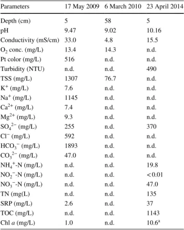
Table 1 Basic characteristics of the studied anonymous soda pan water and measured physicochemical parameters during the dual bloom Previous data (2009 and 2010) were taken from Boros et al.
(2013)
NTU nephelometric turbidity unit, TN total nitrogen, TOC total organic carbon, SRP soluble reactive phosphorous, n.d. not deter- mined
a Data represent only the upper, green layer
Parameters 17 May 2009 6 March 2010 23 April 2014
Depth (cm) 5 58 5
pH 9.47 9.02 10.16
Conductivity (mS/cm) 33.0 4.8 15.5
O2 conc. (mg/L) 13.4 14.3 n.d.
Pt color (mg/L) 516 n.d. n.d.
Turbidity (NTU) n.d. n.d. 490
TSS (mg/L) 1307 76.7 n.d.
K+ (mg/L) 7.6 n.d. n.d.
Na+ (mg/L) 1145 n.d. n.d.
Ca2+ (mg/L) 7.4 n.d. n.d.
Mg2+ (mg/L) 9.3 n.d. n.d.
SO42− (mg/L) 255 n.d. 370
Cl− (mg/L) 592 n.d. n.d.
HCO3− (mg/L) 1893 n.d. n.d.
CO32− (mg/L) 47.0 n.d. n.d.
NH4+-N (mg/L) n.d. n.d. 19.8
NO2−-N (mg/L) n.d. n.d. < 0.01
NO3−-N (mg/L) n.d. n.d. 47.0
TN (mg(L) n.d. n.d. 135
SRP (mg/L) 2.6 n.d. 37
TOC (mg/L) n.d. n.d. 1143
Chl a (mg/L) 1.0 n.d. 10.6a
For pigment analyses, the samples were filtered through a GF-5 glass fiber filter (Whatman). Chlorophyll a (Chl a) and bacteriochlorophyll a (Bchl a) were extracted from the filters as described in Felföldi et al. (2016), while another filter set per se was used for in vivo absorption measure- ments. Chlorophyll concentration was measured using the method of Wellburn (1994), and the concentration of Bchl a was determined according to Biel (1986). In vivo absorption spectra were recorded with a 160A UV–Vis spectrophotom- eter (Shimadzu) between 380 and 900 nm.
Native preparations from the two layers were photo- graphed using an Olympus BX51 microscope with a CCD camera (Olympus DP71).
Determination of BCC based on total DNA analysis Community DNA was extracted according to Szabó et al.
(2017). For the determination of BCC, next-generation DNA sequencing (NGS) was applied using the protocol and sequence analysis pipeline as described previously (Szabó et al. 2017). For this, the V3–V4 region of the 16S rRNA gene was amplified with S-D-Bact-0341-b-S-17 forward (5′-CCT ACG GGNGGC WGC AG-3′) and S-D-Bact-0785- a-A-21 reverse (5′-GAC TAC HVGGG TAT CTA ATC C-3′) primers (Klindworth et al. 2013). Based on the proposed species-level sequence similarity threshold (Tindall et al.
2010), operational taxonomic units (OTUs) were picked at 97% similarity. Raw sequence data are available under the NCBI BioSample ID SAMN10724752. Sequences of cul- tivated strains (see below), and those (strains and clones) from the purple bacterial bloom published in Borsodi et al.
(2013) were also analyzed using the same pipeline. Since plastid 16S rRNA gene references are underrepresented in the ARB-SILVA database, chloroplast sequences were ana- lyzed separately as described in detail by Kalwasińska et al.
(2017).
The ratio of Archaea and Bacteria was determined with qPCR targeting the 16S rRNA gene. All reactions were carried out in triplicates in a StepOnePlus Real-Time PCR System (Thermo Fisher Scientific) using TaqMan Gene Expression Master Mix (Thermo Fisher Scientific). A final volume of 16 μL was applied with 1 × Taqman Gene Expression Master Mix and 1 μL DNA sample. In case of the Bacteria-specific reaction, the following primers and probe were applied: 1.2 μM BACT1369F (5′-CGG TGA ATA CGT TCY CGG -3′), 1.0 μM PROK1492R (5′-GGW TAC CTT GTT ACG ACT T-3′), 0.5 μM TM1389F (5′-FAM-CTT GTA CAC ACC GCC CGT C-BHQ-3′) (Suzuki et al. 2000); while for Archaea, 0.8 μM Arch349F (5′-GYGCASCAGKCGM- GAAW-3′), 0.8 μM Arch806R (5′-GGA CTA CVSGGG TAT CTAAT-3′) and 0.5 μM Arch516F (5′-FAM-TGY CAG CCG CCG CGG TAA HACCVGC-BHQ-3′) primers and specific probe were used (Takai and Horikoshi 2000). Reactions
were carried out using the thermal profile: 50 °C for 2 min, 95 °C for 10 min, followed by 40 cycles at 95 °C for 15 s and 60 s at 56 °C for the Bacteria- and at 59 °C for the Archaea- specific reaction. Standard curves were used for the estima- tion of 16S rRNA gene numbers using the StepOne v2.3 software (Thermo Fisher Scientific) based on serial tenfold dilution (109–103 copy) of genomic DNA from Nitrincola lacisaponensis DSM 16316T and Thermoplasma acidopilum DSM 1728T. Gene copy numbers were determined based on the molar mass values of the standard amplicons.
Analyses based on bacterial strain cultivation
For cultivation of bacteria, three different media were used:
‘R’ [DSMZ medium 830 (R2A; see details: http://dsmz.
de), pH 10, adjusted with 1 M NaOH], ‘C’ [1 L autoclaved sample water, 16 g gellan gum (Gelzan CM, Sigma), 0.6 g MgSO4 × 7H2O and 0.3 g CaCl2 × 2H2O)] and ‘S’ (1 L dH2O, 16 g gellan gum, 1 g yeast extract, 0.6 g MgSO4 × 7H2O, 0.3 g CaCl2 × 2H2O and 10 g NaHCO3; pH 10, adjusted with 1 M NaOH). A tenfold dilution series using sterile distilled water was prepared from the sample, and aliquots were subsequently spread onto solid media. Incubations were carried out under both aerobic and anaerobic (Microbiol- ogy Anaerocult® A mini, Merck) conditions at room tem- perature under scattered light. Colony counts were recorded after 19 days, then randomly picked colonies were isolated and purified; the obtained strains were used for downstream analyses (the first letter of the stain codes is according to the applied media).
DNA was extracted from bacterial strains with the G-spin™ Total DNA Extraction Kit (iNtRON Biotechnol- ogy). The 16S rRNA gene was amplified by PCR using the primers 27F (Lane 1991) and 1492R (Polz and Cavanaugh 1998). PCR products were purified and sequenced by LGC Genomics (Berlin, Germany). Chromatograms were manu- ally corrected with Chromas (Technelysium). For taxonomic identification, EzBioCloud’s online service was used (Yoon et al. 2017). Sequences were screened for chimeras using Pintail 1.0 (Ashelford et al. 2005). GenBank accession num- bers of the obtained sequences are: KR233183–KR233245.
Measuring the Bchl concentration using the spectros- copy-based method of Biel (1986) was not possible due to the weak growth rate of strains which yielded low amount of biomass. Therefore, microscopy- and PCR-based tech- niques were applied to assess the phototrophic potential of the strains. Briefly, the cells were detected with an Olym- pus BX51 epifluorescence microscope upon excitation at 350–550 nm (excitation of Bchl a) using a 780-nm high-pass filter and a monochrome CCD (Olympus XM10) camera according to Jiao et al. (2006). All strains were checked for the presence of the pufM gene (coding the M subunit of the heterodimeric cores of the photosynthetic reaction center
complex) with PCR using the primers pufM_uniFfresh and pufM_uniRfresh (Martinez-Garcia et al. 2012). The 25 µL reaction volume contained 1 × Taq buffer (Fermentas), 2 mM MgCl2, 0.2 mM dNTP, 0.325 μM of each primer, 1 U Taq polymerase (Fermentas), 10 μg BSA (Fermentas) and 1 µL DNA. Cycling conditions were as given by Martinez-Gar- cia et al. (2012). Amplicons were sequenced and processed as described above. GenBank accession numbers of the obtained sequences are: KX361312–KX361323.
Results
Results of limnological analyses
At the time of sampling, pH 10.2 and 15.5 mS/cm conductiv- ity were measured (Table 1), which is equivalent to 12.4 g/L salinity according to the conversion coefficient determined for these pans by Boros et al. (2014). Other measured param- eters of lake water are shown in Table 1. High levels of total organic carbon (TOC), nitrogen forms, orthophosphate and sulfate were present. The upper layer of the water con- tained 10.6 mg/L Chl a and 1.9 mg/L Bchl a, while the lower contained 4.0 mg/L Chl a and 7.8 mg/L Bchl a. Oocystis submarina Lagerheim, a unicellular green alga, was identi- fied microscopically as the sole organism causing the algal bloom (Supplementary Figure S2). Based on microscopy, high density of vibrio-shaped bacteria was observed in the bottom layer. These bacteria showed phototaxis and sulfur globules were also observed (Supplementary Figure S3).
Results of cultivation‑based analyses
The three media used for the determination of hetero- trophic bacterial viable cell counts in the purple layer resulted in different plate counts. Under aerobic condi- tions 1.7 × 10⁶, 2.8 × 10⁷ and 3.5 × 10⁷ CFU (colony form- ing units)/mL were recorded after 19 days of incubation on medium ‘C’, ‘R’ and ‘S’, respectively. Anaerobic cul- tivation yielded lower or similar values: 4.2 × 10⁴ and 3.6 × 10⁷ CFU/mL were obtained after 19 days in the case of media ‘C’ and ‘S’, respectively, while no colony forma- tion was observed using medium ‘R’. Therefore, medium
‘S’ was the most suitable for the growth of facultative anaerobic bacteria present in the sample.
From the three different solid media, 63 pure cultures were obtained, 51 with aerobic, 12 with anaerobic cultiva- tion: 6 and 4 strains from medium ‘C’, 17 and 8 strains from medium ‘S’, 28 from ‘R’ agar plates, respectively. According to their 16S rDNA sequences, 37 strains belonged to phylum Proteobacteria (Alphaproteobacteria, 17 and Gammaproteo- bacteria, 20) and 26 strains to Bacteroidetes (Fig. 1). Iso- lates showed 89.0–100% sequence similarity values with type strains of validly published species (Table 2). No clear selectivity of the media was observed, while anaerobic cul- tivation resulted in similar bacteria as the aerobic one (e.g.
Halomonas, Porphyrobacter, Roseicitreum), only few taxa were isolated exclusively under anaerobic conditions (repre- sented by three strains showing the highest 16S rRNA gene similarities to Erythromicrobium, Ectothiorhodospira and Adhaeribacter type strains).
'14 bloom (reads) '08 bloom (clones) '14 bloom (strains) '08 bloom (strains) 0
10 20 30 40 50 60 70 80 90 100
relative abundance (%)
other genera (<5%) Pseudomonas Nitrincola Marinospirillum Halomonas Alkalimonas Thiorhodospira Ectothiorhodospira Porphyrobacter
unclassified Rhodobacteraceae Rhodobaca
Loktanella unc. Lentisphaerae
Heliorestis unc. Bacillaceae
Gracilibacillus Bacillus
unc. Cyclobacteriaceae Indibacter
Belliella
ML635J-40 aquatic group (Bacteroidetes) Fig. 1 BCC of purple layer at genus level from Böddi-szék in 2008
(re-analysis of data published in Borsodi et al. 2013) and from an anonymous soda pan near Soltszentimre in 2014 based on different
methods (cultivation, cloning and NGS). Only genera or equal ranks of uncultured bacteria with abundance above 5% are shown. Term
‘unc.’ stands for ‘unclassified’
Vast majority of alphaproteobacterial strains were red colored (e.g. members of genera Erythromicrobium, Por- phyrobacter, Loktanella and Rhodobaculum), four of them produced Bchl (Supplementary Figure S4). One of these strains, Roseinatronobacter sp. S4-16 stopped expressing its pigment(s) during the cultivation process. An anaero- bic gammaproteobacterial strain showed 100% sequence similarity to an alkalophilic purple sulfur bacterium, Ecto- thiorhodospira shaposhnikovii. Other strains from this class were typical heterotrophs, with high similarities to halo- philic or alkalophilic representatives of genera Alkalimonas, Halomonas, Nitrincola, Pseudomonas and Vibrio. All Bac- teroidetes strains belonged to order Cytophagales, and were pink or bright red colored. Five strains were closely (99.8%) related to the type strain of Belliella pelovolcani, while another five strains probably represent a new species within this genus (~ 97% similarity to type strains). Fifteen strains were affiliated to Mongoliibacter, while strain S4B-3 may represent a new family, as the shared sequence similarity values to type strains were less than 89%.
Strains were screened for the presence of the pufM gene, and amplicons were obtained in the case of 12 strains (19%
of total strains). As in many cases a longer fragment was co-amplified with the deposited amplicon (< 150 nt length), we presume an upstream, secondary annealing position of the forward (pufM_uniFfresh) primer inside the pufL gene.
Taxon identification (against the GenBank database) based on pufM gene sequences was consistent to the 16S rDNA- based results. The pufM-positive strains belonged to the three purple bacterial groups [purple sulfur bacteria (PSB), purple nonsulfur bacteria (PNSB) and aerobic anoxygenic phototrophs (AAP)]. With the exception of the Porphyro- bacter strains (one out of five was positive), strains from the same genus were either positive or negative. All pufM- containing isolates and representatives of sequence groups from pufM-negative strains were investigated with epifluo- rescence microscopy to detect the presence of Bchl. Not all of the pufM-positive strains were found to synthesize Bchl pigments, but Bchl was not detected in any of the pufM-neg- ative strains. Interestingly, one strain (Roseinatronobacter
Table 2 Taxonomic affiliation of the bacterial strains isolated from the purple layer based on 16S rRNA gene sequence similarity
aLoktanella vestfoldensis was recently reclassified as Yoonia vestfoldensis by Wirth and Whitman (2018)
b This phylotype was recently described as N. schmidtii (Borsodi et al. 2017)
Phylum/Class Order Strain code Closest species 16S rRNA
gene similarity (%)
Presence of the pufM gene
Number of strains in sequence group
Proteobacteria
Alphaproteobacteria Sphingomonadales S4B-2 Erythromicrobium ramosum 98.53 + 1
S5B-2 Porphyrobacter
colymbi/donghaensis 99.17 – 1
R4-10P Porphyrobacter neustonensis 99.05 only in C4B-1 4
Rhodobacterales S4-11 Loktanella vestfoldensisa 99.36 + 4
R4-14 Rhodobaca barguzinensis 97.71 + 2
S4-16 Roseinatronobacter monicus 97.71 + 1
R3-13B Roseicitreum antarcticum 99.58 + 2
S5-6A Rhodobaculum claviforme 99.87 – 1
Rhizobiales S4-27B Chelatococcus composti 95.24 – 1
Gammaproteobacteria Chromatiales C3B-1 Ectothiorhodospira shaposhnikovii 100 + 1
Oceanospirillales R3-4 Halomonas shengliensis 98.97 – 1
Oceanospirillales S4-1 Halomonas ventosae 98.46 – 3
R4-8b Nitrincola alkalisediminis 98.56 – 2
Pseudomonadales R3-8 Pseudomonas salegens 98.87 – 1
R4-10 Pseudomonas salegens 98.75 – 3
Vibrionales R4-7 Vibrio metschnikovii 99.70 – 3
Alteromonadales R4-13 Alkalimonas amylolytica 99.90 – 6
Bacteroidetes
Cytophagia Cytophagales S4-3 Belliella pelovolcani 99.80 – 5
R3-1 Belliella aquatica 97.28 – 5
Cytophagales R3-9 Mongoliibacter ruber 98.29 – 15
Cytophagales S4B-3 Adhaeribacter aerophilus 88.82 – 1
sp. S4-16) stopped producing Bchl pigments during the cul- tivation process.
Results of cultivation‑independent analyses
Overall, 3398 high-quality sequences were obtained with next-generation DNA sequencing, which were clustered to 162 bacterial OTUs. Two OTUs with 27 sequences were identified as eukaryotic plastid and belonged to the chloro- phyte genus Oocystis (unfortunately no O. submarina 16S rRNA gene sequence is available in GenBank currently;
Supplementary Figure S5).
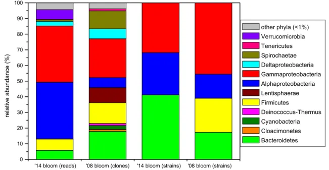
Altogether 18 bacterial phyla were detected (Fig. 2).
Proteobacteria was the most abundant phylum, dominated by Alpha- (36.2% of total bacterial reads) and Gammapro- teobacteria (35.9%), while only low relative abundance of Deltaproteobacteria was detected (2.9%), and the contribu- tion of Betaproteobacteria was negligible (< 0.1%). Other bacterial phyla represented only a minor fraction of the bacterial community: Firmicutes (7.1%), Verrucomicrobia (6.3%), Bacteroidetes (5.8%), etc. (Figure 2). Almost one- third of the sequences was affiliated with the PSB family Ectothiorhodospiraceae (Gammaproteobacteria) and the vast majority of these sequences belonged to a single OTU assigned to the genus Thiorhodospira, while other five OTUs containing two to four sequences clustered into a Thioal- kalivibrio, two Ectothiorhodospira and two unclassified groups (Fig. 1). Another third of the reads was affiliated with PNSB and AAP groups of the Rhodobacteraceae family
(Alphaproteobacteria), dominated by a single OTU belong- ing to genus Rhodobaca, while a Loktanella and a Porphy- robacter OTU turned out to be also significant (~ 3% both).
Other notable groups (with 1–4% relative abundance) were:
two unclassified OTUs from each Firmicutes and Alphapro- teobacteria; two verrucomicrobial OTUs (from Haloferula and a closely related unclassified genus) and one OTU from genera Desulfonatronum (Deltaproteobacteria), Indibacter (Bacteroidetes) and group ML602 J-51 (Actinobacteria). In total, 11 OTUs showed higher relative abundance than 1%, together they contributed 75.7% of the bacterial community.
From the 162 bacterial OTUs, 10 were shared between cultivated strains and the NGS library, which included 43 strains (68% of isolates) and 1221 sequences (36% of total reads). 20 strains forming seven OTUs were not detected by NGS.
Based on the result of qPCR, the 16S rRNA gene copy number of Archaea was below the detection limit (40 PCR cycles).
Discussion
Features of the dual bloom
At the time of sampling, this small pan was close to desic- cation with a maximum depth of 5 cm. The low water level resulted in high conductivity, though the salinity value still fell into the hyposaline category according to Hammer’s
'14 bloom (reads) '08 bloom (clones) '14 bloom (strains) '08 bloom (strains) 0
10 20 30 40 50 60 70 80 90 100
relative abundance (%)
other phyla (<1%) Verrucomicrobia Tenericutes Spirochaetae Deltaproteobacteria Gammaproteobacteria Alphaproteobacteria Lentisphaerae Firmicutes
Deinococcus-Thermus Cyanobacteria Cloacimonetes Bacteroidetes
Fig. 2 BCC of purple layer at phylum level from Böddi-szék in 2008 (re-analysis of data published in Borsodi et al. 2013) and from an anonymous soda pan near Soltszentimre in 2014 based on different
methods (cultivation, cloning and NGS). Only phyla or classes (in case of Proteobacteria) with abundance above 1% are shown
(1986) classification system. Salinity is thought to have a major influence on microbial community composition (Wu et al. 2006; Sorokin et al. 2014), exceeding the effect of temperature and pH (Lozupone and Knight 2007). The high levels of TOC and inorganic nutrients (Table 1) suggest no bottom-up limitation, and are originated from the droppings of livestock (buffalo) or migrating aquatic birds (Boros et al.
2008). Therefore, algal blooms occur frequently in shallow soda pans (Somogyi et al. 2009), but the observed dual bloom (simultaneous presence of algae and purple bacteria) is uncommon (Borsodi et al. 2013).
The green layer observed on the surface was virtually a monoculture of the unicellular green alga Oocystis sub- marina. In general, pico-sized algae dominate the phyto- plankton, and O. submarina is found to be rare in the soda pans of the Carpathian Basin (Somogyi et al. 2009, 2017;
Pálffy et al. 2014), though it bloomed in a neighboring soda pan in 2008 with a purple layer formed beneath (Borsodi et al. 2013). Phytoplankton blooms undergo succession stages including the exudate profiles of the cells. At the developmental phase, low-molecular weight (LMW) com- pounds (including amino acids with high nitrogen content) dominate, while during the aging, algae release mainly high- molecular weight (HMW) macromolecules (such as polysac- charides, lipids and proteins) (Buchan et al. 2014).
As postulated by Tank et al. (2011), calm, sunny and warm conditions favor PSB blooms. The dense layer of algae hinders mixing and makes favorable light conditions for phototrophic purple bacteria in the deeper layer (Vörös et al. 1998; Stomp et al. 2007). Additionally, as heterotrophs consume all the oxygen, the chemocline rose from the sedi- ment just below the green layer, turning the bottom of the pan anaerobic, being advantageous to the growth of (gener- ally) anaerobic PSB from the Ectothiorhodospiraceae family (Imhoff 2006). Moreover, the proximity of sediment and the activity of sulfate-reducing bacteria provide sulfide for sulfur bacteria. As these habitats are turbid due to wind-induced mixing and groundwater upwelling (Boros et al. 2017) which provide competitive advantage to picoalgae (Somogyi et al.
2017), we assume that besides calm weather, other factors (e.g. other meteorological and hydromorphological condi- tions, geographical position, selective zooplankton grazing;
Eiler et al. 2003; Horváth et al. 2014) had contributed to the development of the Oocystis bloom. Contrary to many saline lakes (e.g. Lake Shira or Lake Shunet; Rogozin et al. 2005), the presence of the purple layer is not permanent, but only an occasional event in the studied region.
Purple bacteria in the purple layer
In the purple layer, the observed motile vibrios had similar cellular characteristics as the family Ectothiorhodospiraceae (Imhoff 2006). The purple layer was dominated by two
OTUs, a PSB belonging to genus Thiorhodospira and a PNSB from genus Rhodobaca. Although PSB prefer anaero- bic or microaerobic conditions (Imhoff, 2006), these bacteria have been reported previously from the proximity of algal blooms or cyanobacterial mats (Kompantseva et al. 2009;
Borsodi et al. 2013). Because PSB are considered to have the major role in purple layer formation (Ollivier et al. 1994), we suppose that in this case, members of Thiorhodospira were the main bloom-formers, while other detected red-pigmented taxa (Rhodobaca, Porphyrobacter, Indibacter) found favora- ble milieu created by the algae or the PSB.
Members of the Ectothiorhodospiraceae family are well known from alkaline aquatic environments (Sorokin et al.
2004; Imhoff, 2006; Vavourakis et al. 2016); additionally, Sorokin et al. (2004) showed moderate halotolerance of isolated Thiorhodospira species. Interestingly, in an earlier blooming event in 2008 in a nearby soda pan (Böddi-szék;
Borsodi et al. 2013, Fig. 1), no Thiorhodospira sequences were found, but Ectothiorhodospira was the major PSB com- ponent in the purple layer (with 14.6% relative abundance).
However, regarding the bloom observed in 2014, one of the isolates showed 100% sequence similarity with the type strain of Ectothiorhodospira shaposhnikovii, but based on NGS this was a negligible PSB group in this case.
It should be noted that the second most abundant OTU based on NGS (‘Rhodobaca’ in Fig. 1) harbored two valid genera, Rhodobaca and Roseinatronobacter, as type strains belonging to these genera share high (98.3–99.0%) 16S rRNA gene sequence similarity values. Another problem of proper taxon assignment was observed in the case of
‘unclassified Rhodobacteraceae’ (Fig. 1), since bootstrap values of Rhodobaca and related strains did not reach the threshold required for genus-level identification. Order Rho- bobacterales [including both PNSBs (e.g. Rhodobaca) and AAPs (e.g. Roseinatronobacter and Loktanella)] occur fre- quently in soda lakes (Vavourakis et al. 2016; Szabó et al.
2017). This is a metabolically diverse group with the ability to utilize a wide range of substrates (Moran et al. 2007).
Several members favor LMW organic matter, including by- products of algae, and the association of these bacteria with phytoplankton was supported by many studies (e.g. Moran et al. 2007; Buchan et al. 2014). However, Sarmento et al.
(2016) studying marine microbial communities found that not the quality but the quantity of organic matter has the major effect on Alphaproteobacteria.
Most of our isolates from this group (in total 12 bacte- rial strains) contained the pufM gene, which encodes one of the core proteins of the photosynthetic reaction center.
The expression of the photosynthetic reaction center genes is considered to be highly dependent on environmental condi- tions (Jeanthon et al. 2011), which is a possible explanation regarding the lack of Bchl pigments in strains containing
the pufM gene besides having an incomplete photosynthetic gene cluster.
Other bacteria in the purple layer
Several isolated strains represented the major contributors of the bacterial community (Rhodobaca, Loktanella, Porphy- robacter and Indibacter), while others represented poten- tially new taxa (one of these has been described as a new species recently, Nitrincola schmidtii; Borsodi et al. 2017).
This could be the result of the high nutrient and organic matter content of the sample and the application of newly designed media. The complete absence of certain isolated heterotrophic genera (Alkalimonas, Halomonas, Nitrincola, Pseudomonas and Vibrio) from the NGS amplicon library can be explained by their relatively low abundance and easy cultivability. On the other hand, a few highly similar sequences to these strains were obtained from other soda lakes in this region (Szabó et al. 2017), and these genera were among the first cultivated bacteria from soda lakes (Grant and Sorokin 2011).
In our study, relatively few groups dominated the bacte- rial community (two OTUs from genera Thiorhodospira and Rhodobaca proportionated 55% of the obtained NGS reads, representatives of Gammaproteobacteria and Alphaproteo- bacteria, respectively). Our earlier investigations on nearby soda pans (Szabó et al. 2017; Szabó A., Korponai K., and Felföldi T., unpublished results) showed that Actinobacte- ria is the most abundant phylum along with Bacteriodetes and class Alphaproteobacteria. All these results support that dynamic changes of environmental parameters at these sites (Kirschner et al. 2002; Felföldi et al. 2009; Somogyi et al. 2009; Boros et al. 2017) induce drastic changes in the composition of planktonic prokaryotes (including occasional blooms).
The different oxygen tolerance of genera detected in the purple layer (Loktanella and Indibacter are obligate aerobic, Thiorhodospira and Desulfonatronum are obligate anaer- obes, while Rhodobaca can switch from one to another using different electron acceptors; Koblížek 2015) suggests that an aerobic–anaerobic gradient was present within this 2-cm- thick layer. Desulfonatronum is the most widely reported sulfate-reducing bacterial genus from soda lakes (Sorokin et al. 2011), which supports the electron donor (sulfide) to PSB and PSNB.
Bacteroidetes species have a major role in organic matter biodegradation with the preference of complex, recalcitrant (HMW) molecules (Cottrell and Kirchman 2000; Pérez and Sommaruga 2006). While most of the detected Bacteroi- detes bacteria are considered to be obligate aerobes (e.g.
Indibacter, Belliella, Aquiflexum), members of the proba- bly anaerobic ML635 J-40 aquatic group (eight OTUs with 9.8% relative abundance altogether) (Humayoun et al. 2003)
was also identified. We suppose that aerobic Bacteroidetes bacteria were associated with algae; their presence in the deeper zone can be explained by gravitational sinking from the green layer.
The dominant Actinobacteria OTU (ML602 J-51) was found to be an infrequent member of the otherwise preva- lent phylum in the Central European soda pans (Szabó et al.
2017, Szabó A., Korponai K. and Felföldi T., unpublished results). Phylum Verrucomicrobia (e.g. Haloferula) have been reportedly found to be associated with blooming events (Eiler and Bertilsson 2004), and members of this phylum could contribute with 2–10% to soda pan bacterial commu- nities (Szabó et al. 2017; this study).
Comparing to other soda lake studies (Grant 2006;
Antony et al. 2013; Borsodi et al. 2013), the complete absence of Bacillus and related species in our isolates and NGS reads is striking, though the applied method is suitable to their identification (Kalwasińska et al. 2017). Most of the detected Firmicutes sequences (66%) fell into a yet uncul- tured genus of order Clostridiales, while the phylum itself seems to be negligible (7%), though this value exceeds our previous findings (0.2–1.5%) (Szabó et al. 2017).
The negligible proportion of Archaea can be a result of the relatively low salinity (between 3.8 and 24.6 g/L;
Table 1) of the pan (Oren 1994).
Dual blooms in the region
Comparing the revealed BCC in this anonymous soda pan with a previous Oocystis-associated purple bacterial bloom in a neighboring soda lake (Böddi-szék), significant differ- ences were found (Figs. 1 and 2). While both communities were dominated by PSB, different genera were identified.
Other, functionally similar groups also showed habitat sepa- ration: Belliella was present in the previous, while in the former blooming event it was replaced by its close relative Indibacter. Rhodobaca was absent and other alphaproteo- bacteria composed only a minor fraction in Böddi-szék, where a yet uncultured Bacteroidia group (ML635 J-40 aquatic group) showed higher relative abundance. Some important groups in the BCC of Böddi-szék, such as helio- bacteria, Lentisphaerae, and heterotrophic gammaproteo- bacteria were absent in the community of the anonymous soda pan. On the contrary, Loktanella was present only in the latter. The re-analysis of the originally published data (Borsodi et al. 2013) enabled us to compare the two datasets generated through different methods. It is also noteworthy, that in the re-analyzed dataset many taxa were assigned to different genera than originally, which highlights the need to avoid outdated taxonomy, and the fact, that the field of soda lake research is still thriving.
It seems that temperature (Kirschner et al. 2002) could be the key selection factor for PSB, not only in the Chromaticeae
(Tank et al. 2011) but in the Ectothiorhodospiraceae fam- ily as well, since water temperature was higher in the case of Böddi-szék (33 °C; Borsodi et al. 2013) than in the case of the anonymous soda pan (23 °C) bloom, and the growth temperature optimum of Ectothiorhodospira strains is usually above, while for Thiorhodospira is below 30 °C (Oren 2014).
Another explanation could be the different ionic compositions of the two lakes, since Böddi-szék is a ‘soda-saline’ type (Na+, Cl− > HCO3−), while the anonymous pan belongs to the ‘soda’
type (Na+, HCO3− > Cl−) according to the classification of Boros and Kolpakova (2018).
Summary
A dual bloom of green algae and purple bacteria in a Central European soda pan was characterized in this study. While algal blooms are common in these waters, the presence of a purple layer is only an occasional event in the studied region. The high level of oxygen produced by the algae was consumed by heterotrophic bacteria in a few centimeters, and a sulfure- tum was developed, where sulfate reducers created hydrogen sulfide, which was oxidized to sulfate by PSBs, PNSBs and AAPs. Probably the warm, calm and sunny conditions enabled both the Oocystis bloom, and the development of a sulfure- tum in the water as the chemocline rose into the water column from the sediment–water boundary. These special conditions resulted in an uncommon microbial community, dominated by PSB (Thiorhodospira) and PNSB (Rhodobaca) that oxidize sulfide under anaerobic conditions. The purple layer develop- ment was probably initiated by the sulfide genesis of sulfate- reducing bacteria (Desulfonatronum).
It can be concluded that such purple layer formation in a shallow lake is a result of the combination of several biotic and abiotic factors (e.g. favorable light climate, ionic composition, calm weather, sediment proximity).
Acknowledgements Open access funding provided by Eötvös Loránd University (ELTE). The authors are thankful to Anikó Lajosné Balogh, Nóra Tugyi and Balázs Németh for their technical assistance and to József Kukolya and Erzsébet Baka for providing DNA reference sam- ples. We are also grateful to the anonymous reviewers, who played a significant role in improving the manuscript.
Funding This work was supported by the Hungarian Scientific Research Fund (PD 105407 and PD 112449). Purchase of equipment was financed by the National Development Agency (Grants KMOP- 4.2.1/B-10-2011-0002 and TÁMOP-4.2.2/B-10/1-2010-0030).
Open Access This article is distributed under the terms of the Crea- tive Commons Attribution 4.0 International License (http://creat iveco mmons .org/licen ses/by/4.0/), which permits unrestricted use, distribu- tion, and reproduction in any medium, provided you give appropriate credit to the original author(s) and the source, provide a link to the Creative Commons license, and indicate if changes were made.
References
Antony CP, Kumaresan D, Hunger S, Drake HL, Murrell JC, Shouche YS (2013) Microbiology of Lonar Lake and other soda lakes.
ISME J 7:468–476
Ashelford KE, Chuzhanova NA, Fry JC, Jones AJ, Weightman AJ (2005) At least 1 in 20 16S rRNA sequence records currently held in public repositories is estimated to contain substantial anoma- lies. Appl Environ Microbiol 71:7724–7736
Biel AJ (1986) Control of bacteriochlorophyll accumulation by light in Rhodobacter capsulatus. J Bacteriol 168:655–659
Boros E, Kolpakova M (2018) A review of the defining chemical properties of soda lakes and pans: an assessment on a large geo- graphic scale of Eurasian inland saline surface waters. PLoS One 13:e0202205
Boros E, Nagy T, Pigniczki Cs, Kotymán L, Balogh KV, Vörös L (2008) The effect of aquatic birds on the nutrient load and water quality of soda pans in Hungary. Acta Zool Acad Sci Hung 54:207–224
Boros E, Ecsedi Z, Oláh J (eds) (2013) Ecology and management of soda pans in the Carpathian Basin. Hortobágy Environmental Association, Balmazújváros
Boros E, Horváth Zs, Wolfram G, Vörös L (2014) Salinity and ionic composition of the shallow astatic soda pans in the Carpathian Basin. Ann Limnol Intern J Limnol 50:59–69
Boros E, V-Balogh K, Vörös L, Horváth Zs (2017) Multiple extreme environmental conditions of intermittent soda pans in the Car- pathian Basin (Central Europe). Limnologica 62:38–46 Borsodi AK, Knáb M, Czeibert K, Márialigeti K, Vörös L, Somo-
gyi B (2013) Planktonic bacterial community composition of an extremely shallow soda pond during a phytoplankton bloom revealed by cultivation and molecular cloning. Extremophiles 17:575–584
Borsodi AK, Korponai K, Schumann P, Spröer C, Felföldi T, Márial- igeti K, Szili-Kovács T, Tóth E (2017) Nitrincola alkalilacustris sp. nov. and Nitrincola schmidtii sp. nov. novel alkaliphilic bacte- ria isolated from soda pans and emended description of the genus Nitrincola. Int J Syst Evol Microbiol 67:5159–5164
Buchan A, LeCleir GR, Gulvik CA, González JM (2014) Master recy- clers: features and functions of bacteria associated with phyto- plankton blooms. Nat Rev Microbiol 12:686–698
Cottrell MT, Kirchman DL (2000) Natural assemblages of marine pro- teobacteria and members of the Cytophaga–Flavobacter cluster consuming low-and high-molecular-weight dissolved organic mat- ter. Appl Environ Microbiol 66:1692–1697
Eiler A, Bertilsson S (2004) Composition of freshwater bacterial com- munities associated with cyanobacterial blooms in four Swedish lakes. Environ Microbiol 6:1228–1243
Eiler A, Farnleitner AH, Zechmeister TC, Herzig A, Hurban C, Wesner W, Krachler R, Velimirov B, Kirschner AK (2003) Factors con- trolling extremely productive heterotrophic bacterial communities in shallow soda pools. Microb Ecol 46:43–54
Felföldi T, Somogyi B, Márialigeti K, Vörös L (2009) Characteriza- tion of photoautotrophic picoplankton assemblages in turbid, alka- line lakes of the Carpathian Basin (Central Europe). J Limnol 68:385–395
Felföldi T, Somogyi B, Márialigeti K, Vörös L (2011) Notes on the biogeography of non-marine planktonic picocyanobacteria: re- evaluating novelty. J Plankton Res 33:1622–1626
Felföldi T, Ramganesh S, Somogyi B, Krett G, Jurecska L, Szabó A, Vörös L, Márialigeti K, Máthé I (2016) Winter planktonic micro- bial communities in highland aquatic habitats. Geomicrobiol J 33:494–504
Grant WD (2006) Alkaline environments and biodiversity. In: Gerday C, Glansdorff N (eds) Extremophiles (life under extreme environ- mental conditions). Eolss Publishers, Oxford, pp 1–19
Grant WD, Sorokin DY (2011) Distribution and diversity of soda lake alkaliphiles. In: Horikoshi K, Antranikian G, Bull AT, Robb FT, Stetter KO (eds) Extremophiles handbook. Springer, Tokyo, pp 27–54
Hammer UT (1986) Saline lake ecosystems of the world. Dr W. Junk Publishers, Dordrecht
Horváth Zs, Vad CsF, Tóth A, Zsuga K, Boros E, Vörös L, Ptacnik R (2014) Opposing patterns of zooplankton diversity and func- tioning along a natural stress gradient: when the going gets tough, the tough get going. Oikos 123:461–471
Humayoun SB, Bano N, Hollibaugh JT (2003) Depth distribution of microbial diversity in Mono Lake, a meromictic soda lake in California. Appl Environ Microbiol 69:1030–1042
Imhoff JF (2006) The family Ectothiorhodospiraceae. In: Dworkin M, Falkow S, Rosenberg E, Schleifer K-H, Stackebrandt E (eds) The prokaryotes. Springer, New York, pp 874–886
Jeanthon C, Boeuf D, Dahan O, Gall FL, Garczarek L, Bendif EM, Lehours AC (2011) Diversity of cultivated and metabolically active aerobic anoxygenic phototrophic bacteria along an oli- gotrophic gradient in the Mediterranean Sea. Biogeosciences 8:1955–1970
Jiao N, Zhang Y, Chen Y (2006) Time series observation based Infra- Red Epifluorescence Microscopic (TIREM) approach for accu- rate enumeration of bacteriochlorophyll-containing microbes in marine environments. J Microbiol Methods 65:442–452 Kalwasińska A, Felföldi T, Szabó A, Deja-Sikora E, Kosobucki P,
Walczak M (2017) Microbial communities associated with the anthropogenic, highly alkaline environment of a saline soda lime, Poland. Antonie Van Leeuwenhoek 110:945–962 Kirschner AK, Eiler A, Zechmeister TC, Velimirov B, Herzig A,
Mach R, Farnleitner AH (2002) Extremely productive micro- bial communities in shallow saline pools respond immediately to changing meteorological conditions. Environ Microbiol 4:546–555
Klindworth A, Pruesse E, Schweer T et al (2013) Evaluation of general 16S ribosomal RNA gene PCR primers for classical and next- generation sequencing-based diversity studies. Nucleic Acids Res 41:e1
Koblížek M (2015) Ecology of aerobic anoxygenic phototrophs in aquatic environments. FEMS Microbiol Rev 39:854–870 Kompantseva EI, Komova AV, Rusanov II, Pimenov NV, Sorokin DY
(2009) Primary production of organic matter and phototrophic communities in the soda lakes of the Kulunda steppe (Altai krai).
Microbiology 78:643–649
Lane DJ (1991) 16S/23S rRNA sequencing. In: Stackebrandt E, Good- fellow M (eds) Nucleic acid techniques in bacterial systematics.
Wiley, New York, pp 115–175
Lozupone CA, Knight R (2007) Global patterns in bacterial diversity.
Proc Natl Acad Sci 104:11436–11440
Martinez-Garcia M, Swan BK, Poulton NJ, Gomez ML, Masland D, Sieracki ME, Stepanauskas R (2012) High-throughput single-cell sequencing identifies photoheterotrophs and chemoautotrophs in freshwater bacterioplankton. ISME J 6:113–123
Moran MA, Belas R, Schell MA, González JM, Sun F, Sun S, Binder BJ, Edmonds J, Ye W, Orcutt B, Howard EC, Meile C, Palefsky W, Goesmann A, Ren Q, Paulsen I, Ulrich LE, Thompson LS, Saunders E, Buchan A (2007) Ecological genomics of marine roseobacters. Appl Environ Microbiol 70:4559–4569
Ollivier B, Caumette P, Garcia JL, Mah RA (1994) Anaerobic bacteria from hypersaline environments. Microbiol Rev 58:27–38 Oren A (1994) The ecology of the extremely halophilic archaea. FEMS
Microbiol Rev 13:415–439
Oren A (2014) The family Ectothiorhodospiraceae. In: Rosenberg E, DeLong EF, Loy S, Stackebrandt E, Thompson F (eds) The prokaryotes. Springer, Berlin, pp 199–222
Pálffy K, Felföldi T, Mentes A, Horváth H, Márialigeti K, Boros E, Vörös L, Somogyi B (2014) Unique picoeukaryotic algal commu- nity under multiple environmental stress conditions in a shallow, alkaline pan. Extremophiles 18:111–119
Pérez MT, Sommaruga R (2006) Differential effect of algal- and soil- derived dissolved organic matter on alpine lake bacterial com- munity composition and activity. Limnol Oceanogr 51:2527–2537 Polz MF, Cavanaugh CM (1998) Bias in template-to-product ratios
in multitemplate PCR. Appl Environ Microbiol 64:3724–3730 Rogozin DY, Pimenov NV, Kosolapov DB, Chan’kovskaya YV, Deger-
mendzhy AG (2005) Thin-layer vertical distributions of purple sulfur bacteria in chemocline zones of meromictic lakes Shira and Shunet (Khakassia). Dokl Biol Sci 400:54–56
Sarmento H, Morana C, Gasol JM (2016) Bacterioplankton niche partitioning in the use of phytoplankton-derived dissolved organic carbon: quantity is more important than quality. ISME J 10:2582–2592
Simon SZ, Mádl-Szőnyi J, Müller I, Gy Pogácsás (2011) Conceptual model for surface salinization in an overpressured and a super- imposed gravity-flow field, Lake Kelemenszék area, Hungary.
Hydrogeol J 19:701–717
Somogyi B, Felföldi T, Vanyovszki J, Ágyi Á, Márialigeti K, Vörös L (2009) Winter bloom of picoeukaryotes in Hungarian shallow turbid soda pans and the role of light and temperature. Aquat Ecol 43:735–744
Somogyi B, Felföldi T, Solymosi K, Makk J, Homonnay ZG, Hor- váth Gy, Turcsi E, Böddi B, Márialigeti K, Vörös L (2011) Chloroparva pannonica gen. et sp. nov. (Trebouxiophyceae, Chlorophyta)—a new picoplanktonic green alga from a turbid, shallow soda pan. Phycologia 50:1–10
Somogyi B, Felföldi T, Balogh KV, Boros E, Pálffy K, Vörös L (2016) The role and composition of winter picoeukaryotic assem- blages in shallow Central European great lakes. J Gt Lakes Res 42:1420–1431
Somogyi B, Pálffy K, Balogh KV, Botta-Dukát Z, Vörös L (2017) Unusual behaviour of phototrophic picoplankton in turbid waters.
PLoS One 12:e0174316
Sorokin DY, Gorlenko VM, Namsaraev BB, Namsaraev ZB, Lysenko AM, Eshinimaev BT, Khmelenina VN, Trotsenko YA, Kuenen JG (2004) Prokaryotic communities of the north-eastern Mongolian soda lakes. Hydrobiologia 522:235–248
Sorokin DY, Kuenen JG, Muyzer G (2011) The microbial sulfur cycle at extremely haloalkaline conditions of soda lakes. Front Micro- biol 2:44
Sorokin DY, Berben T, Melton ED, Overmars L, Vavourakis CD, Muyzer G (2014) Microbial diversity and biogeochemical cycling in soda lakes. Extremophiles 18:791–809
Stomp M, Huisman J, Stal LJ, Matthijs HC (2007) Colorful niches of phototrophic microorganisms shaped by vibrations of the water molecule. ISME J 1:271
Suzuki MT, Taylor LT, DeLong EF (2000) Quantitative analysis of small-subunit rRNA genes in mixed microbial populations via 5′-nuclease assays. Appl Environ Microbiol 66:4605–4614 Szabó A, Korponai K, Kerepesi Cs, Somogyi B, Vörös L, Bartha D,
Márialigeti K, Felföldi T (2017) Soda pans of the Pannonian steppe harbor unique bacterial communities adapted to multiple extreme conditions. Extremophiles 21:639–649
Takai K, Horikoshi K (2000) Rapid detection and quantification of members of the archaeal community by quantitative PCR using fluorogenic probes. Appl Environ Microbiol 66:5066–5072 Tank M, Blümel M, Imhoff JF (2011) Communities of purple sulfur
bacteria in a Baltic Sea coastal lagoon analyzed by pufLM gene libraries and the impact of temperature and NaCl concentration
in experimental enrichment cultures. FEMS Microbiol Ecol 78:428–438
Tindall BJ, Rosselló-Mora R, Busse HJ, Ludwig W, Kämpfer P (2010) Notes on the characterization of prokaryote strains for taxonomic purposes. Int J Syst Evol Microbiol 60:249–266
Vavourakis CD, Ghai R, Rodriguez-Valera F, Sorokin DY, Tringe SG, Hugenholtz P, Muyzer G (2016) Metagenomic insights into the uncultured diversity and physiology of microbes in four hypersa- line soda lake brines. Front Microbiol 7:211
Vörös L, Callieri C, Balogh KV, Bertoni R (1998) Freshwater pico- cyanobacteria along a trophic gradient and light quality range.
Hydrobiologia 369:117–125
Vörös L, Somogyi B, Boros E (2008) Birds cause net heterotrophy in shallow lakes. Acta Zoo Acad Sci Hung 54:23–34
Wellburn AR (1994) The spectral determination of chlorophylls a and b, as well as total carotenoids, using various solvents with spectro- photometers of different resolution. J Plant Physiol 144:307–313 Wirth JS, Whitman WB (2018) Phylogenomic analyses of a clade
within the roseobacter group suggest taxonomic reassignments
of species of the genera Aestuariivita, Citreicella, Loktanella, Nautella, Pelagibaca, Ruegeria, Thalassobius, Thiobacimonas and Tropicibacter, and the proposal of six novel genera. Int J Syst Evol Microbiol 68:2393–2411
Wu QL, Zwart G, Schauer M, Kamst-van Agterveld MP, Hahn MW (2006) Bacterioplankton community composition along a salinity gradient of sixteen high-mountain lakes located on the Tibetan Plateau, China. Appl Environ Microbiol 72:5478–5485
Yoon SH, Ha SM, Kwon S, Lim J, Kim Y, Seo H, Chun J (2017) Introducing EzBioCloud: a taxonomically united database of 16S rRNA and whole genome assemblies. Int J Syst Evol Microbiol 67:1613–1617
Publisher’s Note Springer Nature remains neutral with regard to jurisdictional claims in published maps and institutional affiliations.