R E V I E W P A P E R
Challenges of unculturable bacteria: environmental perspectives
Attila Bodor .Naila Bounedjoum.Gyo¨rgy Erik Vincze.A´ gnes Erdeine´ Kis . Krisztia´n Laczi.Ga´bor Bende.A´ rpa´d Szila´gyi.Tama´s Kova´cs.
Katalin Perei.Ga´bor Ra´khely
Published online: 1 February 2020 ÓThe Author(s) 2020
Abstract Environmental biotechnology offers sev- eral promising techniques for the rehabilitation of polluted environments. The modern industrialized world presents novel challenges to the environmental sciences, requiring a constant development and deep- ening of knowledge to enable the characterization of novel pollutants and a better understanding of the bioremediation strategies as well as their limiting factors. The success of bioremediation depends
heavily on the survival and activities of indigenous microbial communities and their interaction with introduced microorganisms. The majority of natural microbiomes remain uncultivated; therefore, further investigations focusing on their intrinsic functions in ecosystems are needed. In this review, we aimed to provide (a) a comprehensive overview of the presence of viable but nonculturable bacteria and yet-to-be- cultivated cells in nature and their diverse awakening strategies in response to, among other factors, sig- nalling extracellular metabolites (autoinducers, resus- citation promoting factors, and siderophores); (b) an outline of the trends in isolating unculturable bacteria;
and (c) the potential applications of these hidden players in rehabilitation processes.
Keywords Uncultured bacteriaViable but nonculturable bacteriaBacterial resuscitation Environmental application of VBNC bacteria Exploitation of microbial metabolic potential
1 Introduction
Since the dawn of microbiology in the eighteenth century, microbiologists have primarily relied on obtaining pure cultures via traditional plating tech- niques. However, culture-dependent methods, which require the cultivation of microorganisms, are not capable of thoroughly depicting the current microbial diversity in the biosphere (Austin 2017). Although A. BodorN. BounedjoumG. E. Vincze
A´ . Erdeine´ KisK. LacziG. Bende A´ . Szila´gyiK. Perei
G. Ra´khely (&)
Department of Biotechnology, University of Szeged, Ko¨ze´p Fasor 52, Szeged 6726, Hungary
e-mail: rakhely.gabor@brc.mta.hu
A. BodorN. BounedjoumA´ . Erdeine´ KisK. Perei G. Ra´khely
Institute of Environmental and Technological Sciences, University of Szeged, Szeged, Hungary
A. BodorA´ . Erdeine´ KisG. Ra´khely
Institute of Biophysics, Biological Research Centre, Szeged, Hungary
G. E. VinczeG. Bende
Doctoral School of Environmental Sciences, University of Szeged, Szeged, Hungary T. Kova´cs
Department of Biotechnology, Nanophagetherapy Center,
Enviroinvest Corporation, Pecs, Hungary https://doi.org/10.1007/s11157-020-09522-4(0123456789().,-volV)( 0123456789().,-volV)
microbial diversity on Earth is impressively rich, more than 99% of the potentially 1011–1012 microbial species remain undiscovered to date (Locey and Lennon 2016) and only a small fraction can be cultured by current techniques (Rappe´ and Giovannoni 2003; Pedro´s-Alio´ and Manrubia 2016; Hofer2018;
Hahn et al. 2019). Limited microbial availability by cultivation has created an emerging need to learn more about the missing species and their functions (Epstein 2013), since they have great environmental sustain- ability potential for bioremediation and bio-waste industries.
Regardless of the culturing techniques, culture- independent molecular based methods, such as, dena- turing and temperature gradient gel electrophoresis (DGGE/TGGE), terminal restriction fragment length polymorphism (T-RFLP), 16S rDNA clone library preparation (Marzorati et al.2008), fatty acid methyl esters (FAME) analysis (Cavigelli et al. 1995), and next generation sequencing (NGS) technology (Smets et al. 2016), are suitable for obtaining overall infor- mation about the genetic diversity and community structures of microorganisms, including unculturable bacteria. As a culture-independent technology, metagenomics requires the harvesting of the complete genetic material from the examined sample (Handels- man 2004; Rayu et al. 2012). Combined with other omics, such as; metatranscriptomics, metabolomics, or even single-cell genomics, it can contribute to the development and production of new enzymes, sup- ported by metabolic engineering (ME) and recombi- nant DNA technology. Moreover, it broadens our knowledge of yet-to-be-cultured bacteria and their potentially novel pathways for degrading environ- mental pollutants; therefore, it affords new insights into their promising environmental applications (Schloss and Handelsman 2003; Riesenfeld et al.
2004; Villas-Boˆas and Bruheim2007; Wilson and Piel 2013; Overmann et al.2017).
2 The role of microorganisms in bioremediation Due to extensive industrial and agricultural activities, accidents during transportation or improper storage, large quantities of contaminants and xenobiotics are
released into the environment (Vidali 2001). The pollution of soil, water, and air is one of the most serious current environmental issues. Pollutants (in- cluding persistent organic pollutants, heavy metals, nutrients, etc.) may be very diverse in their chemical structures and toxicological effects but most of them pose a serious risk to human health and jeopardise ecological systems (Gilden et al.2010; Kang2014).
To date, a great variety of physico-chemical treat- ments have been available for removing these contam- inants from the environment (Scullion 2006), but biological approaches are still among the most promis- ing methods. Bioremediation based on natural processes utilizes the metabolic pathways of microorganisms (Laczi et al.2015; Hegedu¨s et al.2017; Heged}us et al.
2017) (or plants) to neutralize pollutants and, thus, can be performed either ex situ or in situ (Vidali2001; Perei et al. 2001; Kang 2014; Kis et al. 2015, 2017). A decrease in contamination can be achieved by stimu- lating the indigenous microflora of the polluted site with nutrient addition (i.e. biostimulation) or by introducing microbial degraders (i.e. bioaugmentation) into the site that requires remediation (Kis et al.2017). In the case of complete mineralization of environmental pollutants, the whole process generates carbon dioxide, water and biomass as by-products, making bioremediation the most cost-effective and environmentally friendly reme- diation approach (Singh et al.2011; Tyagi et al.2011).
Bioremediation can be inhibited by several abiotic (temperature, pH, moisture, available electron accep- tors or electron donors, etc.) and biotic (competition, predation, etc.) (Labana et al. 2005; Alvarez et al.
2006; Singh et al.2011) factors and it has often been observed that, even though the environmental condi- tions for biodegradation are apparently optimal, efficiency is far below expectations. Recent studies have pointed out that strains exhibiting high pollutant biodegradation rates under controlled conditions in laboratories might be less effective and survive poorly in field-scale bioremediation (Megharaj et al. 2011;
Kuppusamy et al.2016) further supporting the exis- tence of viable but nonculturable (VBNC) states in pollutant-degrading bacteria (Su et al.2015b). Bacte- ria entering this a state in which they are alive, but have lost their culturability, could provide one of the explanations for the above observations.
3 VBNC state in bacteria
Until the first description of the phenomenon known as the ‘viable but nonculturable’ state in 1982 (Xu et al.
1982), bacterial cells deprived of their ability to grow on routinely-used laboratory media were considered to be dead. This study made the first attempt to distin- guish viability from culturability. By now, many reports have revealed different physiological states in bacteria (Fig.1) ranging from unstressed living cells to dead cells (Kell et al.1998; Bergkessel et al.2016;
Hegedu¨s et al.2017); a range which includes VBNC states. VBNC cells maintain their viability but unable to grow on routinely-used laboratory media (Oliver 2005).
Like other living organisms, microorganisms are able to interact with changing environmental condi- tions by triggering specific stress responses and survival mechanisms if the environmental parameters become suboptimal (Heimann 2002; Hegedu¨s et al.
2017). Bacteria can survive in extreme environments by forming spores (Hutchison et al.2014) or entering a non-sporulating dormant state (Lennon and Jones 2011; Shoemaker and Lennon2018). According to our
current knowledge, a VBNC state is an adaptive strategy that serves long-term bacterial subsistence under adverse conditions (Li et al.2014a; Oliver2016) such as extreme temperatures; nutrient starvation;
increased salinity (Oliver 1993); pH changes (Cun- ningham et al.2009); osmotic stress; reactive oxygen species (ROS); and exposure to food preservatives, heavy metals (Li et al.2014a), organic pollutants (Su et al. 2016), chlorination (Oliver et al. 2005; Chen et al. 2018), UV radiation (Ben Said et al. 2010) or even white light (Oliver1993).
Although VBNC cells fail to grow or form colonies on laboratory media, they are very different from dead cells. VBNC cells preserve their cellular integrity, their membranes are not injured and they retain genomic or plasmid DNA. In contrast to dead cells, VBNC cells exhibit metabolic and respiratory activ- ities, have high ATP levels, and may perform transcription and gene expression (Oliver 2005, 2010; Li et al. 2014a). Entering this survival state causes many physiological and molecular changes in VBNC cells compared to normal viable and culturable cells. Nutrient transport, and metabolic and respiratory activities are reduced in VBNC cells;
Fig. 1 Schematic diagram of the transition of bacterial cells between different physiological states
cell wall and membrane composition, morphology, or gene expression level are also modified. Environmen- tal factors, which cause VBNC states in bacteria, lead to increased crosslinking in peptidoglycan cell walls, alterations in the profile of outer membrane proteins or modifications in the composition of fatty acids in cytoplasmic membranes (Oliver 2010). Many bacte- rial species exhibit cell dwarfing or form coccoid- shaped cells in a VBNC state (Oliver2000,2005; Li et al. 2014a; Zhao et al. 2017b), suggesting that a reduced surface/volume ratio might help to minimize the energy requirement of cells (Li et al. 2014a).
Morphological changes, however, cannot be consid- ered as the sole decisive properties for defining a VBNC state.
Diverse bacterial genera were demonstrated to be capable of forming VBNC cells, implying the exis- tence of various regulatory mechanisms underlying this process, but our knowledge of the genetic background is still limited. Two basic stress regula- tors, RpoS and OxyR, were found to play important role in inducing a VBNC state. Sigma factor, RpoS, is an essential regulator of general stress response.
Expression of the rpoSgene is continuous in VBNC cells (Li et al.2014a) but a lack of RpoS easily leads to cell death (Boaretti et al.2003). RpoS plays a role in the survival of cells exposed to H2O2(Oliver 2005) and may also affect the expression of other stress regulated genes that promote more effective stress responses and survival. OxyR is involved in regulating genes relating to oxidative stress (Oliver2005; Li et al.
2014a).
Since the majority of microorganisms live in nutrient-limited natural habitats surrounded by bio- logical competitors it can be hypothesized that this environmental context is responsible for the domi- nance of the unculturable microbial population on Earth, and a considerable proportion might be found in a VBNC state. According to a recent review, VBNC states have been described in more than 100 bacterial species belonging to almost 60 genera (Oliver2016);
however, some studies have revealed that eukaryotic cells, such as yeastSaccharomyces cerevisiae(Salma et al.2013) and the vine spoilage yeastBrettanomyces bruxellensis could also enter this survival state in response to sulphite stress (Serpaggi et al.2012).
4 The detection of VBNC bacteria
Although there are no specific methods available for the direct detection of VBNC bacteria, a combination of certain techniques may reveal the viability of VBNC bacteria (Table1).
Despite the fact that they are alive, VBNC cells are no longer able to grow either on liquid or semi-solid standard bacteriological media; thus, a lack of colony formation by certainly living cells can be confirmed by using simple plating methods (Zhao et al.2017b).
Kogure’s procedure (Kogure et al. 1979), which applied acridin orange staining to elongated living cells after exposure to a cell division inhibitor, nalidixic acid, allow the detection of normal-shaped VBNC or dormant cells under a microscope (Fakrud- din et al.2013). Once unculturability is confirmed, a wide range of fluorescent stains is available for fluorescent microscopy, which are mostly based on detecting the membrane integrity or enzymatic activ- ity of tested cells. Counterstaining of microbes with 5-cyano-2,3-ditolyl tetrazolium chloride (CTC) and 4,6-diamino-2-phenyl indole (DAPI) is suitable for the simultaneous detection of total and viable bacteria (Ramamurthy et al. 2014). The LIVE/DEADÒ BacLightTM bacteria viability kit combines two nucleic acid stains: green fluorescent SYTOÒ 9 for stainig living bacteria and red-fluorescent propidium iodide (PI), which passes only through the injured membrane of dead cells (Fakruddin et al. 2013).
Owing to these properties, a LIVE/DEADÒ BacLightTMkit is often used not only in fluorescent microscopy, but in fluorescent microplate readers, flow cytometers, and fluorimeters (Ramamurthy et al.
2014).
The combination of the direct viable count method (DVC), which applies the DNA-gyrase inhibitor novobiocin, and fluorescent in situ hybridization (FISH) made both the detection and enumeration of VBNC Helicobacter pylori cells possible (Piqueres et al.2006).
Molecular methods that exclusively target DNA, such as normal PCR or qPCR, are not capable of differentiating viable cells from tdead ones. Combin- ing qPCR with the microbial dye propidium mono- azide (PMA), which penetrates only damaged membranes and inhibits PCR amplification by binding to the DNA of dead cells or extracellular DNA, enables the detection of nucleic acids exclusively in
living cells (Ramamurthy et al. 2014; Zhao et al.
2017b). Since bacterial mRNA has a short half-life and is present only within the metabolically active cells, the latest studies have proposed a novel method for detecting cell viability by monitoring stress-related genes with reverse transcriptase PCR (RT-PCR) (Oliver 2010; Ramamurthy et al. 2014). Based on these considerations, molecular methods are not only suitable for distinguishing viable and dead cells, but when applied after a failed culturability test, can be used to detect VBNC bacteria.
5 The microbial scout hypothesis
Dormancy is a widespread phenomenon in nature that facilitates survival under hostile environmental con- ditions. Bacteria, including spore forming and non- sporulating species, are capable of entering a zero- or low-activity dormant state (Lennon and Jones2011), but when conditions become optimal again, they reverse their dormancy. This reversal may be a result of external inducing effects (Dworkin and Shah2010) or random awakening events (Paidhungat and Setlow
2000; Epstein 2009b). According to the theory proposed by Epstein (Epstein2009a), dormant micro- bial populations periodically, but stochastically, form so-called ‘scout’ cells. A scout is a newly reactivated cell that has left dormancy in respone to infrequent and essentially random unknown internal events (Fig.2).
A scout is not a genetic variant and it does not differ from the typical growing cells of the population (Buerger et al.2012a). A scout explores the available resources and initiates the transition from dormancy in the rest of the population by secreting intercellular metabolites as signals [e.g. autoinducers (AIs), resus- citation promoting factors (Rpfs), siderophores, cata- lases, etc.] when growth conditions are once again suitable (Epstein2013; Pande and Kost2017). Other- wise, the scout dies and a new scout will randomly arise in due course in the same dormant population (Buerger et al.2012a).
Although some aspects of the hypothesis were controversial (Janssen2009; Kell2009) and the exact mechanism regulating scout formation remained undiscovered, Buerger and collegues experimentally supported the scout model of the microbial life cycle (Buerger et al.2012a,b). The authors highlighted that Table 1 Methods used for detection of VBNC bacteria
Purpose of reaction Mechanism References
Fluorescence microscopy methods Acridine orange staining after the
addition of nalidixic acid
Distinguishing actively growing cells from VBNC or dormant cells
Actively growing cells become elongated and fluoresce reddish orange, while VBNC or dormant cells remain normal- shaped and have greenish white fluorescence
Fakruddin et al.
(2013)
Counterstaining with 5-cyano-2,3- ditolyltetrazolium chloride (CTC) and 40,6-diamidino-2-phenylindole (DAPI)
Differential staining of VBNC and dead cells, once unculturability is confirmed
Viable cells have red fluorescence, while dead cells fluoresce blue
Ramamurthy et al. (2014)
Combination of SYTOÒ9 and propidium iodide (PI) e.g. LIVE/
DEAD BacLight kit
Differential staining of VBNC and dead cells, once unculturability is confirmed
Viable cells have green fluorescence, while dead cells fluoresce red
Fakruddin et al.
(2013)
Molecular methods
qPCR combined with propidium monoazide (PMA) treatment
Detection of nucleic acids exclusively from living cells, once unculturability is confirmed
PMA penetrates only damaged membranes and inhibits PCR amplification by binding to DNA of dead cells or extracellular DNA
Ramamurthy et al. (2014) and Zhao et al.
(2017b) Monitoring stress-related genes with
RT-PCR
Detection of viability, once unculturability is confirmed
Bacterial mRNA has short life-time and is mainly detectable in metabolically active cells
Oliver (2010) and Ramamurthy et al. (2014)
the stochastic appearance of microbial scouts in a dormant bacterial community could explain, not only the random pattern of latent infections, but also the riddle of why a few cultivable bacteria exist in all VBNC populations (Bogosian and Bourneuf 2001).
Recent studies produced evidence of spontaneous awakening (van Vliet2015; Sturm and Dworkin2015;
Xu and Vetsigian 2017) and suggested that gene expression stochasticity, as a key factor in bacterial epigenetics, could impact transitions between states such as dormancy and scout formation independently of environmental cues (Bury-Mone´ and Sclavi2017).
In the future, both human health sciences and environmental biotechnology could profit from the artificial awakening of inactive cells into scouts by using intercellular signalling molecules.
6 Bacterial resuscitation
Unlike dead cells, VBNC cells exhibit detectable metabolic activity, which allows them to recover culturability; hence, the whole process is
reversible. The term ‘resuscitation’ was first used by Roszak and collegues in 1984 to describe the awak- ening of nonculturable Salmonella enteriditis cells (Roszak et al.1984). The main challenge of resusci- tation is to distinguish the regrowth of residual undetected culturable cells in a bacterial culture from the real recovery of VNBC cells (Zhao et al.2017b).
It is hypothesized that bacteria have a so-called
‘resuscitation window’, defined by Pinto and col- legues as ‘the period of time in which VBNC cells can resuscitate in response to the stimulus under study’
(Pinto et al.2015). Although little is known about the precise length of the resuscitation window in bacteria, it varies considerably between different strains: in the case of Salmonella enteriditis the window lasts for 4 days (Roszak et al.1984); inMicrococcus luteus,for 6 months (Mukamolova et al.1998a) but inCitrobac- ter freundiifor 11 years (Dhiaf et al.2008). Supposing that a bacterial culture at a given time consists of a mixture of culturable cells and VBNC cells, under VBNC state-inducing conditions the number of cul- turable cells decreases, while the number of the latter increases; thus, cells are not the same age and Fig. 2 Life cycle of unculturable microorganisms and their
environmental potential. From time to time, uncultured and VBNC microorganisms, which make up the vast majority of microbial populations, randomly form scouts. These cells explore the available resources and can initiate the transition
from a zero- or low-activity state in the rest of the population (resuscitation) by secreting certain signalling molecules. Arti- ficial resuscitation of previously unculturable microorganisms, and utilization of their potential abilities, can open up novel prospects for environmental rehabilitation
resuscitation efficiency decreases over time (Pinto et al.2015).
Since a VBNC state is triggered by changes in the environmental conditions mentioned previously, elim- inating or neutralizing these stress factors can help to regain culturability. Favourable growth parameters, involving a favourable temperature, an increased energy source, a suitable nutrient concentration (with an ideal C/N ratio), chemical agents (e.g. ROS scavengers, antioxidants, the supernatant of actively growing cells), or co-culturing with other species, might contribute to the recovery of VBNC cells (Ramamurthy et al.2014; Zhao et al.2017b). In most of the cases, however, bacterial resuscitation is more complicated than simply removing the adverse factors.
The resuscitation process differs between acteria (Dworkin and Shah 2010) and can be provoked by several stimuli (Table2).
6.1 Autoinducers
A wide range of microorganisms have the ability to excrete the secondary metabolites or pheromones involved in regulating cellular differentiation, main- taining virulence, or communicating with conspecifics (Kell et al. 1995). Gene expression regulator
autoinducers (AIs) are small, heat-stable, hormone- like signalling molecules mostly produced by Gram- negative bacteria that play an important role in cell-to- cell interspecies communication known as quorum sensing (Sperandio et al.2003; Rutherford and Bassler 2012; Li et al. 2014a). AIs were discovered in Escherichia colicultures growing in a norepinephrine containing serum-SAPI medium, in which growth rate stimulation and a reduction in lag phase were observed (Freestone et al.1999).
Quorum sensing can be affected by several envi- ronmental factors (e.g. temperature, salinity, or pH) that have also been proved to trigger a VBNC state in bacteria, suggesting that some AIs might be involved in resuscitation (Ayrapetyan et al. 2014). Multiple bacterial strains produce autoinducer 2 (AI-2) mole- cules (Waters and Bassler2006) further corroborating their potential cross-species activity (Bassler et al.
1997; Sperandio et al. 2003). Unculturable Vibrio choleraecells were found to be resuscitated by AI-2 in aquatic reservoirs (Bari et al.2013). Ayrapetyan and collegues proposed an AI-2-mediated resuscitation in V. vulnificus,in which the AI-2 level increased before regaining culturability and after reaching a threshold, it was able to serve as a resuscitation signal for the dormant population by stimulating rpoS expression Table 2 Resuscitation effects of different stimuli
Signalling molecule
Function Microorganisms References
Autoinducer (AI)
Cell-to-cell communication (quorum sensing)
Escherichia coli, Vibrio cholerae, Vibrio vulnificus
Pinto et al. (2011), Bari et al. (2013) and Ayrapetyan et al. (2014)
Resuscitation promoting factor (Rpf)
Muralytic activity (peptidoglycan hydrolase)
Amycolatopsis echigonensis, Amycolatopsis niigatensis, Arthrobacter niigatensis, Rhodococcus kyotonensis, Arthrobacter alkaliphilus, Arthrobacter echigonensis, Arthrobacter albidus, Curvibacter fontana, Curvibacter fontana, Curvibacter fontana, Gordonia jinhuaensis, Rhodococcus biphenylivorans, Rhodococcus soli, Arthrobacter liuii, Bacillussp., Castellaniella ginsengisoli
Ding et al. (2007), Li et al. (2007), Ding et al.
(2009), Li et al. (2014b), Su et al. (2015a), Li et al. (2015), Yu et al. (2015), Li et al.
(2018) and Su et al. (2019a)
Siderophore Ferric ion (Fe3?) chelation
Bacillus megaterium, Cyclobacteriumsp., Maribactersp., Winogradskyellasp., Hyphomonassp., Reinekeasp., Simiduida sp., Sulfitobactersp., Micrococcus polysiphoniae
Lankford et al. (1966) and D’Onofrio et al.
(2010)
(Ayrapetyan et al.2014). Apart from purified autoin- ducers, Pinto and collegues demonstrated that the supernatant of growing cells in the late logarithmic phase also had a positive effect on bacterial resusci- tation (Pinto et al.2011).
6.2 Resuscitation promoting factors
Resuscitation promoting factor (Rpf) is a small, cytokine-like extracellular protein obtained fromMi- croccus luteus cultures of actively growing cells on lactate minimal medium (LMM). It was first discov- ered in 1998 by Mukamolova and collegues (Muka- molova et al.1998b,2002,2006; Su et al.2014).
The gene familyrpf is wide-spread across Gram- positive bacteria with high DNA G?C content.
Aside from M. luteus, rpf gene homologues were found in other species of Actinobacteria such as Mycobacterium tuberculosis,M. leprae,M. smegma- tis, Corynebacterium glutamicum and someStrepto- mycesspecies (Keep et al.2006), which express Rpf- like proteins that share a highly conserved 70 amino acid segment called the Rpf-domain (Gupta and Srivastava 2012) and show similar characteristics and mechanisms to Rpf ofM. luteus(Zhao et al.2015).
Despite the great abundance of rpf genes in Gram- positive bacteria, Panutdaporn and collegues explained the resuscitation of a food pathogen, Salmonella enterica, by a recombinant resuscitation promoting factor derived from the Gram-negative bacterium Salmonella typhimurium (Panutdaporn et al.2006).
Like autoinducers, bacterial growth factor Rpf (or Rpf-containingM. luteusculture supernatant) also has a cross-species effect on stimulating cell growth, reducing lag phase, or helping dormant cells to revert from a VBNC state, even at picomolar concentrations (Shleeva et al. 2002) but it is sensitive to high temperatures and tryptic digestion (Mukamolova et al.
1998b). Rpf proteins display sequence homology with lysozymes and lytic transglycosylases (Cohen-Gon- saud2004; Mukamolova et al.2006; Nikitushkin et al.
2015), and they exhibit muralytic activity as a peptidoglycan hydrolase (Mukamolova et al. 2006;
Telkov et al.2006). In addition, a structure analysis of Rpf B obtained from M. tuberculosisrevealed ubiq- uitin-like domains (Ruggiero et al.2016). Boiling or exposure to ethanol can inactivate Rpf (Mukamolova et al.2006).
Although Rpf-mediated resuscitation of VBNC cells is presumably based on the induction of cell expansion and division through the muralytic activity of Rpf, by making the cells more sensitive to environmental stimuli that support their growth (Keep et al.2006; Mukamolova et al.2006; Nikitushkin et al.
2016), the exact mechanism is still unclear. As an explanation for resuscitation by Rpfs, Pinto and collegues (Pinto et al.2015) proposed three different models (Fig.3). In the first, Rpf was considered to be a signalling molecule produced by actively-growing cells that bound to specific receptors on the surface of VBNC cells inducing resuscitation (Mukamolova et al. 1998b; Pinto et al. 2015); however, these receptors have not, so far, been detected. The second suggested that cleavage or remodelling of the pepti- doglycan cell walls of dormant cells by Rpf was the initiating step for leaving VBNC state (Pinto et al.
2015). Sequence homology of Rpf with lysozymes and lytic transglycosylases further supported this theory (Telkov et al.2006). The third model was based on the idea that, instead of being secreted into the growth medium, Rpf could be cell-bound (Mukamolova et al.
2002; Koltunov et al.2010) and acts on the cell wall of producing cells, causing them to release small frag- ments of peptidoglycan that could bind to specific receptors on the surface of dormant cells and trigger reactivation from the VBNC state (Pinto et al.2015).
6.3 Siderophores
As a central element of redox enzymes, iron is essential for basic cellular processes such as electron transport or synthesis of amino acids and DNA.
Despite its important role in living organisms, the biological availability of soluble ferrous iron (Fe2?) is quite limited (Wandersman and Delepelaire 2004).
Siderophores are small ferric ion (Fe3?) chelator molecules produced by microorganisms, scavenging insoluble iron then transporting it into the cells in case of iron shortage (Neilands1995).
Siderophores also promote cell division; for exam- ple, the siderophore schizokinen can act as a growth factor by reducing the lag phase of Bacillus mega- terium(Lankford et al.1966). A lack of an available form of iron or siderophores may limit bacterial growth even in nutrient rich environments, suggesting that siderophores play an important role in the resuscitation of unculturable bacteria (Saha et al.
2016). D’Onofrio and collegues demonstrated the reactivation of dormant cells by accessing low molecular weight compounds produced by neighbour- ing members of the original microbial community (D’Onofrio et al. 2010). Lewis and collegues also identified siderophores from E. coli and M. luteus KLE1011 as growth factors for uncultured strains (Lewis et al.2010). Many bacteria seem to be able to use the siderophores of other species despite their inability to produce them. This siderophore-depen- dence called ‘siderophore piracy’ is a widespread phenomenon in bacteria (D’Onofrio et al. 2010;
Traxler et al. 2012). Considering the low cost of maintenance or the relatively high abundance of cases in which the horizontal transfer of the genes respon- sible for siderophore synthesis has been observed, losing these systems could provide a reductive evo- lutionary strategy in bacteria, known as the ‘black queen hypothesis’. According to this hypothesis, initial fast-growing strains are much more exposed to adverse environmental conditions, while slow
growers are dependent on these co-existing strains and remain dormant until they can take advantage of their metabolites. This theory also supports the claim that previously unculturable bacteria cannot grow without external help (D’Onofrio et al. 2010; Morris et al.2012).
7 Trends in isolating unculturable bacteria Uncultivated bacteria are unable to grow in standard laboratory media due to their slow growth rates or transitions into dormancy. These species might be considered as K-strategists being adapted to limited resources and exhibit slow growth rates but having a stable existence in their habitat. Despite these bacterial species, r-strategist fast growers rapidly respond to nutrient flushing by blooming (Schmidt and Konopka 2009; Janssen2009),; therefore, using standard culti- vation techniques can result in an overgrowth of fast Fig. 3 Overview of the possible mechanisms for Rpf-mediated
resuscitation according to Pinto and collegues (Pinto et al.
2015).aRpf may be a signalling molecule produced by actively- growing cells that binds to specific receptors on the surface of VBNC cells, inducing resuscitation.bCleavage or remodelling of peptidoglycan cell walls of dormant cells by Rpf can be an
initiating step for transition from VBNC state.cCell-bound Rpf may act on the cell walls of producing cells, causing them to release small fragments of peptidoglycan, which could bind to specific receptors on the surface of dormant cells and trigger reactivation from a VBNC state
growers masking slow-growing or rare species (Poin- dexter1981).
Based on these considerations, the first attempts to isolate previously uncultured bacteria were performed by mimicking natural conditions via decreased nutri- ent concentrations, decreased inoculum sizes and extended incubation times (Connon and Giovannoni 2002; Davis et al. 2005). Notably, besides the modification of isolating media formulation (sub- strates and nutrients) and factors that can be easily manipulated in the laboratory (e.g. pH, temperature, and oxygen availability), biotic factors such as syner- gistic interactions, secondary metabolite dependence or competition between species should also be taken into account during the development of new cultiva- tion methods (Dewi Puspita et al.2012; Pham and Kim 2012). According to the microbial scout hypothesis, Buerger and collegues suggested that given the same growth medium, a culturable but rare environmental species, with limited scout formation, might elude isolation for a long time; therefore, longer incubation is favourable for capturing novel species due to their stochastic awakening characteristics (Buerger et al.
2012b).
Since the natural milieu usually contains every nutrient required for bacterial growth, isolation tech- niques that stimulate the environment in the laboratory and in situ cultivation have been attracting attention during the last decade (Table3). The main focus, however, has been on the resuscitation of VBNC bacteria (Table4) in response to the addition of recombinant Rpf or extracellular organic matter (EOM) from M. luteus (also called supernatant Rpf or SRpf) (Su et al.2012,2015a; Li et al.2014b,2015;
Zou et al. 2014; Jin et al. 2017). EOM is the sterile filtered culture supernatant of M. luteus grown on LMM and composed mainly of proteins and polysac- charides with concentrations of 25.1 and 405.7 mg L-1, respectively. Rpf has been revealed to be the dominant protein component in the EOM of M. luteus(Su et al.2014).
8 Significance and environmental application of unculturable bacteria
Most of the microorganisms in natural environments live under suboptimal conditions. Lack of energy or resources and overcoming various stresses, such as
unfavourable temperature and/or pH, the presence of competitors and predators, and similar can suppress their ability either to divide or survive (Haruta and Kanno2015; Locey et al.2017). Entering a dormant or low activity state serves as a survival strategy for microbes and hence a genetic and phenotypic diversity preserving mechanism. Diverse microbial seedbanks, which are large reservoirs of inactive individuals, deserve great attention and effort with a view to resuscitating these microorganisms and exploiting their highly promising potential for relevant ecolog- ical processes (Lennon and Jones 2011; Locey et al.
2017; Shoemaker and Lennon2018).
Since the original paper regarding VBNC bacteria in 1982, many publications have attempted to describe the mechanisms underlying the formation, survival and resuscitation of VBNC bacteria. For pathogenic bacteria, the phenomenon has been deeply investi- gated and has become more popular, due to studies revealing the nature of latent infectious diseases in humans (Oliver 2010; Li et al.2014a; Ramamurthy et al.2014), focusing on the importance of food safety (Serpaggi et al. 2012; Fakruddin et al. 2013;
Ayrapetyan and Oliver 2016; Shi et al. 2017; Zhao et al. 2017b) and seeking to develop new pharmaco- logical products (Demain and Sanchez2009). In spite of the abundance of information regarding VBNC pathogens, little is known about VBNC bacteria in polluted environments and their role in biological remediation techniques (Su et al.2013a; Tripathi et al.
2017; Su et al.2018c; Murugan and Vasudevan2018).
Given that most of the bacteria naturally occur in a VBNC state in the environment, these yet-to-be- cultured species are considered to be a vast unexplored pool of microbial resources (e.g. metabolic enzymes, biosurfactants, etc.) for environmental rehabilitation (Fig.2).
8.1 Exploring the environmental potential of uncultivated bacteria
Recent studies have further supported the claim that uncultured bacteria may play an important role in the biodegradation of environmental pollutants. Metage- nomic analysis showed a great abundance of aromatic- ring-hydroxylating oxygenase (RHO) genes, which were potentially responsible for polycyclic aromatic hydrocarbon (PAH) biodegradation, in uncultured microorganisms in chronically-polluted coastal
sediments (Loviso et al. 2015). Exploitation of metagenomic data has proved to be beneficial for the enzymatic characterization of RHOs from yet-to-be- cultured microorganisms in polluted soils (Singleton et al.2012; Martin et al.2013; Chemerys et al.2014) and marine environments (Musumeci et al. 2019) providing a better understanding of the catabolic versatility of the vast majority of uncultured microbes.
Dong and collegues used shotgun metagenomics to discover uncultured bacterial and archaeal phyla capable of the anaerobic degradation of aliphatic and aromatic hydrocarbons from deep-sea sediments.
Their results indicated that a rapid turnover of acetate and hydrogen can promote oxidization of organic compounds (Dong et al.2019). Examining the bacte- rial composition of hydrocarbon-contaminated
tropical African soil, Salam and collegues found uncultured genera that were involved in the natural attenuation of hydrocarbons (Salam et al. 2017).
Furthermore, Chandra and Kumar detected uncultured bacterial members of the microbial community in a post-methanated distillery sludge (PMDS), which potentially played a significant role in neutralizing recalcitrant pollutants in distillery wastes and decreas- ing phyto- and genotoxicity (Chandra and Kumar 2017).
In addition to molecular approaches, the targeted isolation and resuscitation of previously uncultured bacteria by growth promoting factors have been gaining more attention in the past decade.
For a comparison of traditional plating and diffu- sion chamber methods, Bollmann and collegues Table 3 Trends in isolating unculturable bacteria
Isolation method Microorganisms Original matrix References
Cultivation in seawater Strains HTCC1002, 1004, 1013, 1016, 1025, 1040, 1056, 1057 and 1062 belonging to SAR11 bacterioplankton clade ofAlphaproteobacteria
Seawater Rappe´ et al.
(2002) Combination of flow
cytometry and gel microdroplets (GMD)
Microcolonies belonging to uncultivated clades obtained from encapsulated cells
Marine and soil samples Zengler et al.
(2002) Diffusion chamber Strains MSC1 and MSC2 (showing highest homology to
Lewinella persica and Arcobacter nitrofigilis, respectively)
Intertidal marine sediment Kaeberlein et al.
(2002) Diffusion chamber Strains mainly belonging to the genera ofStreptomyces,
Agromyces, CellulomonasandCellulosimicrobium
Garden soil Gavrish
et al.
(2008) Soil substrate membrane
system (SSMS)
Strains PCF1, PCF2, PCF4, PCF10, PCF12, PCF13, PCF14, PCF18, PCF24, PCF25, PCF26, PCF28, PCF29, PCF30, PCF33 and PCF39 (showing highest homology toAminomonas aminovorus, Nitrosococcus mobilis, Pseudomonas saccharophila, Methylophilus
methylotrophus, Dechlorosoma suillum, Methylophilus methylotrophus, Pseudomonas citronellolis, Schlegelella thermodepolymerans, Methylophilus methylotrophus, Enterobacter ludwigii, Nocardioides jensenii, Aquabacteriumsp., Aminomonas aminovorus, Pseudomonas alcaligenes and Massiliaa timonae, respectively)
Garden soil Ferrari
et al.
(2005, 2008)
Isolating chip (ichip) Ichip-reared microorganisms mainly belonging to the taxa ofDeltaproteobacteria, Firmicutes,and
Gammaproteobacteria(in soil samples) andFirmicutes, DeltaproteobacteriaandSpirochaetes(in seawater)
Seawater and soil Nichols et al.
(2010) Magnetic nanoparticle-
mediated isolation (MMI)
MMI-recovered cells mainly belonging toBurkholderiales spp.
Raw biosludge from a biological wastewater treatment plant
Zhang et al.
(2015) Isolation methods mediated by resuscitation promoting factor (Rpf) are summarized in Table4
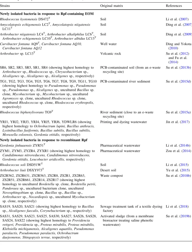
Table 4 Studies on exploiting the resuscitation effect of resuscitation promoting factor (Rpf) fromMicrococcus luteus for the isolation of previously viable but nonculturable (VBNC) bacteria
Strains Original matrix References
Newly isolated bacteria in response to Rpf-containing EOM
Rhodococcus kyotonensisDS472T Soil Li et al. (2007)
Amycolatopsis echigonensisLC2T,Amycolatopsis niigatensis LC11T
Soil Ding et al. (2007)
Arthrobacter niigatensisLC4T,Arthrobacter alkaliphilusLC6T, Arthrobacter echigonensisLC10T,Arthrobacter albidusLC13T
Soil Ding et al. (2009)
Curvibacter fontanaAQ9T, Curvibacter fontana AQ10, Curvibacter fontana AQ12
Well water Ding and Yokota
(2010)
Arthrobactersp. LC13T Volcanic rock Su et al. (2012)
and Fu et al.
(2014) SR6, SR2, SR3, SR5, SR1, SR4 (showing highest homology to
Arthrobactersp.,Rhodococcussp.,Chryseobacteriumsp., Alcaligenessp.,Alcaligenessp.,Alcaligenessp., respectively)
PCB-contaminated soil (from an e-waste recycling site)
Su et al. (2013b)
TG1, TG2, TG3, TG4, TG5, TG6, TG7, TG8, TG9, TG11, TG10 (showing highest homology toPseudomonassp.,Pseudomonas sp.,Pseudomonassp.,Alcaligenessp., unculturedBacillussp.
clone,Mycobacteriumsp.,Mycobacteriumsp., uncultured Agromycessp. clone, unculturedRhodococcussp. clone, unculturedRhodococcussp. clone,Rhodococcus erythropolis, respectively)
PCB-contaminated river sediment Su et al. (2015d)
Rhodococcus biphenylivoransTG9T River sediment (close to an e-waste recycling site)
Su et al. (2015a) YRJ1, YRJ2, YRJ3, YRJ4, YRJ5, YRJ6, YDWLR6 (showing
highest homology toOchrobactrum lupini, Bacillus anthracis, Lysinibacillus fusiformis, Bacillus subtilis, Bacillus subtilis, Moraxella osloensis, Gordonia otitidis,respectively)
Printing and dyeing wastewater Jin et al. (2017)
Newly isolated bacteria in response to recombinant Rpf
Gordonia jinhuaensisZYR51T Pharmaceutical wastewater Li et al. (2014b)
ZYM1, ZYM3, ZYZR4, ZYXR1 (showing highest homology to Candidimonas nitroreducens, Candidimonas nitroreducens, Gordonia otitidis, Leucobacter aridicollis,respectively)
Pharmaceutical wastewater Zou et al. (2014)
Rhodococcus soliDSD51WT Soil Li et al. (2015)
Arthrobacter liuiiDSXY973T Desert soil Yu et al. (2015)
ZS2R562, ZS2R631, ZS2R563, ZS2R8, ZS2R1, ZS2R63, ZS2R51, ZS2R661, ZS2R14, ZS2R17 (showing highest homology to unculturedBordetellasp. clone,Bordetella petrii, Pandoraeasp., uncultured bacterium clone, uncultured Novosphingobiumsp. clone,Bacillussp.,Bacillussp., Arthrobactersp.,Nocardiopsissp., unculturedMycobacterium sp. clone, respectively)
Waste compost Su et al. (2018b)
SAS19, SAS20, SAS21 (showing highest homology toBacillus sp.,Alcaligenes faecalis,Corynebacteriumsp., respectively)
Sewage treatment tank of a textile dyeing factory
Li et al. (2018) SAS31, SAS29, SAS23, SAS25, SAS30, SAS27, SAS26, SAS28,
SAS24, SAS22 (showing highest homology toProvidencia rettgeri, Providenciasp.,Proteus mirabilis, Proteus mirabilis, Klebsiella michiganensis, Alcaligenes aquatilis, Pseudomonas paralactis, Pseudomonas paralactis, Ochrobactrum
daejeonense, Shingopyxis terrae,respectively)
Activated sludge (from a membrane bioreactor treating saline phenolic wastewater)
Su et al. (2019b)
isolated novel strains from subsurface sediments contaminated with uranium, heavy metals, nitrate, and organic pollutants using the chambers instead of the plates, and recovered isolates with potential for advantageous bioremediation (Bollmann et al.2010).
Zhao and collegues modified the microcultivation system of Ferrari and collegues (Ferrari et al.2005) to obtain pollutant-degraders from crude oil contami- nated soil. Comparing the in situ SSMS method to traditional nutrient-rich technique for isolation of phenanthrene-degrading bacteria, revealed that an in situ method that mimicks the original environment is more suitable for the growth ofSphingomonas. By contrast, traditional method resulted in the isolation of Pseudomonas. Both strains were able to degrade phenanthrene but the former seemed to be previously uncultured by nutrient-rich methods (Zhao et al.
2017a).
8.2 Potential environmental functions of VBNC bacteria
Although, there have been several examples for the resuscitation of previously VBNC bacteria from environmental samples (Ding et al. 2007, 2009; Li et al. 2007; Ding and Yokota 2010), isolation of a novel bioflocculant-producing actinomycete Arthrobacter sp. LC13T by resuscitation from a VBNC state was one of the first attempts to use Rpf for environmental purposes (Su et al.2012; Fu et al.
2014). Since then, many studies have been carried out on the resuscitation (Table 4) and stimulation effects
of Rpf (Table5), with regard to the Rpf-containing EOM or recombinant Rpf of M. luteus. Jin and collegues resuscitated VBNC bacteria from dyeing wastewater, in which the Rpf-treated group proved to be more diverse than the Rpf-lacking control group indicating the high sensitivity of VBNC bacteria to Rpf in dyed water. All strains degraded more than 70%
Congo red after 8 days. (Jin et al. 2017). A novel approach applied an Rpf-containing supernatant ofM.
luteus culture to stimulate VBNC or uncultured indigenous bacteria in PCB (polychlorinated biphe- nyl) contaminated environments. Rpf-responsive enrichment cultures degraded nearly 1500 mg L-1 biphenyl in 24 h at the optimal EOM dosage of 15%
(v/v). This was the first case when Rpf was used to promote biodegradation of toxic compounds (Su et al.
2013b). Further exploring the potential environmental applications of supernatant Rpf, Liu and collegues achieved promising results for biological nutrient removal from wastewater. Although optimal dosage assessment is still needed, the presence of Rpf increased the abundance of the phyla Proteobacteria and Actinobacteria, which were responsible for the enhanced removal of phosphorus and nitrogen in activated sludge systems (Liu et al. 2016). Bouned- joum and collegues assessed a potentially functional hydrocarbon-degrader community enriched from oil- contaminated soil in response to M. luteus EOM addition (Bounedjoum et al. 2018). Among other strains,Rhodococcus biphenylivoransTG9Twas iso- lated from a PCB-contaminated river sediment using the resuscitating and stimulating effects of EOM.
Table 4 continued
Strains Original matrix References
SSPR8, SSPR5, SSPR6, SSPR2, SSPR11, SSPR13, SSPR12, SSPR1, SSPR9, SSPR10, SSPR4, SSPR7, SSPR3 (showing highest homology toBacillussp.,Bacillussp.,Bacillus wiedmannii, Bacillussp.,Bacillus tequilensis, Bacillussp., Bacillussp.,Pseudomonassp.,Stenotrophomonas maltophilia, Acinetobacter oleivorans, Acinetobactersp.,Acinetobactersp., Acinetobactersp., respectively)
Nitrogen-polluted river sediment Su et al. (2019c)
SPC1, SPC2, SPC3, SPC4, SPC5, SPC10, SPC11, SPC12, SPC13, SPC18, SPC19 (showing highest homology toBacillus altitudinis, Acinetobacter lwoffii, Bacillus wiedmannii, Castellaniella ginsengisoli, Sphingomonas paucimobilis, Pseudomonas beteli, Stenotrophomonas maltophilia, Bacillus megaterium, Cupriavidus necator, Exiguobacterium enclense, Enterobacter muelleri,respectively)
e-waste recycling town Su et al. (2019a)
EOM significantly increased both the abundance of potentially difficult-to-culture biphenyl-degraders in enrichment cultures and the biodegradation of biphe- nyls (Su et al.2015a,c,d). Transcriptome analysis of R. biphenylivoransTG9T,Rhodococcussp. TG13 and TN3 not only verified that pollutant-degrading bacte- ria could enter a VBNC state under cold stress and revert from it in response to the addition of supernatant Rpf but also pointed out molecular mechanisms behind these reversible processes that may vary with different bacterial species (Su et al. 2015b, 2016).
Their work drew attention to the importance of preventing and controlling the formation of VBNC bacteria during bioremediation, which can be crucial in the case of bioaugmentation.
Analysing the fate of transformant soil bacteria capand colleaguesble of degrading the agricultural insecticide lindene (c-hexachlorocyclohexane), Zhang and colleagues found that cell numbers decreased below the detection limit and after the addition of nutrients to the microcosms, they became detectable again demonstrating the low-activity state Table 5 Studies on exploiting the stimulation effect of resuscitation promoting factor (Rpf) fromMicrococcus luteusfor enhanced biodegradation of environmental pollutants
Purpose of treatment Materials and methods Applied microorganisms References
Enhanced biodegradation in response to Rpf-containing EOM Biodegradation of BPs Liquid mineral salt medium
contaminated with BPs
Enrichment culture obtained from PCB-contaminated soil
Su et al.
(2013b) Biodegradation of BPs Liquid mineral salt medium
contaminated with BPs
ES1-T and ES2-T enrichment cultures (composed mainly of the following genera:Achromobacter, Rhodococcus, Pseudomonas, Bacillus,
Stenotrophomonas, Dysgonomonas, Acinetobacter, Solibacillus, Ochrobactrum, Burkholderia,
Paenibacillus, Dechloromonas, Pandoraea) obtained from PCB-contaminated sediments
Su et al.
(2015c)
Biodegradation of BPs Liquid mineral-salts medium contaminated with BPs
Enrichment culture (composed mainly of the following genera:Achromobacter, Rhodococcus,
Pseudomonas, Stenotrophomonas, Leucobacter, Pontibacter, Clostridium, Sphingobacterium, Dechloromonas, Ochrobactrum, Paenibacillus, Hylemonella, Mycobacterium) obtained from PCB- contaminated river sediment
Su et al.
(2015d)
Nutrient (P and N) removal
Sequencing batch reactor Activated sludge (with the predominant phylum of Proteobacteria) from domestic wastewater treatment plant
Liu et al.
(2016) Hydrocarbon
biodegradation
Liquid mineral-salts medium contaminated with artificial diesel oil
Enrichment culture obtained from oil-contaminated soil
Bounedjoum et al.
(2018) Enhanced biodegradation in response to recombinant Rpf
Assessing extracellular cellulase activity
Crude culture supernatant on filter paper and
carboxymethyl-cellulose
Enrichment culture obtained from waste compost Su et al.
(2018b) Biodegradation of
phenol under high- salinity conditions
Liquid high salt medium contaminated with phenol
Domesticated sludge (with the predominant phyla of Proteobacteria, Bacteriodetes, Actinobacteria and Firmicutes) from a wastewater treatment plant of a textile factory
Su et al.
(2018a)
Biodegradation of phenol under high- salinity conditions
Artificial high-saline phenolic wastewater as influent in membrane bioreactor
Activated sludge (composed mainly of the following genera:Achromobacter, Rhizobium, Pseudomonas, Reyranella, Halomonas, Gemmobacter, Thauera) from a membrane bioreactor treating saline phenolic wastewater
Su et al.
(2019b)
of these engineered strains when exposed to adverse environmental conditions (Zhang et al.2012). Higher cell numbers ofRhodococcussp. D310-1::gfpdetected by fluorescence microscopy compared to colony forming units (CFUs) indicated the formation of VBNC cells in chlorimuron-ethyl-contaminated soil (Xiong et al. 2013). Fida and collegues had similar observations when a GFP-tagged PAH-degrader Novosphingobium sp. LH128 was inoculated into phenanthrene spiked soil. A rapid decline was detected in CFUs without any reduction in the number of GFP- expressing cells. Transcriptome analysis showed high expression levels of stress-related genes and phenan- threne biodegradation activity suggesting that strain LH128 entered a VBNC-like state (Fida et al.2017).
Performing proteomic analysis on the soil bacterium Cupriavidus metalliduransCH34 under water scarcity and nutrient starvation, Giagnoni and colleagues monitored metabolic changes (e.g. expression of genes related to cell shape formation and stress proteins) in C. metallidurans CH34 during its transition into a VBNC state, then its reversion to culturable cells after the addition of water or gluconate as the sole carbon source (Giagnoni et al. 2018). Their results further proved the importance of sigma factors in the forma- tion of VBNC cells, but also suggested that such soil properties as water content or carbon sources could be associated with the intracellular metabolisms of soil bacteria, hence influencing the survival, recoloniza- tion or spatial distribution of microbial communities in restored soils.
In addition to the use of Rpf-containing EOM, the application of purified recombinant Rpf protein is starting to attract more attention. Recovery of Rhodococcus sp. DS471 cells from a VBNC state was the first report of the resuscitation ability of the recombinant Micrococcus luteus Rpf expressed in Escherichia coli(Ding et al.2012). Aiming to map the importance of VBNC bacteria in industrial wastewater and improve sewage treatment systems, Gordonia jinhuaensisZYR51T(as a novel species of the genus Gordonia) was isolated, along with other strains, from pharmaceutical wastewater, using Rpf (Fu et al.2014;
Li et al. 2014b; Zou et al. 2014). The novel strains Rhodococcus soli DSD51WT (Li et al. 2015) and Arthrobacter liuii DSXY973T (Yu et al. 2015) belonging to the genusRhodococcusandArthrobac- ter, respectively, were resuscitated from soil samples.
Use of recombinant Rpf protein promoted the isolation
of unique bacterial species from waste composting sample and enhanced the cellulase activity of enrich- ment cultures. Filter paper cellulase (FPCase) and carboxymethyl-cellulase (CMCase) were detected in both pure and mixed cultures. Cellulase activities were higher in both mixed and Rpf-supplemented cultures than in the pure and Rpf-lacking ones, respectively (Su et al. 2018b). Su and colleagues resuscitated func- tional strains from a highly nitrogen-polluted river sediment that reached ammonium and nitrate removal rates of 2.23 and 0.86 mg L-1h-1, respectively. The results highlighted that indigenous VBNC bacteria in polluted rivers and water bodies can be considered useful resources for biological nitrogen removal (Su et al.2019c). A 1% (v/v) concentration of purified Rpf proved to be an enhancing additive for bacteria in activated sludge facilitating their activity during the biological treatment of a phenolic wastewater under high salinity conditions. Rpf-treatment not only shortened the whole domestication process for acti- vated sludge, but promoted the biodegradation of 1800 mg L-1phenol at 60 g L-1NaCl concentration in 18 days (Su et al. 2018a). Better phenol removal performance was also achieved in membrane biore- actors (MBRs) with Rpf addition. Nearly 1500 mg L-1 of phenol was degraded within 100 h in phenol-laden saline wastewater probably due to the resuscitation and stimulation of gammaproteobacte- rial and alphaproteobacterial populations (Su et al.
2019b). In a search for further halotolerant phenol- degrader bacteria, several strains were isolated from a sewage treatment tank (Li et al.2018) and activated sludge samples (Su et al.2019b) by Rpf. Neutraliza- tion of phenolic wastewaters by applyingBacillussp.
SAS19, either in a mixed consortium withCorynebac- terium sp. SAS21 (Li et al.2018) or immobilized in porous gels (Ke et al. 2018), represented the first application of a previously VBNC bacterium.Castel- laniella sp. SPC4, another former VBNC bacterium resuscitated by Rpf, was found to be capable of effectively catabolizing 3,30,4,40-tetrachlorobiphenyl (PCB 77). The strain maintained the capability of PCB biodegradation even without Rpf supplementation, further proving the degradative potential of VBNC bacteria in contaminated environments (Su et al.
2019a). The study of their capabilities is one of the major future tasks of environmental biotechnology.
Considering the results mentioned above and the acknowledged fact that Rpf is able to resuscitate
potential pollutant-degrading VBNC bacteria, its application can be integrated into biostimulation methods of bioremediation (which raises the question of the role of non-protein components in EOM). By contrast, it is assumed that the outcome of bioaug- mentation, which involves the inoculation of efficient pollutant-degrader bacteria into polluted, heavily depends not only on the survival and efficiency of these strains, but also on their synergistic degradation aptitudes and transitions into VBNC states (Su et al.
2015b).
9 Conclusion
Nowadays, the most crucial challenges of bioremedi- ation are to recover the hidden players in microbial diversity and overcome the obstacles to moving pollutant-degrader microorganisms from laboratory flasks to field sites. It is essential to enhance their survival and biodegradation capabilities in order to intensify their bioconversion rates, even for field remediation processes. The future success of environ- mental biotechnology depends heavily on developing and improving databases of the microorganisms and enzymes that are responsible for biodegradation of environmental pollutants. A deeper understanding of the metabolic pathways involved in the microbial degradation of recalcitrant contaminants requires a better knowledge about the unculturable majority of bacteria, their relationships with coexisting members of the microbial community, and how they can be integrated as key players in environmental rehabilita- tion processes.
Acknowledgements Open access funding provided by University of Szeged (SZTE, University of Szeged Open Access Fund 4587). The project was supported by the European Union and co-financed by the European Social Fund (Grant Agreement No. EFOP-3.6.2-16-2017-00010) in the frame of the Sze´che´nyi program of the Hungarian State.
Open Access This article is licensed under a Creative Com- mons Attribution 4.0 International License, which permits use, sharing, adaptation, distribution and reproduction in any med- ium or format, as long as you give appropriate credit to the original author(s) and the source, provide a link to the Creative Commons licence, and indicate if changes were made. The images or other third party material in this article are included in the article’s Creative Commons licence, unless indicated otherwise in a credit line to the material. If material is not included in the article’s Creative Commons licence and your
intended use is not permitted by statutory regulation or exceeds the permitted use, you will need to obtain permission directly from the copyright holder. To view a copy of this licence, visit http://creativecommons.org/licenses/by/4.0/.
References
Alvarez PJJ, Illman WA, (Walter A, Wiley InterScience (Online service) (2006) Bioremediation and natural attenuation:
process fundamentals and mathematical models. Wiley, Chichester
Austin B (2017) The value of cultures to modern microbiology.
Antonie van Leeuwenhoek Int J Gen Mol Microbiol.
https://doi.org/10.1007/s10482-017-0840-8
Ayrapetyan M, Oliver JD (2016) The viable but non-culturable state and its relevance in food safety. Curr Opin Food Sci 8:127–133.https://doi.org/10.1016/j.cofs.2016.04.010 Ayrapetyan M, Williams TC, Oliver JD (2014) Interspecific
quorum sensing mediates the resuscitation of viable but nonculturable vibrios. Appl Environ Microbiol 80:2478–2483.https://doi.org/10.1128/AEM.00080-14 Bari SMN, Roky MK, Mohiuddin M et al (2013) Quorum-
sensing autoinducers resuscitate dormantVibrio cholerae in environmental water samples. Proc Natl Acad Sci USA 110:9926–9931.https://doi.org/10.1073/pnas.1307697110 Bassler BL, Greenberg EP, Stevens AM (1997) Cross-species induction of luminescence in the quorum-sensing bac- teriumVibrio harveyi. J Bacteriol 179:4043–4045.https://
doi.org/10.1128/JB.179.12.4043-4045.1997
Ben Said M, Masahiro O, Hassen A (2010) Detection of viable but non cultivableEscherichia coli after UV irradiation using a lytic Qbphage. Ann Microbiol 60:121–127.https://
doi.org/10.1007/s13213-010-0017-4
Bergkessel M, Basta DW, Newman DK (2016) The physiology of growth arrest: uniting molecular and environmental microbiology. Nat Rev Microbiol 14:549–562.https://doi.
org/10.1038/nrmicro.2016.107
Boaretti M, Lleo` MDM, Bonato B et al (2003) Involvement of rpoS in the survival ofEscherichia coliin the viable but non-culturable state. Environ Microbiol 5:986–996.https://
doi.org/10.1046/j.1462-2920.2003.00497.x
Bogosian G, Bourneuf EV (2001) A matter of bacterial life and death. EMBO Rep 2:770–774. https://doi.org/10.1093/
embo-reports/kve182
Bollmann A, Palumbo AV, Lewis K, Epstein SS (2010) Isola- tion and physiology of bacteria from contaminated sub- surface sediments. Appl Environ Microbiol 76:7413–7419.
https://doi.org/10.1128/AEM.00376-10
Bounedjoum N, Bodor A, Laczi K et al (2018) Assessment of potentially functional hydrocarbon-degrader bacterial communities in response to Micrococcus luteus EOM using culture-dependent and culture-independent methods.
New Biotechnol 44:S134–S135.https://doi.org/10.1016/j.
nbt.2018.05.1091
Buerger S, Spoering A, Gavrish E et al (2012a) Microbial scout hypothesis, stochastic exit from dormancy, and the nature of slow growers. Appl Environ Microbiol 78:3221–3228.
https://doi.org/10.1128/AEM.07307-11