Analytical Approaches for the
Quantitation of Redox-active Pyridine Dinucleotides in Biological Matrices
Anna Somogyi
1, George Horvai
1,2, Miklós Csala
3, Blanka Tóth
1*Received 12 May 2016; accepted after revision 24 May 2016
Abstract
Some of the main electron carriers in the metabolism are mono- or dinucleotides and they play crucial roles in main- taining a balanced redox homeostasis of cells, and in coupling many anabolic and catabolic reactions. Altered cellular redox status can be an indicator of various metabolic disorders such as obesity, the metabolic syndrome, or type 2 diabetes and of other pathological conditions, which involve oxidative stress, such as cardiovascular diseases. Adequate NAD+/NADH and NADP+/NADPH ratios are fundamental for normal cellular functions, thus accurate measurement of these pyridine dinu- cleotides is essential in biochemical research. Liquid chroma- tography coupled to tandem mass spectrometry has become the leading analytical technology in (targeted) state-of-the-art metabolic profiling. Main difficulties that hamper quantifica- tion of metabolites are chemical similarities, high polarity, and chemical and biological instability of the molecules to be measured. In this review, some critical steps of studying cellular redox status are described, in particular, different techniques of sample preparation and challenges in chroma- tographic separation.
Keywords
nicotinamide adenine dinucleotide, redox couple, separation, HPLC, mass spectrometry, method
1 Introduction
In the last few decades, more and more attention has been paid to the role of redox active nucleotides in the metabolism.
Their enormous importance in the cellular antioxidant defense is undebated; flavin and nicotinamide nucleotides are essential in energy transduction as main electron transfer molecules and substrates for over 700 oxidoreductase enzymes [1]. Severe disturbance in the redox balance, e.g. oxidative stress, which is often caused by an external impact is an important component of the pathomechanism of cardiovascular diseases such as heart failure, myocardial ischemia, unstable angina and ischemia re- perfusion injury [2]. The largely autonomous redox homeostasis of the endoplasmic reticulum (ER) is based on the co-localization of a reduced pyridine dinucleotide NAD(P)+/NAD(P)H system and an oxidized thiol-disulfide system. The former maintains a local cortisol production in many glucocorticoid target cells (e.g. hepatocytes, muscle cells and adipocytes), while the latter is indispensable for generation of intrachain and interchain pro- tein disulfide bridges and hence for the appropriate protein fold- ing in the ER lumen. When the ER redox conditions are affected by cellular disturbance, inadequacy of the protein processing machinery leads to the luminal accumulation of immature pro- teins. This ER stress stimulates a complex signalling network referred to as the unfolded protein response (UPR), which is primarily adaptive, but it can also lead to different destructive downstream effects including inflammations, apoptosis and insulin resistance [3]. Ratio of oxidized and reduced forms of pyridine dinucleotides gives precious information about redox metabolism disorders or energetic alterations [2]. Thus, precise measurement of these nucleotides has become essential.
Since the development of liquid chromatography (LC), its increasing penetration in biological and clinical analysis is obvi- ous. LC coupled to UV detection was a common tool in analytics, but its application is limited by its relatively low sensitivity and specificity. Development of mass spectrometry (MS) and its cou- pling to LC seems to solve the majority of these problems. MS is up to several orders of magnitudes more sensitive than UV detec- tion, and it simplifies separation as well, as it can differentiate compounds of equal retention time. However, MS needs changes
1 Department of Inorganic and Analytical Chemistry, Budapest University of Technology and Economics, H-1111 Budapest, Hungary
2 MTA-BME Research Group of Technical and Analytical Chemistry, H-1111 Budapest, Hungary
3 Department of Medical Chemistry, Molecular Biology and
Pathobiochemistry, Semmelweis University, H-1144 Budapest, Hungary
* Corresponding author, e-mail: tblanka@mail.bme.hu
60(4), pp. 218-230, 2016 DOI: 10.3311/PPch.9470 Creative Commons Attribution b research article
PP Periodica Polytechnica
Chemical Engineering
in separation techniques since use of non-volatile salts such as phosphates, is not advisable in MS. The most suitable mobile phases usually consist of methanol or acetonitrile and water with a buffer of volatile salts (i.e. ammonium-acetate, ammonium for- mate) and in some cases, volatile ion-pair reagents, among oth- ers dibutylamine (DBA) [4], tributylamine (TBA) [5-7], dimeth- ylhexylamine (DMHA) [8]. High resolution is fundamental in untargeted metabolomics, but in targeted metabolic profiling, lower resolution tandem mass spectrometers, such as triple quad- rupoles also provide the information needed [9].
The novel detection mode requires new or modified tech- niques to be considered for sample pre-treatment as well.
Although a generic method consisting of protein precipitation and extraction may sometimes be sufficient, the efficiency of the analysis can be largely improved by optimization of sample preparation for the selected metabolites.
The purpose of this review is to collect and critically com- pare the most common practices in profiling cellular metabolic and redox status using LC coupled to tandem MS, focusing particularly on sample pre-treatment and different separation techniques such as using an ion-pair reagent, or HILIC-HPLC.
2 Metabolism and roles of pyridine nucleotides
Pyridine dinucleotide coenzymes, such as NAD+ and NADP+, play crucial roles in intracellular metabolism as they serve as electron carriers for several enzymes. External impacts, for instance overfeeding, starving, alcohol ingestion or drug treatments usually affect the intracellular redox state of these coenzymes and therefore monitoring the levels of their oxidized and reduced forms (i.e. NAD(P)+ and NAD(P)H, respectively) (Fig. 1) can provide information on the effect of dietary factors or drug candidates [2].
Fig. 1 Chemical structure of NAD+ and NADP+, and their nicotinamide group carrying the hydride ion.
One of the most important coenzymes in hydride transfer reactions is nicotinamide adenine dinucleotide (NAD+). It is a substrate of most dehydrogenase enzymes involved in nutrient catabolism, and its reduced form, NADH delivers the collected electrons preferentially to mitochondrial oxidative phosphoryl- ation. As NAD+ is essential in fuel utilization, protein modifica- tion and in cell signalling, information on its concentration and redox state in different cells and tissues can help understand biochemical functions, metabolic state of cells and effects of drugs in the organism [9].
Cytoplasmic NAD+/NADH ratio normally lies around 1100 [10] in aerobic cell; and it shows that NAD+ usually functions as an oxidative agent in biochemical processes such as fatty acid oxidation, glycolysis, and citrate cycle. Thus, change in NAD+/NADH ratio can indicate changes in metabolic processes and in several diseases [1]. In 2013, Gomes et al. proved that NAD+ plays an essential role in anti-aging processes, and eleva- tion of NAD+ levels in old mice restores mitochondrial function to that of a young mouse in a SIRT1-dependent manner [11].
Therefore regular monitoring of NAD+ levels can improve our knowledge about the mechanism of age-associated diseases [12].
3 Endoplasmic reticulum and its redox environment The ER is a metabolic compartment which participates in many fundamental pathways of the intermediary metabo- lism [3]. The organelle is a continuous membrane network separating a narrow lumen from the cytosol. A large number of enzymes are embedded in the ER membrane, and many of them expose their active sites on the cytosolic surface.
However, the ER lumen also gives place to the active centres of several membrane-bound or particulate enzymes, includ- ing many oxidoreductases [13]. The concentration and redox state (i.e. the ratio of oxidized and reduced forms) of the redox couples can be remarkably different between the cytosolic and luminal environment [14]. Although the NADP+-NADPH couple is dominantly reduced in both compartments, the main- tenance of this reduced state is based on different and inde- pendent enzyme activities. Moreover, the thiol-disulfide cou- ple is actively oxidized in the ER lumen, and consequently the disulfide/thiol ratio is much higher in the ER than in the cyto- sol. In case of varying metabolic conditions, cells can modify ER functions. Extreme conditions such as over- or undernutri- tion, hypoxia or lipotoxicity [15] can cause redox imbalance and ER stress, which contributes to apoptosis, inflammation and insulin resistance via activation of the UPR. The main reason of obesity, metabolic syndrome and type 2 diabetes is the stimulation of local activation of glucocorticoids [3, 16].
Cytoplasmic NADP+/NADPH ratio was shown to be around 0.01 [10]; in accordance with the fundamental role of this redox couple as a readily available reducing source for biosyn- thesis, biotransformation and antioxidant defense. It is widely accepted that the major redox buffer of the ER is composed
of pyridine dinucleotides, especially NADP+ and its reduced form, NADPH, but presence of flavin nucleotides, ascorbate and other redox-agents was shown too [17]. The cytosolic NADP+-NADPH pool is practically separated from that of the ER because the ER membrane has a limited and selective per- meability, and the transmembrane traffic of pyridine dinucleo- tides is very slow compared to their metabolic interconversion [18]. In other words, the ER luminal reductases are uncoupled from the cytosolic NADPH generating mechanisms, and they cooperate with local NADP+-dependent dehydrogenases. It is due to the membrane impermeability that the microsomes, i.e.
the artificial vesicles produced from the ER upon cell or tis- sue fractioning, stably retain the luminal pyridine dinucleotides and still possess the luminal redox couplings.
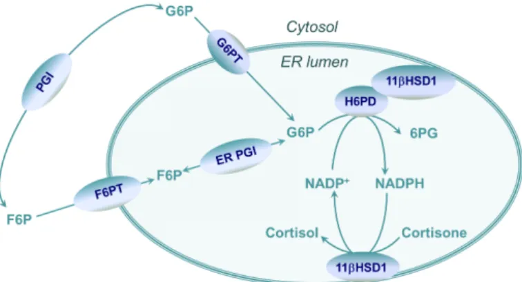
Measurement of all the linked redox components and deter- mination of a redox environment in its entirety is not pos- sible, hence changes of a complex redox system are studied by monitoring a representative redox couple as an indica- tor [14]. Measurement of the microsomal (ER luminal) pyri- dine dinucleotides would be greatly informative regarding the local redox conditions. In vivo or cellular monitoring of NAD+/NADH and NADP+/NADPH cannot be performed yet, therefore all available data have been collected from isolated microsomes. However, the precise and reliable quantitative analysis of microsomal pyridine dinucleotides has not been solved yet either, hence the intravesicular pyridine dinucleo- tide redox state is usually monitored in an indirect manner by using enzymatic methods [17]. These methods take advantage of the presence of intraluminal oxidoreductases which use pyridine nucleotides as co-substrates. Decades ago, pyridine nucleotide content of ER-derived microsomal vesicles was proved by Bublitz et al. [19] Odermatt et al. [20] performed a study in which functions of the ER enzyme 11β hydroxysteroid dehydrogenase type 1 (11βHSD1) were observed. 11βHSD1 is responsible for the interconversion of cortisone and cortisol in the lumen of ER (Fig. 2). Although in vitro this enzyme can act both in oxidative and reductive reaction, it was shown that in vivo, this reversible reaction is shifted towards cortisone reduc- tion [18]. This observation suggests the dominance of NADPH in the lumen, and hexose-6-phosphate dehydrogenase (H6PD) can help maintain this ratio [14]. Lavery et al. found that in H6PD knockout mice, 11βHSD1 mediated glucocorticoid gen- eration was not performed [13], hence separate luminal pyri- dine nucleotide pool must exist to satisfy NADPH need of the reduction [14]. Alterations in the ER luminal pyridine dinucle- otide redox state are usually deduced from the changes in the intrinsic cortisone reducing and cortisol oxidizing capacity of the microsomes. The results of such measurements are usually in accord with the modifications of endogenous NAD(P)(H) fluorescence of the intact microsomal vesicles; nevertheless, these approaches do not yield data of appropriate accuracy and reproducibility, and a significant improvement can be expected
from direct measurements of individual redox components (NAD+, NADP+, NADH and NADPH).
Fig. 2 Pyridine dinucleotide homeostasis in the ER lumen. F6P, fructose 6-phosphate; F6PT, F6P transporter; G6P, glucose 6-phosphate; G6PT, G6P
transporter; H6PD, hexose 6-phosphate dehydrogenase; 11βHSD1, 11β hydroxysteroid dehydrogenase type 1; 6PG, 6-phosphogluconate;
PGI, phosphoglucose isomerase.
4 Analysis of endogenous pyridine dinucleotides As mentioned above, endogenous concentration of pyridine dinucleotides can vary within wide ranges. Numerous meth- ods were studied for measurement of pyridine and adenine nucleotides, in which the leading methods were HPLC meth- ods coupled to different detecting systems, such as HPLC-UV [2, 21-24] HPLC-NMR, or enzymatic assays. Beside the most common methods, other type of measurement of nucleotides were suggested, such as capillary zone electrophoresis [25, 26], or spectrophotometric method for the determination of intracel- lular NAD(P)H [27]. But recently, LC coupled to mass spec- trometry (LC-MS) has become the most important analytical method in metabolic profiling and quantitation of these small molecules in complex matrices. Untargeted metabolomics need high mass resolution instruments such as mass spectrometers with time of flight or ion-cyclotron resonance detecting sys- tems, in targeted quantitative LC-MS, use of triple quadrupole (QQQ) as a lower resolution tandem mass spectrometers can satisfy the requirements. QQQ systems can provide multidi- mensional data (m/z, MS2 transitions in MRM mode and reten- tion time), thus metabolites which differ only in one hydrogen atom can also be distinguished. With the development of sta- tionary phases and separation columns and the use of sub-2-µm particles, chromatographic resolution has been enhanced [28].
Most of the published studies aimed a multiple metabolite profiling, which involves the examination of many differ- ent metabolite groups. Several groups assess the phosphoryl metabolites, such as adenine nucleotides, but pyridine dinucle- otides, NAD(H) and NADP(H) rarely constitute the main sub- ject of these studies. Hence, in many cases, sample pre-treat- ment and LC are not optimised directly to these compounds.
This review focuses on the methods for the determination of often neglected pyridine dinucleotides.
5 Sample pre-treatment
Nucleotide profile has been investigated in various matri- ces in recent years. Metabolism of Escherichia coli (E.coli) [5, 6, 29-31], Saccharomyces cerevisiae (Sacch. cer.) [9, 31, 32], and Methylobacterium extorquens AM1 [33] was moni- tored, and whole blood [2, 12], serum and plasma [6, 7, 34]
urine [6], cultured cells [6-9, 30, 34, 35], cerebrospinal fluid [34], and tissues [4, 6, 7, 31, 34] were used as samples.
The measurement usually starts with the quick quenching of metabolism. In living cells, concentration and ratio of the moni- tored molecules can change within seconds, so keeping the orig- inal state of cells is fundamental in profiling. Several methods are described to stop metabolism, such as rapid cooling, espe- cially quick freezing in liquid nitrogen for tissues, and mixing the cultured cells with organic solvents [29] (in many cases with cold organic solvent) [5] are often used. Less common methods, but still with importance, are the heat treatment of the system [31, 32], and acidification. But while considering the use of these techniques, it must be ensured that all the analytes to be exam- ined are heat or acid stable. After quenching the metabolism, the extraction procedure is the critical step. A wide variety of extrac- tion methods can be found; the nature of the metabolites to be determined and the sample type have the biggest impact in choos- ing the most appropriate protocol. When the analytes have been extracted, samples are usually kept at -20 °C or -80 °C.
A generally accepted extraction method for Escherichia coli usually has a quenching step with the use of 80:20 methanol:
water [29, 30], ethanol [5], methanol: acetonitrile: water [6].
After several centrifugation – resuspension – ultrasound cycles, in which the supernatant is always removed and stored, the final extract is then stored at -20 °C or -80 °C. In one case, 0.3 M KOH was added to the extract which was later neutralized with glacial acetic acid [5]. For whole broth extraction, aqueous 75% (v/v) ethanol solution preheated to 78 °C was mixed with E.coli cul- ture, and was incubated at 78 °C for 1 min, then frozen in liquid nitrogen. After thawing and centrifugation, the supernatant was dried and stored at -80 °C until re-suspension [31].
Extraction methods for Saccharomyces cerevisiae are usu- ally very similar to methods for E.coli. Preheated at 95 °C, 75%
ethanol was used for extraction followed by drying – resuspen- sion – centrifugation to obtain nucleotides from yeast culture [31]. In another case, precooled, aqueous 60% (v/v) methanol buffered with 10 mM ammonium acetate (pH 7.5) was used as an extraction solvent [32].
Heat treatment was applied for extraction of nucleotides from Methylobacterium extorquens AM1 [33]. After centrifu- gation of quenched biomass, boiling HEPES buffered etha- nol: water 75:25 at pH 5.2 was mixed with cell pellet. After two centrifugation cycles, supernatant was dried and stored at -80 °C until analysis.
In case of cell cultures, the quenching metabolism is also fun- damental. For Chinese hamster ovary (CHO) cell culture different
extraction solvents were tested [8]. Acetonitrile, ethanol, metha- nol, acetonitrile: water (8:2), ethanol: water (8:2), methanol: water (8:2), 0.1 M formic acid, 0.5 M perchloric acid (PCA) and 0.1 M formic acid in methanol were used. It was found that methanol provided the most efficient extraction (recovery of 53%). Other interesting result is that extraction solvents containing water caused very broad, split chromatographic peaks. Perchloric acid, neutralised with KOH was found to be incompatible with LC-MS because of presence of precipitate. Sonication and hexane extrac- tion were also tested; hexane extraction provided greater sample clean-up and better peak shapes; sonication also improved recov- ery, but as the cooling of plates was unsolved, the sonication was disregarded. Methanol: acetonitrile: water (40:40:20) at -20 °C was used for cancer cell lines HCT116 and Calu 6 and methanol:
acetonitrile (50:50) at -20 °C for cell medium [6]. Sonication was used for cell cultures such as for Hela cells [34] and HCT116 [30]. In an experiment for metabolomic analysis of Plasmodium falciparum lipid related metabolites, two different methods were used depending on ionisation mode in mass spectrometry. After two extraction step by methanol: water (80:20), in the last resus- pension, acetonitrile: water (75:25) was used.
The second step of tissue or cell analysis after arresting metabolism is homogenization. Frozen tissues can be homog- enized using a mortar or other physical effect [4, 31, 34].
Sometimes, direct extraction [6] is used where methanol: ace- tonitrile: water (40:40:20) are the organic solvents, and the extraction is followed by shaking cycle. Extraction solvents are usually the same as for cell cultures and PCA can also be found in some procedures [4] and this needs neutralising steps as well; heated ethanol buffered with 10 mM ammonium acetate can also provide a suitable recovery [31].
Human plasma [6, 34], serum [6, 34], urine [6, 34] and cer- ebrospinal fluid [34] usually need only a protein precipitation step, which can be performed with methanol:acetonitrile [6]
(50:50) or by a simple filtration with a centrifuge filter unit [34].
In a recent publication [12], human acidified blood samples were analysed where collection of blood samples was per- formed in a sample tube containing 0.5 N PCA solution. After the sample preparation process 0.5 mM ammonium formate was added to supernatant.
Caruso et al. investigated the preanalytical phase of blood adenine and pyridine nucleotide determination [2]. They per- formed a double extraction method and two different chroma- tographic runs for reduced and oxidized forms of nucleotides.
In acid extraction for oxidized form, K+/EDTA treated whole blood was mixed with 7.2% PCA, and after centrifugation, 1 M borate buffer was used as a neutralizing agent. Borate buffer was compared to carbonate buffer and KOH as neutralizing agent for acidic extraction, and it was demonstrated that 1 M borate buffer provides a stable pH 6.5 supernatant.
In alkaline extraction for reduced forms, cold, 0.5M KOH was added to K+/EDTA treated whole blood. After treatment
with cold distilled water, ultrafiltration membranes were used to separate the nucleotide containing phase. After ultrafiltration, 1 M KH2PO4 was used to neutralise the solution. Ultrafiltration time and different ultrafiltration conditions were tested.
6 Liquid chromatography
Difficulties in the separation of nucleotides arise from two main sources: the high polarity and the similar chemical struc- ture of these molecules. In developing useful methods, it must be considered that in some cases, molecules only differ in one hydrogen atom. The phosphate moiety also makes the use of reverse phased (RP) LC more difficult.
Anion exchange chromatography (AEX) could be used to separate anionic metabolites, but it has a poor compatibility with MS since in AEX, non-volatile salts are generally used.
Salt concentration must be reduced with a membrane suppres- sor if it is coupled with MS [36].
Normal phase (NP) nanoflow LC was used to separate phosphor-related metabolic changes. Uehara et al. [30] found an amino-propyl silica gel column with basic ammonium car- bonate buffer appropriate for the separation of pyridine nucleo- tides. As ionic and hydrophilic interaction was present between the phosphorus compounds and the solid phase, this method provided good retention and separation (Fig. 3).
However, the use of normal phase is less common in cur- rent metabolomics, when LC is coupled to MS. Reversed phase liquid chromatography (RP-HPLC) without ion-pair reagent has not shown good performance either. As mentioned above, nucleotides are highly polar, hence interaction with the station- ary phase is insufficient, and metabolites are minimally retained, and poor peak shapes are detected. When separating 160 metab- olites of E.coli, Bajad et al. [29] tested two different silica based reversed phase columns, one with embedded polar group and one with ether linked phenyl at three different pH values, but they found that lack of phosphate in the buffer (phosphate buffer is non-volatile thus incompatible with MS) yielded poor peak shape. However, in case of UV detection which allows use of phosphate buffers, RP-HPLC could be a useful method. An extensive report about preanalytical phase of nucleotide assay applied RP-HPLC with UV detection successfully [2].
Ion-pair (IP) reagents seem to solve the mentioned problems of RP-HPLC, therefore IP-RP-HPLC is one of the most stud- ied method in metabolomics. Use of volatile salts is compatible with MS detection. The principle of ion-pairing is the interaction between the negatively charged nucleotide and the positively charged ion-pair reagent. When using IP reagents, the stationary phase is a usual reversed phase column. Ion-pair reagents are nor- mally alkylamines, such as dibutylammonium acetate (DBAA) [32], tributylamine (TBA) [5-7, 29, 31], dimethylhexylamine (DMHA) [8, 34], dibutylamine (DBA) [4], hexylamine (HA) [7]
for negative ionization mode. As nucleotides are detected in nega- tive mode, ion suppression is less problematic. The effect of add- ing DBAA as an ion-pair reagent is shown in Fig. 4.
Luo et al. [5] investigated the effect of several volatile alkylamine ion-pair reagents, different pH ranges, and the effect of using methanol and acetonitrile as organic mobile phase.
Five different alkylamines, triethylamine (TEA), tripropylamine (TPrA), tributylamine (TBA), tripentylamine (TPeA), and trihexylamine (THA) were tested in the first step at pH 6.8 in the aqueous eluent. Systematic increase of retention was observed with the increase of the length of the alkyl chain. This is caused by the hydrophobic interaction of the alkyl chain with the station- ary phase. TEA and TPrA which are alkylamines with short alkyl chain did not offer good resolution, and the use of longer alkyl chains such as TPeA and THA resulted in long retention times and poor peak shapes. After choosing TBA as the best IP reagent, effect of pH was tested, and pH 4.95 was found to be the opti- mal pH to separate 29 metabolites including sugar phosphates, nucleotides and carboxyl acids. It must be mentioned here that changing the pH between 4.95 and 6.8 did not have effect on the separation of nucleotides due to their low pKa. Decreasing the pH was necessary because of their interest in carboxylic acid metab- olites. The last step was to optimise the organic eluent. Methanol was chosen as its aspect of being a weaker eluent gave the oppor- tunity of the fine tuning of the mobile phase. Figure 5 shows the combined chromatogram of 29 negatively charged compounds (sugar phosphates, nucleotides, and carboxylic acids), where the peaks of NAD(P)(H) are highlighted. If the main object is the determination of pyridine dinucleotides, changing the gradient of the elution can shorten the separation.
Fig. 3 The MRM chromatograms obtained by using normal phase nanoflow liquid chromatography of pyridine dinucleotides of human HCT116 cells, log phase E.coli and stationary phase E.coli cells [30].
Fig. 5 Combined selective ion chromatogram of 29 metabolites in a standard mixture. The LC–MS/MS method is described in Table 1. Peaks of pyridine
dinucleotides: (10) NAD; (23) NADP (26) NADH; (27) NADPH [5].
Optimization of the concentration of IP is fundamental as it can represent a source of contamination of the MS. The concen- tration can be reduced if the alkyl chain of the used IP is longer as it helps interaction between IP reagent and the apolar station- ary phase. The effect of different concentrations of DMHA was tested by Cordell et al. [8]. They varied the concentration of the ion-pair agent between 0.5 mM and 5 mM. It was found that the increasing concentration of IP agent caused increasing reten- tion time and better peak shape. Concentration greater than 5 mM did not improve the separation. Effect of mobile phase pH was also tested. It was noticed that increasing pH increased
retention time and the pH had influence on peak shapes as well.
At pH below 3, compounds eluted near dead time, and peak broadening was observed. Figure 6 represents the simultane- ous separation of 24 nucleotides for standard solution (a), and for biological sample (b). The chromatogram shows the most important advantage of MS detection: the peaks which overlap in the combined chromatograms, in fact do not overlap.
To avoid contamination, Seifar et al. [32] diverted the efflu- ent of the analytical column to waste at the beginning and in the end of the separation, which prevented the ESI source and the MS from being contaminated.
The other most important chromatographic system, which is used in metabolic profiling, is hydrophilic interaction chroma- tography (HILIC). In HILIC, the retention mechanism is parti- tioning, and water is used as the strong eluent [37]. There can be found three main types of HILIC columns, the neutral (no electrostatic interaction), charged (strong electrostatic interac- tion) and zwitterionic (weak electrostatic interaction). A very successful application of HILIC was reported by Bajad et al. [29]. Three different HILIC columns were compared, cyano column at acidic pH, silica column and amino column at pH 9.
They found that the cyano and silica column had minimal reten- tion and poor peak shape for multi-phosphorylated compounds, but an amino-modified HILIC column at pH 9 gave detectable peaks for triphosphates as well with a good peak shape. After optimization, 69 metabolites from E. coli were quantified. In the metabolomic analysis of Plasmodium falciparum, which
Fig. 4 Effect of DBAA ion-pair reagent on peak height of NADPH [32].
is the causative agent of malaria, a Luna NH2 HILIC column under alkaline conditions in negative ionization mode was used [35]. Their method was complementary with ion-pair chroma- tography, and a total of 35 compounds were quantified. Many reports were published about comparison between IP-RP-HPLC and HILIC-HPLC. Michopoulos et al. [6] found that use of ion- pair agents provided better separation and peak shapes than HILIC. Peak tailing was observed for multi-phosphorylated compounds, which was increasing with the degree of phospho- rylation. In spite of these facts, good resolution was observed for reduced and oxidized forms of NAD+ and NADP+.
Myint et al. [34] reported a practical method for polar ani- onic metabolites using nano-LC/MS system. A polyamine- bonded polymer-based apHera NH2 column was applied, and during optimization, it was observed that traces of metal caused a binding to the column for highly polar compounds (citric acid or ATP). A chelating agent, EDTA was added in 85 pmol concentration to remove metallic impurities which may come from the stainless steel of HPLC hardware. By using EDTA, no further peak tailing was observed and the pre- viously disappearing peaks were detected in every analysis.
A titania column was used to separate nucleotides and their pathway intermediates [21]. Titania’s mechanical and pH sta- bility is better than of silica. It acts as Lewis acid and this way it has a strong affinity for phosphonate groups. This property makes it a very good possibility in separating nucleotides. In their report, Zhou et al. examined the effects of mobile phase composition (% (v/v)) of ACN in sample, eluent pH and elu- ent anion. By optimizing the titania column, fifteen nucleotides and their intermediates were separated.
IP-RP-HPLC and HILIC can act as complementary meth- ods in profiling metabolites. The need for high proportions of
organic solvents in separations by HILIC can confer benefit as it intensifies MS signal by facilitating ionization, but it can be a disadvantage as well. In many cases, nucleotides are present in a very low concentration in biological samples, and diluting them to the same solvent composition as the eluent can lower the concentration below the working concentration range.
Recently, beside the common stationary phases, many other possibilities are gaining popularity. One of the most promising phases is graphitic carbon column. The retention mechanism is very complex, pi-pi interactions and dispersive interactions between the stationary phase and the aromatic solutes seem to be the main components [38]. PGC is still not so much used, but there can be found reports about its possible applications. When comparing HILIC and IP-RP-HPLC, Michopoulos et al. [6]
examined PGC columns as well, and they found it applicable for pyridine nucleotides. When 50 mM ammonium hydrogen car- bonate was added to the eluent containing 60% ACN and 40%
water, improvement was observed in elution of NAD+, NADH, NADP+, NADPH and AMP. Despite the promising results in separation of pyridine nucleotides, the separation method was not examined in further studies since they were focusing on a greater number of metabolites to be determined.
Another approach was presented with respect to quanti- tative metabolomics in Methylobacterium extorquens [33].
Comparison was performed between a pentafluorophenyl-pro- pyl column and a Luna NH2 HILIC column. Reversed phase mode provided a suitable separation of all tested analytes. After examining the effect of buffers, they proved that ammonium formate and ammonium acetate in acidic pH lowered reten- tion and the separation deteriorated. The use of 0.1% aqueous formic acid/acetonitrile gradient provided the best results with good retention, and good peak shapes. Although NADP+ and
Fig. 6 Results of MRM scan (dwell 0.2 s) of simultaneous detection of 24 nucleotides in standard solution (a) and for biological sample in control conditions (b) [8]. LC-MS method is described in Table 1.
ATP showed 4 times lower sensitivity, compared to NADH and NADPH, 5-10 times higher sensitivities were observed than with the use of the HILIC column.
7 Mass spectrometry
Detection in complex biological matrices, such as blood, urine and tissues, is complicated by the presence of numer- ous contaminants, which makes the precise determination of selected components more difficult or even impossible without a highly sensitive and selective method. This is where the dis- advantages of UV detection are the most evident. MS can dif- ferentiate molecules based upon their mass, which allows the use of isotope labelled internal standards. The biggest draw- back of MS is the inability to distinguish isobaric components, such as sugar phosphates or cis/trans molecules. Despite this drawback, coupling LC to MS can provide a highly sensitive and widely usable method in qualification and quantification of metabolome. Coupling the two instruments was the big- gest challenge due to the high flow rate and to the possibil- ity of contaminating the MS. Electrospray ionization (ESI) has become the most important ionization technique in bioanalyti- cal studies. For detection of nucleotides, usually negative ESI is applied since these molecules have acidic groups, but reports describing positive mode also can be found [12].
In untargeted metabolomics, very high resolution is essen- tial, thus use of FTIR MS is established, but in targeted profil- ing, a triple quadrupole (QQQ) may be the best choice. QQQ has a lower resolution, but can reduce the background noise hence its sensitivity is outstanding. Triple quadrupole is usu- ally used in selected reaction monitoring (SRM) [5, 29, 35] or in multiple reaction monitoring (MRM) [6-9, 12, 30-34] Bajad et al. [29] profiled the metabolome of E.coli, and since many of the metabolites can be ionized in positive mode, the 50-minute LC runs were divided into nine time segments, five of which were in positive mode and four segments were in negative mode. Compounds eluting at the boundaries had SRM scan in both time segments.
Modern softwares provide the possibility of automatic opti- mization, which can facilitate the selection of the best daughter ion(s), but in some cases, manual optimization is recommended, especially in case of compounds with very similar structure – this is the case of nucleotides. NAD+ and NADH differ by only one hydrogen, and the only difference between NAD+ and NADP+ is a phosphate group. When Cordell et al. [8] devel- oped a method for determination of twenty nucleotides, effi- ciency of both ESI+ and ESI- were examined. Because DMHA was used as ion-pair agent, the possibility of detecting adduct ions of nucleotides and DMHA allowed the testing of ESI+.
However, protonated DMHA caused a high background inter- ference in the ESI+ spectra. [M+DMHA]+ was found to be the most abundant relevant ion, but since in ESI- mode the back- ground was much lower, signal/noise ratio was much higher in
ESI-. After determining dominant product ions of each nucleo- tide, different ionization parameters were optimized.
Heated and unheated electrospray ionization were compared by Lu et al. [7]. While in the unheated ESI source, the auxil- iary gas and sheath gas is at ambient temperature, in heated ESI sources (HESI) temperature is usually between 200–
600 °C. As high temperature accelerates the evaporation of sample solution emitted from the metal needle, it increases the ionization efficiency. During their experiment, Lu et al. inves- tigated the effect of HESI for targeted metabolomics. E. coli cellular extract was studied, and two different mass spectrom- eters (Thermo Scientific TSQ Quantum Ultra instrument with unheated or heated ESI sources, and an AppliedBiosystems API 4000 instrument with Turbo VTM source). The experi- ment was carried out in positive and in negative mode as well.
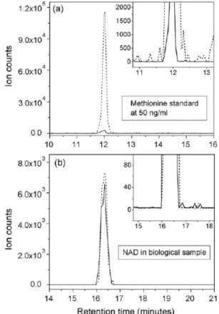
They found that with the use of heated ESI, the sensitivity was at least five times greater than with unheated ESI. Although noise increased, the signal-to-noise ratio was enhanced sig- nificantly. It must be noted that the effect of HESI depends on the compound: for methionine, a 25-fold increase in absolute ion counts was observed, but for NAD, HESI did not provide improvement as seen in Fig. 7. Heat degradation of metabo- lites also must be taken into accounts, but this is a subject of further experiments.
Fig. 7 Comparison of heated and non-heated ESI. As reported, for methio- nine standard (a), a huge increase in intensity was observed, while for NAD+
in biological sample (b) the increase was insignificant. However, as this is compound-dependent, it is worth to check the effect of heating [7].
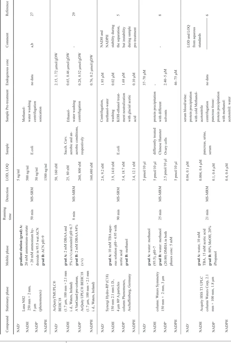
Table 1 Summary of the most important methodological parameters of the attempts to measure pyridine dinucleotides in different biological samples. CompoundStationary phaseMobile phaseRunning timeDetectionLOD, LOQSampleSample Pre-treatmentEndogenous concCommentReference NAD+ Luna NH2 250 mm × 2 mm, 5 μm (phenomenex) gradient elution (grad) A: 20 mM ammonium-acetate + 20 mM ammonium hy- droxide in 95:5 wat:ACN grad B: ACN pH=9
50 minMS-SRM
5 ng/ml E.coli
Methanol- water washing centrifugation sonication
no dataa,b27NADH500 ng/ml NADP+50 ng/ml NADPH1500 ng/ml NAD+AcQuityTMUPLC® BEHC18 (1.7 μm, 100 mm × 2.1 mm i. d., Waters, Ireland) A VanGuard pre-column, AcQuity UPLC® BEHC18 (1.7 μm, 100 mm × 2.1 mm i. d., Waters, Ireland)
grad A: 2 mM DBAA and 5% (v/v) acetonitril pH=6.7 grad B: 2 mM DBAA 84% (v/v)
8 minMS-MRM
50, 160 nM Sacch. Cerv. aerobic and an- aerobic conditions, respestively Ethanol- water washing centrifugation
2.15, 1.72 μmol/gDW -29NADH25, 80 nM0.65, 0.46 μmol/gDW NADP+260, 800 nM0.28, 0.52 μmol/gDW NADPH160,480 nM0.76, 0.2 μmol/gDW NAD+ Synergi Hydro-RP (C18) 150 mm 2.1 mm I. D., 4 μm 80o A particles column Phenomenex, Aschaffenburg, Germany
grad A: 10 mM TBA aque- ous solution pH= 4.95 with acetic acid grad B: methanol
90 minMS-SRM
2.6, 9.2 nM E.coli
Centrifugation, methanol-water washing KOH-ethanol treat- ment neutralization with glacial acetic acid 1.95 μMNADH and NADPH stability during the separation, but instability during sample pre-treatment
5NADH3.9, 14.0 nM0.02 μM NADP+5.4, 18.7 nM1.68 μM NADPH3.4, 12.1 nM0.10 μM NAD+ column: Waters Symmetry 150 mm × 2 mm, 3 μm
grad A: water: methanol (95:5), pH=7 grad B: water: methanol (20:80) DMHA in both phases conc: 5 mM
25 minMS-MRM
5 pmol/10 μl differently treated Chinese Hamster Ovary cells protein precipitation with different solvents
37–70 μM -8NADH5 pmol/10 μl- NADP+2.5 pmol/10 μl2.40–5 μM NADPH5 pmol/10 μl46–75 μM NAD+ Acquity HSS T3 UPLC column Waters Corp, 2.1 mm × 100 mm, 1.8 μm
grad A: water, 10 mM TBA, 15 mM acetic acid grad B: 80% MeOH, 20% iPropanol
21 minMS-MRM
0.04, 0.1 μM pancreas, urine, serum
serum blood/urine: protein precipitation with cold Methanol- acetonitrile centrifugation pancreas tissue: protein precipitation with methanol: acetonitril: water
no data
LOD and LOQ from aqueous standards 6NADH0.004, 0.4 μM NADP+0.1, 0.4 μM NADPH0.4, 0.4 μM
CompoundStationary phaseMobile phaseRunning timeDetectionLOD, LOQSampleSample Pre-treatmentEndogenous concCommentReference NAD+ Luna NH2 250 mm × 2 mm, 3 μm Phenomenex, Le Pecq, France grad A: 20 mM TEAAc + 20 mM Nh4OH in water/ACN (95:5) grad B: ACN
31 minMS-SRM
5 pmol/3*10^7 cells Uninfected RBCs, Parasites, respectively parasite pellet resuspension in cold methanol-water centrifugation in two steps solvent for injection: ACN-water
36.3 pmol/3*10^7 cells -33NADH10 pmol/3*10^7 cellsbelow limit of quantification (blq) NADP+5 pmol/3*10^7 cellsblq NADPH10 pmol/3*10^7 cellsblq NAD+ Luna PFP (2), 150 mm × 2 mm, 3 μm Phenomenex, Torrance, CA, USA
grad A: water with 0,1% formic acid grad B: ACN24 minMS-MRM 1.0 pmol/10 μl M. extorquensHEPES-buffered methanol-water centrifugation
1.66, 1.82 μmol/gDW NADH and NADPH were not stabile dur- ing the cell extraction31NADH0.2 pmol/10 μl0.22, 0.21 μmol/gDW NADP+2.0 pmol/10 μl0.33, 0.36 μmol/gDW NADPH0.8 pmol/10 μl0.49, 0.64 μmol/gDW NAD+Nanoflow HPLC capillary column: 20 cm length, 8 μm opening filled with apHera NH2 gel (5 μm, Supelco, PA)
grad A: 5 mM ammonium acetate in 80% MeOh grad B: 50 mM ammonium carbonate in 20% ACN pH=10 with 25% ammonia solution 70+30 min equi- librationMS-MRMno data Human plasma, CSF, mouse brain, Hela cells
Hela cells: methanol- water, sonication, centrifugation Brain tissue: methanol, sonication, centrifugation Plasma and CSF: filtration with centrifuge filter 0.04 ng/μl, 0.61 ng/μl, 1 ng/mg wet weight 14.62 ng/ 1^4 cells Addition of sodium- free EDTA to the extracted samples32 NADP+Mouse brain, Hela cells
0.22 ng/mg wet weight 1.64 ng/ 1^4 cells NAD+ Luna 5-μm C-8(2) column (150 mm × 4.6 mm) Phenomenex, Torrance, CA, USA
grad A: 50 mM aqueous TEA buffer (adjusted with phosphoric acid to pH 6.5) grad B: acetinitrile 20+25 min equi- librationUV 0.4, 0.9 ng/20 μl Penicillium simplicissimum in different growing conditions
90o ethanol-water, filtration 0.094, 1.463, 1.527, 0.938 mM
NADH is sensitive to pH thus decomposes at the extraction conditions, so recovery was between 40.5–44.8%. But an estimation was done.
22 NADH1.9, 4.1 ng/20 μl0.01, 0.013, 0.012, 0.133 mM NAD+Supelcosil LC-18, 150 mm × 4.6 mm, 3 μm (Sigma-Aldrich/ Supelco, USA) Supelguard, LC-18 2 cm
grad A: 0.1 M, KH2PO4, 8 mM TBAs, 30% (v/v), ACN, pH 6.0 grad B: 0.1 M, KH2PO4, 8 mM TBAs, 30% (v/v), ACN, pH 6.0 22+2 minUVno datacerebellar granule cells ice-cold 0.5 M PCA, neutralization by KOH, centrifugation 0.62 nmol/mg proteinNADH disappeared after treating the std with PCA, no increase in NAD
21 NADH- a) NADH: Unstable (>20% loss) when stored for 1 week at 4oC at pH 2.8, stable (≤20% loss) when stored for 1 week at 4oC at pH 6.8 b) NADPH: Unstable (>20% loss) when stored for 1 week at 4oC at pH 2.8 or pH 6.8, t 1/2>24 hrs at 4oC
8 Calibration curves and endogenous concentration Stock solutions are usually prepared in pure water [4, 5, 22, 24, 31, 35] or in water: methanol 50:50 [30]. After the prepara- tion of a 10 mM standard solution, Klawitter et al. [4] stored it at -80 °C, and prepared working solutions every day which had to be used within two hours at room temperature.
Calibration concentration during the measurements vary among the experiments. In some cases, 1.5 nM was the lower limit of calibration [5, 29], but usually the calibration curves lie between 0.1 µM and 10 µM. Top concentration of calibra- tion was 200 µM in one case [9]. Linearity is usually tested with standard solutions, or treating standard solutions with the sample preparation protocol [2]. Endogenous concentration is generally determined by spiking the sample with a mix of the compounds at different concentrations, or by using isotope labelled internal standards, which are obtained from isotope labelled cell extract [9, 32, 33].
Despite the significant variability in the endogenous pyri- dine dinucleotide levels, the concentrations of these cofactors are always in a very low range in the biological samples. Hence, having a good lowest limit of detection (LOD) and lowest limit of quantification (LOQ) is essential. LOD is usually defined as the concentration where the signal/noise ratio is 3, and at LOQ concentration, the ratio is 10. The results obtained by several research groups using different samples can be seen in Table 1.
It may be noticed, that LOD, LOQ and endogenous concentra- tions are given in different units; the choice usually depends on the type of the sample (tissue, cell culture, etc.), or – especially for LOD and LOQ – whether it is given in concentration or in amount of substance on column.
9 Stability of pyridine dinucleotides
Instability of pyridine dinucleotides is one of the biggest challenges to be met when studying these molecules. Stability of nicotinamide cofactors in solution was tested by Wong et al. [39]. It was shown that reduced forms are more stable in alkaline solution, and oxidized forms are more stable in acidic solutions. This is caused by an acid-catalysed hydration of NADH. Other strong nucleophiles, such as SO32- or CN- also induce decomposition of the oxidized forms. At low pH, where the concentration of sodium-acetate buffer is 0.1 M, the decomposition of NADPH is three times faster than of NADH.
In some cases, disappearance of the reduced forms was observed during sample preparation [23, 33]. Treating the standard solu- tion of NADH with perchloric acid, it disappeared, but increase of NAD+ level was not observed [23]. Yang et al. applied a sam- ple preparation method that turned out to be too drastic for the measurement of the level of reduced forms [33]. As NADPH was unstable at pH 2.8 and 6.8, Bajad et al. decided to leave it out from further analyses. Furthermore, poor performance was observed for NADH as well. When examining E.coli, the
applied LC–MS/MS method provided good separation for all of the four nucleotides in standard solution due to the mild con- ditions. Despite this, the observed concentration of NADH and NADPH in E.coli samples was very low. The conditions during the sample preparation (the use of KOH and glacial acetic acid) might have caused the degradation of the reduced forms [5].
10 Application and conclusion
Pyridine dinucleotides play an essential role in metabolism, and their ratio is fundamental in normal functioning of cells or cell compartments. By monitoring levels of these nucleotides under varying conditions, more information could be achieved about the pathomechanism of metabolic disorders, aging pro- cess, and effects of drug candidates, hence it could help develop treatment for a variety of diseases.
Concentration of NAD+, NADH, NADP+ and NADPH indi- cates the current status of cells hence it can be a marker of numerous disorders. Determination of adenine dinucleotide levels under different nutritional conditions clearly revealed the effect of starvation or a high-sucrose diet [10]. Thus, accurate measurement of pyridine dinucleotide redox couples would largely facilitate researches about metabolic disorders and it could be applied in clinical diagnosis as well.
In the last decade, a wide variety of studies have been con- ducted in order to profile metabolic status of different bacteria, yeast, mammalian cell cultures and tissues. A great number of techniques were used, and it can be declared that HPLC-MS has become the leading method. Several studies were per- formed in order to substitute RP-HPLC, or HILIC, such as pentafluorophenylpropyl column, titania column, or graphitic carbon columns. Capillary electrophoresis may be a potential separation technique, as pyridine dinucleotides are negatively charged molecules. Nano-LC-MS can also represent a potential future technique. Despite the numerous reports, there are still challenges to face. Several studies focused only on oxidized forms of nucleotides, and among those who decided to deter- mine the concentration of both oxidized and reduced forms, many groups reported poor performance or were unable to quantify the concentration of reduced forms due to instability of these compounds [6, 9, 22, 29, 32, 33]. Due to the different characteristics of reduced and oxidized forms, simultaneous determination of both forms is problematic, hence it may be possible that biological matrices need to be prepared in two different ways for separate analysis of NAD(P)+ and NAD(P) H. Therefore, the sample preparation protocols suitable for HPLC-MS quantification of all the four relevant pyridine dinu- cleotides still remain to be elaborated.
Acknowledgement
The authors thank the Hungarian Scientific Research Fund (OTKA 106060) for its support.
References
[1] Sun, F. F., Dai, C. Y., Xie, J. S., Hu, X. "Biochemical Issues in Estimation of Cytosolic Free NAD/NADH Ratio." PLoS One. 7(5), e34525. 2012.
DOI: 10.1371/journal.pone.0034525
[2] Caruso, R., Campolo, J., Dellanoce, C., Mariele, R., Parodi, O., Accinni, R. "Critical study of preanalytical and analytical phases of adenine and pyridine nucleotide assay in human whole blood." Analytical Biochemistry. 330(1), pp. 43-51. 2004. DOI: 10.1016/j.ab.2004.03.063 [3] Kereszturi, E., Kalman, F. S., Kardon, T., Csala, M., Banhegyi, G.
"Decreased prereceptorial glucocorticoid activating capacity in starva- tion due to an oxidative shift of pyridine nucleotides in the endoplasmic reticulum." FEBS Letters. 584(22), pp. 4703-4708. 2010.
DOI: 10.1016/j.febslet.2010.10.053
[4] Klawitter, J., Schmitz, V., Klawitter, J., Leibfritz, D., Christians, U.
"Development and validation of an assay for the quantification of 11 nucleotides using LC/LC-electro spray ionization-MS." Analytical Biochemistry. 365(2), pp. 230-239. 2007. DOI: 10.1016/j.ab.2007.03.018 [5] Luo, B., Groenke, K., Takors, R., Wandrey, C., Oldiges, M. "Simultaneous determination of multiple intracellular metabolites in glycolysis, pentose phosphate pathway and tricarboxylic acid cycle by liquid chromatog- raphy-mass spectrometry." Journal of Chromatography A. 1147(2), pp.
153-164. 2007. DOI: 10.1016/j.chroma.2007.02.034
[6] Michopoulos, F., Whalley, N., Theodoridis, G., Wilson, I. D., Dunkley, T. P. J., Critchlow, S. E. "Targeted profiling of polar intracellular metabo- lites using ion-pair-high performance liquid chromatography and -ultra high performance liquid chromatography coupled to tandem mass spec- trometry: Applications to serum, urine and tissue extracts." Journal of Chromatography A. 1349, pp. 60-68. 2014.
DOI: 10.1016/j.chroma.2014.05.019
[7] Lu, W., Bennett, B. D., Rabinowitz, J. D. "Analytical strategies for LC-MS-based targeted metabolomics." Journal of Chromatography B - Analytical Technologies in the Biomedical and Life Sciences. 871(2), pp.
236-242. 2008. DOI: 10.1016/j.jchromb.2008.04.031
[8] Cordell, R. L., Hill, S. J., Ortori, C. A., Barrett, D. A. "Quantitative profiling of nucleotides and related phosphate-containing metabolites in cultured mammalian cells by liquid chromatography tandem electro- spray mass spectrometry." Journal of Chromatography B - Analytical Technologies in the Biomedical and Life Sciences. 871(1), pp. 115-124.
2008. DOI: 10.1016/j.jchromb.2008.07.005
[9] Trammell, S. A., Brenner, C. "Targeted, LCMS-based Metabolomics for Quantitative Measurement of NAD(+) Metabolites." Computational and Structural Biotechnology Journal. 4, e201301012. 2013.
DOI: 10.5936/csbj.201301012
[10] Veech, R. L., Eggleston, L. V., Krebs, H. A. "The redox state of free nic- otinamide-adenine dinucleotide phosphate in the cytoplasm of rat liver."
Biochemical Journal. 115(4), pp. 609-619. 1969.
[11] Gomes, A. P., Price, N. L., Ling, A. J. Y., Moslehi, J. J., Montgomery, M.
K., Rajman, L., White, J. P., Teodor, J. S., Wrann, C. D., Hubbard, B. P., Mercken, E. M., Palmeira, C. M., de Cabo, R., Rolo, A. P., Turner, N., Bell, E. L., Sinclair, D. A. "Declining NAD(+) Induces a Pseudohypoxic State Disrupting Nuclear-Mitochondrial Communication during Aging."
Cell. 155(7), pp. 1624-1638. 2013. DOI: 10.1016/j.cell.2013.11.037 [12] Liu, L. L., Cui, Z. Y., Deng, Y. Z., Dean, B., Hop, C. E. C. A., Liang, X.
R. "Surrogate analyte approach for quantitation of endogenous NAD(+) in human acidified blood samples using liquid chromatography coupled with electrospray ionization tandem mass spectrometry." Journal of Chromatography B - Analytical Technologies in the Biomedical and Life Sciences. 1011, pp. 69-76. 2016. DOI: 10.1016/j.jchromb.2015.12.040
[13] Csala, M., Banhegyi, G., Benedetti, A. "Endoplasmic reticulum: A metabolic compartment." FEBS Letters. 580(9), pp. 2160-2165. 2006.
DOI: 10.1016/j.febslet.2006.03.050
[14] Banhegyi, G., Benedetti, A., Csala, M., Mandl, J. "Stress on redox."
FEBS Letters. 581(19), pp. 3634-3640. 2007.
DOI: 10.1016/j.febslet.2007.04.028
[15] Zambo, V., Simon-Szabo, L., Szelenyi, P., Kereszturi, E., Banhegyi, G., Csala, M. "Lipotoxicity in the liver." World Journal of Hepatology.
5(10), pp. 550-557. 2013. DOI: 10.4254/wjh.v5.i10.550
[16] Senesi, S., Legeza, B., Balazs, Z., Csala, M., Marcolongo, P., Kereszturi, E., Szelenyi, P., Egger, C., Fulceri, R., Mandl, J., Giunti, R., Odermatt, A., Banhegyi, G., Benedetti, A. "Contribution of Fructose-6-Phosphate to Glucocorticoid Activation in the Endoplasmic Reticulum: Possible Implication in the Metabolic Syndrome." Endocrinology. 151(10), pp.
4830-4839. 2010. DOI: 10.1210/en.2010-0614
[17] Margittai, E., Enyedi, B., Csala, M., Geiszt, M., Banhegyi, G.
"Composition of the redox environment of the endoplasmic reticulum and sources of hydrogen peroxide." Free Radical Biology & Medicine.
83, pp. 331-340. 2015. DOI: 10.1016/j.freeradbiomed.2015.01.032 [18] Piccirella, S., Czegle, I., Lizak, B., Margittai, E., Senesi, S., Papp, E.,
Csala, M., Fulceri, R., Csermely, P., Mandl, J., Benedetti, A., Banhegyi, G. "Uncoupled redox systems in the lumen of the endoplasmic reticu- lum - Pyridine nucleotides stay reduced in an oxidative environment."
Journal of Biological Chemistry. 281(8), pp. 4671-4677. 2006.
[19] Bublitz, C., Lawler, C. A. "The Levels of Nicotinamide Nucleotides in Liver-Microsomes and Their Possible Significance to the Function of Hexose Phosphate Dehydrogenase." Biochemical Journal. 245(1), pp.
263-267. 1987.
[20] Odermatt, A., Atanasov, A. G., Balazs, Z., Schweizer, R. A. S., Nashev, L. G., Schuster, D., Langer, T. "Why is 11 beta-hydroxy steroid dehydro- genase type 1 facing the endoplasmic reticulum lumen? Physiological relevance of the membrane topology of 11 beta-HSD1." Molecular and Cellular Endocrinology. 248(1-2), pp. 15-23. 2006.
DOI: 10.1016/j.mce.2005.11.040
[21] Zhou, T., Lucy, C. A. "Hydrophilic interaction chromatography of nucleotides and their pathway intermediates on titania." Journal of Chromatography A. 1187(1-2), pp. 87-93. 2008.
DOI: 10.1016/j.chroma.2008.02.027
[22] Cichna, M., Daxecker, H., Raab, M. "Determination of 18 nucleobases, nucleosides and nucleotides in human peripheral blood mononuclear cells by isocratic solvent-generated ion-pair chromatography." Analytica Chimica Acta. 481(2), pp. 245-253. 2003.
DOI: 10.1016/S0003-2670(03)00081-3
[23] Giannattasio, S., Gagliardi, S., Samaja, M., Marra, E. "Simultaneous determination of purine nucleotides, their metabolites and beta-nicoti- namide adenine dinucleotide in cerebellar granule cells by ion-pair high performance liquid chromatography." Brain Research Protocols. 10(3), pp. 168-174. 2003. DOI: 10.1016/S1385-299x(02)00215-5
[24] Ganzera, M., Vrabl, P., Worle, E., Burgstaller, W., Stuppner, H.
"Determination of adenine and pyridine nucleotides in glucose-lim- ited chemostat cultures of Penicillium simplicissimum by one-step ethanol extraction and ion-pairing liquid chromatography." Analytical Biochemistry. 359(1), pp. 132-140. 2006. DOI: 10.1016/j.ab.2006.09.012 [25] Markuszewski, M. J., Britz-McKibbin, P., Terabe, S., Matsuda, K.,
Nishioka, T. "Determination of pyridine and adenine nucleotide metabo- lites in Bacillus subtilis cell extract by sweeping borate complexation capillary electrophoresis." Journal of Chromatography A. 989(2), pp.
293-301. 2003.
[26] Soga, T., Ueno, Y., Naraoka, H., Ohashi, Y., Tomita, M., Nishioka, T.
"Simultaneous determination of anionic intermediates for Bacillus subti- lis metabolic pathways by capillary electrophoresis electrospray ionization mass spectrometry." Analytical Chemistry. 74(10), pp. 2233-2239. 2002.
[27] Palfi, M., Halasz, A. S., Tabi, T., Magyar, K., Szoko, E. "Application of the measurement of oxidized pyridine dinucleotides with high-perfor- mance liquid chromatography-fluorescence detection to assay the uncou- pled oxidation of NADPH by neuronal nitric oxide synthase." Analytical Biochemistry. 326(1), pp. 69-77. 2004. DOI: 10.1016/j.ab.2003.11.010 [28] Becker, S., Kortz, L., Helmschrodt, C., Thiery, J., Ceglarek, U.
"LC-MS-based metabolomics in the clinical laboratory." Journal of Chromatography B - Analytical Technologies in the Biomedical and Life Sciences. 883-884, pp. 68-75. 2012. DOI: 10.1016/j.jchromb.2011.10.018 [29] Bajad, S. U., Lu, W., Kimball, E. H., Yuan, J., Peterson, C., Rabinowitz,
J. D. "Separation and quantitation of water soluble cellular metabolites by hydrophilic interaction chromatography-tandem mass spectrometry."
Journal of Chromatography A. 1125(1), pp. 76-88. 2006.
DOI: 10.1016/j.chroma.2006.05.019
[30] Uehara, T., Yokoi, A., Aoshima, K., Tanaka, S., Kadowaki, T., Tanaka, M., Oda, Y. "Quantitative phosphorus metabolomics using nanoflow liquid chromatography-tandem mass spectrometry and culture-derived comprehensive global internal standards." Analytical Chemistry. 81(10), pp. 3836-3842. 2009. DOI: 10.1021/ac9002062
[31] Buescher, J. M., Moco, S., Sauer, U., Zamboni, N. "Ultrahigh perfor- mance liquid chromatography-tandem mass spectrometry method for fast and robust quantification of anionic and aromatic metabolites." Analytical Chemistry. 82(11), pp. 4403-4412. 2010. DOI: 10.1021/ac100101d [32] Seifar, R. M., Ras, C., Deshmukh, A. T., Bekers, K. M., Suarez-Mendez,
C. A., da Cruz, A. L., van Gulik, W. M., Heijnen, J. J. "Quantitative analysis of intracellular coenzymes in Saccharomyces cerevisiae using ion pair reversed phase ultra high performance liquid chromatography tandem mass spectrometry." Journal of Chromatography A. 1311, pp.
115-120. 2013. DOI: 10.1016/j.chroma.2013.08.076
[33] Yang, S., Sadilek, M., Lidstrom, M. E. "Streamlined pentafluorophe- nylpropyl column liquid chromatography-tandem quadrupole mass spectrometry and global (13)C-labeled internal standards improve performance for quantitative metabolomics in bacteria." Journal of Chromatography A. 1217(47), pp. 7401-7410. 2010.
DOI: 10.1016/j.chroma.2010.09.055
[34] Myint, K. T., Uehara, T., Aoshima, K., Oda, Y. "Polar anionic metabo- lome analysis by nano-LC/MS with a metal chelating agent." Analytical Chemistry. 81(18), pp. 7766-7772. 2009. DOI: 10.1021/ac901269h [35] Duy, S. V., Besteiro, S., Berry, L., Perigaud, C., Bressolle, F., Vial, H.
J., Lefebvre-Tournier, I. "A quantitative liquid chromatography tandem mass spectrometry method for metabolomic analysis of Plasmodium falciparum lipid related metabolites." Analytica Chimica Acta. 739, pp.
47-55. 2012. DOI: 10.1016/j.aca.2012.06.016
[36] Seifar, R. M., Ras, C., van Dam, J. C., van Gulik, W. M., Heijnen, J. J., van Winden, W. A. "Simultaneous quantification of free nucleotides in complex biological samples using ion pair reversed phase liquid chro- matography isotope dilution tandem mass spectrometry." Analytical Biochemistry. 388(2), pp. 213-219. 2009. DOI: 10.1016/j.ab.2009.02.025 [37] Alpert, A. J. "Hydrophilic-Interaction Chromatography for the Separation of Peptides, Nucleic-Acids and Other Polar Compounds." Journal of Chromatography. 499, pp. 177-196. 1990.
DOI: 10.1016/S0021-9673(00)96972-3
[38] Hanai, T. "Separation of polar compounds using carbon columns."
Journal of Chromatography A. 989(2), pp. 183-196. 2003.
DOI: 10.1016/S0021-9673(02)02017-4
[39] Wong, C. H., Whitesides, G. M. "Enzyme-Catalyzed Organic-Synthesis - Nad(P)H Cofactor Regeneration by Using Glucose-6-Phosphate and the Glucose-6-Phosphate-Dehydrogenase from Leuconostoc-Mesenteroides."
Journal of the American Chemical Society. 103(16), pp. 4890-4899. 1981.
DOI: 10.1021/Ja00406a037

![Fig. 3 The MRM chromatograms obtained by using normal phase nanoflow liquid chromatography of pyridine dinucleotides of human HCT116 cells, log phase E.coli and stationary phase E.coli cells [30].](https://thumb-eu.123doks.com/thumbv2/9dokorg/1348265.109538/5.892.190.718.73.241/chromatograms-obtained-normal-nanoflow-chromatography-pyridine-dinucleotides-stationary.webp)

![Fig. 6 Results of MRM scan (dwell 0.2 s) of simultaneous detection of 24 nucleotides in standard solution (a) and for biological sample in control conditions (b) [8]](https://thumb-eu.123doks.com/thumbv2/9dokorg/1348265.109538/7.892.136.770.74.416/results-simultaneous-detection-nucleotides-standard-solution-biological-conditions.webp)