THE ELABORATION OF GLYCOPROTEINS INTO CULTURE MEDIUM BY HUMAN OVARIAN TUMOR CELLS
K, J, ALLEN J. J, BARLOW Å, A, Z. JOHNSON
Roswell Park Memorial Institute Department of Gynecology
Buffalo, New York
I. INTRODUCTION
Ovarian cancer is the fourth most frequently fatal cancer in women in the United States. There are generally no early symp- toms for this disease and diagnosis is usually made at an ad- vanced stage when prognosis is poor (Lingeman, 1974). Epithelial cancers of the ovary are the predominant types of which serous tumors account for 40-60%; mucinous tumors account for 10-15%; un- differentiated tumors account for 16%; and endometrioid tumors account for 14% (Lingeman, 1974; Barber et al., 1975; Curling et al., 1975; and Young et al., 1974).
Some of the clinical features of this disease include (1) high cure rate (75-90%l for early cancer; C2) low cure rate
(<10%) for advanced cancer; C3) tumor implantation on the peri-
689
690 H. J. ALLEN et al.
toneal surface; C4) plugging of the lymphatics draining the peri- toneal cavity; C5) generation of large volumes of ascites with occasionally large numbers of free tumor islets present; and (6) occasional generation of pleural effusions due to metastatic spread.
As part of a program to elucidate the role of cell surface glycoproteins in carcinogenic and metastatic processes, we have chosen ovarian cancer as one of our model systems. It is well known that a variety of cells elaborate glycoproteins into their growth medium and that at least some of these glycoproteins are derived from the cell surface (Molnar et al., 1965; Cooper et al., 1974; Ruoslahti et al., 1975; Kraemer, 1967). It has also been demonstrated that some cells derived from human cancer can elaborate tumor-specific (associated) products into their culture medium (Hickok et al,, 1974; Breborowicz et al., 1975). We have therefore made the assumption that ovarian tumor cells elaborate glycoproteins into their growth medium in vivo. Since ascites may result from the movement of fluids into the peritoneal cavity but not from the cavity (Feldman et al., 1974; and Feldman, 1975), the tumor-derived glycoproteins might accumulate in this fluid.
The amount of tumor-derived glycoproteins present would depend upon many factors, including tumor load, but is expected to be small.
In order to develop a method for detecting all tumor-derived glycoproteins present in pleural and peritoneal effusions inde- pendent of antigenicity, we have carried out studies on the bio- synthesis and secretion of glycoproteins in vitro by human ovarian tumor cells. With the use of radioactive glycoprotein precursors, it was hoped to obtain radiolabeled glycoproteins from in vitro cultures which could be used as markers for the isolation of tumor-derived glycoproteins present in the patient effusions.
GLYCOPROTEINS INTO CULTURE MEDIUM 691 II. EXPERIMENTAL
A, Materials
D-l6-3H] glucosamine CSp, act. 10.13 Ci/mM, L-[6-3H] fucose (Sp. act. 12.066 Ci/mM) and Aquasol were obtained from New England Nuclear, Boston, Mass. Sephadex and Sepharose were purchased from Pharmacia Fine Chemicals, Piscataway, N. J. RPMI 1640 culture medium with HEPES buffer was obtained from Grand Island Biologi- cal Co., Grand Island, Ν. Y.
Â. Methods
1. Cell Work-up from Patient Effusions
Fresh pleural and peritoneal effusions obtained from ovarian cancer patients were centrifuged for 15 minutes, 500 xg at 4°C.
Cell pellets were examined microscopically and slide smears were prepared.
The red blood cells in excessively bloody pellets were lysed by suspending the cells in 20 volumes of 0.83% ammonium chloride and incubating for 10 minutes at 4°C. The cells were washed 2 times with 20 volumes of cold PBS (0.15 M NaCl, 0.01 M phosphate, pH 7.2). For some cell preparations, the tumor cell population was enriched by incubating 10 ml portions of a PBS cell suspen-
sion in 4 oz Blake bottles at 37°C for 30 minutes. The tumor cells were poured from the bottles while the normal cells re- mained attached to the glass. The cells were suspended in 10 volumes PBS and the cell concentration was estimated by counting the cells in a hemocytometer. Cell viability was determined by the Trypan Blue method.
2. Cell Culture Conditions
Large cultures were set up in 4 oz glass Blake bottles con- taining 30 * 1 06 cells in 10 ml medium consisting of 90% RPMI
692 H. J. ALLEN et al.
1640 medium with. HEPES buffer, 10% heat inactivated autologous patient effusion, 0.3 mg streptomycin and 0.5 mg penicillin. To each were added 25 yCi of D-[6-3H] glucosamine or L-[6-3H] fucose.
The bottles were capped tightly and laid flat in a 37°C incu- bator for 15 to 40 hours.
For time course experiments, 1 ml cultures were set up in 20 x 40 mm serum vials with one-tenth the quantities used for the large cultures.
3. Work-up of Cell Cultures
The culture medium from large cultures was separated from the cells by centrifugation, dialyzed against deionized water and lyophilized.
At given time intervals, time course cultures were centri- fugea . To 0.7 ml culture medium were added 2,5 ml ice cold 12.5%
TCA. TCA precipitates were washed 4 times with cold 10% TCA, dissolved in 0.5 ml of 0.5 M KOH, bleached with 50 μÀ of 50%
H202 and transferred to polyethylene Mini-vials with 5 ml Aquasol for tritium determination.
TCA supernatants were dialyzed exhaustively against deionized water and lyophilized in glass scintillation vials. To the vials were added 10 ml Aquasol.
The cells were washed 4 times with cold PBS and suspended in 0.7 ml of 0.5% bovine serum albumin solution. From this stage, TCA-precipitable and soluble fractions of the cells were processed as described for the culture medium. All radioactivity deter- minations were carried out in a Packard 3375 Tri-Carb scintilla- tion spectrometer.
4. Gel Filtration Chromatography
Sephadex and Sepharose chromatography was carried out with a buffer consisting of 0,15 M NaCl, 0,015 M phosphate, pH = 7.0, 0.05% NaN3, A constant ascending flow was maintained with a Gil-
GLYCOPROTEINS INTO CULTURE MEDIUM 693 son Mini^pulse pump. Column temperature was maintained at 18°C.
Load samples were dissolved in 5 ml column buffer and the solu- tions were clarified by centrifugation before applying to gel columns. Column effluents were monitored for U.V. absorbing material. Aliquots of effluent fractions were placed in poly- ethylene Mini-vials with 4 ml Aquasol and their radioactivity was measured.
Ill. RESULTS
A. Cell Preparations
The biosynthesis and secretion of glycoproteins by four dif- ferent cell populations were investigated. The cells shown in Fig. 1 were obtained from a peritoneal effusion of a patient with a serous cystadenocarcinoma of the ovary. About 97% of the cell population were non-malignant cells which included some leuko- cytes, mesothelial cells and a large number of activated histio- cytes. Many of the latter had phagocytized erythrocytes. Since there were a very small number (less than 3%) of malignant cells present in this preparation, it was used in these studies as a
"normal" cell population and has served as a control for compara- tive purposes.
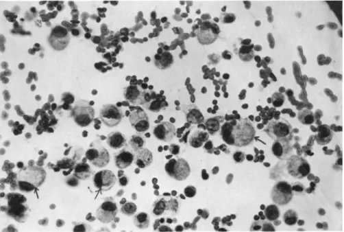
In Fig. 2 are shown the cells from a pleural effusion of a patient with a primary Krukenberg tumor of the ovary. A minimum of 75% of these cells were malignant. The malignant cells were characteristically large, single cells with a highly vacuolated cytoplasm and a large, irregular nucleus.
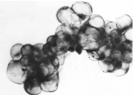
Tumor cells obtained from peritoneal effusions of patients with serous and mucinous cystadenocarcinoma of the ovary are
shown in Figs. 3 and 4, respectively, Tumor cells in the effu- sions of these two types of ovarian cancer were usually present
694 H. J. ALLEN et al.
FIGURE 1 Íïç-malignant cells obtained from the peritoneal effusion of a patient with serous cystadenocarcinoma of the ovary.
Papanicolaou staining. Magnification 630 X.
as clusters which had from several to about 50 cells. The cells contained large vacuoles, large irregular nuclei and often, promi- nent nucleoli were observed.
It was found that the tumor cell clusters did not adhere to glass as did the viable normal cells. This difference was used to enrich the tumor cell population to more than 90% in these preparations as described in Methods B. 1.
From viability determinations it was observed that 90% or more of the tumor cells and of normal cells which adhered to glass were viable. Invariably, a very low viability was observed for the normal cells which did not attach to glass surfaces.
GLYCOPROTEINS INTO CULTURE MEDIUM 695
FIGURE 2 Mixture of malignant and non-malignant cells ob- tained from pleural effusion of patient with primary Krukenberg tumor of the ovary. Several malignant cells are indicated by ar- rows. Papanicolaou staining. Magnification 630 X.
B. Time Course Incorporation of D-[6-3H] Glucosamine
Time course experiments were carried out to examine the abili- ty of cell preparations to incorporate radiolabeled glycoprotein precursor into cellular components and macromolecules elaborated into the culture medium.
The radioactivity present in the TCA-precipitable and TCA- soluble non-dialyzable fractions from cells cultured with triti- ated glucosamine has been plotted as a function of time in Fig.
5. Very little radioactivity was incorporated into the TCA- soluble non-dialyzable fraction from normal cells; whereas, there was a significant amount of radioactivity in corresponding frac- tions from both types of tumor cells.
696 H. J. ALLEN et al.
FIGURE 3 Cluster of malignant cells obtained from peritoneal effusion of patient with serous cystadenocarcinoma of the ovary.
Papanicolaou staining. Magnification 630 X.
The incorporation of tritiated glucosamine into TCA-precipi- table cellular glycoproteins continues for at least 120 hours with respect to the normal cell population. In contrast, for the serous tumor cells, the incorporation of tritiated glucosamine into corresponding fractions reaches a plateau after 50-60 hours.
For the Krukenberg tumor cell population a plateau was reached after 30-40 hours. The experiment with the Krukenberg tumor cells was repeated on a different date and similar results were ob- tained. The decline in cellular TCA-precipitable glycoproteins for the Krukenberg tumor cells appeared to be due to cellular leakage during the washing of the cultured cells. The time course of elaboration of glycoproteins into the culture medium is shown in Fig. 6. As was the case for the cellular glycoproteins, the normal cells incorporated no tritiated glucosamine into the TCA- soluble non-dialyzable fraction of the culture medium. However, a significant portion of the radiolabeled glycoproteins elaborated
FIGURE 4 Cluster of malignant cells obtained from peritoneal effusion of patient with mucinous cystadenocarcinoma of the ovary.
Papanicolaou staining. Magnification 630 X.
by the tumor cells into the culture medium were in the TCA soluble non-dialyzable fraction.
The incorporation of label into the acid-precipitable extra- cellular glycoproteins plateaued after about 120 hours for the serous tumor cells, plateaued after 60-80 hours for the Kruken- berg tumor cells whereas no plateau was evident up to 120 hours for the normal cell population.
C. Sephadex Gel Filtration of Radiolabeled Glycoproteins in Culture Medium
From the time course experiments it was apparent that the cultured cells elaborated glycoproteins into the culture medium.
To determine the molecular weight distribution of the radiolabeled glycoproteins, the dialyzed and lyophilized culture media were subjected to gel filtration on columns of Sephadex G-150 or G- 200, The elution profiles for glucosamine-labeled glyco- proteins are shown for four different cell types in Figs, 7, 8, 9 and 10, Essentially identical profiles were obtained for all culture media examined. Virtually all of the radioactivity was
697
698 H. J. ALLEN et al.
FIGURE 5 Time course incorporation of [JE] glucosamine into TCA-precipitable (·} and TCA-soluble non-dialyzable £xj fractions of cells incubated for 0 to 140 hours.
eluted in the excluded fraction while only a small amount of ra- dioactivity was distributed in the retarded fractions. No dif- ference was observed in the radioactivity elution profile when 5 mg/ml cold D-glucosamine was included in the column load.
The protein elution profiles were also determined for these experiments. Protein profiles for all culture media were similar and the protein peaks corresponded to the three major peaks ob-
GLYCOPROTEINS INTO CULTURE MEDIUM 699
FIGURE 6 Time course incorporation of [ E] glucosamine into TCA-precipitable (·) and TCA-soluble non-dialyzable (x) fractions of culture medium from cells incubated for 0 to 140 hours.
served when either normal human serum or plasma was chromato- graphed on the same columns.
In Fig. 11 is shown a comparison of the elution profiles for the [3H] glucosamine-labeled and [3H] fucose-labeled glycoproteins present in the culture media obtained from serous tumor cell cul- tures. The profile for the I3H] fucose-labeled glycoproteins was identical to that obtained for the I3H] glucosamine-labeled glyco- proteins with the exception that a somewhat greater proportion of the [3H] fucose label was eluted in retarded fractions. This difference in elution profiles has been consistently observed for all culture media studied.
700 H. J. ALLEN et al.
R.N. NORMAL
ELUTION VOLUME (ML)
FIGURE 7 Sephadex G-150 chromatography of culture medium from normal cell cultures incubated with [ H] glucosamine. The culture medium from five 10 ml cultures incubated for 40 hours was prepared for chromatography as described in Methods B.4.
Column: 2.6 x 95.5 cm. (Vo = 201.6 ml); flow rate = 17.1 ml/hr;
fraction folume = 5 ml. Radioactivity in 50 yl aliquots from fractions was determined.
Sephadex gel filtration profiles for [3H] glucosamine and [3H] fucose labeled glycoproteins have been obtained for the cul- ture media derived from cultures of (1) cells collected from the same patient on different dates; (2) cells collected from several patients with serous cystadenocarcinoma of ovary; and (3) cells collected from patients with different histological types of o- varian cancer. All of these profiles for the [3H] glucosamine- labeled glycoproteins were similar to those shown in Figs. 7-11.
As was noted previously, all of the elution profiles for the [3H]
fucose-labeled glycoproteins were similar to each other with a consistently greater proportion of the radioactivity present in
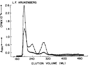
GLYCOPROTEINS INTO CULTURE MEDIUM 701 L.F. KRUKENBERG
400 480 ELUTION VOLUME (ML)
FIGURE 8 Sephadex G-150 chromatography of culture medium from cultures of Krukenberg tumor cells. Culture medium from five 10 ml cultures incubated with [-%] glucosamine for 18 hours was prepared for chromatography as described in Methods B.4.
Column: 2.6 x 95.5 cm (Vo = 201.6 ml); flow rate = 17.0 ml/hr;
fraction volume = 5 ml. Radioactivity in 100 \il aliquots from fractions was determined.
the retarded fractions as compared to the [3H] glucosamine-labeled glycoproteins.
D. Sepharose 6B Gel Filtration of the Excluded Fractions from Sephadex Columns
To further resolve the high molecular weight radiolabeled glycoproteins elaborated by the cultured cells, the excluded frac- tion from Sephadex chromatography of the four different culture media were chromatographed on columns of Sepharose 6B. As shown in Figs. 12-14, the radioactivity and protein of the 3 tumor cell- derived fractions were both resolved into an excluded fraction and a broad retarded fraction in which the radioactivity was somewhat displaced from the protein toward the high molecular weight re- gion. In Fig. 15 is shown the protein and radioactivity profile obtained for thß normal cell-derived fraction, In contrast to the tumor cell-derived fractions, neither protein nor radioactivi-
702 H. J. ALLEN et al.
FIGURE 9 Sephadex G-200 chromatography of culture medium from serous cystadenocarcinoma tumor cell cultures. Half of the culture medium from five 10 ml cultures incubated with \3H\ glu- cosamine for 14.5 hours was prepared as described in Methods B.4.
Column: 2.6 x 94.5 cm (Vo = 218 ml); flow rate= 17.0 ml/hr; frac- tion volume = 5 ml. Radioactivity in 300 \il aliquots from frac- tions was determined.
ty were eluted in the excluded fraction from the Sepharose 6B column. A similar displacement of retarded radioactivity in re- lation to retarded protein was observed but the radioactivity ap- peared to be less heterogenous than that obtained from the tumor cell-derived fractions.
In Fig. 16 is shown a comparison of the Sepharose 6B elution profiles for the [3H] gludosamine-labeled and [3H] fucose-labeled glycoproteins of the excluded fractions from the Sephadex chro- matography shown in Fig. 11. No significant differences in the elution profiles were observed.
GLYCOPROTEINS INTO CULTURE MEDIUM 703
D.K. MUCINOUS
40.75
0.25
180 220 260 300 340 380 ELUTION VOLUME (ML)
420
FIGURE 10 Sephadex G-200 chromatography of culture medium from cultures of mucinous cystadenocarcinoma tumor cells. Culture medium from ten 10 ml cultures incubated with [-%] glucosamine for 24 hours was prepared for chromatography as described in Methods B.4. Column: 2.6 x 94.5 cm (Vo = 218 ml); flow rate = 16.9 ml/hr;
fraction volume 5.0 ml. Radioactivity in 50 μÀ aliquots from fractions was determined.
IV. DISCUSSION
For the characterization of ovarian tumor glycoproteins, it has been proposed that pleural and peritoneal effusions may serve as a source of tumor-derived glycoproteins. This followed from the postulate that tumor cells present in the effusions and/or im- planted on the pleura or peritoneum may elaborate glycoproteins into the effusion and that these glycoproteins will accumulate there since the drainage of the effusion from the pleural and peritoneal cavities has been severely restricted or completely blocked.
In the studies reported here, viable cells were collected from effusions and cultured in media containing radiolabeled gly-
τ 1-0
704 H. J. ALLEN et ai
FIGURE 11 Comparison of radioactivity profiles from Sephadex G-150 chromatography of culture medium from cultures of serous cyst adenocarcinoma tumor cells. Culture medium from four 10 ml cultures incubated with [%] glucosamine for 15 hours and another nine 10 ml cultures incubated with [^H] fucose for 15 hours were prepared separately for chromatography as described in Methods B.4. Sample load volume = 10 ml; Column: 2.5 x 89.2 cm (Vo = 140 ml); flow rate = 7.0 ml/hr; fraction volume = 4 ml and 7.75 ml for
[^H~\ glucosamine and [^H] fucose labeled culture medium, respec- tively. Radioactivity in 100 yl aliquots from fractions was de- termined .
coprotein precursors. The results of the time course experiments shown in Figs. 5 and 6 indicate that both normal cells (i.e. non- malignant cells) and tumor cells are quite capable of synthesiz- ing and elaborating glycoproteins into the culture medium for a period of 20-120 hours. When comparing different cell popula- tions, differences were observed between the normal and tumor cells with respect to the amount and kinetics of radiolabel ap^
pearing in the TCA-precipitable and TCA-soluble non-dialyzable fractions from both, cells and culture medium. However, since we do not know what affect the chemotherapy administered to the donor
GLYCOPROTEINS INTO CULTURE MEDIUM 705 L.F KRUKENBERG
160 240 320 400 ELUTION VOLUME (ML)
FIGURE 12 Sepharose 6B chromatography of excluded fraction from Sephadex chromatography of culture medium from Krukenberg tumor cell culture. Column: 2.6 x 93.1 cm (Vo = 186.2 ml); flow rate = 16.4 ml/hr; fraction volume = 5 ml. Radioactivity in 100
\il aliquots from fractions was determined.
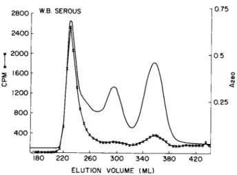
W.B. SEROUS
160 240 320 400 ELUTION VOLUME (ML)
FIGURE 13 Sepharose 6B chromatography of excluded fraction from Sephadex chromatography of culture medium from serous cysta~
denocarcinoma tumor cell cultures, Chromatography conditions as described for Fig. 12. Radioactivity in 300 μÀ aliquots from fractions was determined.
706 H. J. ALLEN et al.
FIGURE 14 Sepharose 6B chromatography of excluded fraction from Sephadex chromatography of culture medium from mucinous cys- tadenocarcinoma tumor cell cultures. Chromatography conditions as described for Fig, 12, Radioactivity in 100 yl aliquots from fractions was determined.
R.N. NORMAL
160 240 320 4 00 ELUTION VOLUME (ML)
FIGURE 15 Sepharose 6B chromatography of excluded fraction from Sephadex chromatography of culture medium from normal cell cultures. Chromatography conditions as described for Fig, 12, Radioactivity in 100 yxl aliquots from fractions was determined.
GLYCOPROTEINS INTO CULTURE MEDIUM 707
FIGURE 16 Comparison of radioactivity profiles from Sepharose 6B chromatography of excluded fractions from Sephadex chromatog- raphy described for Fig. 11, Sepharose chromatography conditions the same as described for Fig. 12. Radioactivity in 50 \il ali- quots from fractions was determined.
patient may have on the cells, no definitive conclusions may be made at this time about differences observed in the kinetics of
[3H] glucosamine incorporation.
To assess the ability of cells to replicate in the culture system described, several time course experiments were carried out in which the incorporation of [3H] thymidine into DNA was de- termined. The results indicated that some DNA synthesis occurred during the first several hours in culture.
Sephadex gel filtration of the culture media indicated that most of the radiolabeled precursor was incorporated into high molecular weight glycoproteins as evidenced by their elution in
the excluded fractions from the columns. Since more than two- thirds of the unlabeled protein was eluted in the retarded frac- tion, a significant enrichment of the high molecular weight radi- olabeled glycoprotein was achieved.
708 H. J. ALLEN et al.
It should be made note of that when the dialyzed and lyophi- lized culture medium was reconstituted with column buffer, usually a small portion of the lyophilizate was insoluble. This was also true of the dialyzed and lyophilized high molecular weight frac- tions obtained from the Sephadex chromatography of culture media.
The nature of these fractions has not been studied.
Sepharose 6B chromatography of the high molecular weight fractions from Sephadex elution of the culture media showed the presence of a radioactive component Cs) excluded by the gel and a retarded radioactive fraction. The elution profile of the latter indicated the presence of molecular weight heterogeneity. The displacement of the retarded peak of radioactivity from the re- tarded protein peak suggested that the radiolabeled glycoproteins are distinctly different from the bulk protein in the effusions and are characteristic of the cultured cells. The absence of an excluded radiolabeled fraction and the symmetrical retarded peak of radioactivity obtained from the Sepharose 6B chromatography of the high molecular weight fraction from the normal cell culture medium was in contrast to the elution profiles obtained for the Sepharose chromatography of the high molecular weight fraction from the tumor cell culture media. The significance of these dif- ferences awaits further study.
The isolation and charact erization of the radiolabeled gly- coproteins are now in progress.
ACKNOWLEDGMENTS
We are grateful to Dr. Marie Gamarra of the Department of Cy- tology for her valuable assistance in classifying cell popula- tions, We thank Miss Jessica Pawlowicz for her technical assis- tance .
This work was supported by Grant DRG-1221 awarded by the Damon Runyon Memorial Fund for Cancer Research Incorporated and
GLYCOPROTEINS INTO CULTURE MEDIUM 709 by NIH Grant 1R01 CA 14854-OlAl from the National Cancer Insti-
tute.
REFERENCES
Barber, H. R. K., Sommers, S. C , Snyder, R. and Kwon, T. H.
(1975). Am. J. Obstet. Gynec. 121: 795-807.
Breborowicz, J., Easty, G. C , Birbeck, Ì., Robertson, D., Nery, R. and Neville, A. M. (1975). Br. J. Cancer 31, 559-569.
Cooper, A. G., Codington, J. F. and Brown, M. (1974). Proc. Nat.
Acad. Sei. U.S.A. 71, 1224-1228.
Curling, Ο. M. and Hudson, C. N. (1975). Br. J. Obstet. Gynaec.
82, 405-411.
Feldman, G. Â. (1975). Cancer Res. 35, 325-332.
Feldman, G. Â. and Knapp, R. C. (1974). Am. J. Obstet. Gynec.
119: 991-994.
Hickok, D. F. and Miller, L. 0-974). In Vitro. 10, 157-166.
Kraemer, P. M. (1967). J. Cell Physiol. 69, 199-208.
Lingeman, C. H. (1974). J. Nat. Cancer Inst. 53, 1603-1618.
Molnar, J., Teegarden, D. W. and Winzler, R. J. (1965). Cancer Res. 25, 1860-1866.
Ruoslahti, E. and Vaheri, A. (1975). J. Exptl. Med. 141, 497-501.
Young, R. C. and DeVita, V. T. (1974). Am. J. Obstet. Gynec. 120, 1012-1024.


![FIGURE 5 Time course incorporation of [ J E] glucosamine into TCA-precipitable (·} and TCA-soluble non-dialyzable £xj fractions of cells incubated for 0 to 140 hours](https://thumb-eu.123doks.com/thumbv2/9dokorg/1109754.77380/10.669.157.486.114.631/figure-incorporation-glucosamine-precipitable-soluble-dialyzable-fractions-incubated.webp)
![FIGURE 6 Time course incorporation of [ E] glucosamine into TCA-precipitable (·) and TCA-soluble non-dialyzable (x) fractions of culture medium from cells incubated for 0 to 140 hours](https://thumb-eu.123doks.com/thumbv2/9dokorg/1109754.77380/11.669.180.500.118.559/figure-incorporation-glucosamine-precipitable-soluble-dialyzable-fractions-incubated.webp)
![FIGURE 7 Sephadex G-150 chromatography of culture medium from normal cell cultures incubated with [ H] glucosamine](https://thumb-eu.123doks.com/thumbv2/9dokorg/1109754.77380/12.669.128.529.97.456/figure-sephadex-chromatography-culture-medium-cultures-incubated-glucosamine.webp)