FISH PARASITOLOGY - ORIGINAL PAPER
Henneguya (Cnidaria: Myxosporea: Myxobolidae) infections of cultured barramundi, Lates calcarifer (Perciformes: Latidae) in an estuarine wetlands system of Malaysia: description
of Henneguya setiuensis n. sp., Henneguya voronini n. sp.
and Henneguya calcarifer n. sp.
Muhammad Hafiz Borkhanuddin1&Gábor Cech2&Kálmán Molnár2&Faizah Shaharom-Harrison3&
Tran Nguyen Duy Khoa5&Muhammad Arif Samshuri1&Suhairi Mazelan4&Stephen D. Atkinson6&Csaba Székely2
Received: 28 November 2018 / Accepted: 5 November 2019
#The Author(s) 2019
Abstract
Examination of 35 barramundi (Lates calcarifer) from aquaculture cages in Setiu Wetland, Malaysia, revealed a single fish infected with three Henneguyaspp. (Cnidaria: Myxosporea). Characterization of the infections using tissue tropism, myxospore morphology and morphometry and 18S rDNA sequencing supported description of three new species:
Henneguya setiuensisn. sp., Henneguya voronini n. sp. and H. calcarifer n. sp.Myxospores of all three species had typicalHenneguyamorphology, with two polar capsules in the plane of the suture, an oval spore body, smooth valve cell surfaces, and two caudal appendages. Spores were morphometrically similar, and many dimensions overlapped, but H. voroninin. sp. had shorter caudal appendages compared withH. calcarifern. sp. andH. setiuensisn. sp. Gross tissue tropism distinguished the muscle parasiteH. calcarifern. sp. from gill parasitesH. setiuensisn. sp. andH. voroninin. sp.;
and these latter two species were further separable by fine-scale location of developing plasmodia, which were intra- lamellar forH. setiuensisn. sp. and basal to the filaments forH. voroninin. sp. small subunit ribosomal DNA sequences distinguished all three species: the two gill speciesH. setiuensisn. sp. andH voroninin. sp. were only 88% similar (over 1708 bp), whereas the muscle speciesH. calcarifern. sp. was most similar toH. voroninin. sp. (98% over 1696 bp). None of the three novel species was more than 90% similar to any known myxosporean sequence in GenBank. Low infection prevalence of these myxosporeans and lack of obvious tissue pathology from developing plasmodia suggested none of these parasites are currently a problem for barramundi culture in Setiu Wetland; however additional surveys of fish, particularly at different times of the year, would be informative for better risk assessment.
Keywords Cnidaria .Henneguya. Setiu Wetlands . Barramundi . South China Sea
Section Editor: Astrid Holzer
* Csaba Székely
szekely.csaba@agrar.mta.hu
1 Faculty of Science & Marine Environment, Universiti Malaysia Terengganu, 21030 Kuala Nerus, Malaysia
2 Institute for Veterinary Medical Research, Centre for Agricultural Research, Hungarian Academy of Sciences, POB 18,
Budapest H-1581, Hungary
3 Faculty of Fisheries & Food Science, Universiti Malaysia Terengganu, 21030 Kuala Nerus, Malaysia
4 Institute of Tropical Aquaculture & Fisheries Research (AKUATROP), Universiti Malaysia Terengganu, 21030 Kuala Nerus, Malaysia
5 Department of Coastal Aquaculture, Can Tho University, Ninh Kieu District, Can Tho City, Viet Nam
6 Department of Microbiology, Oregon State University, Corvallis, Oregon 97330, USA
https://doi.org/10.1007/s00436-019-06541-1
Introduction
Myxosporeans (Cnidaria: Myxosporea) are common, diverse parasites of marine and freshwater fishes (Lom and Dyková 2006). The genus Henneguya Thélohan, 1892, in family Myxobolidae, is the third most speciose myxosporean genus afterMyxidiumandMyxobolus, with > 200Henneguyaspe- cies described (Eiras2002; Eiras and Adriano2012; Székely et al.2018). While mostHenneguya species do not cause overt disease in their hosts, a few are economically important pathogens. Infections can cause mortalities in fish populations if parasites replicate to high intensities on the gills and cause respiratory insufficiency, especially in juvenile fish (Haaparanta et al.1994; Whitaker et al.2005). For example, H. ictaluriPote, Hanson et Shivaji, 2000, causes proliferative gill disease in cultured channel catfish (Ictalurus punctatus Rafinesque) with up to 50% mortality (Bowser and Conroy 1985; Bosworth et al.2003), andH. lateolabracisYokoyama, Kawakami, Yasuda et Tanaka, 2003, is the causative agent of fatal cardiac henneguyosis in cultured Japanese seabass (Lateolabraxspp.) (Pote et al.2000; Yokoyama et al.2003).
Henneguyainfection in the muscle can reduce the quality and marketability of the fish (Patashnik and Groninger1964;
Boyce et al.1985), while otherHenneguyaspecies, for exam- ple,H. testicularisand H. oviperda, which are commonly found in the gonads of farmed fish, may affect reproductive health (Sitjá-Bobadilla2009; Sokolov et al.2019).Henneguya prevalence of infection can reach 75–100%, with an intensity of up to 8,000 cysts in one fish (Nie1996; Fomena and Bouix 1996). Whether intense or not, exposure of the fish host to other stressors can exacerbate disease effects of the parasite, for example, under stressful or aggregated conditions, mortal- ity of 95–100% has been reported in farm-reared channel cat- fish (Bowser and Conroy1985; Minchew1972).
Previous studies have demonstrated thatHenneguyaspp.
are the most common myxosporean parasites in non-cyprinid fishes in Southeast Asia (Molnár et al.2006a,2006b; Székely et al. 2009a, 2009b). From Malaysia, the few known Henneguyaspecies are from freshwater host species and/or habitats (Shariff1982; Molnár et al.2006a,2006b; Székely et al.2009a,2009b). Six species are described from non- cyprinid fishes in Malaysia:H. shariffi Molnár, Székely, Mohamed and Shaharom-Harrison, 2006, inPangasianodon hyphopthalmus Sauvage (Pangasidae: Siluriformes);
H. mystusia Sarkar, 1985, in Hemibagrus nemurus Valenciennes (Bagridae: Siluriformes);H. basifilamentalis Molnár, Székely, Mohamed and Shaharom-Harrison, 2006, inH. nemurus; H. shaharini Shariff 1982, in Oxyeleotris marmorata (Bleeker) (Eleotridae: Perciformes); and H. hemibagriTchang et Ma, 1993, inH. nemurus, all from the municipality of Kuala Terengganu, andH. daoudiSzékely, Shaharom-Harrison, Mohamed and Molnár, 2009 in Trichopodus trichopterus (Pallas) (Osphronemidae:
Perciformes), from Machang municipality. This research on the biodiversity ofHenneguyaspp. in freshwater habitats con- trasts sharply with the paucity of information on these para- sites from marine or estuarine fishes (Eiras 2002; Dyková et al.2011).
Fishes of the genusLatesinclude species that are econom- ically important such as Lates calcarifer (Barramundi) or Lates niloticus(Nile perch), which have been cultured glob- ally as popular food fish (Tantikitti et al.2005; Biswas et al.
2010; Aloo et al.2017). Barramundi is cultured primarily in sea cages near river mouths or estuaries and harvested t h r o u g h o u t t h e y e a r i n t h e I n d o - p a c i f i c r e g i o n (Boonyaratpalin1997; Tantikitti et al. 2005; Biswas et al.
2010). Despite their importance, very little myxosporean re- search has been conducted on these fishes. Only one Henneguya species is known from barramundi, H. latesi Tripathi 1952, from India, together with several other myxosporeans: Myxidium calcariferi Chakravarty, 1943, Myxidium procerumvar. calcariferiChakravarty, 1943, and Myxobolus calcariferum Basu and Haldar, 2003 (Tripathi 1952; Eiras et al.2005; Eiras et al.2011).
Henneguya has never been documented formally in cul- tured barramundi in Setiu Wetland, Terengganu, Malaysia, despite recurring observations (2009–2012) of spores in gills, but never from the muscle (Shaharom-Harrison pers. comm.).
These observations prompted us to formally identify the spe- cies and determine if the parasite/s were causing any patholo- gy in the barramundi, which are cultured at high densities with other important fish species like groupers (Epinephelussp.).
We conducted a limited survey of myxosporean parasites in cultured barramundi and identified three newHenneguyaspe- cies:H. setiuensisn. sp. andH. voroninin. sp. from different locations in the gills andH. calcarifern. sp. from the skeletal muscle.
Materials and methods
Host and parasite samplesCultured barramundi,Lates calcarifer(n = 35; length 15–30 cm), were sampled from estuarine net cages from Setiu Wetland, Malaysia (5040’53.30”N, 1020 42’43.93”E).
Specimens were transported live to the Institute of Tropical Aquaculture (AKUATROP), University Malaysia Terengganu (UMT), in March 2013, and maintained in an aerated fish tank. Within 5 days of collection, fish were sedated using clove oil and killed by a cervical cut, according to institutional animal treatment protocols. Fish were necropsied and sur- veyed for myxosporean parasites with a Leica EZ4 stereomi- croscope and a Leica DM750 compound microscope. We ex- amined the body surface, oral cavity, gills, heart, gall bladder, kidney and skeletal muscle for overt myxosporean
pseudocysts (plasmodia). When suspected myxosporean plasmodia were found, they were excised using fine forceps and then ruptured in a drop of phosphate-buffered saline (PBS) and the contents examined in wet mount preparations.
Parasites were examined and photographed using a Nikon Model Eclipse 80i advanced light microscope. Spores were checked for the presence of an iodinophilous vacuole after adding a drop of Lugol’s iodine solution. At least 2 dimen- sions (length, width or thickness) were measured from 30 or more spores from each plasmodium, according to the guide- lines of Lom and Arthur (1989), with the exception that we use the more structurally accurate term“polar tubule”instead of“polar filament”. Spores from individual plasmodia were preserved in 80% ethanol for subsequent molecular analysis.
For histological examination, tissue samples from infected organs containing mature plasmodia were fixed in Bouin’s solution, embedded in paraffin wax, sectioned at 5–6 μm and stained with haematoxylin and eosin.
Molecular and phylogenetic analyses
At the IVMR CAR HAS, Hungary, DNA was extracted from the ethanol fixed spores using a DNeasy tissue kit (animal tissue protocol; Qiagen, Germany) according to the manufacturer’s in- structions. Two rounds of PCR were performed: round 1 with universal eukaryotic small subunit ribosomal DNA (18S rDNA) primers ERIB1 and ERIB10 and fully nested round 2 with primers Myx1F and SphR (Table1). First-round amplifications were performed in 25μl reactions that comprised 2μl extracted DNA ( ~ 0.2μg), 0.2 mM of each dNTP (MBI Fermentas), 0.5μM each of the forward and reverse primers, 2.5 μl 10×
Taq buffer (MBI Fermentas), 2 U Taq polymerase (MBI Fermentas) and 15μl of water. Thermal cycling comprised initial denaturation at 95 °C for 3 min, followed by 35 cycles of 1 min at 95 °C, 1 min at 55 °C and 2 min at 72 °C, completed with terminal extension for 7 min at 72 °C and then resting at 4 °C.
Second-round PCR was conducted in 50 μl, which consisted of 1 μl amplified DNA ( ~ 0.5 μg), 0.2 mM of each dNTP (MBI Fermentas), 0.5 μM each of the for- ward and reverse primers, 5 μl 10 × Taq buffer (MBI Fermentas), 2 U Taq polymerase (MBI Fermentas) and 33 μl of water. Amplification conditions in the second round were 95 °C for 3 min, followed by 35 cycles of 95 °C for 50 s, 50 °C for 50 s and 72 °C for 1 min 40 s, with terminal extension of 72 °C for 10 min, and then rest at 4 °C. All PCRs were performed in a PTC-200 thermocycler (MJ Research).
Amplified DNA was purified with EZ-10 Spin Column PCR Purification Kit (Bio Basic Inc., USA) and then se- quenced in both directions using an ABI BigDye Terminator v3.1 Cycle Sequencing Kit with an ABI 3100 Genetic Analyser (IVMR, HAS). Initial short ( ~ 800 bp) sequences inspired development of new Henneguya-specific sequencing primers HMF1, HMF2, HMR1 and HMR2 (Table 1).
BLAST searches were conducted to determine affini- ties to other myxosporeans in NCBI GenBank, and we selected the 33 18S rDNA sequences that had above 80% similarity to our three novel Henneguya species.
Thes e seque nces were aligned using Clustal W (Thompson et al.1994). Alignments were corrected man- ually in MEGA X (Kumar et al. 2018). The dataset was tested using MEGA X for the nucleotide substitution m o d e l o f b e s t f i t a s i n d i c a t e d b y t h e A k a i k e Information Criterion (AIC). Phylogenetic relationships were inferred using the Maximum Likelihood (ML) method with the GTR + G + I substitution model and bootstrapping with 1000 replicates. Chloromyxum cyprini (AY604198) was chosen as the outgroup, as this repre- sents one of the most primitive lineages of the myxosporean group that includes Henneguya spp.
Genetic distances were determined using the p-distance model matrix in MEGA X.
Table 1 Primers used for PCR and sequencing
Primer Sequence Application Reference
ERIB1 5’-ACCTGGTTGATCCTGCCAG-3’ 1st round PCR Barta (1997)
ERIB10 5’-CTTCCGCAGGTTCACCTACGG-3’ 1st round PCR Barta (1997)
Myx1F 5′-GTG AGA CTG CGG ACG GCT CAG-3′ 2nd round PCR Hallett and Diamant (2001)
SphR 5′-GTT ACC ATT GTA GCG CGC GT-3′ 2nd round PCR and sequencing Eszterbauer and Székely (2004)
MC5 5′-CCT GAG AAA CGG CTA CCA CAT CCA-3′ Sequencing Molnár et al. (2002)
MC3 5′-GAT TAG CCT GAC AGA TCA CTC CAC A-3′ Sequencing Molnár et al. (2002)
HMF1 5′-GAT CTG GTG ATG AGT GGT GCA T-3′ Sequencing Present study
HMF2 5′-GAG TTG TTC AAT GCT CGG GAT-3′ Sequencing Present study
HMR1 5′-GGC CAT CCT TAC GCG CAA TTA -3′ Sequencing Present study
HMR2 5′-GCA ACG TCG AAC CAA AGC GAT-3′ Sequencing Present study
Scanning electron microscopic (SEM)
Plasmodia ofHenneguya voroninin. sp. were compressed and ruptured onto 25 × 40 mm glass slides, fixed in 2.5% glutar- aldehyde, then washed several times in 0.1 M sodium cacodylate buffer and postfixed in cold 1% (w/v) osmium tetroxide. Fixed tissue was then dehydrated in an alcohol se- ries of 35% to 100% for 10- to 15-min each step, and then critical point dried in carbon dioxide (Baltac CPD 030). The glass slide containing the dried specimens was cut into smaller pieces, mounted onto aluminium stubs with double-sided tape and then sputter-coated with gold (Jeol JFC 1600). The spec- imens were examined in a Jeol SEM (Model JSM 6360LA).
Results
Thirty-five barramundi were examined, and myxosporean in- fections were found in only a single fish. Plasmodia were found in two different organs, the gills and the muscle.
Plasmodia in the gills occurred in two well-differentiated lo- cations: spherical plasmodia 50–70μm in diameter developed intra-lamellarly, whereas ellipsoidal plasmodia 250–300 × 130–150μm were located basi-filamentally between gill fila- ments. A third type of plasmodium, which was ovoid and 300–400μm long, was found within the skeletal muscle. All three types of plasmodia contained mature myxospores, which were morphologically similar, with compact, ellipsoidal spore bodies of typicalHenneguyamorphology, with two polar cap- sules in the plane of the suture, smooth surfaced valve cells, with caudal appendages that extended about four times longer than the spore body. Morphometrics (Table2) overlapped among myxospores from the three different tissue locations.
Our diagnosis of three different species was based on morpho- logical comparisons with existing records, specific tissue site of development and 18S rDNA sequence data (see below for taxonomic summaries).
Henneguya setiuensisn. sp.
Type host: barramundi,Lates calcarifer(Bloch 1790).
Site of infection: Within gill lamellae.
Locality: Setiu Wetlands, Terengganu, Malaysia.
Prevalence of infection: 2.8% (1/35).
Vouchers: Digital images of syntype spores and histologi- cal sections deposited in the parasitological collection of the Zoological Department, Hungarian Natural History Museum, Budapest, collection no. HNHM-71892. The 18S rDNA se- quence was deposited in GenBank under accession number MH743111.
Etymology: The species is named after the collection loca- tion, Setiu Wetlands, Malaysia.
Description of spores: Spores symmetric, with two equal caudal appendages, fusiform sporebody, two equal-sized pyr- iform polar capsules (Fig.1). Spore wall thin (0.3–0.4μm), smooth and composed of two equal valves. Apical end of spore body blunt, caudal end tapered and extends into the caudal appendages. Fresh myxospores 36–41μm long, spore length 8.9 ± 0.4 (8.3–9.5)μm, width 5.9 ± 0.3 (5.8–6.0)μm and thickness 4.1 ± 0.1 (4.1–4.2)μm. Two polar capsules, pyriform, blunt at the posterior end and taper anteriorly, length 3.3 ± 0.2 (3.1–3.5) μm and width 2.1 ± 0.1 (2.0–2.2) μm.
Polar tubules coiled in 6 turns perpendicular to the long axis of the polar capsules. Sporoplasm binucleate with a small iodinophilous vacuole. Caudal appendages straight, tapering, 30.5 ± 1.6 (28.0–32.0)μm. Plasmodia spherical, diameter 50–
75μm.
Remarks: We identified H. setiuensis n. sp. as a new species based on host, site of development (intra-lamellar in gills), morphology and morphometrics. The spores were most similar morphometrically and by tissue tropism (gill) to a previously described species H. latesi Tripathi 1952, from barramundi in India; however, the description of this earlier species has limited informative characters for comparison of spore measurements, and no 18S rDNA sequence data (Table 2). From the limited data available for comparison,H. setiuensis n. sp. was distinct in spore body shape (rounder vs oval), total length (36–41μm vs 26–36 μm) and spore thickness (4.1 μm vs 5.4 μm).
Determination of whetherH. latesirepresents a taxon dis- tinct from any that we observed in Malaysian barramundi would require rediscovery of parasite material from the type locality in India, with molecular and fine-scale tissue tropism observations. Compared with Henneguya spp.
known from Latesspecies hosts, H. setiuensisn. sp. was unique in most dimensions (Table2) and had the smallest plasmodia; however molecular data and specific tissue site of infection are not available for these other species for more precise comparison. H. setiuensis n. sp. was mor- phometrically similar to the other two species found in the same fish in the current study, except spore width, polar capsule width and plasmodium size, but distinguishable based on fine-scale tissue tropism and DNA sequence.
Molecular analysis: Our consensus 18S rDNA se- quence of 1708 bp was shown by BLAST search to be most similar to otherHenneguyaspecies in GenBank, but all < 89%. Pairwise comparison showed H. setiuensis n.
sp. to be no more than 91% similar to the other Henneguya species found in the same fish: 89.9%,p-dis- tance 0.101 with H. calcarifer n. sp.; 90.1%, p-distance 0.099 with H. voroninin. sp.
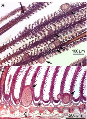
Histology: Plasmodia observed in different parts of the gill filaments, inside the multilayered epithelium between lamel- lae (Fig.4a–b). Only local damage of the lamellae was ob- served, with no gross pathology of the gill filaments.
Henneguya voroninin. sp.
Type host: barramundi,Lates calcarifer(Bloch 1790).
Site of infection: Base of the gill filament.
Type locality: Setiu Wetlands, Terengganu, Malaysia.
Prevalence of infection: 2.8% (1/35).
Type material: Digital images of syntype spores and histo- logical sections were deposited in the parasitological collec- tion of the Zoological Department, Hungarian Natural History Museum, Budapest, collection no. HNHM-71893. The 18S rDNA sequence was deposited in GenBank under accession number MH743110.
Etymology: The species is named in honour of V. A.
Voronin, an eminent Russian fish parasitologist.
Description of spores: Myxospores symmetric, with two equal caudal appendages and equal-sized polar capsules (Fig.2). Spore wall thin (0.3–0.4μm), smooth and composed of 2 equal valves. Apical end of spore body blunt, the caudal end tapers and extends into the caudal appendages. Total length 35–39μm, length of spore 9.9 ± 0.3 (9.5–10.3) μm and width 5.9 ± 0.3 (5.8–6.0) μm; thickness could not be measured as no spores were observed in sutural plane. Polar capsules pear shaped, blunt at the posterior end and tapered anteriorly, length 3.7 ± 0.2 (3.5–4.0)μm and width 2.1 ± 0.1 (2.0–2.2)μm. Polar tubules coiled in 6 turns perpendicular to the long axis of the polar capsules. Sporoplasm binucleate, with a small iodinophilous vacuole. Caudal appendages straight, tapering, length 27.2 ± 1.4 (25.0–29.0)μm, about 4 times as long as the spore body. Plasmodium ellipsoidal 250–
300μm × 130–150μm.
Remarks: Myxospores ofH. voroninin. sp. could not be distinguished morphometrically using most measurements with the two other species observed in the host. Both H. voroninin. sp. andH. setiuensisn. sp. were found in the gills but the specific tissue locality differed: the large ellipsoi- dal plasmodia (up to 300 μm) ofH. voronini n. sp. were localized to the cartilaginous base of the gill filaments, while the smaller spherical plasmodia ofH. setiuensisn. sp. (up to 75 μm) developed between lamellae of the gill filaments.
ConsideringHenneguyaspp. from otherLatesspecies, spores of H. voroninin. sp. could be differentiated in at least two dimensions (Table2).
Molecular analysis: 1696 bp 18S rDNA were sequenced, including the primers. A BLAST search indicated that highest sequence similarities were to other Henneguya species in GenBank, but all < 90%. Pairwise analysis showed H. voronini n. sp. was molecularly very similar to H. calcarifer n. sp. (97.7%; 1658/1969 bp; p-distance 0.013), described from the same fish (below).
Histology: Ellipsoidal plasmodia were located in the carti- laginous gill arch between gill filaments (Fig.4c). We suspect that development began in the multilayered connective tissue covering the gill arch and then a large part of the plasmodium Table2Comparisonofhosts,localities,tissuesandmyxosporedimensionsofHenneguyaspeciesdescribedfromLatessp.SLsporelength,SWsporewidth,ALcaudalappendagelength,STspore thickness,PCLpolarcapsulelength,PCWpolarcapsulewidth,NCnumberofcoilsofunfiredpolartubule.Allmeasurementsareinμm SpeciesSLSWSTALPCLPCWNCPlasmodium dimensionPlasmodium shapeSiteof infectionHostLocalityReferences H.mandouri11–13*6.0–7.5–40–533.1–4.31.5–2.23–5200–1000 ×100–500EllipsoidalorovalGillfilamentsL.niloticusEgyptRabieetal. 2009 H.massii8–9 (8.3±0.1)**5–6 (5.6±0.1)–12–14 (13.6±0.3)2–3 (2.8±0.1)1–2 (1.6±0.1)––SphericalGillfilamentsL.niloticusChadKostoïngue etal.2001 H.ghaffari11.8–14 (13.0±0.6)6.9–7.9 (7.5±0.4)4.9–5.9 (5.2±0.5)36.3–53.0 (43.2)4.8–5.9 (5.2±0.5)2.8–3.9 (3.2±0.3)4–51000×400Spindleshapedin thegill;round toovalinthe intestine
Gillfilaments, intestinal muscles
L.niloticusEgyptAli1999 H.mbakaouensis10–12 (10.8)7–9.9 (7.5)4.8–5.2 (5.0)51.5–69.2 (61.8)3.5–4.7 (4.0)2–3(2.5)4–5120–470 ×60–230Ovoidor subsphericalgillsL.niloticusCameroonFomenaand Bouix2000 H.latesi9.0–10.86.3–8.25.417.2–25.43.62–––Gills,mouthL.calcariferIndiaTripathi1952 H.setiuensisn.sp.8.3–9.5 (8.9±0.4)*5.8–6.0 (5.9±0.3)4.1–4.2 (4.1±0.1)28.0–32.0 (30.5±1.6)3.1–3.5 (3.3±0.2)2.0–2.2 (2.1±0.1650–75 ×50–75SphericalBetweenthegill lamellaeL.calcariferMalaysiaPresentstudy H.voroninin.sp.9.5–10.3 (9.9±0.3)*6.3–7.3 (6.8±0.3)–25.0–29.0 (27.2±1.4)3.5–4.0 (3.7±0.2)2.0–2.2 (2.1±0.1)6250–300 ×130–150EllipsoidalAtthebaseof gillfilamentsL.calcariferMalaysiaPresentstudy H.calcarifern.sp.8.3–10.0 (9.4±0.6)*4.8–5.5 (5.2±0.3)3.7–4.0 (3.8±0.128.0–35.0 (30.9±3.0)3.1–3.7 (3.4±0.2)1.1–1.7 (1.4±0.2)6300×400SphericalSkeletalmusclesL.calcariferMalaysiaPresentstudy *ovalinfrontalviewwithroundedanteriorend **ovalinfrontalviewwithroundedanteriorandposteriorends
moved into the gill filaments and was covered by a multilay- ered epithelium as it matured.
Microscopy: SEM revealed that the valve cell surfaces were smooth, which is a morphological feature typical of ge- nusHenneguya.
Henneguya calcarifern. sp.
Type host: barramundi,Lates calcarifer(Bloch 1790).
Site of infection: Skeletal muscle.
Type locality: Setiu Wetlands, Terengganu, Malaysia.
Prevalence of infection: 2.8% (1/35).
Type material: Digital images of syntype spores were de- posited in the parasitological collection of the Zoological Department, Hungarian Natural History Museum, Budapest, collection no. HNHM-71894. The 18S rDNA sequence was deposited in GenBank under accession number MH743109
Etymology: The species is named after the host.
Description of spores: Fig. 3. Myxospores symmetric, with two equal caudal appendages, and equal-sized polar
capsules. Spore wall 0.3–0.4 μm, smooth and composed of two equal valves. Apical end of spore body blunt, the caudal end tapers and extends into the caudal append- ages, total length 34–45 μm. Spore length 9.4 ± 0.6 (8.3–10.0) μm, width 5.2 ± 0.3 (4.8–5.5) μm and thick- ness 3.8 ± 0.1 (3.7–4.0) μm. Two polar capsules pear shaped, blunt at the posterior end and taper anteriorly, length 3.4 ± 0.2 (3.1–3.7) μm and width 1.4 ± 0.2 (1.1–1.7) μm. Polar tubules coiled in 6 turns perpendic- ular to the long axis of the capsule. Sporoplasm binucle- ate with a small iodinophilous vacuole. Caudal append- ages straight, tapering, length 30.9 ± 3.0 (28.0–35.0)μm,
~ 4 times longer than the spore body. Plasmodia spheri- cal 300 × 400 μm.
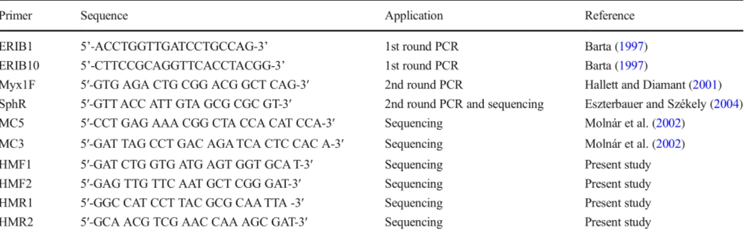
Remarks:H. calcarifern. sp. resembles morphological- ly and morphometrically both H. setiuensis n. sp. and H. voronini n. sp. (Table 2) but has different tissue site of development (muscles not gill). This is the first muscle-infecting Henneguya described from Lates calcarifer and is relatively distinct from the only other Fig. 1 Henneguya setiuensis n.
sp. (a–b) Line drawings of mature myxospores in frontal view showing polar capsules with coiled polar tubules. (c) Fresh, unstained myxospores in frontal (arrow) and sutural (*) views, with divergent caudal appendages
species from a Lates congener: H. ghaffari from muscle and gills ofL. niloticusin Egypt (Table2).
Molecular analysis: 1696 bp 18S rDNA were sequenced. A BLAST search indicated that the most similar species were otherHenneguya species, but all < 89%. Pairwise analysis showedH. calcarifer n. sp. was molecularly very similar (97.7% over 1696 bp) withH. voroninin. sp., described from the same fish (above).
Histology: Low intensity of parasite plasmodia in the host skeletal muscle meant no plasmodia were visible in histolog- ical sections.
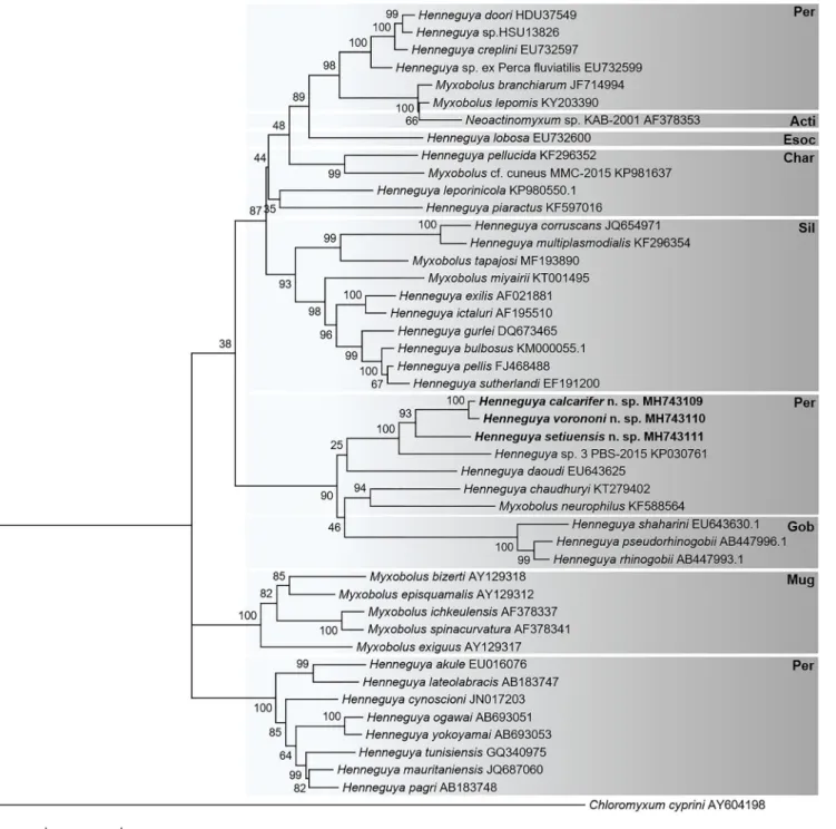
Phylogenetic analysis
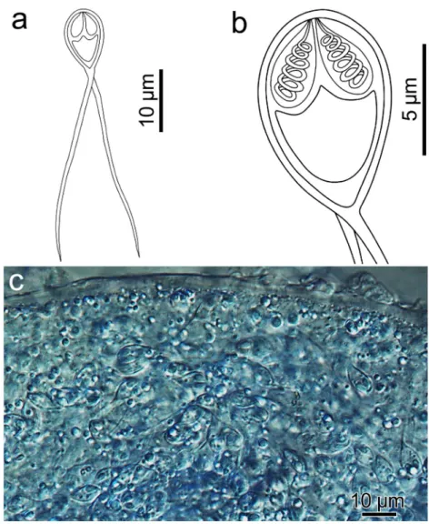
Phylogenetic analyses of 43 myxosporean sequences revealed a topology that showed correlation of myxosporeans Fig. 2 Henneguya voronini n. sp.
(a–b) Line drawings of mature myxospores in frontal view showing polar capsules with coiled polar tubules. (c) Fresh, unstained myxospores in frontal view showing the two pyriform polar capsules. (d) Scanning electron microscope image of the spores showing simple, smooth valve cell surfaces, each contiguous with a caudal process;
features typical of the genus
clustering with host order, withHenneguya spp. that infect Perciformes clustering into three distinct clades. The three species described herein cluster together within a subclade that includes another species that infect Perciformes (Fig.5).
Discussion
Henneguyais histozoic myxosporean parasite of freshwater and marine fishes and one of the most species-rich genus of myxosporeans with more than 200 species described (Lom and Dyková2006; Eiras2002; Eiras and Adriano2012). We used characters of specific host, tissue, myxospore morpholo- gy and 18S rDNA sequences to describe threeHenneguya species in barramundi,Lates calcarifer, from the east coast of Peninsula Malaysia. Henneguya setiuensis n. sp. and H. voroninin. sp. developed between gill lamellae and at the base of the gill filaments, respectively, whileH. calcarifern.
sp. developed in skeletal muscle from the flank of the fish.
Discovery of theHenneguyainfections in gills was not unex- pected, as this organ is a common site of myxobolid
infections; however, H. calcarifer n. sp. is the first myxosporean with a preference for muscle in a species of Lates. Muscle tropism ofHenneguyaspecies is less common than gills, being a character of 8 of some 180 species, includ- ing only 1 from a host in the Asian region: H. ophicephali Chakravarty, 1939, inOphiocephalus punctatusfrom India (Eiras2002; Eiras and Adriano2012).
Henneguyahas the distinctive morphological character of two caudal appendages, which distinguishes it from the other genera of the family Myxobolidae. However, molecular (SSU rDNA) evidence shows that genusHenneguyais polyphyletic, and this does not support the hypothesis that the presence of caudal appendages is a valid character for distinguishing spe- cies of Henneguya from Myxobolus (see comprehensive analysis in Liu et al.2019). While we acknowledge the weak support forHenneguyaas a distinct genus, formal taxonomic revision of genera within Myxobolidae is still lacking and is outside the scope of the present study. Accordingly we de- scribed the three novel species herein as Henneguya, based on the historical morphological character-based definition, augmented with DNA data.
Fig. 3 Henneguya calcarifern.
sp. (a–b) Line drawings of mature myxospores in frontal view showing polar capsules with coiled polar tubules. (c) Unstained, compressed
plasmodium, densely packed with mature myxospores
Our PCR approach for genetically characterizing the parasites was based on previous work to amplify DNA from another MalaysianHenneguya species,H. daoudi, in which its 18S rDNA was sequenced using“freshwater”myxosporean primers SphF-SphR, MC3-MC5 and MB5r-MB5f (Székely et al.2009a).
Although we were able to amplify ~ 800 bp fragments using SpHR, MC3 and MC5, these did not overlap sufficiently to construct contigs. Accordingly, we used our sequence informa- tion from H. calcarifer n. sp. to design new barramundi- Henneguya-specific primer pairs HMF-HMF2 and HMR1- HMR2, to give sufficient coverage to assemble contigs of 1696–1708 bp, including terminal primers MYX1f and SphR.
Henneguyaspecies can cause disease and mortality in farmed fish species, for example,H. exilisin catfish (Current and Janovy1977) andH. piaractus in pacu (Martins et al.
1997). Our study of parasites ofL. calcariferwas to assess the baseline occurrence of myxosporeans and any associated pathology in this important aquaculture species in Malaysia.
We observed infection in only 1/35 fish (2.8%), but this indi- vidual was positive for all threeHenneguyaspecies. This was lower than prevalences of otherHenneguyainfections inLates
spp.:H. mandouri32/40, 80%; H. ghaffari65/188, 34.6%;
H. latesi6/20, 30%; andH. massii3/67 (4.4%) (Tripathi1952;
Ali1999; Kostoïngue et al.2001; Rabie et al.2009). In addi- tion to low prevalence, the single fish that we observed with the infections did not have any observable pathology associ- ated with the parasites. This is consistent with theHenneguya infections from congeneric hosts, with none of these reported to cause henneguyosis (Tripathi1952; Ali1999; Kostoïngue et al.2001; Rabie et al.2009).
Correlation between particular myxosporean species and tis- sue tropism in the vertebrate host is well-established (e.g. Molnár 1994), particularly in species of Myxobolidae, with fine-scale tissue preferences being observed in the gills (Molnár2002;
Eszterbauer 2004). Alternatively, many myxobolids and myxosporean species in general tend to cluster according to the family/order of the fish host, with this character being useful to distinguish genetically closely related species (e.g. Ferguson et al. 2008; Carriero et al.2013; Vieira et al.2017; Naldoni et al.2018; Holzer et al.2018; Liu et al.2019). In the present study, our phylogenetic analysis showed that the three novel Henneguya species formed a well-supported sub-clade of perciform-infecting myxobolids, which includedHenneguya sp. 3 PBS-2015 (KP030761), from gills ofLates niloticusfrom Lake Turkana, Kenya, and Henneguya daoudi(EU643625), from gills of another perciform fish, Trichogaster trichopterus from Malaysia (Fig. 5). The nearest sister clade included Myxobolus neurophilus (KF588564) and Henneguya chaudhuryi (KT279402), both of which infect perciform fish:
Perca flavescens(yellow perch) andChanna punctata(spotted snakehead), respectively. The analysis thus revealed a stronger correlation between all three novelHenneguyaspp. and their host, rather than the specific tissue in which they developed a pattern consistent across multiple groups of Myxobolidae as demonstrated previously (Liu et al.2019).
Our taxonomic diagnosis incorporated consideration of whether either gill-infectingHenneguyaspecies had already been described. Specifically, we considered their similarity with the only Henneguya known from gills (and mouth) of L. calcarifer:H. latesiTripathi1952, in barramundi from India.
The description of that parasite is limited to host, tissue and morphometry (no DNA, no micrographs; Tripathi 1952). The taxon shares host and one of the two reported tissues (gills) with H. setiuensisn. sp. andH. voroninin. sp. but differs in most myxospore dimensions. We concluded that there was insufficient evidence to unambiguously identify either of our Malaysian gill- infecting species asH. latesi. Furthermore, given that we ob- served two genetically distinctHenneguya species within the gills, it is possible that the original description of H. latesifor
“gills and mouth”could actually represent two distinct species.
Determination of whether H. latesi represents a single taxon, distinct from any that we observed in Malaysian barramundi, would require redescriptionH. latesifrom the type locality in India, with molecular and fine-scale tissue tropism observations.
Fig. 4 Histological sections of gill filaments fromLates calcarifer infected by plasmodia ofH. setiuensisn. sp. andH. voroninin. sp. (a) Plasmodia of H. setiuensis n. sp. (arrows) producing compression and damage to the lamellae (*). (b) Development of plasmodia of H. voroninin. sp. (arrows) in the sub-epithelial layer at the base of the filament. Note that the plasmodia also impinge into the gill arch (g)
Based on our limited sampling, we demonstrated the pres- ence of at least threeHenneguyaspecies that parasitize barra- mundi in Malaysia. In our target population, these parasites occurred only at low prevalence and intensity, and without overt pathology. The health effects of any of these Henneguyaspecies either singly or combined are unknown in populations of fish under conditions of stress or exposed
to other health impacts from intense aquaculture, where Henneguya-related disease could emerge. Future work should concentrate on surveys from additional cultured fishes (both barramundi and grouper) from this region, under different stress levels and rearing conditions, to determine what other myxosporean species might be present and might be causing disease impacts.
Fig. 5 Phylogenetic tree generated by maximum likelihood analysis of 18S ribosomal DNA sequences of Henneguya species from perciform hosts and other closely related myxosporean species identified by BLAST; GenBank accession numbers shown after the species name, including the three novel data in bold (H. setiuensisn. sp.,H. voronini
n. sp. andH. calcarifern. sp.). Numbers at nodes indicate the bootstrap confidence values (ML). Taxonomic orders of the fish hosts are shown at right:CharCharaciformes,SilSiluriformes,EsocEsociformes,Mug Mugiliformes, Gob Gobiiformes, Per Perciformes and Acti Actinospores.Chloromyxum cypriniwas used as an outgroup
Acknowledgements We are grateful to AKUATROP officers for their excellent support during the field work and their help in the collection of fish specimens. We are grateful for the SEM work of Kartini Mohamed. We would also like to thank the staff of Biodiversity Laboratory (FSME) for their assistance in dissecting the fish and to the anonymous reviewers for their helpful suggestions that improved this article.
We acknowledge the Malaysian Ministry of Higher Education (MOHE) for the Niche Research Grant Scheme NRGS/2015/53131/33 granted to MHB, and the GINOP 2.3.2–15–2016–00004 project Establishing the sustainable angling-aimed management of Lake Balaton’for support provided to SzCs.
Funding Information Open access funding provided by MTA Centre for Agricultural Research (MTA ATK).
Open AccessThis article is distributed under the terms of the Creative C o m m o n s A t t r i b u t i o n 4 . 0 I n t e r n a t i o n a l L i c e n s e ( h t t p : / / creativecommons.org/licenses/by/4.0/), which permits unrestricted use, distribution, and reproduction in any medium, provided you give appro- priate credit to the original author(s) and the source, provide a link to the Creative Commons license, and indicate if changes were made.
References
Ali MA (1999)Henneguya ghaffarisp. n. (Myxozoa: Myxosporea), in- fecting the Nile perchLates niloticus(Teleostei: Centropomidae).
Dis Aquat Org 28:225–230
Aloo PA, Njiru J, Balirwa JS, Nyamweya CS (2017) Impacts of Nile Perch,Lates niloticus, introduction on the ecology, economy and conservation of Lake Victoria, East Africa. Lakes Reserv Res Manag 22(4):320–333
Barta JR (1997) Investigating phylogenetic relationships within the Apicomplexa using sequence data: the search for homology.
Methods 13(2):81–88
Biswas G, Thirunavukkarasu AR, Sundaray JK, Kailasam M (2010) Optimization of feeding frequency of Asian seabass (Lates calcarifer) fry reared in net cages under brackish water environment.
Aquaculture 305(1–4):26–31
Boonyaratpalin M (1997) Nutrient requirements of marine food fish cul- tured in Southeast Asia. Aquaculture 151(1–4):283–313
Bosworth BG, Wise DJ, Terhune JS, Wolters WR (2003) Family and genetic group effects for resistance to proliferative gill disease in channel catfish, blue catfish and channel catfish x blue catfish back- cross hybrids. Aquac Res 34(7):569–573
Bowser PR, Conroy D (1985) Histopathology associated of gill lesions withHenneguyain channel catfish. J Wildl Dis 21:177–179 Boyce NP, Kabata Z, Margolis L (1985) Investigations of the distribution,
detection, and biology ofHenneguya salminicola (Protozoa, Myxozoa), a parasite of the flesh of Pacific salmon. Can Tech Rep Fish Aquat Sci No 1405
Carriero MM, Adriano EA, Silva MRM, Ceccarelli PS, Maia AAM (2013) Molecular phylogeny of theMyxobolusandHenneguyagen- era with several new South American species. PLoS ONE 8(9) Current WL, Janovy JJ (1977) Sporogenesis inHenneguya exilisinfect-
ing the channel catfish: an ultrastructural study. Protistol 13:157–
167
Dyková I, de Buron I, Roumillat WA, Fiala I (2011)Henneguya cynoscionisp. n. (Myxosporea: Bivalvulida), an agent of severe cardiac lesions in the spotted seatrout,Cynoscion nebulosus (Teleostei: Sciaenidae). Folia Parasitol 58(3):169–177
Eiras JC (2002) Synopsis of the species of the genus Henneguya Thélohan, 1892 Myxozoa: Myxosporea: Myxobolidae. Syst Parasitol 52(1):43–54
Eiras JC, Adriano EA (2012) A checklist of new species ofHenneguya Thélohan, 1892 (Myxozoa: Myxosporea, Myxobolidae) described between 2002 and 2012. Syst Parasitol 83:95–104
Eiras JC, Molnár K, Lu YS (2005) Synopsis of the species ofMyxobolus Bütschli, 1882 (Myxozoa: Myxosporea: Myxobolidae). Syst Parasitol 61:1–46
Eiras JC, Saraiva A, Cruz CF, Santos MJ, Fiala I (2011) Synopsis of the species ofMyxidiumBütschli, 1882 (Myxozoa: Myxosporea:
Bivalvulida). Syst Parasitol 80:81–116
Eszterbauer E (2004) Genetic relationship among gill-infecting Myxobolusspecies (Myxosporea) of cyprinids: molecular evidence of importance of tissue-specificity. Dis Aquat Org 58:35–40 Eszterbauer E, Székely C (2004) Molecular phylogeny of the kidney-
parasiticSphaerospora renicolafrom common carp (Cyprinus carpio) andSphaerospora sp. from goldfish (Carassius auratus auratus). Acta Vet Hung 52(4):469–478
Ferguson JA, Atkinson SD, Whipps CM, Kent ML (2008) Molecular and morphological analysis ofMyxobolusspp. of salmonid fishes with the description of a newMyxobolusspecies. J Parasitol 94:1322– 1334
Fomena A, Bouix G (1996) New species ofHenneguyaThélohan, 1892 (Myxozoa: Myxosporea) parasites of freshwater fishes in Cameroon. J Afr Zool 110:413–423
Fomena A, Bouix G (2000) Henneguya mbakaouensissp. nov., Myxobolus nounensissp. nov. andM. hydrocyniKostoingue &
Toguebaye, 1994, Myxosporea (Myxozoa) parasites of Centropomidae, Cichlidae and Characidae (Teleosts) of the Sanaga Basin in Cameroon (Central Africa). Parasite 7:209–214
Haaparanta A, Valtonen ET, Hoffman RW (1994) Pathogenicity and sea- sonal occurrence ofHenneguya creplini(Protozoa, Myxosporea) on the gills of perchPerca fluviatilisin central Finland. Dis Aquat Org 20:15–22
Hallett SL, Diamant A (2001) Ultrastructure and small-subunit ribosomal DNA sequence ofHenneguya lesterin. sp.. (Myxosporea), a para- site of sand whitingSillago analis(Sillaginidae) from the coast of Queensland, Australia. Dis Aquat Org 46(3):197–212
Holzer AS, Bartošová-Sojková P, Born-Torrijos A, Lövy A, Hartigan A, Fiala I (2018) The joint evolution of the Myxozoa and their alternate hosts: A cnidarian recipe for success and vast biodiversity. Mol Ecol 27:1651–1666
Kostoïngue B, Diebakate C, Faye N, Toguebaye BS (2001) Presence of myxosporidea (Myxozoa: Myxosporea) of the genusHenneguya Thélohan, 1892 in freshwater fishes from Chad (Central Africa).
Acta Protozool 40:117–123
Kumar S, Stecher G, Li M, Knyaz C, Tamura K (2018) MEGA X:
Molecular evolutionary genetics analysis across computing plat- forms. Mol Biol Evol 35(6):1547–1549
Liu Y, Lövy A, Gu Z, Fiala I (2019) Phylogeny of Myxobolidae (Myxozoa) and the evolution of myxospore appendages in the Myxobolusclade. Int J Parasitol 49:523–530
Lom J, Arthur JR (1989) A guideline for the preparation of species de- scriptions in Myxosporea. J Fish Dis 12:151–156
Lom J, Dyková I (2006) Myxozoan genera : definition and notes on taxonomy, life-cycle terminology and pathogenic species. Folia Parasitol 53:1–36
Martins ML, Souza VN, Moraes FR, Moraes JRE, Costa AJ, Rocha UF (1997) Pathology and behavioral effects associated withHenneguya sp. (Myxozoa: Myxobolidae) infections of captive pacuPiaractus mesopotamicusin Brazil. J World Aquacult Soc 28:297–300 Minchew CD (1972) Identification and frequency of occurrence of four
forms ofHenneguyafound in channel catfish. Proceedings of the 26th Annual Conference of the Southeastern Fish and Game Commissions
Molnár K (1994) Comments on the host, organ and tissue specificity of fish myxosporeans and on the types of their intrapiscine develop- ment. Parasitol Hung 27:5–20
Molnár K (2002) Site preference of fish myxosporeans in the gill. Dis Aquat Org 48:197–207
Molnár K, Eszterbauer E, Székely C, Dán Á, Harrach B (2002) Morphological and molecular biological studies on intramuscular Myxobolusspp. of cyprinid fish. J Fish Dis 25(11):643–652 Molnár K, Székely C, Mohamed K, Shaharom-Harrison F (2006a)
Myxozoan pathogens in cultured Malaysian fishes. I. Myxozoan infection of the sutchi catfish. Dis Aquat Org 68:209–218 Molnár K, Székely C, Mohamed K, Shaharom-Harrison F (2006b)
Myxozoan pathogens in cultured Malaysian fishes. II. Myxozoan infections of redtail catfishHemibagrus nemurusin freshwater cage cultures. Dis Aquat Org 68:219–226
Naldoni J, Maia AAM, Correa LL, Silva MRM, Adriano EA (2018) Two new myxosporean species parasite of Phractocephalus hemioliopterusfrom the Brazilian Amazon: morphology, ultrastruc- ture and SSU-rDNA sequencing. Dis Aquat Org 128:37–49 Nie P (1996) Co-occurrence and microhabitat ofAncyrocephalus
mogurndae (Monogenea) and Henneguya weishanensis (Myxosporea) on gills of the mandarin fish,Siniperca chuatsi.
Folia Parasitol 43:272–276
Pote LM, Hanson LA, Shivaji R (2000) Small subunit ribosomal RNA sequences link the cause of proliferative gill disease in channel cat- fish toHenneguyan. sp.. (Myxozoa: Myxosporea). J Aquat Anim Health 12(3):230–240
Rabie SA, Mohammed NI, Hussein AN, Hussein NM (2009) The infec- tion of freshwater fishes with three species ofHenneguyain Qena.
Upper Egypt Egypt Acad J Biol Sci 1:11–19
Shariff M (1982)Henneguya shaharinisp. nov. (Protozoa: Myxozoa), a parasite of marble goby,Oxyeleotris marmoratus(Bleeker). J Fish Dis 5:37–45
Sitjá-Bobadilla A (2009) Can myxosporean parasites compromise fish and amphibian reproduction? P Roy Soc B-Biol Sci 276:2861–2870 Sokolov SG, Lebedeva DI, Murzina SA, Parshukov AN, Bystrova KA, Ieshko EP (2019) Morphology and phylogeny ofHenneguya oviperdainfecting oocytes of Esox lucius, with description of parasite-induced histopathology. Dis Aquat Org 133(2):91–98
Székely C, Shaharom-Harrison F, Cech G, Mohamed K, Molnár K (2009a) Myxozoan pathogens of Malaysian fishes cultured in ponds and net-cages. Dis Aquat Org 83(1):49–57
Székely C, Shaharom-Harrison F, Cech G, Ostoros G, Molnár K (2009b) Myxozoan pathogens of fishes of the Tasik Kenyir Water Resevoir, Terengganu, Malaysia. Dis Aquat Org 83(1):37–48
Székely C, Borzák R, Molnár K (2018) Description ofHenneguya jaczoi sp. n. (Myxosporea, Myxobolidae) fromPerca fluviatilis(l.) (Pisces, Percidae) with some remarks on the systematics ofHenneguyaspp.
of European fishes. Acta Vet Hung 66(3):426–443
Tantikitti C, Sangpong W, Chiavareesajja S (2005) Effects of defatted soybean protein levels on growth performance and nitrogen and phosphorus excretion in Asian seabass (Lates calcarifer).
Aquaculture 248(1–4):41–50
Thompson JD, Higgins DG, Gibson TJ (1994) CLUSTAL W: improving the sensitivity of progressive multiple sequence alignment through sequence weighting, position-specific gap penalties and weight ma- trix choice. Nucleic Acids Res 22:4673–4680
Tripathi YR (1952) Studies on parasites of Indian fishes I. Protozoa:
Myxosporidia together with a check list of parasitic protozoa de- scribed from Indian fishes. Rec. Indian Museum 50:63–88 Vieira DHMD, Alama-Bermejo G, Bartholomew JL, Abdallah VD,
Azevedo RK (2017) Morphological and molecular description of Myxobolus batalhensisn. sp.. (Myxozoa, Myxosporea), a liver and ovary parasite ofSalminus hilariiin Brazil. Parasitol Res 116:3303–
3313
Whitaker JW, Pote LM, Hanson LA (2005) Assay to detect the actinospore and myxospore stages of proliferative gill disease in oligochaetes and pond water. N Am J Aquac 67:133–137 Yokoyama H, Kawakami H, Yasuda H, Tanaka S (2003)Henneguya
lateolabracis(sp. n. Myxozoa: Myxosporea), the causative agent of cardiac henneguyosis in Chinese sea bassLateolabraxsp. Fish Sci 69(6):1116–1120
Publisher’s noteSpringer Nature remains neutral with regard to jurisdic- tional claims in published maps and institutional affiliations.