DESCRIPTION OF HENNEGUYA JACZOI SP. N. (MYXOSPOREA, MYXOBOLIDAE) FROM PERCA FLUVIATILIS (L.) (PISCES, PERCIDAE) WITH SOME REMARKS ON THE SYSTEMATICS
OF HENNEGUYA SPP. OF EUROPEAN FISHES
Csaba SZÉKELY*, Réka BORZÁK and Kálmán MOLNÁR
Institute for Veterinary Medical Research, Centre for Agricultural Research, Hungarian Academy of Sciences, Hungária krt. 21, H-1143 Budapest, Hungary
(Received 6 June 2018; accepted 25 July 2018)
A new Henneguya species, H. jaczoi sp. n., is described from perch (Perca fluviatilis) from Lake Balaton, Hungary. This species infects the palatal region of the fish, forming large plasmodia in the thickened caudal part of the buccal cavity and at the dorsal ends of the cartilaginous gill arches. The species differs from the gill-dwelling Henneguya species of perch and pike (Esox lucius) both morpholog- ically and in molecular aspects. The authors conclude that the type species H.
psorospermica Thélohan is a specific parasite of pike, while the species forming plasmodia in the gills of perch corresponds to H. texta Cohn, which was hitherto regarded as a synonym of H. psorospermica. Besides the above-mentioned spe- cies, H. creplini was frequently found in pikeperch (Sander lucioperca) and Vol- ga pikeperch (Sander volgensis), but no Henneguya infection has been recorded in ruffe (Gymnocephalus cernua), which is a common percid fish of the lake and is known to be the type host species for H. creplini.
Key words: Myxozoa, perch, pikeperch, pike, occurrence, molecular phy- logeny, histology
Henneguya spp. are common myxozoan parasites of pike (Esox lucius L.) and percid fishes (Table 1). The type species, H. psorospermica was described by Thélohan (1895) from the gills of both pike and perch (Perca fluviatilis L.).
Of them the pike, mentioned first by Thélohan, should be regarded as type host.
Other pike-infecting Henneguya species were recorded from the gills (H. lobosa Cohn, 1895), the ovary (H. oviperda Cohn, 1895), the epidermis (H. schizura Gurley, 1893) and the intestine (H. periintestinalis Cépéde, 1906) (Table 1).
From perch, H. texta Cohn, 1895 and H. minuta Cohn, 1895 were described from the gills and H. wolinensis Romuk-Wodoracki, 1990 from the epidermis beneath the scales (Cohn, 1895; Romuk-Wodoracki, 1990). The latter author also men- tioned the occurrence of H. lobosa and H. creplini Gurley, 1894 in perch, alt- hough the original descriptions of these parasites were from different fish hosts
*Corresponding author; E-mail: szekely.csaba@agrar.mta.hu; Phone: 0036 (1) 467-4065
(Kudo, 1919). Henneguya creplini was first observed in the gills of ruffe (Gym- nocephalus cernua L.) by Gurley (1894). From this fish two more Henneguya species were described: H. acerinae Schröder, 1906 from the gills and H. tenuis Vaney & Conte, 1901 from the intestine of ruffe (Kudo, 1919; Donec and Shul- man, 1984). Henneguya gigantea Nemeczek, 1911 and H. nemeczeki Tripathi, 1952 were described and the occurrence of H. creplini was reported from the gills of pikeperch (Sander lucioperca L.) and Volga pikeperch (Sander volgensis Gmelin) (Donec and Shulman, 1984; Lom and Dyková, 1992, 2006) (Table 1).
In addition to these European species, Fantham et al. (1939) described H. percae in Canada from yellow perch [Perca flavescens (Mitchill)], and another species, H. dogieli Akhmerov, 1960, was recorded from a Far Eastern percid fish, Sini- perca chuatsi (Basilewsky) (Donec and Shulman, 1984).
Table 1
Henneguya spp. described from pike and percid fishes
Pike
(Esox lucius) Perch
(Perca fluviatilis)
Pikeperch (Sander lucioperca)
Volga-pikeperch (Sander volgensis)
Ruffe (Gymnocephalus
cernua) H. psorospermica
(Thélohan, 1895) G
H. psorospermica (Thélohan, 1895)
G
H. nemeczeki (Tripathi, 1952)
G
H. creplini H. creplini (Gurley, 1894)
G H. oviperda
(Cohn, 1895) O
H. texta (Cohn, 1895)
G
H. gigantea (Nemeczek, 1911)
G
H. acerinae (Schröder, 1906)
G H. lobosa
(Cohn, 1895) G
H. minuta (Cohn, 1895)
G
H. psorospermica H. tenuis
(Vaney et Conte, 1901) I
H. schizura (Gurley, 1893) E
H. wolinensis
(Romuk-Wodoracki, 1990) H. oviperda
H. periintestinalis (Cépéde, 1906) I
H. creplini H. creplini
H. gigantea H. lobosa H. acerinae
H. nemeczeki H. dogieli (Akhmerov, 1960)
G (Siniperca)
G = gills, I = intestine, O = ovary, E = eyes; Names with bold letters = good hosts; names with non-bold letters
= inadequate identifications
In Hungary, the myxozoans of Lake Balaton fishes were first studied by Jaczó (1940) who found a Myxobolus and a Chloromyxum spp. in the gills and the gallbladder, respectively, and a Henneguya species in the tongue of perch but did not describe it. On the basis of illustrations, however, this last mentioned
species appears to be the same as the one we recorded and present here. Based on morphological data, habitat preference, tissue specificity and molecular data, in this paper we describe a new species, Henneguya jaczoi sp. n. from perch, and re-describe H. texta and H. psorospermica spp. from perch and pike, respectively.
Materials and methods
Most of the fish were collected at a fish trap built into the water gate of Lake Balaton, while some others were captured with a seine at the city of Siófok (46°54'29.4"N 18°02'46.4"E) from 12 March to 1 April 2015 and from 24 Febru- ary to 12 April 2016). A total of 8 pikes [33–41 cm standard length (SL)] and 104 percid fishes (9 to 20 cm SL) were collected and examined for myxozoans.
The percid fishes included 48 perch, 17 pikeperch, 7 Volga perch and 32 ruffe specimens.
The fish were brought to the laboratory alive in oxygenated plastic bags, kept in aerated aquaria and subjected to complete parasitological dissection with- in three days. They were sedated with a drop of clove oil into their water and eu- thanised by a cervical cut. When mature plasmodia were found, some of the spores were studied as fresh preparations. Subsamples of spores from mature plasmodia were studied as fresh preparations, collected and stored in 70% etha- nol in Eppendorf tubes until further molecular analysis or preserved as glycerine- gelatin slide preparations. Tissue samples from organs infected by mature plas- modia were fixed in Bouin’s solution, processed for histology through a series of ethanol, embedded in paraffin (Molar Chemicals Ltd., Hungary), cut to 4–5 μm thick sections, and stained with haematoxylin and eosin. The maturity of plas- modia and the vitality of the spores were assessed by adding a few hundred spores into a 0.4% solution of urea in water (Lom and Dyková, 1992). Plasmodia were considered mature when at least 90% of the spores extruded polar filaments in this solution. Unfixed spores were studied using Nomarski differential inter- ference contrast with an Olympus BH2 microscope. The spores were photo- graphed with an Olympus DP 20 digital camera. All measurements are expressed in micrometres and given as the range followed by the mean ± standard devia- tion, with the number of measurements in parentheses.
Molecular data
Myxospores preserved in ethanol were centrifuged at 10,000 g for 10 min and the supernatant was removed. Genomic DNA was extracted from the pellet- ed spores using the DNeasy® Blood & Tissue Kit (Qiagen, Hilden, Germany) ac- cording to the manufacturer’s instructions.
Amplification and sequencing of the 18S rDNA were conducted as de- scribed by Cech et al. (2015).
The sequence fragments were assembled using MEGA 5.2 software (Tamura et al., 2011). The contiguous 18S rDNA sequences and the most similar myxozoan sequences from the GenBank based on BLAST matches were aligned with the software CLUSTAL W (Thompson et al., 1994). DNA pairwise distanc- es were calculated with the MEGA 5.2 software using the Maximum Composite Likelihood model. Phylogenetic analysis was performed via Maximum Likeli- hood (ML) Ceratonova shasta (Noble, 1950) was used as the outgroup. The da- taset was tested using MEGA 5.2 for the nucleotide substitution model of best fit and the model shown by the Akaike Information Criterion (AIC) as the best- fitting one was chosen (TN93+G model). Bootstrap values based on 1,000 resampled datasets were generated.
Results
Large, globular myxosporean cysts, about 1 to 2 mm in diameter, were found in the palatal region of the mouth and in the gill arches of perch. The cysts contained mature spores of a Henneguya species and occurred exclusively in this specific habitat. No similar infection was found in other percid fishes, e.g. in pikeperch, Volga pikeperch and ruffe (Molnár, 1998; present study). Based on the shape and size of spores, the specific habitat of cysts and molecular data of the 18S rDNA of spores, this parasite is described here as Henneguya jaczoi sp. n.
Twelve out of the examined 48 perch specimens were infected with H. jaczoi sp.
n. plasmodia. Among fish of different size only specimens measuring 13 to 16 cm in length showed infection. The plasmodia of H. jaczoi sp. n. were located in a specific sclerotic area of the palate located dorso-caudally in the buccal cavity.
This area, divided into a right and a left side, is a thickened part of the buccal cavity covered by stratified epithelium containing mucous cells and small ossi- fied spines (Fig. 1). The epithelium is supported by a dense connective tissue un- der which a zone of loose connective tissue and a muscle layer are located. This specific part of the palate incorporates the dorsal ends of the cartilaginous gill arches (Fig. 2). Two types of plasmodia were found. Some large globule-shaped plasmodia 1 to 2 mm in diameter were located between the dense and the loose connective tissue. Other, less regular shaped plasmodia were located at the end of the cartilaginous gill arches, where they joined the muscle. Some of the large plasmodia surrounded by a very thin connective tissue were recovered in the muscular layer. Less frequently round plasmodia were found also in the cartilag- inous gill arch, which is also regarded as part of the caudal portion of the oral cavity in its function.
Besides infection of the palatal region, small plasmodia containing Hen- neguya spores but differing in shape and size from H. jaczoi sp. n. were found in the gill lamellae of four perch specimens collected with a seine. By their size and shape these spores corresponded to H. texta. In addition to perch, Henneguya in-
fection was also found in 2 out of the 8 pike specimens examined. In this fish, large plasmodia were located in the gill filaments. Spores found in these plasmo- dia were identified as the species H. psorospermica.
The gill lamellae of 17 pikeperch and 2 Volga pikeperch specimens har- boured immature cysts of H. creplini, while none of the ruffe specimens showed Henneguya infection.
Description of the new species:
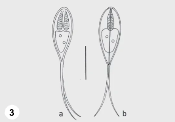
Henneguya jaczoi sp. n. (Figs 3 and 4) Type-host: Perch, Perca fluviatilis (L.)
Type locality: Lake Balaton, Siófok (46°54’29,8’’N, 18°2’,46,2’’E) Habitat of tissue development: Palatal region of the buccal cavity, gill arches Prevalence: 25% of examined perches, 12/48, and 100% of specimens measuring 13–16 cm in standard length, 12/12
Type-material: Spores in glycerine-gelatin, photo-types and histological sections were deposited in the parasitological collection of the Zoological De- partment, Hungarian Natural History Museum, Budapest, Coll. No. HNHM-19792.
Etymology: The name of the species comes from the name of the late Hungarian fish parasitologist, Imre Jaczó, who first reported this infection.
Vegetative stages: Large round or roundish plasmodia 1.0–2.0 mm in di- ameter developed in the dorso-caudal part of the palate and in the gill arches. Ir- regular-shaped plasmodia were also found around the cartilaginous ends of the gill arches.
Spores: Spores elongated, spindle-shaped both in frontal and sutural view (Figs 3a, 4 and 3b). Length of the spore body 13.2–15.2 (14.1 ± 1.1) (n = 50), width 5.2–7 (6.2 ± 0.64) (n = 50), thickness 4.8–5.2 (5.1 ± 0.19) (n = 11). Polar capsules elongated, equal or somewhat different in size, tapering posteriorly, 4.8–7.2 (6.4 ± 0.83) long (n = 50) and 1.6–2.4 (2 ± 0.23) wide (n = 50). Twelve to 13 filament coils arranged perpendicular to the capsule length, coiled densely in the polar capsule. The straight, continuously tapering caudal extensions 20.8–
27.2 (24 ± 2.4) long. They are in most cases equal in length, less frequently somewhat different. Sutural edge markings not seen. No intercapsular appendix found. In the sporoplasm no iodinophilous vacuole found, but a distinct, single, bright nucleus present (Fig. 4).
Histology: Large, round plasmodia developed in the connective tissue of the thickened dorso-caudal region of the buccal cavity at the border of the dense and the loose connective tissue. They extended deep in the muscular layer (Fig. 1).
Some other smaller irregular-shaped plasmodia were also found in the dense connective tissue joining the cartilaginous ends of the gill arches (Fig. 2) and in the gill arch.
Fig. 1. Palatal region of a perch infected with H. jaczoi sp. n.: a) layer of epithelial cells and goblet cells, b) dense connective tissue, p) M. jaczoi plasmodium, t) ossified spines, l) loose connective tissue. Arrows = blood vessels. Histological section, haematoxylin and eosin (HE). Bar = 100 µm
Fig. 2. Palatal region at the dorsal end of the cartilaginous gill arch: a) end of the cartilaginous gill arch, b) dense connective tissue covering the gill arch, m) muscle cells attaching to the gill arch, p)
plasmodium of H. jaczoi. Histological section, HE. Bar = 100 µm
Fig. 3. Schematic drawing of Henneguya jaczoi sp. n.: a) spore in frontal view, b) spore in sutural view. Bar = 10 µm
1
2
3
Fig. 4. Spores of Henneguya jaczoi sp. n. from the palate of a perch. A single nucleus can be seen in the sporoplasm. Bar = 10 µm
Fig. 5. Gill of a perch infected with an intralamellar plasmodium (p) of Henneguya texta. The ca- pillary (arrows) runs at one side of the plasmodium. The other side is covered only by a single lay-
er of membrana basalis. Histological section, HE. Bar = 100 µm
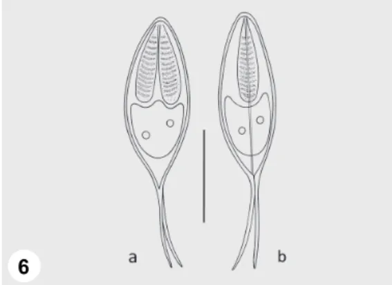
Fig. 6. Schematic drawing of Henneguya texta: a) spore in frontal view, b) spore in sutural view.
Bar = 10 µm 4
5
6
Fig. 7. Spores of Henneguya texta from the gills of a perch. Two or three nuclei can be seen in the sporoplasms. Bar = 10 µm
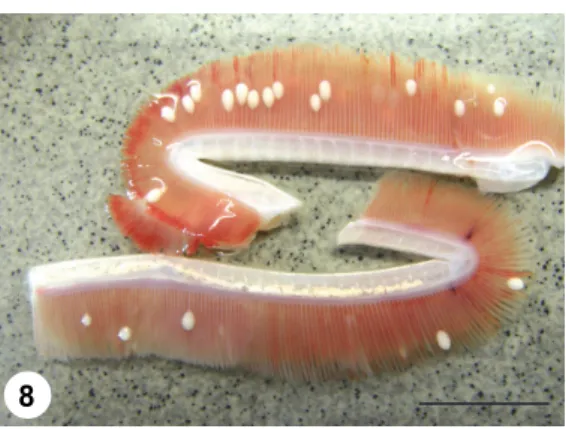
Fig. 8. Gill filaments of a pike infected by plasmodia of H. psorospermica. Fresh mount picture.
Bar = 2 cm
Fig. 9. Schematic drawing of Henneguya psorospermica: a) spore in frontal view, b) spore in su- tural view. Bar = 10 µm
7
8
9
Fig. 10. Spores of Henneguya psorospermica from the gills of a pike. Bar = 10 µm
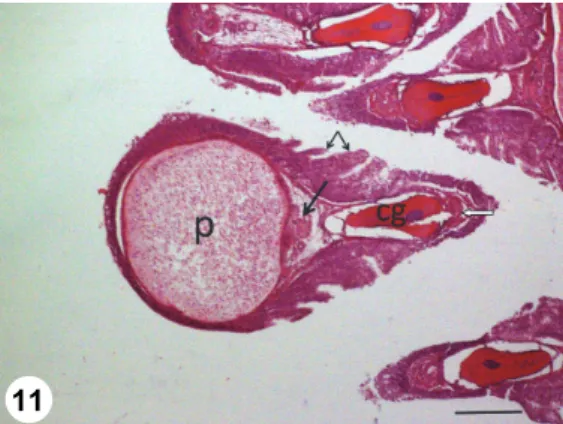
Fig. 11. Gill filament of a pike with a cross-sectioned Henneguya psorospermica plasmodium (p).
The plasmodium is located in the outer part of the filament close to the efferent branchial artery (arrow). The cartilaginous gill ray (cg) and the afferent branchial artery (open arrow) can be found in the inner part of the filament. The edges of the filament are covered by multilayered epithelium, but in the middle some lamellae are seen (small arrows). Histological section, HE. Bar = 200 µm
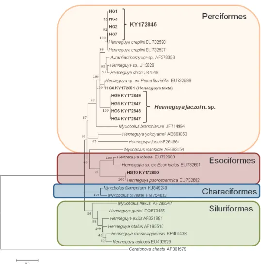
Molecular data: The partial sequence of the 18S rRNA gene of four samples of H. jaczoi sp. n. were identified. HG4 and HG5 samples (KY172847) were identical, HG6 (KY172848) and HG9 (KY172849) differed in 2 nucleotides (0.1%), so they can be considered the same myxozoan species. Their sequences showed 94.5% similarity to H. texta and Henneguya sp. ex Perca fluviatilis (EU732599), parasites of the gill from the same host species. They were also similar (94.7%) to the H. creplini-like spores from pikeperch of the present study and to H.
creplini sequences deposited in the GenBank (EU732597-8). The sequences of the new species showed, however, only very low similarity (82.8%) to H. pso- rospermica of the pike. The 18S rDNA sequences of H. jaczoi sp. n. were depos- ited in the GenBank under the accession number KY172847-9 (Fig. 12).
10
11
Fig. 12. Phylogenetic position of the new species Henneguya jaczoi, H. texta and H. psorospermi- ca based on 18S rDNA analysis by the Maximum Likelihood algorithm. Ceratonova shasta was used as the outgroup. Bootstrap values are given at the nodes. The scale bar indicates the number
of expected substitutions per site
Remarks: The species seems to have an affinity to the connective tissue, forming large plasmodia in this palatal region of the mouth. The morphology of the spores of H. jaczoi sp. n. was very similar to that of H. texta, the specific par- asite of the gill lamellae of perch, but they had a smaller spore body size (Table 2) and longer caudal extensions, and their plasmodia had a different location in the organs and tissues of the host. The approximately 5% differences between the 18S rDNA sequences of H. jaczoi sp. n. and those of H. texta and H. creplini, re- spectively, indicate that H. jaczoi sp. n. should be regarded as a new species. The size and shape of H. jaczoi sp. n. spores also resembled the spores of H. pso-
rospermica, which infects the pike, but their caudal extensions were longer and they substantially differed genetically. Besides genetic differences of the hosts, there were differences in the location of plasmodia, tissue affinity and molecular sequences of the spores. In infecting the connective tissue, H. jaczoi sp. n. re- sembles H. wolinensis, which forms large plasmodia and develops in similar tis- sues under the scales. However, the spores of H. jaczoi sp. n. are significantly smaller than those of H. wolinensis. Henneguya percae has been described from the gills of a genetically closely related fish, Perca flavescens, but the polar cap- sules in the spores of this American species are short and differ from the elongat- ed capsules of the European Henneguya species of percid fishes. In the morphol- ogy of spores, H. jaczoi sp. n. also resembles H. creplini, but this latter species has longer caudal extensions and its spores are formed during the winter (Molnár, 1998). Moreover, the 18S rDNA sequences of the two species show 5% difference.
Redescription of Henneguya texta Cohn, 1895 (Figs 5, 6 and 7) Host: Perch, Perca fluviatilis (L.)
Locality: Lake Balaton, Siófok (46°54’29,8’’ N, 18°2’,46,2’’E) Habitat of tissue development: Gill lamellae
Prevalence: 8.3% of examined perches, 4/48
Material: Spores in glycerine-gelatin, photo-types and histological sec- tions were deposited in the parasitological collection of the Zoological Depart- ment, Hungarian Natural History Museum, Budapest, Coll. No. HNHM-19793.
The 18S rDNA sequences of H. texta were deposited in the GenBank under the accession number KY172851.
Vegetative stages: Small round plasmodia located in the gill lamellae (Fig. 5) Spores: Spores elongated, spindle-shaped both in frontal and sutural view (Figs 6a,b and 7). Length of the spore body 17.5–22 (19.7 ± 1.8) (n = 50), width 7.2–9.6 (8.3 ± 1) (n = 50), thickness 7.2–9.6 (8 ± 0.9) (n = 11). Polar capsules elongated, equal or somewhat different in size, tapering posteriorly, 8.1–12 (9.5 ± 1.4) long (n = 50) and 2.4–3.2 (2.9 ± 0.4) wide (n = 50). Twelve to 13 filament coils arranged perpendicular to the capsule length, coiled densely in the polar capsule. The straight, continuously tapering caudal extensions are shorter than the spore body, they measure 11–20 (15 ± 1.4) in length. They are in most cases equal in length, less frequently somewhat different. Sutural edge markings, inter- capsular appendix and iodinophilous vacuole in the sporoplasm not found, but 2 or 3 nuclei in the sporoplasm present (Fig. 7).
Histology: Small, ellipsoidal plasmodia located dissymmetrically in the capillary network, so that the rest of the capillary with blood cells runs at one side of the plasmodium, while the other side is covered only by a thin basement membrane (Fig. 5).
Molecular data: The 18S rDNA sequence of H. texta was 100% identical with that of the specimen from Perca fluviatilis deposited in the GenBank
(EU732599). A relatively high similarity (94.9%) exists to H. doori from yellow perch (Perca flavescens) and to sequences of H. creplini of the pikeperch (95.6%) and an Aurantiactinomyxon (AF378356, 94.7%). Its similarity to H. jaczoi n. sp.
was 94.5% (Fig. 12).
Remarks: Henneguya texta differs from H. jaczoi by its larger spores and its specific location in the gill lamellae, and their molecular similarity is 94.5%.
It also differs from M. psorospermica of the pike by its larger spores and the 84% differences of their 18S rDNA sequences. Although Thélohan (1895) de- scribed H. psorospermica from pike and perch, and Donec and Shulman (1984) regarded H. texta as a synonym of the above species, our molecular data support that H. texta described by Cohn (1895) is a valid species from perch.
Redescription of Henneguya psorospermica Thélohan, 1895 (Figs 8, 9, 10 and 11) Host: Pike, Esox lucius (L.)
Locality: Lake Balaton, Siófok (46°54’29,8’’N, 18°2’,46,2’’E) Site of tissue development: gill filaments
Prevalence: 25% of examined pikes (2/8)
Type-material: Spores in glycerine-gelatin, photo-types and histological sections were deposited in the parasitological collection of the Zoological De- partment, Hungarian Natural History Museum, Budapest, Coll. No. HNHM-19794.
The 18S rDNA sequence of H. psorospermica from our study was deposited in the GenBank under the accession number KY172850.
Vegetative stages: Oval-shaped plasmodia measuring 1–2 mm in length and 0.4–0.6 mm in width are located in the gill filaments (Fig. 8).
Spores: Spores elongated, spindle-shaped both in frontal and sutural view (Figs 9a,b and 10). Length of the spore body 9.6–14.4 (12.4 ± 1.02) (n = 50), width 4.8–7.2 (6.3 ± 0.66) (n = 50), thickness 7.6–7.8 (7.7 ± 0.08) (n = 11). Polar capsules elongated, equal or somewhat different in size, tapering posteriorly, 5–7.2 (6.3 ± 0.74) long (n = 50) and 1.6–2 (1.9 ± 0.19) wide (n = 50). Twelve to 13 fil- ament coils arranged perpendicular to the capsule length, coiled densely in the polar capsule. The straight, continuously tapering caudal extensions are 12.8–16 (13.8 ± 1) long. They are in most cases equal in length, less frequently somewhat different. Sutural edge markings, intercapsular appendix and iodinophilous vacu- ole in the sporoplasm not seen.
Histology: The plasmodia of H. psorospermica were formed in the outer edge of the gill filament close to the efferent branchial artery (arteria branchialis efferens) (Fig. 11). Plasmodia in all cases were surrounded by a relatively thin, dense connective tissue wall, but in the lamella-free parts of the filaments they were covered by stratified epithelium as well. Although the filaments were great- ly deformed by the large plasmodia in their central region, gill lamellae could al- so be seen.
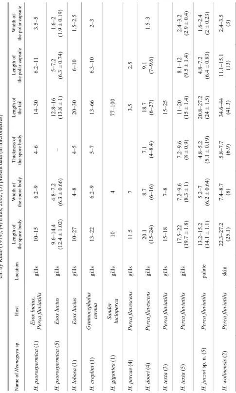
Table 2 Comparative measurements ofHenneguya spp. of pike and percid fishes. Data from: (1) Donec & Shulman, 1984; (2) Romuk-Wodoracki, 1990; (3) Wegener, 1910, cit. by Kudo (1919); (4) Eiras, 2002; (5) present data (in micrometres) Name ofHenneguya sp.Host Location Length of the spore bodyWidth of the spore bodyThickness of the spore bodyLength of the tailLength of the polar capsule Width of the polar capsule H. psorospermica (1) Esox lucius, Perca fluviatilis gills 10–15 6.2–94–614–30 6.2–113.5–5 H. psorospermica (5) Esox lucius gills 9.6–14.4 (12.4 ± 1.02)4.8–7.2 (6.3± 0.66)– 12.8–16 (13.8 ± 1)5–7.2 (6.3± 0.74)1.6–2 (1.9± 0.19) H. lobosa (1) Esox lucius gills 10–27 4–84–520–30 6–101.5–2.5 H. creplini (1) Gymnocephalus cernua gills 13–22 6.2–95–713–66 6.3–102–3 H. gigantea (1) Sander luciopercagills 104 77–100 H. percae (4) Perca flavescensgills 11.5 7 3.5 2.5 H. doori (4) Perca flavescensgills 20.1 (15–24)8.7 (6–16)7.1 (4–8.4)18.7 (6–27)9.1 (7–9.6)1.5–3 H. texta (3) Perca fluviatilis gills 15–18 7–815–25 H. texta (5) Perca fluviatilis gills 17.5–22 (19.7 ± 1.8)7.2–9.6 (8.3± 1)7.2–9.6 (8± 0.9)11–20 (15 ± 1.4)8.1–12 (9.5± 1.4)2.4–3.2 (2.9± 0.4) H. jaczoi sp. n. (5) Perca fluviatilis palate 13.2–15.2 (14.1 ± 1.1)5.2–7 (6.2± 0.64)4.8–5.2 (5.1± 0.19)20.8–27.2 (24 ± 1.5)4.8–7.2 (6.4± 0.83)1.6–2.4 (2± 0.23) H. wolinensis (2) Perca fluviatilis skin22.3–27.2 (25.1)7.4–8.7 (8) 5.8–7.7 (6.9) 34.6–44 (41.3)11.1–15.1 (13)2.4–3.5 (3)
Molecular data: The 18S rDNA sequences of H. psorospermica found by us were completely identical with those of the Henneguya species from Esox lu- cius deposited in the GenBank (EU732602). Their similarity to H. lobosa (EU732600), another parasite of the pike, is only 85.9% and to a Henneguya sp.
of the pike (EU732601) it is 85.4% (Fig. 12).
Remarks: Henneguya psorospermica differs from H. texta by its smaller spores and by the 84.0% difference between their 18S rDNA sequences. Data ob- tained in this study suggest that H. psorospermica is a specific species of the pike and, despite morphological similarities of the spores, it does not infect percid fishes.
Spores of a Henneguya species were found also in the intestine of a perch.
Molecular studies of these spores, however, revealed that the sequences of this species had 100% similarity to the species H. cutanea, a parasite of cyprinid fishes, indicating that the spores originated from a cyprinid prey fish (most prob- ably a young bream, Abramis brama) consumed by the perch.
Comparative measurements of our data and those given by some relevant authors on Henneguya spp. are indicated in Table 2.
Discussion
Henneguya is the second most species-rich genus of myxosporeans after Myxobolus, with over 195 species (Eiras, 2002; Eiras and Adriano, 2012). Most species described recently from different fishes have proper morphological de- scriptions supported by histological and molecular data (Székely et al., 2009; Ye et al., 2012; Yokoyama et al., 2012; Carriero et al., 2013; Rocha et al., 2014).
Some other species described more than a hundred years ago, however, have on- ly scanty data on morphology, insufficiently designated habitats and more than one host in their original description. To add to the problem, researchers often misidentified the myxozoans they encountered, and consequently the host range of known species was erroneously enlarged. For instance, Thélohan (1895) de- scribed the species H. psorospermica from pike and perch which belong to two different fish families. When studying the histopathological changes caused in fish gills by Henneguya spp., Dyková and Lom (1978) – accepting Thélohan’s description – recorded H. psorospermica from pike and perch but remarked that in pike large plasmodia of this species developed in the filament arteries while in perch small plasmodia were formed in intralamellar location. In a similar way, Donec and Shulman (1984) regarded H. texta and H psorospermica as a single species and synonymised the two species. In all probability the above authors came across plasmodia of two different species, namely H. psorospermica in pike and H. texta in perch. These scantly described species need a complete revi- sion which can now be carried out relatively easily, considering the importance
of location and tissue affinity and owing to the availability of molecular tech- niques as an identification tool. Morphological differences among Henneguya species are relatively scarce and the differences in their elongated spindle-shaped spores often rely on the length of the caudal extensions. Then, species infecting the pike and percids are rather similar with their elongated spindle shape. On the other hand, a relatively large difference exists between Henneguya spp. of percids and cyprinids. The spores of H. cutanea, the parasite infecting the fins of the cyprinid bream (Abramis brama), have a wide spore body resembling Myxo- bolus spp. Some authors (Eszterbauer et al., 2005; Fiala and Bartosová, 2010;
Carriero et al., 2013) suggested that Henneguya is a polyphyletic group and mo- lecular data support that some of its members, e.g. H. cutanea with its spore body resembling a Myxobolus, are clustered in a clade composed otherwise only of Myxobolus spp. In its spore structure, H. jaczoi sp. n. resembles most species known from pike and percids, and it is especially similar to H. texta. The specific location of plasmodia in the palatal region of the fish, however, distinguishes it from Henneguya spp. typically developing intravasally in the gills (e.g. H. creplini and H. texta). Histological evidence and molecular data strongly support the va- lidity of H. jaczoi sp. n. as a new species. On the other hand, more data are nec- essary for separating and qualifying some known species described from the pike and from percid fishes. New data on the host specificity of myxobolid myxo- sporeans (Molnár, 1994; Lom and Dyková, 2006; Cech et al., 2012) seem to support the notion that a given myxosporean infects only one host or some close- ly related fish of the same tribe, and genetically far standing species like the pike and the perch are not infected by the same myxosporean species. However, in the case of morphologically similar species described from identical organs of percid fishes, the occurrence of more than a single species cannot be excluded, and only molecular evidence can give a definite answer. Besides molecular data, tissue af- finity and differences in development can serve as useful taxonomic characteris- tics. Considering tissue affinity of the parasite, there is no need for molecular da- ta to state that a species developing in the connective tissue, e.g. H. jaczoi sp. n., differs from H. psorospermica and H. texta, which form perivascular and intra- vascular plasmodia in the gills. In a similar way, differences in developmental cycles help in the proper identification of species. Most Henneguya species have a relatively short developmental cycle and their matured plasmodia can be found in different seasons of the year. Others, like H. creplini of the pikeperch, have a complete one-year developmental cycle and their unmatured plasmodia can be found in all seasons of the year; however, their spores develop at the end of win- ter. Molnár (1998) found an almost 100% infection with this parasite in pike- perch, and Lom and Dyková (1992) also reported its common occurrence in that fish species. Although the occurrence of H. creplini in pikeperch seems to be well documented, its occurrence in this fish species can still be questioned. The species H. creplini was originally described by Gurley (1894) from the gills of
the ruffe (Gymnocephalus cernua), which was intensively studied also in Lake Balaton, but showed no Henneguya infection (Molnár, 1966, 1991; Molnár et al., 2001). It is quite unrealistic that a myxosporean species described as commonly infecting two genetically close standing fishes in a given habitat would infect on- ly one of them (pikeperch) and would never be found in the other (ruffe). Con- sidering the strict host specificity of Henneguya species, H. creplini seems to be a host-specific species of the ruffe, and the species infecting the pikeperch and until now called H. creplini seems to correspond to H. nemeczeki (Tripathi, 1952). While studying Henneguya infection of the perch in Finland, Haaparanta et al. (1994) found an intensive infection with a Henneguya species which they identified as H. creplini. The above authors found small interlamellar plasmodia and large filamental plasmodia in the gills. It is supposed that these authors stud- ied one or two species other than H. creplini, since Dyková and Lom (1978), who studied H. creplini in the original host (the ruffe), described intralamellar cysts for this species.
Henneguya jaczoi sp. n. differs from the Henneguya spp. known from Hungary (H. psorospermica, H. texta, H. creplini, H. cutanea ) in its tissue habi- tat. Its location in a specific part of the oral cavity is unusual, but similar habitats have been reported for other myxosporeans. Liu et al. (2014) has described that Myxobolus oralis infecting the gibel carp [Carassius auratus gibelio (L.)] forms large plasmodia in the palate and induces severe pathogenic effects. Donec and Shulman (1984) also remarked that the plasmodia of H. psorospermica and H.
lobosa can infect the palate of the hosts besides the gill filaments. On the basis of the present investigations we suppose that Donec and Shulman (1984) had misi- dentified their species and actually found H. jaczoi sp. n.
Acknowledgements
This study was supported by the GINOP – 2.3.2-15-2016-00004 project.
References
Carriero, M. M., Adriano, E. A., Silva, M. R., Ceccarelli, P. S. and Maia, A. A. M. (2013): Mo- lecular phylogeny of the Myxobolus and Henneguya genera with several new South Ameri- can species. Plos ONE 8, e73713.
Cech, G., Borzák, R., Molnár, K. and Székely, Cs. (2015): Three new species of Myxobolus Bütschli, 1882 (Myxozoa: Myxobolidae) infecting the common nase Chondrostoma nasus (L.) in the River Danube. Syst. Parasitol. 92, 101–111.
Cech, G., Molnár, K. and Székely, Cs. (2012): Molecular genetic studies on morphologically indis- tinguishable Myxobolus spp. infecting cyprinid fishes, with the description of three new species, M. alvarezae sp. nov., M. sitjae sp. nov. and M. eirasianus sp. nov. Acta Parasitol.
57, 354–366.
Cohn, L. (1895): Über die Myxosporidien von Esox lucius and Perca fluviatilis. Inagural Disserta- tion, Albertus Universität, Königsberg. 48 pp.
Donec, Z. S. and Shulman, S. S. (1984): Knidosporidii (Cnidosporidia). In: Bauer, O. N. (ed.) Key to the Parasites of Freshwater Fishes of the USSR, Vol 1 [in Russian]. Nauka, Leningrad.
pp. 88−251.
Dyková, I. and Lom, J. (1978): Histopathological changes in fish gills infected with myxosporidian parasites of the genus Henneguya. J. Fish Biol. 12, 197–202.
Eiras, J. C. (2002): Synopsis of the species of the genus Henneguya Thélohan, 1892 (Myxozoa:
Myxosporea: Myxobolidae). Syst. Parasitol. 52, 43–54.
Eiras, J. C. and Adriano, E. A. (2012): A checklist of new species of Henneguya Thélohan, 1892 (Myxozoa: Myxosporea, Myxobolidae) described between 2002 and 2012. Syst. Parasitol.
83, 95–104.
Eszterbauer, E., Kallert, D. M., Székely, Cs. and Molnár, K. (2005): The phylogeny of the genus Henneguya (Myxosporea: Bivalvulida) on the basis of 18S rDNA sequences. 12th EAFP International Conference on ‘Diseases of Fish and Shellfish, 11–16 September 2005, Co- penhagen, Denmark.
Fantham, H. B., Porter, A. and Richardson, L. R. (1939): Some Myxosporidia found in certain fresh water fishes in Quebec Province, Canada. Parasitology 31, 1–77.
Fiala, I. and Bartosová, P. (2010): History of myxozoan character evolution on the basis of rDNA and EF-2 data. BMC Evol. Biol. 10, 228–240.
Gurley, R. R. (1894): The myxosporidia or sporosperms of fishes and the epidemies produced by them. Rep. U. S. Fish Comm. 26, 65–304.
Haaparanta, A., Tellervo-Valtonen, E. and Hoffman, R. W. (1994): Pathogenicity and seasonal oc- currence of Henneguya creplini (Protozoa, Myxosporea) on the gills of perch Perca fluvi- atilis in Central Finland. Dis. Aquat. Org. 20, 15–22.
Jaczó, I. (1940): Beiträge zur Kenntnis der Myxosporidien der Balaton-Fische I [in Hungarian, with German abstract]. Works of the Hungarian Biological Research Institute 12, 277–288.
Kudo, R. (1919): Studies on Myxosporidia. A synopsis on genera and species of Myxosporidia. Il- linois Biological Monographies 5, 1–65.
Liu, Y., Whipps, C. M., Nie, P. and Gu, Z. M. (2014): Myxobolus oralis sp. n. (Myxosporea: Bi- valvulida) infecting the palate in the mouth of gibel carp Carassius auratus gibelio (Cy- priniformes: Cyprinidae). Folia Parasitol. 61, 505–511.
Lom, J. and Dyková, I. (1992): Myxosporidia (Phylum Myxozoa). In: Lom, J. and Dyková, I. (eds) Protozoan Parasites of Fishes. Elsevier, Amsterdam. pp. 159–227.
Lom, J. and Dyková, I. (2006): Myxozoan genera: definition and notes on taxonomy, life-cycle terminology and pathogenic species. Folia Parasitol. 53, 1–36.
Molnár, K. (1966): Untersuchungen über die jahreszeitlichen Schwankungen in der Parasitenfauna des Kaulbarsches und des Zanders in Balaton mit besonderer Berücksichtigung der Gat- tung Proteocephalus. Angew. Paras. 7, 65–77.
Molnár, K. (1991): Sphaerospora danubialis sp. n. (Myxosporea: Sphaerosporidae) from the kid- ney of freshwater percid fishes. Parasitol. Hung. 24, 53–58.
Molnár, K. (1994): Comments on the host, organ and tissue specificity of fish myxosporeans and on the types of their intrapiscine development. Parasitol. Hung. 27, 5–20.
Molnár, K. (1998): Taxonomic problems, seasonality and histopathology of Henneguya creplini (Myxosporea) infection of the pikeperch Stizostedion lucioperca in Lake Balaton. Folia Parasitol. 45, 261–269.
Molnár, K., Székely, Cs., Csaba, Gy., Láng, M. and Majoros, G. (2001): Results of veterinary- pathological research of Lake Balaton fishes [in Hungarian]. In: Results of Balaton Re- search in 2000. Hungarian Academy of Sciences, Budapest. pp. 158–166.
Rocha, S., Casal, G., Garcia, P., Matos, E., Al-Quraishy, S. and Azevedo, C. (2014): Ultrastructure and phylogeny of the parasite Henneguya carolina sp. nov. (Myxozoa), from the marine fish Trachinotus carolinus in Brazil. Dis. Aquat. Org. 112, 139–148.
Romuk-Wodoracki, D. (1990): Henneguya wolinensis n. sp. (Myxosporea) from perch Perca fluvi- atilis L. from Szczecin Firth, Poland. Acta Ichthyologica et Piscatoria 20, 91–96.
Székely, Cs., Shaharom-Harrison, F., Cech, G., Ostoros, G. and Molnár, K. (2009): Myxozoan in- fections in fishes of the Tasik Kenyir Water Reservoir, Terengganu, Malaysia. Dis. Aquat.
Org. 83, 37–48.
Tamura, K., Peterson, D., Peterson, N., Stecher, G., Nei, M. and Kumar, S. (2011): MEGA5: Mo- lecular evolutionary genetics analysis using maximum likelihood, evolutionary distance, and maximum parsimony methods. Mol. Biol. Evol. 28, 2731–2739.
Thélohan, P. (1895): Recherches sur les Myxosporidies. Bulletin Scientifique de la France et de la Belgique 26, 100–394.
Thompson, J. D., Higgins, D. G. and Gibson, T. J. (1994): CLUSTAL W: improving the sensitivity of progressive multiple sequence alignment through sequence weighting, position-specific gap penalties and weight matrix choice. Nucleic Acids Res. 22, 4673–4680.
Ye, L. T., Li, W. X., Wu, S. G. and Wang, G. T. (2012): Supplementary studies on Henneguya doneci Schulman, 1962 (Myxozoa: Myxosporea) infecting the gill filaments of Carassius auratus gibelio (Bloch) in China: histologic, ultrastructural, and molecular data. Parasitol.
Res. 110, 1509–1516.
Yokoyama, H., Urawa, S., Grabner, D. and Shirakashi, S. (2012): Henneguya cartilaginis sp. n.
(Myxozoa: Myxosporea) in the head cartilage of masu salmon Oncorhynchus masou ma- sou. Parasitol. Intern. 61, 594–598.