CROSS SECTION OF GILL FILAMENTS IN HISTOLOGICAL PREPARATIONS HELPS BETTER IDENTIFICATION OF
THE LOCATION OF MYXOSPOREAN PLASMODIA IN GILL TISSUES
Kálmán MOLNÁR, Ádám VARGA and Csaba SZÉKELY*
Institute for Veterinary Medical Research, Centre for Agricultural Research, Hungarian Academy of Sciences, Hungária krt. 21, H-1143 Budapest, Hungary
(Received 4 January 2018; accepted 16 April 2018)
Location and tissue preference of filamental-type myxosporean plasmodia in histological slides of the gills can be properly identified only in cross sections of the gill filaments. The authors selected three myxosporeans (Myxobolus rutili, M. dispar and Henneguya psorospermica, parasites of the roach, the common carp and the pike, respectively) for studying the problem. The plasmodia of these spe- cies studied in longitudinal sections were earlier regarded as developing inside the filamental arteries. Cross sections of the filaments showed that all the three spe- cies developed plasmodia in the dense connective tissue constituting the adventi- tia of gill arteries and covering the cartilaginous gill rays. Myxobolus rutili started its development close to the afferent branchial artery but attached to the cartilagi- nous gill ray. More developed plasmodia of this species surrounded the rays.
Plasmodia of M. dispar were formed on the inner side of the afferent branchial ar- tery, while those of H. psorospermica were located at the external side of the ef- ferent branchial artery.
Key words: Myxozoa, plasmodia, histology, site selection, roach, common carp, pike, Lake Balaton, fish ponds
Most myxosporeans are specific parasites which develop in a well-defined tissue of a given organ of a fish (Lom and Dyková, 1992; Dyková and Lom, 2007; Molnár, 2002; Molnár and Eszterbauer, 2015). Identification of the tissue of development is especially important in the case of species infecting the gills.
Regarding the site preference of myxosporeans, Molnár (2002) distinguished species preferring development in the gill lamellae, in the gill filaments and in the gill arch. Of them, some species preferred vascular, epithelial and cartilagi- nous locations in the above parts of the gills. The majority of species infecting the gill filaments and forming elongated large plasmodia have been regarded as vascular species developing inside the filamental arteries. However, Molnár et al.
(2010, 2012) and Liu et al. (2013) have concluded that some seemingly vascular
*Corresponding author; E-mail: szekely.csaba@agrar.mta.hu; Phone: 0036 (1) 467-4065
species like Myxobolus rutili Donec and Tozyyakova, 1984, M. elegans Kash- kovski, 1966 and M. musseliusae Yakovchuk, 1979 develop outside the arterial lumen in the connective tissue but in close contact with the arterial wall. Alt- hough Kovács-Gayer (1975) called attention to the importance of studying the fine structure of the gills in cross sections, longitudinal sectioning of the gill fil- aments has been accepted as the main histological method in diagnostic laborato- ries (Bucke, 1972; Yasutake and Wales, 1983; Genten et al., 2009). In the case of mature cysts of large size, however, this method does not provide sufficient in- formation for identifying the exact location of plasmodia. In these cases cross sections of the filaments provide more details.
In this paper, the authors present histological preparations of Myxobolus and Henneguya spp. where the perivascular location of plasmodia has been demon- strated in cross sections.
Materials and methods
Fishes infected with filamental-type myxosporean cysts were selected from species collected in surveys on parasites of Lake Balaton and pond-cultured fishes. The gills of the examined fish were carefully checked for large-sized myxosporean plasmodia of elongated shape, located lengthwise in the filaments.
Of them, plasmodia of three species (Myxobolus rutili of the roach, M. dispar Thélohan, 1895 of the common carp and Henneguya psorospermica Thélohan, 1895 of the pike) were chosen for further study. Infected filaments with some neighbouring filaments were separated and cut out from the hemibranchia and fixed in Bouin’s solution. The filaments were embedded in paraffin blocks in vertical position so that the tip of the filaments was located at the top. Sections cut to 4–5 μm were stained with haematoxylin and eosin, and studied with an Olympus BH2 microscope. Cross-sectioned lamellae were photographed with an Olympus DP 20 digital camera. As far as possible, cross sections were made at the level of the tapering ends of plasmodia where the neighbouring tissues were less deformed by the pressure of the growing plasmodia.
Results
In cross section of a single filament of an uninfected fish the two filament rows of a hemibranchium are built up so that the afferent branchial artery and the enlarged bulk of the cartilaginous gill ray are located on the inner side of the fil- aments, while the efferent branchial artery runs on their outer side (Fig. 1). In cross-sectioned filaments the location of the afferent and efferent branchial arter- ies, the cartilaginous gill ray and the plasmodium were easily observable. The lo- cation of plasmodia was extravascular for all the three myxosporean species ex-
amined. Of the three examined species, the young plasmodia of M. rutili were located on the inner side of the cartilaginous gill ray in close contact with the af- ferent artery (Fig. 2). At a more advanced stage of plasmodial development the plasmodium encompassed the cartilage, but both the afferent and the efferent ar- teries remained clearly separated (Fig. 3). In the case of M. dispar the plasmodi- um was located at the inner edge of the gill filament beside the afferent branchial artery but had no contact with the gill ray. Externally the plasmodium was cov- ered by multilayered epithelial cells (Fig. 4). The afferent artery became de- formed by the growing plasmodium but its location between the plasmodium and the end of the cartilage was clearly seen (Fig. 5). Contrary to those of M. dispar, the plasmodia of H. psorospermica were formed at the outer edge of the gill fil- aments close to the efferent branchial artery (Fig. 6). Plasmodia were surrounded by a relatively thin, dense connective tissue wall in all cases, but at the lamella- free parts of the filaments they were also covered by a multilayered epithelium (Fig. 7).
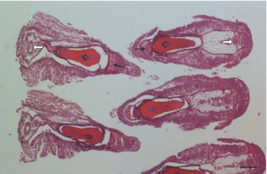
Fig. 1. Cross section of the gill filaments of a pike at their uninfected part. Filaments of the two rows of the hemibranchium are located opposite to each other so that the afferent branchial artery
(black arrows) and the bulk of the cartilaginous gill ray (c) are located at the inner part of the filament, while the efferent branchial artery (white arrow) runs down at the outer side of the
filament. Haematoxylin and eosin (HE) staining. Bar = 100 µm
Fig. 2. Cross section of the filaments of a roach. In one of the filaments a young Myxobolus rutili plasmodium (p) is located at one side of the cartilaginous gill ray (c) close to the afferent branchial
artery (black arrow). On the other side of the filament the efferent branchial artery is seen (white arrow). HE staining. Bar = 300 µm
Fig. 3. Myxobolus rutili infection in a roach. A more developed plasmodium (p) filled by sporogonic stages occupies a large part of the filament. c = cartilaginous gill ray; black arrow =
afferent branchial artery; white arrow = efferent branchial artery, e = multilayered epithelium.
HE staining. Bar = 300 µm
Fig. 4. Myxobolus dispar infection in a common carp. The gill filament infected by a developing plasmodium (p) is bordered by a thin capsule formed by connective tissue (ct). The plasmodium surrounded by multilayered epithelium (e) is located at the inner side of the filament, close to the afferent branchial artery (black arrow) and the cartilaginous gill ray (c). The efferent branchial
artery (white arrow) is found on the outer side of the filament. In this section gill lamellae (open arrows) are also seen. HE staining. Bar = 300 µm
Fig. 5. Higher magnification of the plasmodium seen in Fig. 4, cut at a different level. The plasmo- dium (p) is surrounded by a thin dense connective tissue capsule (black arrow) and by multilayered
epithelium (e). The efferent branchial artery (white arrow) is found between the plasmodium and the cartilaginous gill ray (c). HE staining. Bar = 100 µm
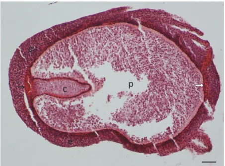
Fig. 6. Henneguya psorospermica infection in a pike. The plasmodium (p) is located at the outer edge of the filament close to the efferent branchial artery (white arrow). The afferent branchial artery (black arrow) and the cartilaginous gill ray (c) are found on the inner side of the filament.
HE staining. Bar = 100 µm
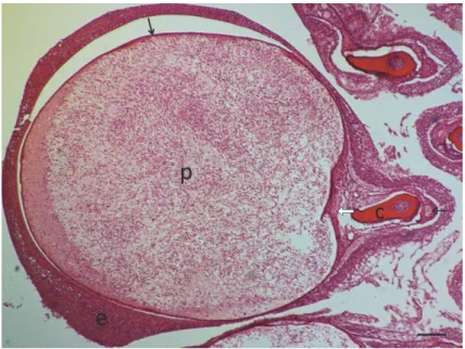
Fig. 7. Henneguya psorospermica plasmodium (p) with spores in the centre and sporogonic stages at the periphery. The plasmodium lies close to the efferent branchial artery (white arrow) and has a thin connective tissue wall (black arrows) covered by multilayered epithelium (e). The afferent branchial artery (open arrow) is found on the outer side of the filament. HE staining. Bar = 100 µm
Discussion
Although presenting data on the tissue location of plasmodial stages is a basic requirement for describing myxosporean species, relatively few papers provide useful information on the intrabranchial location of plasmodia. Some specialists such as Katoch and Kaur (2016) tried to expand the scale of diagnos- tic tools by replacing the widely used haematoxylin and eosin staining with Lu- na’s method which gives a bright colour to myxosporean spores; however, for finding young plasmodia some additional changes are needed in the histological techniques. The identification of plasmodial location is relatively easy in the case of species that develop in or among the gill lamellae in small plasmodia at intral- amellar (vascular) or interlamellar (epithelial) sites. However, for species which develop in large plasmodia inside the gill filaments, identifying the specific site and tissue of plasmodial development is more complicated. While the location of species like M. feisti Molnár, Cech, Székely, 2008 in the cartilaginous gill rays or M. dujardini (Thélohan, 1892) in the multilayered epithelium of the filaments can easily be determined, identifying the location of species developing in arter- ies or in the connective tissue of the gill filaments is rather difficult. In histologi- cal investigations made in the horizontal plane of the gill filaments the lumen of filamental arteries has frequently been designated as the location of plasmodial development. For example, Dyková and Lom (1978), who studied H. pso- rospermica infection of the pike in longitudinal sections, stated in their work on histopathological changes in the gills that H. psorospermica formed plasmodia in the afferent filamental arteries. The results of our present investigations contra- dict that statement. Although when describing the histology of some Myxobolus species (M. margitae Molnár, 2000, M. muelleri Bütschli, 1882, M. sommervillae, Molnár, Marton, Székely, Eszterbauer, 2010, M. arrabonensis Cech, Borzák, Molnár, Székely, 1915) on the basis of longitudinal sections, the present authors (Molnár, 2000; Molnár et al., 2006, 2010; Székely et al., 2015) tried to provide robust evidence for the intravascular development of plasmodia in the afferent branchial artery or the efferent branchial artery, an additional study using cross- sectioned preparations was also recommended. As an exception, in their histolog- ical work Adriano et al. (2009) clearly demonstrated plasmodia of M. salminus Adriano, Arana, Carriero, Naldoni, Ceccarelli, Maia, 2009 in the branchial ves- sels of Salminus brasiliensis (Cuvier) even in longitudinally sectioned prepara- tions. For some other species, however, the extravascular location of plasmodia was clearly proved. Walsh et al. (2012) described plasmodia of M. micropteri Walsh, Iwanowicz, Glenney, Iwanowicz, Blazer, 2012 developing under the epi- thelial layer adjacent to the filament cartilage near the capillary vessel. Liu et al.
(2013) found plasmodia of M. musseliusae located in the loose connective tissue of the filament but in close contact with the branchial vessels. Similar locations were found for M. rutili and M. elegans (Molnár et al., 2010, 2012). In the case
of matured plasmodia the examination of tissue location is difficult because of the large volume of the cysts and, therefore, young developing stages have to be found. In the three cases studied by us, the youngest stages were found for M. ru- tili where cross sections of young plasmodia were found side by side with the cartilage at a certain distance from the afferent branchial artery. In the case of M.
dispar and H. psorospermica, where plasmodia were cross-sectioned close to their tapering, relatively thin region, the extravascular locations of the species could also be established, as the afferent and efferent branchial arteries were clearly separated from the plasmodia. The location of plasmodia both on the in- ner (M. dispar) and on the outer side of the filament (H. psorospermica) proves that plasmodial development takes place outside the afferent and efferent bran- chial arteries.
Acknowledgements
The authors thank the GINOP 2.3.2-15-2016-00004 project for the financial sup- port and Ms Györgyi Pataki for preparing the histological slides.
References
Adriano, E. A., Arana, S., Carriero, M. M., Naldoni, J., Ceccarelli, P. S. and Maia, A. A. M.
(2009): Light, electron microscopy and histopathology of Myxobolus salminus n. sp., a parasite of Salminus brasiliensis from the Brazilian Pantanal. Vet. Parasitol. 165, 25–29.
Bucke, D. (1972): Some histological techniques applicable to fish tissues. In: Mawdesley-Thomas, L. E. (ed.) Diseases of Fish. Proceedings of Symposium no. 30, Zoological Society, Lon- don, May 1971. Academic Press and the Zoological Society, New York and London. pp.
153–189.
Dyková, I. and Lom, J. (1978): Histopathological changes in fish gills infected with myxosporidian parasites of the genus Henneguya. J. Fish Biol. 12, 197–202.
Dyková, I. and Lom, J. (2007): Histopathology of Protistan and Myxozoan Infections in Fishes. An Atlas. Academia, Praha. 219 pp.
Genten, F., Terwinghe, E. and Danguy, A. (2009): Atlas of Fish Histology. Science Publishers, En- field, NH, USA. 215 pp.
Katoch, A. and Kaur, H. (2016): Histological location of myxosporean plasmodia in fish tissue with Luna’s method. Parasitol. Res. 115, 3705–3707.
Kovács-Gayer, É. (1975): Studies on the gills of the fish 1. Anatomy and the microscopic structure of the gills of carp (Cyprinus carpio) [in Hungarian, with English abstract]. Magy. Allator- vosok 30, 707–712.
Lom, J. and Dyková, I. (1992): Protozoan parasites of fishes. In: Developments in Aquaculture and Fisheries Science, Volume 26. Elsevier, Amsterdam. 315 pp.
Liu, Y., Whipps, C. M., Gu, Z. M., Huang, M. J., He, C., Yang, H. L. and Molnár, K. (2013): Myx- obolus musseliusae (Myxozoa: Myxobolidae) from the gills of common carp Cyprinus carpio and revision of Myxobolus dispar recorded in China. Parasitol. Res. 112, 289–296.
Molnár, K. (2000): Survey on Myxobolus infection of the bleak (Alburnus alburnus L.) in the River Danube and in Lake Balaton. Acta Vet. Hung. 48, 421–432.
Molnár, K. (2002): Site preference of myxosporeans in the gill. Dis. Aquat. Org. 48, 197–207.
Molnár, K. and Eszterbauer, E. (2015): Specificity of infection sites in vertebrate hosts. In: Okamu- ra, B., Gruhl, A. and Bartolomew, J. L. (eds) Myxozoan Evolution, Ecology and Develop- ment. Springer International Publishing, Cham. pp. 295–313.
Molnár, K., Cech, G. and Székely, Cs. (2012): Remarks on the seasonal occurrence and identifica- tion of young plasmodial stages of Myxobolus spp. infecting cyprinid fishes in Hungary.
Acta Vet. Hung. 60, 69–82.
Molnár, K., Marton, S., Eszterbauer, E. and Székely, Cs. (2006): Comparative morphological and molecular studies on Myxobolus spp. infecting chub from the River Danube, Hungary, and description of M. muellericus sp. n. Dis. Aquat. Org. 73, 49–61.
Molnár, K., Marton, Sz., Székely, Cs. and Eszterbauer, E. (2010): Differentiation of Myxobolus spp. (Myxozoa: Myxobolidae) infecting roach (Rutilus rutilus) in Hungary. Parasitol. Res.
107, 1137–1150.
Székely, Cs., Molnár, K. and Cech, G. (2015): Description of Myxobolus balatonicus n. sp.
(Myxozoa: Myxobolidae) from the common carp Cyprinus carpio L. in Lake Balaton.
Syst. Parasitol. 91, 71–79.
Walsh, H. L., Iwanowicz, R., Glenney, G. W., Iwanowicz, D. D. and Blazer, V. S. (2012): Descrip- tion of two new gill myxozoans from smallmouth (Micropterus dolomieu) and largemouth (Micropterus salmoides) bass. J. Parasitol. 98, 415–422.
Yasutake, W. T. and Wales, J. H. (1983): Microscopic Anatomy of Salmonids: An Atlas. US Fish and Wildlife Service, Washington, DC. 190 pp.