Bene fi cial impact of Nesfatin-1 on reproductive dysfunction induced by nicotine in male rats:
Possible modulation of autophagy and pyroptosis signaling pathways
N. M MADI
1, R. E. ABO EL GHEIT
1, R. A. BARHOMA
1, A. EL SAADANY
2, G. M. ALGHAZALY
3, K. MAREA
4and M. H. EL-SAKA
1p1Department of Physiology, Faculty of Medicine, Tanta University, Tanta, Egypt
2Department of Pharmacology, Faculty of Medicine, Tanta University, Tanta, Egypt
3Department of Internal Medicine, Faculty of Medicine, Tanta University, Tanta, Egypt
4Department of Pathology, Faculty of Medicine, Tanta University, Tanta, Egypt
Received: October 29, 2020 • Accepted: March 30, 2021 Published online: June 16, 2021
© 2021 Akademiai Kiado, Budapest
ABSTRACT
This study was conducted to explore the beneficial impact of nesfatin-1 on reproductive dysfunction induced by nicotine (NT) in male rats with possible modulation of autophagy and pyroptosis signaling pathways. This research was performed on 40 Wistar male rats. They were distributed into four groups:
control, normalþnesfatin-1, NT, and NTþnesfatin-1. At the end of the experimental period, the serum was separated for assay of testosterone, FSH and LH. Also, sperm parameters were determined. Histopatho- logical examination of testicular tissue and immunohistochemical analysis was done for mammalian target of rapamycin, AMP-activated protein kinase, and mitogen-activated protein kinases including phosphor- ylated extracellular signal regulated kinase and phosphorylated cJun N-terminal kinase. Relative gene expression was determined for testicular nucleotide oligomerization domain (NOD)-like receptors proteins andCaspase-1, and autophagy markers including microtubule-associated protein 1 light chain 3 alpha and Beclin-1. Also, the following testicular parameters were assayed: 3b-hydroxysteroid dehydrogenase, 17b- hydroxysteroid dehydrogenase, malondialdehyde, superoxide dismutase activity, catalase, glucose-6 phosphate dehydrogenase, reactive oxygen species, caspase-3 activity, IL-1b, IL-18, mitochondrial trans- membrane potential and Complex-I activity. The results revealed that the normalþnesfatin-1 group
pCorresponding author. Department of Physiology, Faculty of Medicine, Tanta University, Tanta, Egypt. Tel.:
þ20 01023126578. E-mail: mervat.elsaka1@med.tanta.edu.eg Physiology International108 (2021) 2, 185–201
DOI:10.1556/2060.2021.00176
showed insignificant changes as compared to the control group. Meanwhile, the NT group exhibited prominent reproductive dysfunction in male rats. On the other hand, in the NTþnesfatin-1 group nesfatin- 1 notably attenuated this reproductive dysfunction as evidenced by improvement of hormonal assay, sperm parameters, histopathological picture, immunohistochemical evaluation and real time relative gene ex- pressions. In conclusion: Nesfatin-1 alleviated the impairment of male reproductive functions induced by NT via enhancement of autophagy pathways, suppression of pyroptosis, apoptosis, mitochondrial dysfunction and ROS production. Thus nesfatin-1 may offer a novel protective or therapeutic access for treating male infertility.
KEYWORDS
nesfatin-1, reproductive dysfunction, nicotine, autophagy, pyroptosis
INTRODUCTION
Infertility is a major problem that has harmful impacts on social and psychological aspects [1]. A considerable percentage of infertility is due to males. Male infertility can be induced by numerous factors such as genetic disorders, several diseases, some chemical toxins and cigarette or tobacco smoking [2]. Nicotine (NT) is a toxic pharmacological ingredient in cigarette smoke.
It represents the major percentage of the total constituents of cigarettes. The exposure to NT can occur through smoking, chewing of tobacco or using smokeless tobacco products [3]. Previous studies demonstrated its harmful impacts on the reproductive function and fertility in males [4,5].
Nesfatin-1 is a powerful anorexiogenic neuropeptide that was first recognized in 2006. It is constituted of 82 amino acids. The precursor peptide for nesfatin-1 is called nucleobindin-2 (NUCB2) [6]. Nesfatin-1 has been detected in the central nervous system, e.g. the hypothalamus, and in peripheral tissues such as the digestive system, adipose tissue and reproductive organs in females and males. Previous studies showed that it has anti-inflammatory, anti-apoptotic and antioxidant effects [7, 8].
Autophagy acts as a self-degradative mechanism of cytosolic organelles such as mito- chondria, cellular substrates such as proteins, lipids, and pathogens such as viruses or bacteria. It has a major role in cellular homeostasis and acts as an adapting mechanism for different stresses in the environment such as hypoxia, starvation, oxidative stress and endoplasmic reticulum stress. Therefore it is considered as a mechanism for preserving cell existence [9]. Previous studies have suggested that lack or loss of the autophagic process may contribute to the pathogenesis of some diseases [10]. A previous study reported that the autophagy process in Leydig cells is highly developed and has been implicated in ste- roidogenesis [11].
Pyroptosis is an inflammatory type of organized cell death causing lysis of the cells. A main characteristic of pyroptosis is the need for activation of caspase-1, which subsequently stimulates the active form of pro-inflammatory cytokines as interleukin-1 beta (IL-1b) and interleukin-18 (IL-18) via the inflammasome-relying pathway [12]. The conventional inflammasomes include nucleotide oligomerization domain (NOD)-like receptor proteins (NLRPs) such as NLRP1,
NLRP3 and NLRP6. These inflammasomes are responsible for the activation of caspase-1 with subsequent occurrence of pyroptosis [13].
Few previous studies have demonstrated the effect of nesfatin-1 on testicular dysfunction.
Some previous reports have revealed nesfatin-1 expression in male reproductive organs, even so its exact functions in these organs are still not well defined. Thus, to our knowledge, this is the first study demonstrating the beneficial effect of nesfatin-1 on reproductive dysfunction induced by NT in male rats with possible modulation of autophagy and pyroptosis signaling pathways and possible mechanisms for its action on testicular tissue.
MATERIALS AND METHODS
Experimental animals
This research was performed on 40 Wistar male rats. They were aged 10–12 weeks, and weighed 200–250 g. They were provided by the research lab of experiments, Faculty of Pharmacy, Tanta University. The animals were kept in clean standard cages, three rats per cage, and were freely provided with water and food. They were held at proper temperature (24±28C) with 12h light/
dark cycles. The experimental plan was performed in accordance with the guidelines of the ethical committee of Tanta University, Faculty of Medicine, (Code Approval Number: 3512/02/
27).
Chemicals
Nesfatin-1 and NT were obtained from Sigma Aldrich Co., (MO, USA). NT was in the liquid form and was diluted in normal saline. Nesfatin-1 was dissolved in normal saline. The kits utilized were provided by Abcam (ab),Antibodies-online.com(ABIN), and MyBiosource com (MBS).
Experimental groups
The rats were given one week for adaptation before the start of the experiment. Rats of the groups studied were given all treatments via intraperitoneal injections once daily for 8 weeks.
They were distributed into four groups (10 rats/group) as follows:
1. Control group: animals were administrated normal saline as a vehicle.
2. Normalþnesfatin-1 group: animals were administrated nesfatin-1 in a dose of 0.3
m
g/kg [14].3. NT group: animals were administrated NT in a dose of 0.6 mg/kg [15].
4. NTþnesfatin-1 group: animals were administrated nicotine in a dose 0.6 mg/kg plus nes- fatin-1 in a dose of 0.3
m
g/kg [14].The doses of nesfatin-1 and NT were selected in this study according to former studies [7, 14–18].
At the end of the 8th week of the experiment, the animals were weighed. They were then anaesthetized by inhalation of diethyl ether. Collection of blood samples was done via cardiac puncture, which was followed by centrifugation at 4000 rpm for 15 minutes for separation of the serum. Serum was kept at808C until used for hormonal assays. Eventually, all rats were sacrificed and the testes were taken out and weighed. The relative testicular weight was
Physiology International108 (2021) 2, 185–201 187
determined [19]. The left testes were cut transversely into small portions, homogenized and centrifuged at 2000 rmp for 10 minutes to get the supernatant that was kept at808C till it was analyzed. The right testes were divided transversely into two portions; thefirst portion was kept at808C for RNA extraction by real time gene expression. The second portion was evaluated histopathologically and immunohistochemically.
Evaluation of sperm parameters
For examination of the sperm parameters, the epididymis was carefully disconnected from the testis I. It was then used to assess the sperm count, motility and viability [20].
Hormonal assay of serum
Testosterone, luteinizing hormone and follicle stimulating hormone (FSH) were estimated by ELISA kits, (Cat# No: ABIN6574083, ABIN6574078 and ABIN5670612, respectively) in accord with the manufacturer’s instructions.
Histopathological evaluation of testicular tissue
The excised testes were put in Bouin solution for fixation. They were then inserted into paraffin.
They were sliced into sections with 4–5
m
m thickness. The slides were stained with hematoxylin and eosin (H & E) for evaluation [21].Immunohistochemical examination
The immunohistochemical evaluation of testicular tissue was done for the following markers:
mammalian target of rapamycin (mTOR), (Cat# No: ab2732, dilution 1/1000), AMP-activated protein kinase (AMPK) (Cat# No: MBS2537343, dilution 1/200), and mitogen-activated protein kinases (MAPKs) including phosphorylated extracellular signal regulated kinase (pERK) (Cat#
No: MBS9701266, dilution 1/200) and phosphorylated cJun N-terminal kinase (pJNK), (Cat#
No: MBS624642, dilution 1/100). The expressions were calculated by the image J analysis program and expressed as % area according to the method of Varghese et al. [22].
Testicular tissue homogenate assay
The following parameters were assayed: enzymes of steroidogenesis: both 3b-hydroxysteroid dehydrogenase (3b-HSD) and 17b-hydroxysteroid dehydrogenase (17-bHSD) were assayed according to the method of Bergmeyer [23], the markers for oxidants and antioxidants including malondialdehyde (MDA), superoxide dismutase (SOD) activity, catalase (CAT), and glucose-6 phosphate dehydrogenase (G6PDH) levels were determined colorimetrically (Cat#
No: ab118970, ab65354, ab83464 and ab102529 respectively). Also, testicular reactive oxygen species generation was measured by estimation of hydrogen peroxide [24] and hydroxyl radicals [25]. Apoptotic marker caspase-3 activity was determined colorimetrically (Cat# No: ab39401).
The pro-inflammatory markers IL-1b and IL-18 were estimated by ELISA kits (Cat# No MBS175941, and MBS2701003 respectively). Mitochondria were isolated from testicular tissue [26], then the mitochondrial transmembrane potential (ΔJm) and mitochondrial Complex-I activity were determined by the methods of Mishra and Shaha, [27] and Spinazzi et al. [28]
respectively. Eventually, protein contents of testicular homogenate were estimated (Cat# No 500-0006).
Separation of RNA and PCR Real time quantitative analysis for relative gene expression in testicular tissue
Total RNAs were separated from the testicular tissues of rats with TRIZOL reagent in accord with the instructions of kits (thermo scientific, # 0731 USA) [29]. cDNAs were obtained by reverse transcription of the RNAs by using revert Aid H Minus reverse transcriptase (Thermo Scientific, # Ep 0451, USA). Eventually, cDNAs were used for determination of the relative gene expression for pyroptotic markers; Nlrp3 and caspase-1, and autophagy markers including microtubule-associated protein 1 light chain 3 alpha (Lc3-II) and Beclin-1. b-actin is the housekeeping gene to which the aforementioned genes were normalized using the 2-ΔΔCt method [30]. The primer sequences for the aforementioned genes are presented inTable 1.
Statistical analysis
The data were displayed as the mean ± standard deviation. Unpaired t-test was used for comparing the collected data from two different groups. One–way ANOVA was used for analysis of the collected data between more than two groups, and then Tukey’s test was used to determine the significance. Statistical significance was considered with P values <0.05. The statistical analyses were carried out by SPSS software (Version 24.0).
RESULTS
Changes in body weight and testicular weight
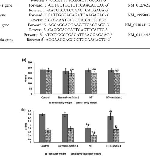
The final body weight showed notable reduction in the normalþnesfatin-1, NT and NTþnesfatin-1 groups in contrast to the control group (Figs 1a & b). Meanwhile, in com- parison with the initial body weight, the final body weight insignificantly changed in the control group, but it showed a remarkable drop in the normalþnesfatin-1, NT and NTþnesfatin-1 groups. Regarding testicular weight and relative testicular weight, both exhibited notable decrease in the NT group as opposed to the control. On the other hand, they showed significant increase in the NTþnesfatin-1 group as compared to the NT group.
But in the normalþnesfatin-1 group, there was insignificant alteration in testicular weigh as compared to the control group.
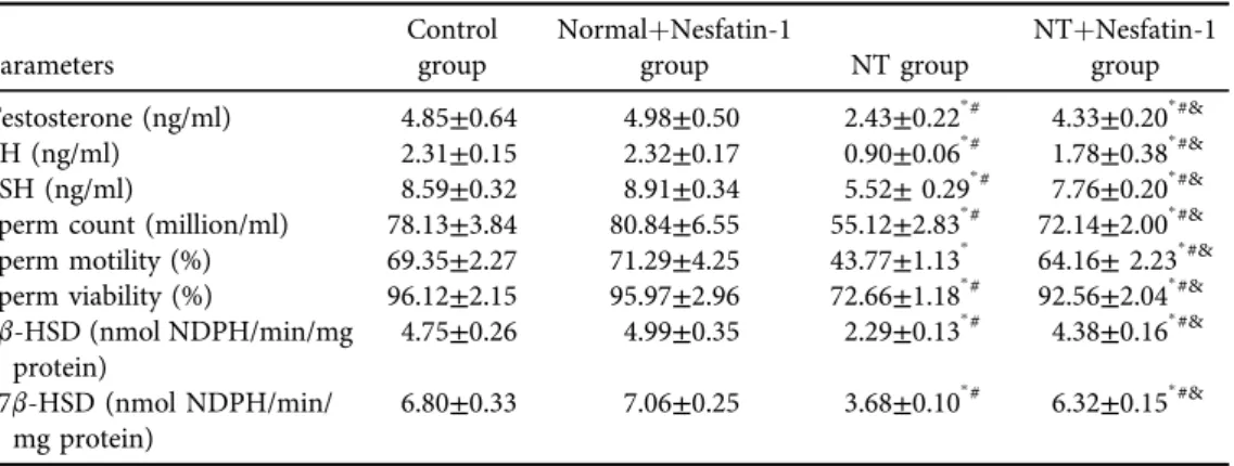
Serum hormonal levels, sperm parameters and testicular steroidogenic enzymes
In contrast with the control group, NT caused dramatic lowering of the serum levels of testosterone, LH and FSH (Table 2). Furthermore, it caused remarkable decline of sperm count, motility and viability with suppression of the testicular enzymes 3b-HSD and 17b-HSD.
Conversely, in the NTþnesfatin-1 group it was observed that treatment with nesfatin-1 caused significant increase of all previously mentioned parameters in comparison with the NT group. It was observed that all these parameters were not modified in the normalþnesfatin-1 group as compared to the control group.
Physiology International108 (2021) 2, 185–201 189
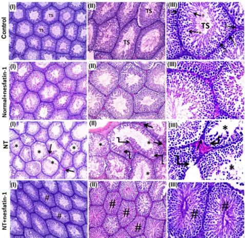
Histopathological analysis of the testicular tissue
In both the control and normalþnesfatin-1 groups, the histopathological inspection of testicular tissues displayed a normal picture (Fig. 2). The NT group displayed degenerative changes of the seminiferous tubules. The NTþnesfatin-1 group showed attenuation of the degenerative changes of seminiferous tubules and restoration of normal structure of testicular tissue.
Fig. 1.Changes in body weight and testicular weight:
Data are given as mean±SD.ΔP< 0.05 vs initial body weight.pPvs control group,#P< 0.05 vs nor- malþnesfatin-1 group,&P< 0.05 vs NT group
Table 1.Real-time PCR primer sequences
Gene name Primer sequence
NCBI GenBank accession number
Nlrp3gene Forward: 59-TCTGTTCATTGGCTGCTGAT-39 NM_001191642.1
Reverse: 59-GCCCTTTTCGAACTTGCCGT-39
Caspase-1gene Forward: 59-CTTGCTGCTCTTCAACACCAG-39 NM_012762.2 Reverse: 59-AATGTCCTCCAAGTCACGAGA-39
Lc3-IIgene Forward: 59CATTGGCACAGATGAAGACAC-39 NM_199500.2
Reverse: 59GCCAAATGTTCATCCACTTTC-39
Beclin-1gene Forward: 59-ACCAGGAGGAACCTCAGTACC-39 NM_001034117.1 Reverse: 59-CAGGCAGCATTGAGTTCATTC-39
b-actin housekeeping gene
Forward: 59-ATCCTGCGTGACATTAAGGAGAAG-39 NM_031144.3 Reverse: 59-AGGAAGGACGGCTGGAAGAGTG-39
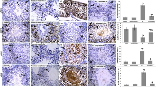
Immunohistochemical expression of mTOR, AMPK, pERK, and pJNK in testicular tissue
As illustrated inFig. 3, the expression of testicular mTOR, pERK and pJNK was significantly upregulated with synchronized significant downregulation of AMPK in the NT group, contrary to the control group. Meanwhile, the NTþnesfatin-1 group showed notable attenuation of expression of testicular mTOR, pERK, and pJNK with accompanying notable augmentation of AMPK expression compared to the NT group. It was noted that all the studied expressions were not altered in the normalþnesfatin-1 group compared to the control group.
As regards immunohistochemical expression of mTOR, in the control group there was mild expression in some primary spermatocytes and a few spermatogonia, whereas the normal- þnesfatin-1 group exhibited mild expression in some spermatogonia. Meanwhile, the NT group displayed marked expression in all germ cells of the seminiferous tublues. On the other hand, the NTþnesfatin-1 group showed moderate expression in some primary spermatocytes.
Immunohistochemical expression of AMPK showed that in both the control and the nor- malþnesfatin-1 groups there was strong positive expression in spermatogonia and primary spermatocytes, whereas the NT group exhibited mild expression only in spermatogonia.
Meanwhile, the NTþnesfatin-1 group displayed strong expression mainly in spermatogonia and primary spermatocytes.
Our study of pERK expression revealed that both the control and the normalþnesfatin-1 groups exhibited mild positive immunoexpression in some spermatogonia and to a lesser extent in a few primary spermatocytes, whereas the NT group exhibited marked expression in all germ cells of seminiferous tubules. On the other hand, the NTþnesfatin-1 group showed mild expression in some spermatogonia.
Concerning immunohistochemical expression of pJNK, both the control and the nor- malþnesfatin-1 groups exhibited mild expression in few spermatogonia, whereas the NT group had strong expression in all germ cells of seminiferous tubules. Meanwhile, the NTþnesfatin-1 group exhibited mild expression in some spermatogonia and primary sper- matocytes.
Table 2.Serum hormonal levels, sperm parameters and testicular steroidogenic enzymes in the studied groups
Parameters
Control group
NormalþNesfatin-1
group NT group
NTþNesfatin-1 group Testosterone (ng/ml) 4.85±0.64 4.98±0.50 2.43±0.22p# 4.33±0.20p#&
LH (ng/ml) 2.31±0.15 2.32±0.17 0.90±0.06p# 1.78±0.38p#&
FSH (ng/ml) 8.59±0.32 8.91±0.34 5.52±0.29p# 7.76±0.20p#&
Sperm count (million/ml) 78.13±3.84 80.84±6.55 55.12±2.83p# 72.14±2.00p#&
Sperm motility (%) 69.35±2.27 71.29±4.25 43.77±1.13p 64.16±2.23p#&
Sperm viability (%) 96.12±2.15 95.97±2.96 72.66±1.18p# 92.56±2.04p#&
3b-HSD (nmol NDPH/min/mg protein)
4.75±0.26 4.99±0.35 2.29±0.13p# 4.38±0.16p#&
17b-HSD (nmol NDPH/min/
mg protein)
6.80±0.33 7.06±0.25 3.68±0.10p# 6.32±0.15p#&
Data are given as mean±SD.pPvs control group,#P< 0.05 vs normalþnesfatin-1 group,&P< 0.05 vs NT group.
Physiology International108 (2021) 2, 185–201 191
Relative gene expression of testicular markers of pyroptosis and autophagy
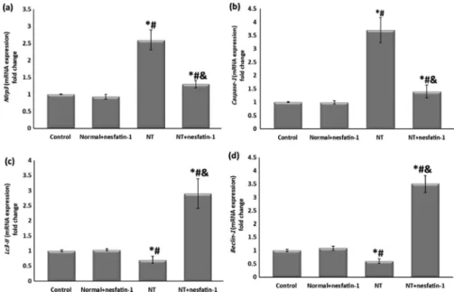
It was noticed that NT exposure for 8 weeks dramatically elevated the testicular gene expression ofNlrp3andCaspase-1(Figs 4a & b). On the other hand, in the NTþnesfatin-1 group nesfatin-1 treatment significantly suppressed the elevation of these markers for pyroptosis in comparison with NT group. Concerning the gene expression for autophagy markers, NT markedly reduced testicular Lc3-II and Beclin-1 compared to the control, whereas in the NTþnesfatin-1 group nesfatin-1 administration for 8 weeks caused remarkable upregulation of the expression ofLc3- IIandBeclin-1as compared to the NT group (Figs 4c & d). It was observed that the testicular
Fig. 2.Histopathological analysis of the testicular tissue:
The control and normalþnesfatin-1 groups displayed normal histopathology of testicular tissues. Semi- niferous tubules (TS), (a) basement membrane, (b) Leydig cells, (c) Sertoli cells, (d) spermatogonia, (e) primary spermatocytes, and (f) spermatids). The NT group exhibited degenerative changes of the semi- niferous tubules and diminishing of all stages of spermatogenesis. Vacuolization (curved arrow). The NTþNesfatin-1 group showed attenuation of the degenerative changes of seminiferous tubules (#) and restoration of normal structure of testicular tissue. I, II & III5magnification 100, 200 &3400 respectively
Fig. 3.Immunohistochemical analysis with estimation of immunoreactivity of mTOR, AMPK, pREK and pJNK. The arrows denote brown staining of mTOR, AMPK, pREK and pJNK. Magnification was3400. Data are given as mean±SD.pPvs control group, #P< 0.05 vs normalþnesfatin-1 group,
&P< 0.05 vs NT group.
Expression of mTOR revealed that, in the control group, there was mild expression in some primary spermatocytes and few spermatogonia. The Normalþnesfatin-1 group exhibited mild expression in some spermatogonia. The NT group displayed marked expression in all germ cells of the seminiferous tubules. The NTþnesfatin-1 group showed moderate expression in some primary spermatocytes. Expression of AMPK showed that, in both control and normalþnesfatin-1 groups, there was strong positive expression in spermatogonia and primary spermatocytes. The NT group exhibited mild expression only in spermatogonia. The NTþnesfatin-1 group displayed strong expression mainly in spermatogonia and primary spermatocytes. pERK expression showed that both control and normalþnesfatin-1 groups exhibited mild positive immunoexpression in some spermatogonia and to a lesser extent in a few primary spermatocytes. The NT group exhibited marked expression in all germ cells of seminiferous tubules. The NTþnesfatin-1 group showed mild expression in some spermatogonia. Expression of pJNK in both the control and normalþnesfatin-1 groups revealed mild expression in few spermatogonia, whereas in the NT group there was strong expression in all germ cells of seminiferous tubules. Meanwhile, the NTþnesfatin-1 group
exhibited mild expression in some spermatogonia and primary spermatocytes
PhysiologyInternational108(2021)2,185–201193
markers of pyroptosis and autophagy were not changed in the normalþnesfatin-1 group compared to the control.
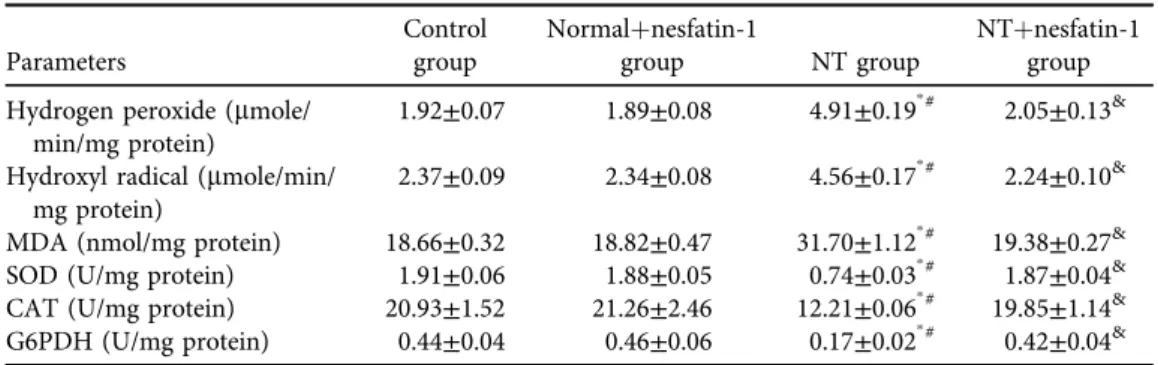
Testicular ROS, oxidant and antioxidant parameters
Testicular hydrogen peroxide, hydroxyl radicals and MDA were significantly elevated with concurrent notable suppression of the activities of antioxidant enzymes including SOD, CAT and G6PDH in the NT group as opposed to the control group (Table 3). Meanwhile, in comparison with the NT group, in NTþnesfatin-1 group nesfatin-1 significantly prohibited the increase in hydrogen peroxide, hydroxyl radicals and MDA with synchronous restoration of normal levels of SOD, CAT and G6PDH activities. It was noted that the studied testicular ROS, oxidant and antioxidant parameters were not altered in normalþnesfatin-1 group compared to the control group.
Testicular apoptosis, pro-inflammatory cytokines, mitochondrial ΔJ m and Complex-I activity
As compared to the control, NT exposure for 8 weeks notably elevated testicular caspase-3, IL- 1band IL18 with accompanying remarkable fall of mitochondrialΔJm and Complex-I activity (Table 4), whereas in the NTþnesfatin-1 group, nesfatin-1 administration dramatically inhibited the increase of caspase-3, IL-1band IL18 with restoration of mitochondrial ΔJm and Com- plex-I activity to their normal values. It was observed that all these parameters were not modified in normalþnesfatin-1 group as compared to the control group.
Fig. 4.Relative gene expression of testicular markers of pyroptosis and autophagy
Data are given as mean±SD.pPvs control group,#P< 0.05 vs normalþnesfatin-1 group,&P< 0.05 vs NT group
DISCUSSION
The data of the present study showed that NT administration for 8 weeks dramatically impaired the reproductive function of male rats. On the other hand, nesfatin-1 administration to NT- treated rats ameliorated the reproductive dysfunction via augmentation of autophagy with attenuation of pyroptosis, apoptosis, oxidative stress and mitochondrial dysfunction of testicular tissue.
The data of the present study showed that NT caused a remarkable diminution of body weight, which was in conformity with previous reports [31]. The precise mechanism of the effect of NT on body weight is not well evident, but it might be related to the reduction of food intake by NT consuming animals [32]. In agreement with previous studies [33], ourfindings docu- mented that in the NTþnesfatin-1 group, nesfatin-1 remarkably caused a decline of body weight in contrast with either the control or NT group, which could be related to its effect as an anorexogenic hormone [34].
In the same way, our results recorded a notable diminution of the testicular weight as well the relative testicular weight in the NT group, which could be explained by the suppressing effect of NT on steroidogenesis and spermatogenesis with subsequent degeneration and reduction of the size of the seminiferous tubules [35]. At the same time our results revealed that in the
Table 3.Testicular ROS, oxidant, and antioxidant parameters in the studied groups
Parameters
Control group
Normalþnesfatin-1
group NT group
NTþnesfatin-1 group Hydrogen peroxide (mmole/
min/mg protein)
1.92±0.07 1.89±0.08 4.91±0.19p# 2.05±0.13&
Hydroxyl radical (mmole/min/
mg protein)
2.37±0.09 2.34±0.08 4.56±0.17p# 2.24±0.10&
MDA (nmol/mg protein) 18.66±0.32 18.82±0.47 31.70±1.12p# 19.38±0.27&
SOD (U/mg protein) 1.91±0.06 1.88±0.05 0.74±0.03p# 1.87±0.04&
CAT (U/mg protein) 20.93±1.52 21.26±2.46 12.21±0.06p# 19.85±1.14&
G6PDH (U/mg protein) 0.44±0.04 0.46±0.06 0.17±0.02p# 0.42±0.04&
Data are given as mean±SD.pPvs control group,#P< 0.05 vs normalþnesfatin-1 group,&P< 0.05 vs NT group.
Table 4.Testicular apoptosis, pro-inflammatory cytokines mitochondrialΔJm and Complex-I activity Parameters
Control group
Normalþnesfatin-1
group NT group
NTþnesfatin-1 group
Caspase-3 0.50±0.06 0.48±0.09 2.84±0.13p# 0.64±0.04p#&
IL-1b(pg/ml) 125.49±5.74 118.78±9.86 176.24±9.52p# 137.67±5.59p#&
IL-18 (pg/ml) 106.67±7.03 99.53±8.30 146.21±7.42p# 117.56±4.37p#&
ΔJm (fluorescence unit) 2.38±0.14 2.41±0.11 1.31±0.06p# 2.51±0.19&
Complex-I activity (U/mg protein)
143.2±8.19 146.20±7.42 101.6±6.35p# 140.6±6.60&
Data are given as mean±SD.pP< vs control group,#P< 0.05 vs normalþnesfatin-1 group,&P< 0.05 vs NT group.
Physiology International108 (2021) 2, 185–201 195
NTþnesfatin-1 group, nesfatin-1 treatment significantly restored the testicular weight to its normal value, which could be attributed to its stimulatory effect on steroidogenesis and sper- matogenesis [17], as shown in our results.
In parallel to the previously mentioned reports [36], the data of our study demonstrated that NT administration markedly reduced the serum levels of testosterone, LH and FSH. The reduction of the serum testosterone level could be explained by the suppressing effect of NT on testicular enzymes for steroidogenesis including 3b-HSD and 17b-HSD [35], as shown in our results. Moreover, apoptotic and pyroptotic inflammatory effects of NT might be additional mechanisms for the decreased testosterone level [37,38], which was also displayed in our results, as well as the stimulatory effect of NT on testicular ERK. ERK was previously reported to suppress the activity of 3b-HSD and 17b-HSD with subsequent diminution of testosterone synthesis [39].
The decline of the serum levels of LH and FSH in NT-administrated rats might be related to the disturbance of the hypothalamo-hypophyseal axis caused by NT exposure [40].
Interestingly, the present study recorded that in the NTþnesfatin-1 group, nesfatin-1 notably elevated the serum levels of testosterone, LH and FSH, in addition to restoring the activities of the steroidogenic enzymes 3b-HSD and 17b-HSD. This might be ascribed to its anti-apoptotic, anti-inflammatory, antioxidant effects [7,8] and its stimulating effect on autophagy in testicular tissue, as shown in our results. It was reported that autophagy enhances the uptake of cholesterol into the Leydig cells; hence it activates the steroidogenic enzymes with subsequent increase of synthesis of testosterone [11]. It was recorded that deficient autophagy in Leydig cells might suppress synthesis of testosterone [41].
Matching previous studies [42], our results showed that NT administration caused a noticeable decline of the sperm count, motility and viability. This effect of NT on sperm pa- rameters could be ascribed to ROS generation. ROS could accelerate apoptosis of the germ cells with subsequent reduction of testosterone level and suppression of sperm production [35], these effects of NT were also documented in our results.
As displayed in our results, nesfatin-1 treatment significantly restored the sperm parameters in NT-treated rats. The restoration of normal sperm parameters by nesfatin-1 might be explained by its antioxidant, anti-apoptotic, anti-inflammatory effects [7,8] and its stimulating effect on autophagy that has a prime role in the conservation of healthy sperms [43], as revealed in our results. The histopatholological analyses of the testicular tissue confirmed our afore- mentionedfindings.
Lc3-II and Beclin-1 are two well-recognized markers for autophagy activation [44].
Intriguingly, our findings revealed that NT notably diminished the expression of autophagy markersLc3-IIandBeclin-1. The exact cause for the downregulating effect of NT on autophagy markers in testicular tissue is not well-defined; it might be attributed to excessive ROS pro- duction by NT with subsequent oxidative stress [45]. These aforementioned factors failed to stimulate autophagy, instead they caused debilitation of antioxidant enzymes and stimulation of both pyroptotic and apoptotic pathways [46, 47]. Meanwhile, our present study showed that nesfatin-1 dramatically upregulated the expression of Lc3-IIand Beclin-1in testicular tissue of NT-treated rats. Thus, nesfatin-1 enhanced the advancement of the autophagy process in this model.
Previous studies demonstrated that the main regulators for the signaling pathways for autophagy could involve mTOR and AMPK. AMPK has been described to regulate autophagy
through suppression of mTOR phosphorylation [48]. It is worthwhile to note that NT administration notably upregulated mTOR with accompanying notable downregulation of AMPK expression in testicular tissue. Nevertheless, our results demonstrated that nesfatin-1 reversed the elevated mTOR with augmentation of AMPK expression in the testicular tissue of NT-treated rats, which suggested activation of autophagy process in testicular tissue.
Our results evince that NT caused remarkable upregulation of the expression of testicular pyroptotic markersNlrp3andCaspase-1. The mechanism by which NT induced pyroptosis in testicular tissue has not been well elucidated. However, it might be explained by enhancement of ROS production and oxidative stress [49], which was shown in our results. Additionally, NT could inhibit mitochondrial Complex I with subsequent augmentation of mitochondrial ROS generation and activation of NLRP3 [50], as shown in our results.
It was observed in our results that nesfatin-1 administration in NT-treated rats caused reduction of the expression ofNlrp3andCaspase-1, hence it attenuated pyroptosis, which might be explained by its suppressive effect on ROS production and oxidative stress [8], as demon- strated in this study. Another explanation for the reduction ofNlrp3is enhancement of auto- phagy by nesfatin-1, as shown in our results. Autophagy is capable of directly inhibiting NLRP3 [51]. In conformity with this, it was previously reported that suppression of NLRP3 could be mediated through activation of AMPK with simultaneous inhibition of mTOR [10], as shown in our results.
It was worthwhile to speculate that one of the probable links between pyroptosis and autophagy could be ROS overproduction caused by NT exposure. The induction of autophagy might help in the protection of various organs against injury by inhibiting NLRP3 [51].
Matching with this view, we assumed that nesfatin-1 in NT-treated rats attenuated the testicular pyroptosis induced by NT exposure, which could be ascribed to enhancing autophagy to eliminate undue ROS overproduction.
The MAPK pathways including ERK, JNK and p38 were reported to be involved in the regulation of cell existence and death [52]. Our results clearly showed that NT exposure remarkably upregulated the expression of pERK and pJNK in testicular tissue. On the other hand, nesfatin-1 administration in the NT-treated group attenuated their expression. It was suggested that the enhanced expression of pERK and pJNK in the NT group might be attributed to ROS and oxidative stress induced by NT [53], as also explored in our results.
Notably, the results of this study showed that the testicular levels of pro-inflammatory cytokines IL-band IL-18 were significantly elevated in the NT group, which might be ascribed to the stimulating effect of NT on NLRP3, which subsequently activated caspase-1. Caspase-1 is a major activator for IL-1b- and IL-18-dependant pyroptosis [47]. Moreover, our results revealed that nesfatin-1, in NT-treated rats, significantly reduced the testicular levels of IL-1b and IL-18. This might be related to its effect to promote autophagy with subsequent inhibition of NLRP3 and suppression of pyroptotic pro-inflammatory cytokines [54], as demonstrated in our results.
The findings of this study verified that NT exposure significantly enhanced testicular ROS generation, as manifested by elevation of the levels of hydrogen peroxide and hy- droxyl radicals. Also, there was prominent augmentation of lipid peroxidation as evi- denced by the increased MDA level with concomitant decline of the antioxidant enzyme activities of SOD, CAT and G6PDH. Furthermore, NT administration caused
Physiology International108 (2021) 2, 185–201 197
mitochondrial dysfunction as evidenced by notable depression of the mitochondrialΔJm and Complex-I activity.
Our results showed that nesfatin-1 administration, in NT-treated rats, suppressed testicular ROS production and returned the antioxidant enzyme activities and the mitochondrial ΔJm and Complex-I activity to their normal levels. The antioxidant effect of nesfatin-1 on testicular tissue might be attributed to its stimulatory effect on autophagy with subsequent elimination of ROS [42], as was revealed in our results.
Our results demonstrated that NT administration caused significant elevation of testicular caspase-3 which denoted apoptosis. Conversely, our results revealed that nesfatin-1 prohibited the testicular caspase-3 activation in NT-treated rats. The anti-apoptotic effect of nesfatin-1 might be ascribed to its promoting effect on autophagy that was previously demonstrated to prevent the apoptotic pathway [55]. Another explanation for the anti-apoptotic effect of nes- fatin-1 was its inhibitory effect on MAPK pathways [56], as verified in our results.
Overall, our results demonstrated that co-treatment with nicotine and nesfatin-1 diminished the deleterious effects of nicotine but did not completely restore them to control levels.
CONCLUSION
Nesfatin-1 has a prominent role in alleviation of the impairment of male reproductive functions induced by NT via enhancement of autophagy pathways, suppression of pyroptosis, apoptosis, mitochondrial dysfunction and ROS production. Thus it improved testosterone, LH and FSH levels with subsequent restoration of sperm parameters. Nesfatin-1 may therefore offer a novel protective or therapeutic access for treating male infertility, and hence further investigations are recommended for exploring more molecular mechanisms for nesfatin-1 in male reproductive dysfunctions.
Limitations for this study
We need further research study to examine if the effect of nesfatine-1 is age-dependent. Further experiments will be needed to study the effect of nesfatin-1 for periods longer than 8 weeks to see whether or not nesfatin-1 has undesirable, strong anorexiogenic side effects.
Funding:The authors did not get any specific donation from funding sources for this research.
Conflict of interest:The authors have no conflicts of interest associated with publication of this article.
REFERENCES
1. Hasanpoor-Azghdy SB, Simbar M, Vedadhir A. The emotional-psychological consequences of infertility among infertile women seeking treatment: results of a qualitative study. Iran J Reprod Med 2014; 12: 131–8.
2. Kovac JR, Khanna A, Lipshultz LI. The effects of cigarette smoking on male fertility. Postgrad Med 2015; 127:
338–41.
3. Benowitz NL, Hukkanen J, Jacob P, 3rd. Nicotine chemistry, metabolism, kinetics and biomarkers. Handb Exp Pharmacol 2009; 29–60.
4. Bundhun PK, Janoo G, Bhurtu A, Teeluck AR, Soogund MZS, Pursun M, et al. Tobacco smoking and semen quality in infertile males: a systematic review and meta-analysis. BMC Public Health 2019; 19:
36.
5. Oyeyipo IP, Raji Y, Emikpe BO, Bolarinwa AF. Effects of nicotine on sperm characteristics and fertility profile in adult male rats: a possible role of cessation. J Reprod Infertil 2011; 12: 201–7.
6. Sundarrajan L, Blanco AM, Bertucci JI, Ramesh N, Canosa LF, Unniappan S. Nesfatin-1-Like peptide encoded in nucleobindin-1 in goldfish is a novel anorexigen modulated by sex steroids, macronutrients and daily rhythm. Sci Rep 2016; 6: 28377.
7. Kolgazi M, Cantali-Ozturk C, Deniz R, Ozdemir-Kumral ZN, Yuksel M, Sirvanci S, et al. Nesfatin-1 alleviates gastric damage via direct antioxidant mechanisms. J Surg Res 2015; 193: 111–8.
8. Solmaz A, G€ulçiçek OB, Erçetin C, Yigitbas¸ H, Yavuz E, Arıcı S, et al. Nesfatin-1 alleviates extrahepatic cholestatic damage of liver in rats. Bosn J Basic Med Sci 2016; 16: 247–53.
9. Murrow L, Debnath J. Autophagy as a stress-response and quality-control mechanism: implications for cell injury and human disease. Annu Rev Pathol 2013; 8: 105–37.
10. Condello M, Pellegrini E, Caraglia M, Meschini S. Targeting autophagy to overcome human diseases. Int J Mol Sci 2019; 20: 725.
11. Gao F, Li G, Liu C, Gao H, Wang H, Liu W, et al. Autophagy regulates testosterone synthesis by facilitating cholesterol uptake in Leydig cells. J Cell Biol 2018; 217: 2103–19.
12. Bolıvar BE, Vogel TP, Bouchier-Hayes L. Inflammatory caspase regulation: maintaining balance between inflammation and cell death in health and disease. FEBS J 2019; 286: 2628–44.
13. Moossavi M, Parsamanesh N, Bahrami A, Atkin SL, Sahebkar A. Role of the NLRP3 inflammasome in cancer.
Mol Cancer 2018; 17: 158.
14. Arabaci Tamer S, Yildirim A, Koroglu MK, Cevik O, Ercan F, Yegen BC. Nesfatin-1 ameliorates testicular injury and supports gonadal function in rats induced with testis torsion. Peptides 2018; 107: 1–9.
15. Budin SB, Kho JH, Lee JH, Ramalingam A, Jubaidi FF, Latif ES, et al. Low-dose nicotine exposure induced the oxidative damage of reproductive organs and altered the sperm characteristics of adolescent male rats. Malays J Med Sci 2017; 24: 50–7.
16. Jana K, Samanta PK, De DK. Nicotine diminishes testicular gametogenesis, steroidogenesis, and steroidogenic acute regulatory protein expression in adult albino rats: possible influence on pituitary gonadotropins and alteration of testicular antioxidant status. Toxicol Sci 2010; 116: 647–59.
17. Ranjan A, Choubey M, Yada T, Krishna A. Direct effects of neuropeptide nesfatin-1 on testicular sper- matogenesis and steroidogenesis of the adult mice. Gen Comp Endocrinol 2019; 271: 49–60.
18. Syam Das S, Nair SS, Indira M. Atorvastatin modulates drug transporters and ameliorates nicotine-induced testicular toxicity. Andrologia 2018; 50: e13029.
19. Motohashi M, Wempe MF, Mutou T, Okayama Y, Kansaku N, Takahashi H, et al. In utero-exposed di(n- butyl) phthalate induce dose dependent, age-related changes of morphology and testosterone-biosynthesis enzymes/associated proteins of Leydig cell mitochondria in rats. J Toxicol Sci 2016; 41: 195–206.
20. Eliasson R. Semen analysis with regard to sperm number, sperm morphology and functional aspects. Asian J Androl 2010; 12: 26–32.
21. Foley GL. Overview of male reproductive pathology. Toxicol Pathol 2001; 29: 49–63.
22. Varghese F, Bukhari AB, Malhotra R, De A. IHC Profiler: an open source plugin for the quantitative eval- uation and automated scoring of immunohistochemistry images of human tissue samples. PLoS One 2014; 9:
e96801.
Physiology International108 (2021) 2, 185–201 199
23. Bergmeyer HU. Methods of enzymatic analysis. 1974.
24. Holland MK, Storey BT. Oxygen metabolism of mammalian spermatozoa. Generation of hydrogen peroxide by rabbit epididymal spermatozoa. Biochem J 1981; 198: 273–80.
25. Puntarulo S, Cederbaum AI. Effect of oxygen concentration on microsomal oxidation of ethanol and gen- eration of oxygen radicals. Biochem J 1988; 251: 787–94.
26. Liao P-C, Bergamini C, Fato R, Pon L, Pallotti F. Isolation of mitochondria from cells and tissues. 2019.
27. Mishra DP, Shaha C. Estrogen-induced spermatogenic cell apoptosis occurs via the mitochondrial pathway:
role of superoxide and nitric oxide. J Biol Chem 2005; 280: 6181–96.
28. Spinazzi M, Casarin A, Pertegato V, Salviati L, Angelini C. Assessment of mitochondrial respiratory chain enzymatic activities on tissues and cultured cells. Nat Protoc 2012; 7: 1235–46.
29. El-Magd MA, Abbas HE, El-kattawy AM, Mokhbatly A. Novel polymorphisms of the IGF1R gene and their association with average daily gain in Egyptian buffalo (Bubalus bubalis). Domest Anim Endocrinol 2013; 45:
105–10.
30. Livak KJ, Schmittgen TD. Analysis of relative gene expression data using real-time quantitative PCR and the 2(-Delta Delta C(T)) Method. Methods 2001; 25: 402–8.
31. Martınez de Morentin PB, Whittle AJ, Fernø J, Nogueiras R, Dieguez C, Vidal-Puig A, et al. Nicotine induces negative energy balance through hypothalamic AMP-activated protein kinase. Diabetes 2012; 61: 807–17.
32. Mercantepe T, Unal D, T€umkaya L, Yazici ZA. Protective effects of amifostine, curcumin and caffeic acid phenethyl ester against cisplatin-induced testis tissue damage in rats. Exp Ther Med 2018; 15: 3404–12.
33. Stengel A, Goebel M, Tache Y. Nesfatin-1: a novel inhibitory regulator of food intake and body weight. Obes Rev 2011; 12: 261–71.
34. Stengel A, Mori M, Tache Y. The role of nesfatin-1 in the regulation of food intake and body weight: recent developments and future endeavors. Obes Rev 2013; 14: 859–70.
35. Mohammadghasemi F, Jahromi SK. Melatonin ameliorates testicular damages induced by nicotine in mice.
Iran J Basic Med Sci 2018; 21: 639–44.
36. Kolawole TA, Oyeyemi WA, Adigwe C, Leko B, Udeh C, Dapper DV. Honey attenuates the detrimental effects of nicotine on testicular functions in nicotine treated wistar rats. Niger J Physiol Sci 2015; 30: 11–6.
37. Kim KH, Joo KJ, Park HJ, Kwon CH, Jang MH, Kim CJ. Nicotine induces apoptosis in TM3 mouse Leydig cells. Fertil sterility 2005; 83 (Suppl 1): 1093–9.
38. Li M-y, Zhu X-l, Zhao B-x, Shi L, Wang W, Hu W, et al. Adrenomedullin alleviates the pyroptosis of Leydig cells by promoting autophagy via the ROS–AMPK–mTOR axis. Cell Death Dis 2019; 10: 489.
39. Adedara IA, Nanjappa MK, Farombi EO, Akingbemi BT. Aflatoxin B1 disrupts the androgen biosynthetic pathway in rat Leydig cells. Food Chem Toxicol 2014; 65: 252–9.
40. Nesseim WH, Haroun HS, Mostafa E, Youakim MF, Mostafa T. Effect of nicotine on spermatogenesis in adult albino rats. Andrologia 2011; 43: 398–404.
41. Li W-R, Chen L, Chang Z-J, Xin H, Liu T, Zhang Y-Q, et al. Autophagic deficiency is related to steroidogenic decline in aged rat Leydig cells. Asian J Androl 2011; 13: 881–8.
42. Poillet-Perez L, Despouy G, Delage-Mourroux R, Boyer-Guittaut M. Interplay between ROS and autophagy in cancer cells, from tumor initiation to cancer therapy. Redox Biol 2015; 4: 184–92.
43. Yin J, Ni B, Tian ZQ, Yang F, Liao WG, Gao YQ. Regulatory effects of autophagy on spermatogenesis. Biol Reprod 2017; 96: 525–30.
44. Xie WY, Zhou XD, Li Q, Chen LX, Ran DH. Acid-induced autophagy protects human lung cancer cells from apoptosis by activating ER stress. Exp Cell Res 2015; 339: 270–9.
45. Arany I, Clark J, Reed DK, Juncos LA. Chronic nicotine exposure augments renal oxidative stress and injury through transcriptional activation of p66shc. Nephrol Dial Transpl 2013; 28: 1417–25.
46. Lan X, Lederman R, Eng JM, Shoshtari SSM, Saleem MA, Malhotra A, et al. Nicotine induces podocyte apoptosis through increasing oxidative stress. PLoS One 2016; 11: e0167071.
47. Wu X, Zhang H, Qi W, Zhang Y, Li J, Li Z, et al. Nicotine promotes atherosclerosis via ROS-NLRP3-mediated endothelial cell pyroptosis. Cell Death Dis 2018; 9: 171.
48. Ryter SW, Mizumura K, Choi AM. The impact of autophagy on cell death modalities. Int J Cell Biol 2014;
502676: 2014.
49. Sharma A, Tate M, Mathew G, Vince JE, Ritchie RH, de Haan JB. Oxidative stress and NLRP3-inflammasome activity as significant drivers of diabetic cardiovascular complications: therapeutic implications. Front Physiol 2018; 9: 114.
50. Abais JM, Xia M, Zhang Y, Boini KM, Li PL. Redox regulation of NLRP3 inflammasomes: ROS as trigger or effector? Antioxid Redox Signal 2015; 22: 1111–29.
51. Yang F, Qin Y, Wang Y, Meng S, Xian H, Che H, et al. Metformin inhibits the NLRP3 inflammasome via AMPK/mTOR-dependent effects in diabetic cardiomyopathy. Int J Biol Sci 2019; 15: 1010–9.
52. Munshi A, Ramesh R. Mitogen-activated protein kinases and their role in radiation response. Genes & Cancer 2013; 4: 401–8.
53. Son Y, Cheong Y-K, Kim N-H, Chung H-T, Kang DG, Pae H-O. Mitogen-activated protein kinases and reactive oxygen species: how can ROS activate MAPK pathways? J Signal Transduction 2011; 2011: 792639.
54. Saitoh T, Akira S. Regulation of inflammasomes by autophagy. J Allergy Clin Immunol 2016; 138: 28–36.
55. Liu W, Yu G, Yu W, Ye X, Jin Y, Shrestha A, et al. Autophagy inhibits apoptosis induced by agrocybe aegerita lectin in hepatocellular carcinoma. Anticancer Agents Med Chem 2017; 17: 221–9.
56. Ramanjaneya M, Tan B, Rucinski M, Kawan M, Hu J, Kaur J, et al. Nesfatin-1 inhibits proliferation and enhances apoptosis of human adrenocortical H295R cells. J Endocrinol 2015; 226.
Physiology International108 (2021) 2, 185–201 201