CARDIAC TRANSMEMBRANE ION CHANNELS AND ACTION POTENTIALS: CELLULAR
PHYSIOLOGY AND ARRHYTHMOGENIC BEHAVIOR
Channel/protein expression
Transmembrane ion channel/pump function
Transmembrane cellular voltage change Single cell action potential
Tissue action potential + impulse conduction Single cell action potential + electrotonic interaction
Whole heart summated electrical signal Electrocardiogram – clinical diagnostic tool
Drugs/Dietary ingredients Pathological settings
Inherited/genetic abnormalities Diabetes
mellitus Heart
failure Myocardial
ischemia Plasma electrolyte
imbalance
HCM/
Athletes heart
TdP Amp
Amp Ce
Genetic mutations, polymorphisms
Gender Hormones
VF AF
Remodeling (disease, chronic drug)
Plasma electrolyte alterations Acute drug effects
Acute diseases (e.g. ischemia)
Impulse Repolarization/ERP
heterogeneity conduction
Enhanced normal Triggered activity
DAD EAD
automaticity SAN Purkinje fiber Arrhythmia
Substrate Trigger X+
X+
X+ X+ X+
AUTHORS
András Varr o, Jakub Tomek, Norbert Nagy, Lászl o Virág, Elisa Passini, Blanca Rodriguez, István Baczk o
CORRESPONDENCE varro.andras@med.u-szeged.hu
KEY WORDS
action potential; arrhythmia; heart; ion channels;
remodeling
CLINICAL HIGHLIGHTS
1. Cardiac arrhythmias are major causes of mortality. They most often arise from pathological changes in the electro- physiological properties of myocardial cells. First, this review summarizes the physiology of cardiac action poten- tials, their regional and species differences, the underlying transmembrane ionic currents, and transporters, with special focus on their human relevance.
2. Progress in computer modeling and vast quantities of experimental data made computerized replication of the action potential, impulse conduction, and simulation of cardiac electrophysiology possible. Current computer models offer improved observability and controllability via utilization of different modeling scales and can assist future individualized anti-arrhythmic therapy as well as drug electrophysiological safety assessment.
3. A number of diseases evoke changes in the con fi guration of the action potential caused by altered function and/or den- sities of transmembrane ion channels and transporters, collectively termed electrical remodeling. Initially, these altera- tions are often compensatory; however, remodeling signi fi cantly contributes to increased arrhythmia susceptibility by impairing impulse generation, conduction, and myocardial refractoriness in these clinical settings. Electrical remodeling in atrial fi brillation, heart failure, hypertrophic cardiomyopathy, myocardial infarction, and advanced age are discussed.
Better understanding of the cellular basis of cardiac electrophysiology, electrical remodeling, and mechanisms of arrhythmias has important implications for future clinical therapeutic strategies.
VARRÓ ET AL., 2021,Physiol Rev101: 1083–1176
CARDIAC TRANSMEMBRANE ION CHANNELS AND ACTION POTENTIALS: CELLULAR
PHYSIOLOGY AND ARRHYTHMOGENIC BEHAVIOR
András Varro,1,2Jakub Tomek,3Norbert Nagy,1,2Lászlo Virág,1Elisa Passini,3Blanca Rodriguez3, and István Baczko1
1Department of Pharmacology and Pharmacotherapy, Faculty of Medicine, University of Szeged, Szeged, Hungary;2MTA-SZTE Cardiovascular Pharmacology Research Group, Hungarian Academy of Sciences, Szeged, Hungary; and3Department of Computer Science, British Heart Foundation Centre of Research Excellence, University of Oxford, Oxford, United Kingdom
Abstract
Cardiac arrhythmias are among the leading causes of mortality. They often arise from alterations in the electro- physiological properties of cardiac cells and their underlying ionic mechanisms. It is therefore critical to further unravel the pathophysiology of the ionic basis of human cardiac electrophysiology in health and disease. In the fi rst part of this review, current knowledge on the differences in ion channel expression and properties of the ionic processes that determine the morphology and properties of cardiac action potentials and calcium dynam- ics from cardiomyocytes in different regions of the heart are described. Then the cellular mechanisms promoting arrhythmias in congenital or acquired conditions of ion channel function (electrical remodeling) are discussed.
The focus is on human-relevant fi ndings obtained with clinical, experimental, and computational studies, given that interspecies differences make the extrapolation from animal experiments to human clinical settings dif fi cult.
Deepening the understanding of the diverse pathophysiology of human cellular electrophysiology will help in developing novel and effective antiarrhythmic strategies for speci fi c subpopulations and disease conditions.
action potential; arrhythmia; heart; ion channels; remodeling
1. INTRODUCTION 1083
2. CARDIAC ACTION POTENTIAL 1086 3. TRANSMEMBRANE ION CHANNELS AND... 1087 4. TISSUE-SPECIFIC ACTION POTENTIALS 1107 5. COMPUTER SIMULATIONS OF ACTION... 1117 6. CELLULAR ARRHYTHMIA MECHANISMS 1120 7. ION CHANNEL AND ACTION POTENTIAL... 1126 8. INHERITED CONDITIONS ASSOCIATED... 1132 9. OTHER FACTORS INFLUENCING... 1134
10. CONCLUSIONS 1136
1. INTRODUCTION
The heart is a mechanical pump with the vital role of supplying blood to other organs. In humans, it contracts and relaxes in a regular fashion 60 times per minute. If the regular heartbeat is interrupted for more than a cou- ple of minutes, the lack of oxygen supply causes irre- versible damage to vital organs, including the heart itself, potentially causing sudden cardiac death (SCD).
Cardiac contractions, the most important function of the heart, are initiated by a bioelectrical signal, the action
CLINICAL HIGHLIGHTS
1. Cardiac arrhythmias are major causes of mortality. They most often arise from pathological changes in the electrophysiolog- ical properties of myocardial cells. First, this review summa- rizes the physiology of cardiac action potentials, their regional and species differences, the underlying transmembrane ionic currents, and transporters, with special focus on their human relevance.
2. Progress in computer modeling and vast quantities of experimen- tal data made computerized replication of the action potential, impulse conduction, and simulation of cardiac electrophysiology possible. Current computer models offer improved observability and controllability via utilization of different modeling scales and can assist future individualized anti-arrhythmic therapy as well as drug electrophysiological safety assessment.
3. A number of diseases evoke changes in the configuration of the action potential caused by altered function and/or densities of transmembrane ion channels and transporters, collectively termed electrical remodeling. Initially, these alterations are often compensatory; however, remodeling significantly contributes to increased arrhythmia susceptibility by impairing impulse genera- tion, conduction, and myocardial refractoriness in these clinical settings. Electrical remodeling in atrialfibrillation, heart failure, hypertrophic cardiomyopathy, myocardial infarction, and advanced age are discussed. Better understanding of the cellu- lar basis of cardiac electrophysiology, electrical remodeling, and mechanisms of arrhythmias has important implications for future clinical therapeutic strategies.
First published October 29, 2020; doi:10.1152/physrev.00024.2019
REVIEW ARTICLE
potential (AP) (1), via a process called excitation-contrac- tion coupling (2). The action potential originates in the sinus node cells, propagates through the whole heart via an active electrophysiological process called impulse conduction, and can be measured as the electrical potential difference between the intra- and extracellular space. The stimulus spreads through both atria, causing their contraction. The next stage of propagation is the atrioventricular (AV) node, which passes the signal to the ventricles with a slight delay to provide enough time for the atria to contract. From the AV node, the bundle of His conducts the stimulus along the septal wall to the subendocardial Purkinje fi bers, which then stimu- late the ventricles, allowing their synchronized contrac- tion. The action potential is determined by the opening and closing of various complex transmembrane pro- teins, which consist of ion channels and transporters, i.e., pumps or exchangers (3–5). Disturbances of action potential generation and/or conduction can lead to changes in the regular heart rhythm called arrhythmias (6, 7). These disturbances can impair contraction to such a degree that thromboembolic stroke of atrial origin or sudden cardiac death may eventually occur. Therefore, understanding the function and regulation of transmem- brane ion channels and transporters, as well as their impact on the cardiac action potential, is essential to understand arrhythmia mechanisms and treat life-threat- ening cardiac arrhythmias. Arrhythmias are usually diag- nosed based on the analysis of electrocardiogram (ECG) recordings, which represent the electrical activity of the
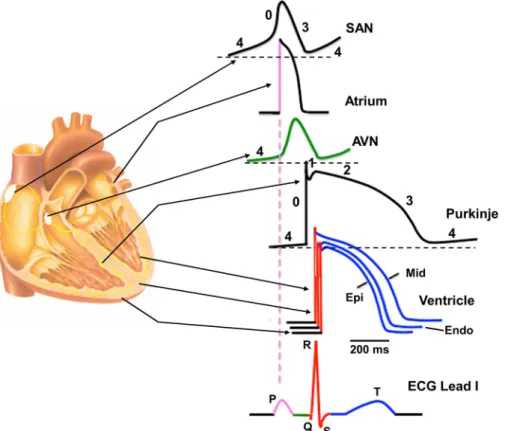
heart as measured on the body surface. The ECG is determined by many variables, including the function of transmembrane ion channels and transporters in the dif- ferent heart cells and the consequent changes in the membrane potential (FIGURE 1).
The P wave corresponds to the activation (depolari- zation) and early repolarization of the atrial cells. The QRS complex re fl ects the time course of the depolari- zation of the ventricles caused mainly by the activation of the fast sodium channels. The PQ segment mainly indicates the impulse conduction from the atria to the ventricles. The PQ segment also contains the HQ inter- val, which re fl ects fast propagation due to the function of the fast sodium current (I
Na). In addition, cell-to-cell coupling is low in the AV node (8), which makes impulse propagation through the AV node relatively unsafe. The isoelectric ST segment re fl ects the plateau phase of the ventricular action potentials. In this phase, membrane potential hardly changes at the cellular level because of the fi ne balance of opening/closing of different ion channels. The con fi guration of the T wave shows the repolarization time course of the ventricles, and it reflects the balance between the slowly activat- ing repolarizing potassium and chloride currents and the depolarizing steady-state, so-called “ window ” so- dium (9) and “ window ” calcium (10) and the slowly decaying, often called “late,” sodium (I
NaLate) and slowly inactivating calcium currents (11–15). Analysis of the PP intervals yields important information regarding heart rate and its regularity.
FIGURE 1. Regional differences in cardiac action potential configurations. Schematic cross section of the heart depicting the corresponding action potential configuration from different regions of the heart indicated by arrows.
Color-coded sections on the action potentials refer to the corresponding sections on the schematic electrocardiogram (ECG). AVN, atrioventricular node; Endo, endocardial; Epi, epicardial; Mid, midmyocardial; SAN, sinoa- trial node.
Cardiac arrhythmia mechanisms are still the sub- ject of intensive research. Because of the large vari- ability in appearances, types (e.g., bradycardia and different types of tachycardia), locations (supraven- tricular or ventricular), and underlying diseases, it is
widely accepted that there is not a single mechanism to explain how arrhythmias originate. Therefore, patients are often treated with little knowledge regarding the mechanisms and/or causes of the arrhythmia.
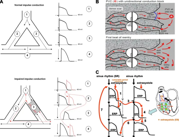
FIGURE 2. A,top: an area of branching cardiac tissue, providing separate paths for impulse propagation from proximal (1) to distal (2–4) directions. The separate paths for impulse conduction can be variable, as branching Purkinjefiber-ventricular junctions, ventricular muscle segments with nonconducting fibrosis, scar, or infarcted tissue in the core. If the tissues in these paths are healthy, the impulse conduction is fast and, because of the relatively long effec- tive refractory period (ERP) in cardiac cells, impulses would collide atsite 4and would propagate only into the distal directions.Bottom: an area of depolar- ized myocardium atsite 3, due to asymmetric severe local myocardial damage, imposing conduction block from the anterograde direction. However, in the case that the impulse travels fromsite 4in the retrograde direction, it can propagate back very slowly into this damaged area, and if its propagation is slow enough to outlast the ERP in front the impulse can reexcite the proximal tissue. Then, this impulse would propagate toward bothsites 1and2, estab- lishing a circus movement and reentry arrhythmia.B: example of a classic mechanism by which a premature ventricular complex (PVC) initiates reentry in thefibrotic border zone of an infarct, due to slow conduction and dispersion of refractoriness.Top: a PVC occurring 250 ms after the previous beat arrives too early to propagate through the upper myocyte strand with a long ERP of 275 ms, but it propagates successfully (red arrows) through the lower strand with a shorter ERP of 225 ms (entry site). The impulse propagates slowly (slow CV), eventually reaching the upper strand from the opposite direction.
Bottom:if the total conduction time is>275 ms, the interface of the upper strand with normal tissue (exit site) has recovered excitability, and the impulse can then propagate through the region of prior conduction block, thus initiating reentry. Dispersion of refractoriness is caused by electrical remodeling.
The slow propagation is due to zig-zag conduction through the myocyte strands as well as gap junction remodeling. (Reproduced from Ref.17with permis- sion.)C: schematic illustration of functional reentry mechanism without a well-defined anatomical obstacle. The arrhythmia substrate is represented by arti- ficially enhanced action potential duration differences. In normal circumstances, impulses originating from the sinus node (black arrows) use physiological pathways to propagate through atrial and ventricular tissue and the conduction system. An early ectopic impulse (trigger, red arrow) can only propagate via pathways where the tissue is not depolarized, and consequently its refractoriness is over, whereas the conduction is blocked in directions where the tissue is not fully repolarized and cells are still in the refractory state. Thus the abnormal impulse can travel in a zig-zag direction through reentry paths cre- ated by heterogeneous repolarization and conduction. The dispersion of repolarization creates a time window called the vulnerable period, where extra stimuli could elicit the reentry arrhythmia. However, outside this window extra stimuli would only cause a single or multiple relatively harmless extrasys- toles. CV, conduction velocity; ES, extrasystole; SR, sinus rhythm. Reproduced from Ref.18with permission.
The majority of cardiac arrhythmias are the result of an enhanced proarrhythmic substrate combined with a trigger (16). Enhanced heterogeneity of repolarization and impaired impulse conduction represent typical ar- rhythmia substrates (conditions that are prerequisites for arrhythmia development) for severe tachyarrhythmia.
Impairment of impulse conduction can be caused by an- atomical (FIGURE 2, A and B) (17) or functional (FIGURE 2C) (18) alterations. The process was described long ago, fi rst in the early twentieth century (19). Impulse con- duction critically depends on the density and kinetics of inward transmembrane ionic currents. Depolarization of the resting membrane potential (RMP), for example, reduces sodium and calcium inward currents and strongly in fl uences their kinetic properties. This can thus slow impulse conduction and cause unidirectional or bidirectional conduction block, and potentially reentry, underpinning a wide range of cardiac arrhythmias.
As mentioned above, reentrant arrhythmias can be caused by functional causes too, without an anatomically well-de fi ned myocardial damage (FIGURE 2C). This form of reentry is more complex and could involve both impulse conduction and repolarization heterogeneities (arrhythmia substrate) as well as enhanced normal or abnormal automa- ticity (trigger).
Reentrant arrhythmias are often initiated by an extrasys- tole formed anywhere in the heart, acting as an arrhythmia trigger (20, 21). Both arrhythmia substrate and trigger, like an extrasystole [premature ventricular complex (PVC)], can be promoted by pathological cardiac conditions (FIGURE 2), e.g., myocardial ischemia, heart failure (7), and genetic diseases (22), or by adverse drug reactions (23). General arrhythmia mechanisms include various cellular aspects, e.g., transmembrane ionic currents, transporters, action potential properties, and automaticity, which are the sub- jects of this review. However, arrhythmia mechanisms at the whole heart level are more complex, since they are also determined by anatomical and structural properties, impulse conduction, and intercellular communication between myocardial and nonmyocardial cells, like fibro- blasts. These factors are beyond the scope of the present work, and the interested reader is referred to other reviews (22, 24–34).
2. CARDIAC ACTION POTENTIAL
The cardiac action potential is a transmembrane poten- tial change, with an amplitude ranging between 60 and 120 mV. It starts from a negative value, i.e., the resting membrane potential (RMP) in working myocardial cells or maximal diastolic potential in spontaneously beating cells (1), ranging from 95 to 40 mV. As in other excita- ble cells, the RMP is mainly defined by the conductance
of inwardly rectifying K
1currents and can be roughly estimated by the Nernst equation from the uneven distri- bution of mainly K
1ions across the cell membrane. The electrogenic ATP-dependent Na
1-K
1pump also con- tributes to the RMP, by exporting 3 Na
1and importing 2 K
1(35–38). In healthy conditions, the duration of the action potential (APD) determines the effective refrac- tory period (ERP), de fi ned as the shortest time interval needed before a new stimulus, or an early extrasystole, can elicit another action potential. The relationship between APD and ERP can be disrupted in pathological conditions, for example, in hyperkalemia, resulting in postrepolarization refractoriness (39).
In the context of the cardiac action potential, two aspects should be emphasized. First, there is no such uniform entity as “ the cardiac action potential, ” since its shape, i.e., the time course of the transmembrane poten- tial changes, differs in the various regions of the heart (FIGURE 1), and therefore different action potentials should be considered and discussed separately.
Second, there are significant interspecies differences (40), even when action potentials are recorded from sim- ilar regions of the heart. This is an important, and often overlooked, issue, since many experimental results have been obtained in small rodents, recently particu- larly in transgenic mice.
In general, the cardiac action potential is divided into
five distinct phases (FIGURE 1). Phase 0 is the fast depo-
larization due to an abrupt increase in sodium influx, and
it is characterized by the upstroke velocity and can result
in an overshoot, i.e., the rapid change of potential from
the negative RMP to positive voltage values, reaching a
peak of up to 1 30 to 1 40 mV. The overshoot is fol-
lowed by a return to negative values, in a process called
repolarization, which includes phases 1, 2, and 3. Phase
1 is characterized by a transient and relatively fast repo-
larization brought about by a decrease in sodium in fl ux
and a transient increase in potassium ef fl ux and chloride
influx. Phase 2 consists of a long-lasting plateau, still at
depolarized voltage, during which the membrane poten-
tial remains almost constant or decreases slowly,
caused by a small net transmembrane current carried by
simultaneous calcium (and some sodium) influx and po-
tassium efflux. Phase 3 represents the large repolariza-
tion toward the diastolic potential, mostly due to
increased potassium ef fl ux and decreased calcium and
sodium in fl ux. Phase 4 represents the resting membrane
potential in diastole in working myocytes and the spon-
taneous depolarization in pacemaker cells. In cardiac
myocytes that do not beat spontaneously the voltage
remains stable at the RMP, whereas in cells exhibiting
automaticity the potential gradually changes toward the
positive values, in a process called spontaneous dia-
stolic depolarization. When the threshold potential is
reached, a new spontaneous action potential is gener- ated, with a certain cycle length.
There are four common methods to record cardiac action potentials:
1) Weidmann and Coraboeuf were the fi rst to record car- diac action potentials in dog ventricular muscle (41) and later in dog Purkinje fi bers, using the sharp glass capil- lary-based microelectrode technique (42). The classical cardiac cellular electrophysiological knowledge gained by using this technique was elegantly summarized in an early monograph entitled Electrophysiology of the Heart by Hoffman and Crane fi eld (1), published in 1960, which is still useful today. This technique is still consid- ered one of the best for accurate cardiac action poten- tial recordings and can be used for both single-cell and tissue recordings. Its major advantages include 1) the ability to accurately record very fast voltage changes and 2) because of the very fi ne tip of the pipette, very little diffusion takes place out of the pipette solution, having negligible effects on the intracellular milieu.
However, since this technique has some limitations (e.g., dif fi culties in maintaining a stable impalement for extended periods of time), other methods have also been developed and used widely.
2) In intact hearts, in in vivo animal experiments, or in clini- cal studies, where the microelectrode technique is dif fi - cult to apply, monophasic action potential recording can also be used, with either a suction electrode (43, 44) or a Franz catheter (45). With this technique, recordings can be easily performed from multiple sites simultaneously, and impalements/attachments are not lost because of vigorous contractions, even in in vivo or ex vivo conditions. However, rapid voltage changes or action potential amplitudes and shapes cannot be determined accurately.
3) Since the introduction of the patch clamp by Neher and Sakmann (46 – 48), the whole cell con fi guration of this technique has been widely used. In the current-clamp mode, it can record action potentials from isolated myo- cytes. Despite its widespread use, this technique has im- portant limitations that should be emphasized. First, measurements are performed in single isolated myo- cytes or, occasionally, cell pairs, and it is uncertain how, and to what degree, different ion channels are in fl u- enced by individual enzymatic digestion during the isola- tion procedure (49). Therefore, even if the recordings show single-cell action potentials with a normal shape, the function of the fi nely regulated ion channels can be drastically altered from their original condition. Also, the cell is dialyzed with the pipette contents, and its intracel- lular composition will change. When carefully and delib- erately applied, however, this point can also be considered an advantage, as it allows control of the intra- cellular milieu. It should also be emphasized that single isolated myocytes are devoid of electrotonic interactions from neighboring cells. Therefore, the stochastic
opening/closing be-havior of ion channels has a more profound effect on membrane potential than in well- coupled tissue preparations (50). In addition, in multicel- lular preparations part of the ionic currents are utilized to depolarize neighboring cells during impulse propaga- tion, and this can considerably reduce the action poten- tial peak compared with single cells (51). Hence, action potential measurements in single isolated myocytes obtained with the patch-clamp technique should be interpreted with caution and not directly extrapolated to intact tissue.
4) The latest approach to recording cardiac action poten- tials is the optical mapping technique, which uses volt- age-sensitive dyes and allows simultaneous recordings from multiple sites (52, 53). This technique is also excel- lent for dynamic studies and for investigations of arrhyth- mia mechanisms (54, 55). Disadvantages of this method include the dif fi culty of calibration to millivolts, phototox- icity, photodegradation, and photon scattering effects (56). Also, the application of excitation-contraction uncoupling compounds, e.g., blebbistatin, is necessary to avoid motion artifacts (57). These compounds may interfere with the experiments, since, e.g., blebbistatin was reported to elicit anomalous electrical activities (58) and prolongation of action potential duration (59), and in- hibition of contraction will also decrease metabolic rate at the concentrations needed for motion artifact red- uction.
3. TRANSMEMBRANE ION CHANNELS AND TRANSPORTERS IN THE HEART
The cardiac action potential is the voltage change caused by ions flowing through transmembrane ion channels, via their dynamic and simultaneous opening and closing (11, 60). Therefore, before addressing differ- ent regional action potential patterns, we describe the various transmembrane ion channels that have been reported to operate in the heart cells.
Transmembrane ionic currents in the heart are usu- ally measured with the patch-clamp technique in enzymatically isolated myocytes. This allows record- ing and analysis of unitary currents through single ion channels or all channels on the sarcolemma.
Before the introduction of the patch clamp, trans-
membrane current recordings were less accurate,
because of the lack of proper voltage control of the
preparation, and fast current changes and gating
kinetics were impossible to determine accurately
(61). Therefore, much of the knowledge gained
before the introduction of the patch-clamp technique
has had to be reevaluated, and some currents have
been renamed. To the interested reader, we recom-
mend an excellent monograph by Denis Noble, The
Initiation of the Heartbeat (62), which deductively and brie fl y summarizes our knowledge before the use of the patch-clamp technique.
It is important to note that a given ionic current, meas- ured with the patch clamp, does not correspond to a unique ion channel. Certain transmembrane ionic cur- rents can be conducted by several different ionic chan- nels (3, 5), e.g., inward rectifier potassium current (I
K1) is carried by inward rectifier potassium (K
ir)2.1, K
ir2.2, K
ir2.3, and K
ir2.4 channels (5, 63) and tandem of pore domains in a weak inward rectifying potassium channel (TWIK)-related acid-sensitive potassium (TASK) channels (64), whereas transient outward current (I
to) is carried by voltage-gated potassium (K
v)4.3, K
v4.2, and K
v1.4 chan- nels. In addition, certain channels like human ether-à- go-go-related gene (hERG) have multiple isoforms (hERG 1a and 1b) (65) with different gating kinetics and drug sensitivities (66). This offers the possibility of phar- macologically modulating specific channel isoforms without interfering with others, and thus avoiding unde- sirable side effects.
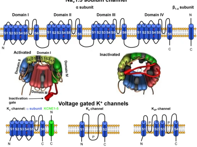
Recent advances in genetics and molecular biology have made it possible to further elucidate the structure of ion channels (FIGURE 3). It is widely known that
most transmembrane ion channels consist of multiple subunits: a pore-forming a and modulatory accessory subunits, which can modify channel gating and serve as possible drug binding or phosphorylation sites (3, 5, 67, 68).
3.1. The Fast Inward Sodium Current
The fast inward sodium current (I
Na) is the most impor- tant current for impulse conduction in cardiomyocytes, with a diastolic potential more negative than 60 mV (69), and is therefore particularly important in atrial, ven- tricular, and Purkinje fiber myocytes. This current is re- sponsible for the influx of Na
1during phase 0 of the action potential (FIGURE 1 and FIGURE 4), and it is con- ducted by voltage-gated sodium (Na
v)1.5 channels, encoded by the gene SCN5A coassembled with b
1–4(SCN1 – 4B)-subunits (70). The high-molecular-weight pore-forming a -subunit contains four repeat domains (la- beled I – IV); each domain consists of six transmembrane segments (S1 – S6), and the S4 segment is responsible for voltage sensing (FIGURE 3). Special extracellular regions (P loops) between the S5 and S6 segments of the four domains form the structure that is responsible
FIGURE 3. Schematic illustration of voltage-gated sodium (Nav) and potassium (Kv) channels.Top: topological models of Nav1.5 channel subunits and their molecular assembly. Four domains (I–IV) of Nava-subunits contribute to individual Navchannel formation.Middle: the activated and inactivated Navchannel configurations.Bottom: the transmembrane topologies of Kv, inward rectifier (Kir), and two-pore domain (K2P) potassium channel subunits.
for the ion selectivity of the channel. The region that links domains III and IV contains the inactivation gate that “plugs” the channel pore after prolonged activation (FIGURE 3). A detailed, comprehensive review on the cardiac sodium channel structure has been published recently (71). Na
v1.5 channels open, within a fraction of a millisecond, at potentials more positive than 60 mV, with strong voltage dependence. Since channel density is high, they carry a large inward current, with an ampli- tude of > 100 pA/pF. They also inactivate very rapidly at voltages more positive than 80 mV (at 10 mV with time constant s
1of 0.6 ms and s
2of 4 ms) (72), with a half-inactivation between 60 and 70 mV. Na
v1.5
channels can recover from inactivation with a time con- stant of 2–20 ms at negative voltages (69, 73, 74). The exact kinetics of their inactivation and recovery are com- plex and still not fully understood. So far, multiple inacti- vation kinetics and recovery kinetics from inactivation have been described (12, 69, 75–77).
In addition to this fast component, slow inactivation, occurring over hundreds of milliseconds, has been described (12, 13, 78) and attributed to late openings of Na
v1.5 channels (79). Recently, a new term, “late sodium current” (I
NaLate), has been used to refer to this current, which, although small in amplitude ( < 0.5% of the peak I
Na) (79, 80), nonetheless represents an important sus- tained depolarizing current during phase 2, thus playing a role in maintaining the relatively long plateau of the cardiac action potential (FIGURE 4) (78). This I
NaLateis more sensitive to tetrodotoxin (TTX) and other sodium channel inhibitors (81) than the peak I
Na(82).
As a steady-state component of I
Na, a so-called “ win- dow sodium current ” in the voltage range from – 65 to – 15 mV based on a different mechanism than the slow inactivation was also suggested earlier by Attwell et al.
(9). This window current was considered to be caused by the overlap between the steady-state activation and inactivation curves. At present, it is still not clear whether such a window current exists, or if the measured overlap is due to an ultraslow inactivation or ultraslow recovery from inactivation when this window current is deter- mined. To better understand the nature of the sodium current during the plateau phase of the action potential, the possible involvement of sodium channels other than the Na
v1.5 channel, which is considered to be the major cardiac sodium channel, was also suggested in several studies (83 – 88). As an example, Na
v2.1 a -, b
1-, and b
2- subunits are highly expressed in human atrial and ven- tricular cells and Purkinje fi bers (84, 85) even if their function has not been explored yet. The expression of various neuronal sodium channel subtypes, e.g., Na
v1.1, Na
v1.2, Na
v1.3, Na
v1.4, Na
v1.6, and Na
v1.8 (encoded by SCN1A, SCN2A, SCN3A, SCN4A, and SCN10A genes, respectively), has been described in different cardiac preparations (89), but again their functional roles are not fully understood (3, 5, 85, 86, 90). Mishra et al. (86) reported that in failing dog and human hearts neuronal Na
v1.1 channels were upregulated and provided signifi- cant I
NaLate, whereas another recent study (91) showed signi fi cant upregulation of Na
v1.8 channels in failing human hearts. In addition, selective inhibition of this Na
v1.8 current by A-803467 (92) abolished arrhythmo- genic Ca
21sparks that were attributed to enhanced in- tracellular Ca
21load due to increased I
NaLate. Mutations in the Na
v1.8 encoding gene, SCN10A, were reported in patients with atrial fi brillation (AF) (88) and also predis- posed to sudden cardiac death (83). Therefore, it is
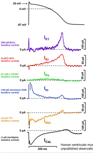
FIGURE 4. Action potential and underlying ionic currents recorded from human ventricular myocytes with the patch-clamp technique applying human ventricular action potential as command pulses at 1 Hz stimulation frequency, in the absence of any sympathetic effects.
Inward rectifier potassium current (IK1), rapid (IKr) and slow (IKs) compo- nents of delayed rectifier potassium current, transient outward current (Ito), and L-type calcium current (ICa,L) were measured as difference cur- rent following application of selective channel inhibitors.INaL, late so- dium current. Unpublished data from our laboratory at the Department of Pharmacology and Pharmacotherapy, University of Szeged.
clear that further studies are needed to elucidate the role of neuronal Na
1channels in normal and diseased heart (93).
It is known that Na
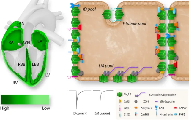
v1.5 channel expression varies in the different regions of the heart, showing a high level of expression in the specialized conduction system, atria and ventricles, while being absent in the central sinus and atrioventricular nodal tissues (FIGURE 5). Also, Na
v1.5 channels gather in clusters and associate with accessory subunits and partner proteins, forming region-speci fi c macromolecular complexes (95). They are not evenly distributed within the myocyte (96). The current densities are large in the intercalated disk area and smaller in the lateral membrane (96, 97). The com- plex nature and the observed regional differences in so- dium channel expression, as well as the functional significance of Na
v1.5 interactions with partner proteins, justify further studies to better understand the patho- physiology of diseases associated with Na
v1.5 dysfunc- tion, including inherited sodium channelopathies (94, 98).
The function of I
Nais regulated by intracellular calcium homeostasis in a complex way that involves multiple
accessory proteins (99). Calmodulin (CaM), the ubiqui- tous Ca
21-sensing protein, plays a central role in intra- cellular Ca
21concentration ([Ca
21]
i)-dependent I
Nafunction alterations by modulating fast inactivation of I
Na(100). Recent data suggest that CaM facilitates the re- covery of the sodium channel from inactivation by inter- acting with its inactivation gate in a Ca
21-dependent fashion (101). The different binding sites of CaM on the sodium channel are important to understand the mecha- nisms linked to disease-associated sodium channel mutations (102 – 104). In addition to CaM, Ca
21/calmodu- lin-dependent kinase II (CaMKII) has been shown to modify I
Nafunction in a Ca
21-dependent manner.
CaMKII phosphorylation regulates cardiac Na
1channels by slowing their recovery from inactivation (105, 106).
This results in reduced availability of fast I
Naat a high rate with enhanced late I
Na(106). The latter contributes to prolonged repolarization and enhanced arrhythmia susceptibility (106, 107) often seen in heart failure, where CaMKII activity is enhanced (107).
In addition to TTX, I
Nais blocked, although not sel- ectively, by a wide range of antiarrhythmic drugs, e.g., lidocaine, mexiletine, quinidine, disopyramide, and
FIGURE 5. Regional and subcellular distribution ofSCN5A/voltage-gated sodium channel (Nav)1.5 in the heart and cardiomyocytes.Left: the expres- sion levels ofSCN5Ain different regions of the heart. Expression ofSCN5Ais highest in the atrioventricular (AV) bundle, His bundle, and right (RBB) and left (LBB) bundle branch (dark green).SCN5Ais broadly expressed in right (RA) and left (LA) atria and right (RV) and left (LV) ventricle with an epi/
endo gradient in the ventricles.SCN5Ais absent from the central sinoatrial node (SAN) and atrioventricular node (AVN).Right: the localization of Nav1.5 with specific regional partner proteins in the microdomains of a cardiomyocyte: intercalated disk (ID), lateral membrane (LM), and T tubules. The sodium current at the ID is larger than the sodium current at the LM. Reproduced from Ref.94with permission.
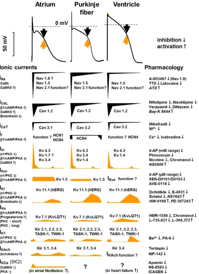
FIGURE 6. Tissue-specific (human) cardiac atrial, Purkinjefiber, and ventricular action potentials and the underlying ionic currents in different action poten- tial phases, indicating their pharmacology and modulation. Black arrows indicate inward and yellow arrows indicate outward current. The contributions of dif- ferent currents to the action potentials are indicated below, with a time course adjusted to the action potential. CaM, calmodulin; CaMKII, Ca21-calmodulin kinase II; hERG, human ether-à-go-go-related gene;IK1, inward rectifier potassium current;IK,Ach, acetylcholine-activated potassium current;INa, sodium current;
ICaL, L-type calcium current;ICaT, T-type calcium current;If, funny/pacemaker current;Ito, transient outward current; IKCa, calcium-activated potassium current;
IKr,IKs, andIKur, rapid, slow, and ultrarapid components of delayed rectifier potassium current; Kir, inward rectifier potassium channel; KV, voltage-gated potas- sium channel; NaV, voltage-gated sodium channel; TASK, Tandem of pore domains in a weak inward rectifying potassium channel (TWIK)-related acid-sensi- tive potassium channel; TTX, tetrodotoxin.
flecainide (FIGURE 6) (108, 109) in a frequency-depend- ent manner. It has recently been reported that certain drugs like ranolazine and GS967 selectively inhibit I
NaLate(110, 111). Therefore, these compounds would be particularly effective for long QT (LQT) syndromes, heart failure (HF), or hypertrophic cardiomyopathy (HCM) (110).
On the other hand, late I
Nacan be pharmacologically augmented by veratrine (112), veratridine, and ATX (113).
Genetic mutations that alter I
Nafunction can lead to severe, potentially lethal, conditions and have been shown to play a significant role in a wide array of inher- ited channelopathies (for recent reviews see Refs. 94, 114–117). As an example, loss-of-function SCN5A muta- tions lead to a reduced I
Napeak, thus slowing impulse conduction and possibly causing conduction block.
These mutations have been identi fi ed in 20% of patients with Brugada syndrome (BrS) (118) as well as in patients with sick sinus syndrome and progressive car- diac conduction defect (119–121). On the other hand, gain-of-function SCN5A mutations have been shown to play key roles in congenital LQT3 syndrome (122). In some cases of familial AF, both loss-of-function and gain-of-function SCN5A mutations have been identified (123, 124).
The late sodium or window current, irrespective of its basic mechanism or molecular background, is particularly important in arrhythmogenesis (125). As an example, in HF and in HCM, I
NaLateis augmented (126, 127). Since this current is an important contributor to the action potential plateau phase, its enhancement prolongs repolarization and increases repolarization heterogene- ity (110, 128). In addition, by increasing intracellular so- dium concentration, it also increases intracellular calcium concentration, via the Na
1/Ca
21exchanger (NCX). This can evoke arrhythmogenic triggered activity such as early and delayed afterdepolarizations (EADs and DADs, respectively) (129 – 131), discussed below in this review. Both triggered automaticity and enhanced dispersion of repolarization are considered major mech- anisms in arrhythmogenesis, and their reduction is a major aim in antiarrhythmic drug development.
3.2. The Transient Outward Current
The main channels providing the transient outward po- tassium current (I
to) in human and dog ventricular muscle are K
v4.3 and K
v4.2 pore-forming a -subunits coas- sembled with KChIP2 and DPP auxiliary subunits (85) encoded by KCND3, KCND2, and KCNIP2 and DPP6/10 genes, respectively (132 – 134). K
v1.4 a channel subunits (encoded by KCNA4) are also expressed with marked regional and interspecies dependence, making up 10 – 20% of I
todensity in humans (135, 136). Accordingly, in humans (unlike in dogs), in addition to the rapid
component ( s
fast50–100 ms), the I
torecovery from inactivation also has a component with a slow recovery time constant (on the order of seconds) that is character- istic of the K
v1.4 channels (137). It is interesting that in rabbit ventricle the I
tocurrent is primarily conducted by K
v1.4 channels (138). However, the functional role of this channel in rabbits is still unclear, as it is inactivated most of the time at their physiological heart rate, which is quite high. In humans, both K
v4.3 and K
v1.4 channels can have functional roles, particularly in the frequency-de- pendent modulation of the APD during both different constant or following abrupt changes in cycle lengths during electrical restitution (i.e., following extrasystoles with different coupling intervals). In addition, there are mRNA expression studies (85, 139) supporting the possi- bility that the K
v1.7 channels also have a functional role for I
toin human atria and ventricles. However, because of the relative paucity of data regarding K
v1.7 channels in the heart, further research is needed to properly eluci- date these issues.
Human and dog I
tostart activating at membrane potentials more positive than 30 mV, rapidly reaching peak values (within 1–2 ms) and then inactivating with a double-exponential time course ( s
fast5 ms, s
slow25 ms) (140). The K
v4.3 channel-mediated I
to, unlike K
v1.4, recovers rapidly from inactivation in the membrane volt- age range of 60 to 80 mV, with a time constant of 50 ms (137). In dog and human Purkinje fi bers, K
v3.4 channels were also described to contribute to I
to, with different kinetic properties compared with K
v4.3 chan- nels (141). This raises the possibility for fi ne-tuning of fre- quency-dependent modulation of repolarization dispersion between Purkinje fi bers and ventricular mus- cle (142).
The transient outward potassium current and the fast inactivation of I
Naare important contributors to the early/fast repolarization of the action potential (phase 1) (137, 143) (FIGURE 4). Phase 1 repolarization and I
toare more prominent in Purkinje fibers and atrial, mid- myocardial, and subepicardial ventricular muscle (137), but they are small, or nonexisting, in subendocardial ventricular cells and sinoatrial (SA) and atrioventricular (AV) nodal cells (137). Interestingly, phase 1 repolariza- tion and I
toare not present in guinea pig ventricular myocytes (144, 145). Also, it has been reported that in mouse ventricle I
tois small and action potential is long after birth and later the action potential shortens as I
todevelops in adult mice (146, 147). The impact of I
toon the shape of the action potential waveform is complex.
In addition to its role in phase 1, I
toalso modulates the
voltage level of the plateau, and consequently it has
an indirect in fl uence on activation, inactivation, and
deactivation of several other transmembrane ionic cur-
rents that operate during the plateau phase. Since I
to,
similarly to I
Na, has activation, deactivation, and inacti- vation properties, it was also suggested that a window I
tocurrent may be generated, in the membrane poten- tial range from 35 to 10 mV. This window current would also contribute to the final repolarization (140). It was also reported that interaction of DPP10a with K
v4.3 channels results in a sustained current compo- nent of human atrial I
tothat can participate in the late repolarization phase (148). However, despite its poten- tial importance, it is dif fi cult to validate or disprove the existence of such current, since selective inhibitors are lacking. This suggests the need for further research to clarify this issue. I
tois a typical transmembrane ionic current that can be attributed to the function of a wide variety of distinctly different potassium and also chlo- ride ion channels (137). Although the role of I
toin arrhythmias is not well defined, it definitely plays a role in Brugada syndrome (117, 149), where a reduced expression level of the auxiliary I
tosubunit, KChIP2, in females most likely underlies the male phenotypic pre- dominance (150, 151). The stronger epicardial I
toin males could further aggravate the consequence of impaired I
Nain Brugada patients; in this case, inhibition of I
towould be bene fi cial. Multiple nonselective I
toinhibitors have been reported (FIGURE 6): 4-aminopyr- idine (4-AP), in millimolar concentrations (152); quini- dine, fl ecainide, and chromanol 293B, in micromolar concentrations (140, 152); and phrixotoxin, in nanomo- lar concentrations (153). However, a selective I
toinhibi- tor is still lacking. This could constitute a potential treatment in atrial fibrillation, by increasing APD and consequently ERP, in atrial but not or less in ventricular tissue (154). An activator for I
towas also reported hav- ing effect on canine ventricular but not atrial myocytes (155).
I
tois downregulated in HF (156–159), HCM (160), and diabetes mellitus (161, 162), possibly contributing to repolarization prolongation in these pathological set- tings. Recently, it was shown that K
v4.3 (fast I
to) and K
v1.4 (slow I
to) were expressed differently in normal and failing hearts, thus contributing to arrhythmogenic regional heterogeneity in action potential waveforms (101). It was also demonstrated that a slowing of phase 1 repolarization, which can be due to the decreased I
tooften observed in failing hearts (163 – 165), decreases the driving force of Ca
21through L-type calcium chan- nels, and it can result in potentially arrhythmogenic asynchronous Ca
21release from the sarcoplasmic reticulum (SR) (166). I
tois subject to a - and b -adrener- gic regulation, both decreasing I
tovia the PKA and PKC pathways (167), whereas CaMKII has been shown to increase I
to(167).
Thyroid-stimulating hormone and thyroid hormones have been shown to modulate I
to(168, 169), thus making
I
toan important contributor to repolarization abnormal- ities in altered thyroid status.
3.3. Inward Calcium Current
The inward calcium current (I
Ca) in the heart was fi rst described by Reuter (170), and it has two major types (171, 172). The most abundant type is the L-type calcium channel (Ca
v1.2), which conducts current through the pore-forming a -subunit ( a
1) encoded by the CACNA1C gene (85). The a
1-subunit consists of 2,000 amino acids, organized into four repeat domains (I – IV), each containing six transmembrane segments (S1 – S6) (173).
The S4 transmembrane helix from each domain collec- tively constitute the voltage sensor of the channel (173).
A region called the “P loop” connecting the S5 and S6 segments is responsible for the Ca
21selectivity of the pore region (174). The pore-forming subunit coassem- bles with the extracellular a
2d and intracellular b auxil- iary (predominantly b
2in cardiac tissue) subunits that modulate kinetics, gating, and traf fi cking properties of the channel (175 – 177). Inward L-type Ca
21current (I
Ca,L) has a very rapid activation (14) and is particularly impor- tant for excitation-contraction coupling (178), since it serves as a trigger for the calcium-induced calcium release (CICR) (179) from the SR and as a source of extracellular calcium when needed. In addition, I
Ca,Lplays a fundamental electrophysiological role in main-
taining the plateau phase of the action potential
(FIGURE 4) and in the depolarization of SA and AV nodal
cells (180). In these cell types, I
Ca,Lis the main contributor
to impulse conduction; therefore its impairment pro-
longs the PQ interval and can result in AV node conduc-
tion block. In addition, since I
Ca,Lprovides depolarizing
current, its decrease would reduce the spontaneous fre-
quency of these cells. In AV nodal cells, this shift in
threshold potential also contributes to the slowing of AV
impulse conduction. The inactivation kinetics of L-type
I
Ca( s
fast2–8 ms, s
slow30–100 ms) depend not only
on membrane voltage but also on the intracellular Ca
21concentration (14, 15, 181 – 184), which is dynamically
changing during the action potential. The recovery from
inactivation is complex and strongly depends on volt-
age. At 40 mV it can be characterized by an exponen-
tial time course ranging between 30 and 60 ms with
s
fast= s
slow(14, 185). Importantly, the recovery kinetics
can be faster in ventricular and Purkinje fibers, which
have a resting potential around 80 mV. The I
Ca,Lwas
originally called slow inward current (I
si), since with the
old experimental methods, before the introduction of
single-cell voltage clamp, a relatively slow I
Caactivation
was measured (170, 186). Later, with more advanced
voltage-clamp techniques, the proper fast activation
kinetics could be determined (187).
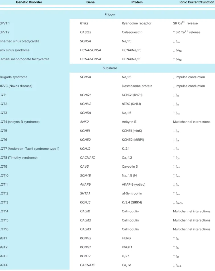
Table 1. Changes of genes, channel/transporter proteins, and ionic currents in various genetic disorders
Genetic Disorder Gene Protein Ionic Current/Function
Trigger
CPVT 1 RYR2 Ryanodine receptor SR Ca21release
CPVT2 CASQ2 Calsequestrin :SR Ca21release
Inherited sinus bradycardia SCN5A Nav1.5 ;INa
Sick sinus syndrome HCN4/SCN5A HCN4/Nav1.5 ;If/INa
Familial inappropriate tachycardia HCN4/SCN5A HCN4/Nav1.5 :If/INa
Substrate
Brugada syndrome SCN5A Nav1.5 ;Impulse conduction
ARVC (Naxos disease) Desmosome protein ;Impulse conduction
LQT1 KCNQ1 KCNQ1 (Kv7.1) ;IKs
LQT2 KCNH2 hERG (Kv11.1) ;IKr
LQT3 SCN5A Nav1.5 :INa
LQT4 (ankyrin-B syndrome) ANK2 Ankyrin-B Multichannel interactions
LQT5 KCNE1 KCNE1 (minK) ;IKs
LQT6 KCNE2 KCNE2 (MiRP1) ;IKr
LQT7 (Andersen–Tawil syndrome type 1) KCNJ2 Kir2.1 ;IK1
LQT8 (Timothy syndrome) CACNA1C Cav1.2 :ICa
LQT9 CAV3 Caveolin 3 :INa
LQT10 SCN4B Nav1.5b4 :INa
LQT11 AKAP9 AKAP-9 (yotiao) ;IKs
LQT12 SNTA1 a1-Syntrophin :INa
LQT13 KCNJ5 Kir3.4 (GIRK4) ;IKACh
LQT14 CALM1 Calmodulin Multichannel interactions
LQT15 CALM2 Calmodulin Multichannel interactions
LQT16 CALM3 Calmodulin Multichannel interactions
SQT1 KCNH2 HERG :IKr
SQT2 KCNQ1 KVQT1 :IKs
SQT3 KCNJ2 Kir2.1 :IK1
SQT4 CACNA1C Cava1 ;ICa,L
Continued
I
Ca,Lis modulated (FIGURE 6) by cAMP-dependent phosphorylation and other factors, including intracellular Ca
21levels (15). It has been shown that CaM supports both inactivation and facilitation of I
Ca(188). The intracel- lular Ca
21enhances I
Cavia CaMKII, involving direct phosphorylation of L-type Ca
21channels (189, 190) inde- pendently of cAMP via PKA (191) involving Rad, a mono- meric G protein that closely interacts with Ca
v1.2 (192).
I
Ca,Lcan be effectively blocked (FIGURE 6) by Cd
21, ve- rapamil, and diltiazem, but the inhibition with these drugs is not selective (193 – 195). Dihydropiridines, e.g., nifedipine (196) and nisoldipine (197), are more selective I
Ca,Lblockers, but they may be sensitive to light (198).
Pharmacological activation of I
Ca,Lis also possible by Bay K8644 (FIGURE 6) (199).
The I
Ca,Lhas key roles in several diseases like HF (200), HCM (27), AF (201), and myocardial ischemia and in other pathophysiological conditions, such as the de- velopment of EADs, DADs, (202–204) and cardiac is- chemia-reperfusion (205), discussed in more detail in other parts of this review.
A gain-of-function G406R mutation of the Ca
v1.2 chan- nel causes type 8 of congenital LQT syndrome, also called Timothy syndrome (TABLE 1). This disease is characterized by slower inactivation of I
Ca,L(206 – 208) and by the fact that small clusters of Ca
v1.2 channels have a larger probability for coordinated opening and closing ( “ coupled gating ” ) (209), thus leading to tachyar- rhythmia and congenital heart defects (ductus arterio- sus, ventricular septal defect, Fallot tetralogy, HCM) (210).
The second type is the T-type calcium current (I
Ca,T), for which signi fi cantly less data are available. Its func- tional role in atrial and ventricular cells and Purkinje
fibers is still unclear. However, it plays an important role in the SA and AV nodal cells (211), where it makes a sig- ni fi cant contribution to the pacemaker function. This cur- rent is conducted by Ca
v3.1 and Ca
v3.2 channels, encoded by CACNA1G and CACNA1H genes, respec- tively (212). I
Ca,Tactivates at more negative membrane potentials than I
Ca,L, and its overlap with I
Namakes it diffi- cult to study (172). I
Ca,Tcan be inhibited by low concen- trations (100 – 200 m M) of Ni
21(172) and by the organic compound mibefradil, which was developed with the aim of decreasing elevated heart rate (213).
3.4. Delayed Recti fi er Potassium Currents
Before the introduction of the patch-clamp technique, a slowly activating current carried by K
1was recorded during the plateau phase. This current was named the delayed recti fi er potassium current (I
x1/2). Later, by apply- ing single-cell patch-clamp technique, Sanguinetti and Jurkiewicz (214) showed that this delayed recti fi er out- ward potassium current can be separated into a rapid (I
Kr) and a slow (I
Ks) component. Molecular biological studies also con fi rmed that these two I
Kcomponents are conducted by distinctly different ion channels.
3.4.1. The rapid delayed-rectifying potassium current.
The rapid delayed rectifier outward potassium current (I
Kr) is conducted by the K
v11.1 pore-forming a -subunit, also called hERG in humans (human ether-à-go-go related gene), which is associated with various acces- sory b and possibly other subunits (85, 215, 216).
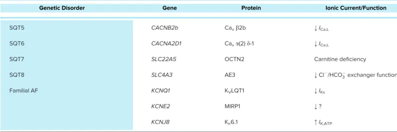
Table 1. — Continued
Genetic Disorder Gene Protein Ionic Current/Function
SQT5 CACNB2b Cavb2b ;ICa,L
SQT6 CACNA2D1 Cava(2)d-1 ;ICa,L
SQT7 SLC22A5 OCTN2 Carnitine deficiency
SQT8 SLC4A3 AE3 ;Cl /HCO3exchanger function
Familial AF KCNQ1 KVLQT1 ;IKs
KCNE2 MIRP1 ;?
KCNJ8 Kir6.1 :IK,ATP
AF, atrialfibrillation; ARVC, arrhythmogenic right ventricular cardiomyopathy; CPVT, catecholaminergic polymorphic ventricular tachycardia;ICa, calcium current;ICa,L, L-type Ca21current;If, funny/pacemaker current;IKACh, acetylcholine-activated potassium current;IK,ATP, ATP-sensitive potassium current;
IKr, rapid component of delayed rectifier potassium current;IKs, slow component of delayed rectifier potassium current;IK1, inward rectifier potassium cur- rent;INa, sodium current; LQT, long QT syndrome; SQT, short QT syndrome; SR, sarcoplasmic reticulum.
Similarly to other K
vchannel pore-forming a -subunits, K
v11.1 consists of six transmembrane segments (S1–S6) and the functional channel contains four a -subunits (FIGURE 3) (217). The K
v11.1 hERG a -subunit has two iso- forms (a and b), which are different in terms of gating and drug sensitivity (65, 66). The wide variety of interact- ing accessory b -subunits include MinK (human minimal potassium ion channel), MiRP1 (mink-related peptide 1), and MiRP2, MiRP3, and MiRP4 proteins encoded by KCNE1, KCNE2, KCNE3, KCNE4, and KCNE5 genes, respectively (85, 215). Initially, it was suggested that MiRP1 interacted with hERG (K
v10.1) to form I
Krchannels (218). This is due to the fact that when hERG (K
v11.1) a channel subunits were expressed alone in HEK cells, a very rapidly activating steady-state-like current was observed, and only coexpression with MiRP1 resulted in currents that resembled native I
Kr. On the basis of this observation MiRP1 was considered the most important accessory subunit to form the native I
Krchannel.
However, later studies indicated very low levels of KCNE2 expression in human heart, whereas genes of
other b -subunits like MinK, MiRP2, MiRP3, and MiRP4 were abundantly expressed in human atrial and ven- tricular tissue and Purkinje fi bers (85). Coexpression of the b - and hERG a -subunits produced currents with kinetics similar to those native currents that can be recorded in different species including human (219, 220). However, the exact role of these b and other possible accessory subunits, how they regulate I
Kr, and whether they are responsible for the observed species-dependent differences and drug sensitivities are unclear at present and need to be elucidated in the future. I
Kractivates in a voltage-dependent man- ner, with an activation s of 31 ms at 130 mV in human ventricular myocyte (221). It also slowly deactivates (222) in a voltage-dependent manner; its deactivation can be fi tted by a double exponential with s
fastof 600 ms and s
slowof 6,800 ms at 40 mV. The ratio of the amplitudes of the fast and slow components increases at more negative potentials. I
Krexhibits a peculiar, very rapid, inactivation (223 – 225), which starts even before it activates.
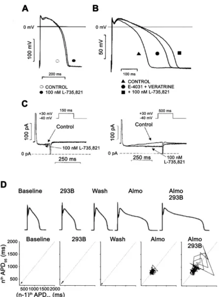
FIGURE 7. The role of the slow component of the delayed rectifier potassium current (I
Ks) in the repolarization in dog ventricular papillary muscle (A–C) and in dog single ventricu- lar myocyte (D). InA, action potential duration (APD) is not, or minimally, changed after fullI
Ksinhibition by 100 nM L- 735,821 without external sympathetic stimulation. InB, in the same preparation fullI
Ksinhibition elicited significant prolon- gation of repolarization in a preparation where the rapid component of the delayed rectifier potassium current (I
Kr) was inhibited by E-4031 and late sodium current (I
NaLate) was augmented by veratrine. In C, transmembrane current recordings show that during a short (150 ms) voltage pulse very little I
Ks develops, but when pulse duration was increased to 500 ms significantI
Ksdeveloped, explaining the lack and significant changes of APD afterI
Ksinhibition in AandB. (Reproduced from Ref.228with permission.) InD, the result of a representative experiment is shown in dog ventricular myocytes. In the baseline (control) situation, small short-term APD variability and normal APD were recorded that did not change significantly afterI
Ksinhibition by chro- manol 293B. Additional application ofI
Krblock by almoka- lant increased both APD and short-term APD variability illustrated by the Poincare plot, which was substantially fur- ther lengthened and increased by additionalI
Ksinhibition, respectively. (Reproduced from Ref.255with permission.)
During the action potential in early plateau and in phase 3, I
Krchannels rapidly recover from inactivation and they reopen as voltage changes toward more nega- tive values, and deactivation progresses. Accordingly, de- spite the rapid activation of the current during the plateau phase, a relatively tiny current develops that gradually increases and decreases during phase 3 repolarization (FIGURE 4); therefore it is a crucially important current to secure repolarization.
Since I
Krdeactivates slowly, it does not have time to fully deactivate during an action potential. Therefore, a residual and gradually decreasing outward current can still flow through this channel, thus shortening the next action potential when diastolic interval is relatively short, i.e., during fast heart rate or during an early extrasystole (222). Consequently, I
Kris considered a key player in fre- quency-dependent APD regulation, and it can influence the pacemaker function as well (226).
I
Krcan be blocked by speci fi c compounds in the sub- micromolar or micromolar range (e.g., dofetilide and E- 4031) (FIGURE 13 and FIGURE 7), causing a marked pro- longation of the action potential (227, 228). It was reported that this current can be modulated (FIGURE 6) by endogenous substances like endothelin, which sup- presses I
Kr(229). Decreased I
Krwas also observed after a
1- and b
1-receptor activation, linked to the PKC and PKA pathways, respectively (230 – 232). There are also some compounds known to enhance I
Kr(FIGURE 6).
In pathophysiological conditions, I
Krcan change. In hy- pokalemia, the magnitude of the current decreases (233 – 235), thus making the heart vulnerable to torsades de pointes (TdP) arrhythmia, especially in the presence
of I
Kr-blocking drugs (236). The effect of ischemia on I
Kris complex, since acidosis was reported to decrease I
Kr(237, 238), particularly by inhibition of the hERG1b iso- form of the channel (239), but hyperkalemia can increase (234, 235) the current—and both are present in ischemia. Reduced I
Krwas also reported in the infarcted zone in dog myocytes (240). In HF, I
Kris generally con- sidered downregulated (241), even if different studies report sometimes contradictory results.
Loss-of-function mutations in hERG channel can cause congenital long QT syndromes (237, 242, 243), whereas gain-of-function mutations lead to short QT syndromes (244–246) (TABLE 1).
3.4.2. The slow delayed-rectifying potassium current.
The slow component (I
Ks) of I
Kis carried by the K
v7.1 chan- nel, consisting of a pore-forming a -subunit (FIGURE 3) (217), encoded by the KCNQ1 gene, that coassembles with various MinK, MiRP1, MiRP2, MiRP3, MiRP4, and other accessory subunits, encoded by KCNE1, KCNE2, KCNE3, KCNE4, and KCNE5 genes, respectively (85, 215). MinK was the first b accessory protein (247, 248) that was identi fi ed for the K
v7.1 channel, but later the im- portance of other b -subunits (e.g., MiRP1, MiRP2, MiRP3, MiRP4) was recognized, suggesting that this variability in b -subunits may serve as the basis for the marked inter- species variability in native I
Ksproperties (67, 68). In guinea pig, the amplitude of I
Ksis large [(3, 214, 249), and its kinetics differs from those measured in rabbit (250, 251), dog (228) or human (252, 253) ventricular muscle. In
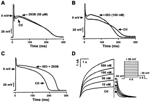
FIGURE 8. A–C: influence of slow compo- nent of delayed rectifier potassium current (IKs) inhibition by chromanol 293B (A), sym- pathetic stimulation by isoproterenol (ISO, B), and combination ofIKsblock and sympa- thetic stimulation (C) in isolated dog cardiac Purkinje cells. Note that in control (Ctl) con- dition plateau phases of the Purkinje cells are at more negative voltage than the acti- vation threshold ofIKs, and accordinglyIKs
inhibition did not change repolarization. InB, isoproterenol shifted the action potential plateau voltage to more positive values because of substantially increasedIKs.D: in this situation,IKsinhibition substantially len- gthened repolarization as shown inC. (Rep- roduced from Ref.157with permission.)