Age-related regulation and region-specific
distribution of ion channel subunits promoting atrial fibrillation in human left and right atria
Peter Biliczki
1,2, Reinier A. Boon
3, Zenawit Girmatsion
2, Alicia Bukowska
4, Bala´zs O ¨ rdo¨g
5, Bernhard M. Kaess
1, Stefan H. Hohnloser
2, Andreas Goette
4, Andra´s Varro ´
5, Anton Moritz
6, Stanley Nattel
7, and Joachim R. Ehrlich
1,2*
1Division of Cardiology, St.Josefs-Hospital, Beethovenstr. 20, 65189 Wiesbaden, Germany;2Division of Cardiology, Goethe-University, Frankfurt, Germany;3Institute for Cardiovascular Regeneration, Goethe-University, Frankfurt, Germany;4Molekularpharmakologische Elektrophysiologie, Universita¨tsklinikum, Magdeburg, Germany;5Institute of Pharmacology and Pharmacotherapy, University of Szeged, Szeged, Hungary;6Cardiothoracic Surgery, Goethe-University, Frankfurt, Germany; and7Research Center, Montreal Heart Institute and University of Montreal, Montreal, Canada
Received 11 November 2018; editorial decision 16 April 2019; accepted 23 April 2019; online publish-ahead-of-print 25 May 2019
Aims Age-induced changes and electrical remodelling are important components of the atrial fibrillation (AF) substrate.
To study regional distribution and age-dependent changes in gene expression that may promote AF in human atria.
...
Methods and results
Human left atrial (LA) and right atrial (RA) tissue samples were obtained from donor hearts unsuitable for trans- plantation and from patients undergoing mitral valve repair. Atrial fibrillation was mimickedin vitroby tachypacing of human atrial tissue slices. Ionic currents were studied by the whole-cell patch-clamp technique; gene expression was analysed by real-time qPCR and immunoblotting. Both healthy RA and RA from older patients showed greater CACNA1c mRNA and CaV1.2 protein expression than LA. No age-dependent changes of Kir2.1 expression in both atria were seen. Remodelling occurred in a qualitatively similar manner in RA and LA.IK1and Kir2.1 protein expression increased with AF. MiR-1, miR-26a, and miR-26b were down-regulated with AF in both atria.ICa,Lwas decreased. CACNA1c and CACNA2b expression decreased and miR-328 increased in RA and LA during AF.Ex vivotachypacing of human atrial slices replicated these findings. There were age-dependent increases in miR-1 and miR-328, while miR-26a decreased with age in atrial tissues from healthy human donor hearts.
...
Conclusion Features of electrical remodelling in man occur in a qualitatively similar manner in both human atria. Age-related miR-328 dysregulation and reducedICa,Lmay contribute to increased AF susceptibility with age.
䊏 䊏 䊏 䊏 䊏 䊏 䊏 䊏 䊏 䊏 䊏 䊏 䊏 䊏 䊏 䊏 䊏 䊏 䊏 䊏 䊏 䊏 䊏 䊏 䊏 䊏 䊏 䊏 䊏 䊏 䊏 䊏 䊏 䊏 䊏 䊏 䊏 䊏 䊏 䊏 䊏 䊏 䊏 䊏 䊏 䊏 䊏 䊏 䊏 䊏 䊏 䊏 䊏 䊏 䊏 䊏 䊏 䊏 䊏 䊏 䊏 䊏 䊏 䊏 䊏 䊏 䊏 䊏 䊏 䊏 䊏 䊏 䊏 䊏 䊏 䊏 䊏 䊏 䊏 䊏 䊏 䊏 䊏 䊏 䊏 䊏 䊏 䊏 䊏 䊏 䊏 䊏 䊏 䊏 䊏 䊏 䊏 䊏 䊏 䊏 䊏 䊏 䊏 䊏 䊏 䊏 䊏 䊏 䊏 䊏 䊏 䊏 䊏 䊏 䊏 䊏 䊏 䊏 䊏 䊏 䊏 䊏 䊏 䊏 䊏 䊏 䊏 䊏 䊏 䊏 䊏 䊏 䊏 䊏 䊏 䊏 䊏 䊏 䊏 䊏 䊏 䊏 䊏 䊏 䊏 䊏 䊏 䊏 䊏 䊏 䊏 䊏 䊏 䊏 䊏 䊏 䊏 䊏 䊏 䊏 䊏 䊏 䊏 䊏 䊏 䊏 䊏 䊏 䊏 䊏 䊏 䊏 䊏 䊏 䊏 䊏 䊏 䊏 䊏 䊏 䊏 䊏 䊏 䊏 䊏 䊏 䊏 䊏 䊏 䊏 䊏 䊏 䊏 䊏 䊏 䊏 䊏 䊏 䊏 䊏 䊏 䊏 䊏 䊏 䊏 䊏 䊏 䊏 䊏 䊏 䊏 䊏
Keywords Aging
•
Atrial fibrillation•
Ion channel•
miRNA•
mRNA•
Protein•
RemodellingIntroduction
Atrial fibrillation (AF) represents a major cardiovascular healthcare challenge. Understanding cellular remodelling processes is important to generate further, effective approaches to treat the arrhythmia.
Involved factors may represent novel therapeutic targets.1,2
Reduced L-type calcium (ICa,L) and increased inward-rectifier cur- rents (IK1) are mechanistically involved in action potential shortening and alterations of intracellular calcium-homoeostasis associated with
AF.3Attenuation of Ca2þcurrent and augmentation of Kþcurrents have been linked to aging in a canine model.4MicroRNAs regulate gene expression levels of the main pore-forming subunits and are in- volved in AF pathogenesis.5Among others, microRNA-1 regulate ex- pression of theIK1subunit Kir2.1 and microRNA-328 is involved in the regulation of the calcium channel subunit CaV1.2.6,7 The microRNA-26 family consists of three distinct genes miR-26a-1, miR- 26a-2, and miR-26b that encode mature microRNAs with identical seed sequences and the same target mRNAs. MicroRNA-26 has
* Corresponding author. Tel:þ49 611 177 1275; fax:þ49 611 177 1276.E-mail address: jehrlich@joho.de
Published on behalf of the European Society of Cardiology. All rights reserved.VCThe Author(s) 2019. For permissions, please email: journals.permissions@oup.com.
Downloaded from https://academic.oup.com/europace/article-abstract/21/8/1261/5498772 by University of Szeged user on 29 August 2019
been shown to be dysregulated in AF and regulate the expression of Kir2.1.5MicroRNAs also regulate age-dependent processes.8Down- regulation ofICa,Lhas been reported in ageing canines.9As AF has an age-dependent demography in man, it is of interest to fill respective knowledge gaps.
Regionally different ion channel expression may underlie specific electrophysiological properties of cardiac chambers.10Alterations in spatial distribution may additionally contribute to the AF substrate.
Only few studies have looked into regional differences in human ion channel expression.11,12No previous study systematically compared changes in matched left atrial (LA) and right atrial (RA) tissues from one patient.
Therefore, the present work sought to analyse region- and age- dependent components in gene expression remodelling associated to AF.
Methods
Patient population
Pairs of LA and RA tissue were obtained from healthy donor hearts and from patients undergoing surgical mitral valve repair (MVR). The study complies with the Declaration of Helsinki and local ethics committees at Goethe-Universita¨t, Frankfurt, Germany (Nr.54/05) and University of Szeged, Hungary (Nr. 63/97) approved the study.
Donor hearts were obtained from six female and nine male organ donors with a mean age of 50.8 ± 9.6 years ranging from 31 to 68, whose non-diseased hearts were explanted to obtain pulmonary and aortic valves for transplant surgery as no matching recipient was avail- able for whole hearts. Before cardiac explantation, organ donors did not receive medication apart from dobutamine, furosemide, and plasma expanders. After explantation, each heart was perfused with cardioplegic solution and kept cold (4–6C) for 2–4 h prior to dissection.
Surgical patients were older than 18 years and had an indication for MVR. All patients provided written information consent to participate.
Exclusion criteria were paroxysmal AF, haemodynamic instability, active endocarditis, systemic inflammation, or treatment with immunosuppres- sive agents. In patients undergoing MVR LA tissue was collected from the site of incision at the roof to anterior LA. RA tissue was from the site of insertion of the heart lung machine at the right atrial appendage (RAA) to RA roof. Donor heart tissue samples were from comparable regions in the anterior LA and RA roof.
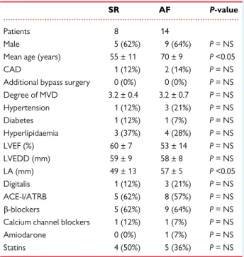
Patients were classified as having persistent/permanent AF if a previous diagnosis of AF was noted in medical records and AF was present on the admission electrocardiogram, whereas sinus rhythm (SR) patients had no mention of AF and were in SR on admission. Patients in SR with a previ- ous diagnosis of AF were classified as having paroxysmal AF. Additional details on surgical patients are provided inTable1.
Electrophysiological and molecular biological methods
Details of these methods are provided in theSupplementary material online.
Statistical analysis
Molecular data were analysed with GraphPad Prism 5 using the Student’s t-tests when comparing two conditions, or an analysis of variance with Bonferroni or Newman–Keuls correction for multiple comparisons.
AP-value <0.05 was considered significant and tests were performed two-sided. Data were tested for outliers using the Grubbs outlier test and, if outliers were present, they were removed. Clampfit (Molecular Devices, Ismaning, Germany) and GraphPad Prism (GraphPad Software, San Diego, CA, USA) were used for patch-clamp data analysis. Data are presented as mean ± SEM.
Results
Regional expression of ion channels, related mRNA, and microRNA—healthy hearts
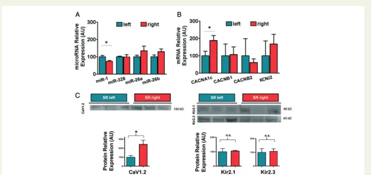
Regional distribution was studied in pairs of LA and RA tissue obtained from each donor heart. miR-1 and miR-26a,b expressions were not different in LA and RA samples (Figure1A). Respective mRNA and protein levels of Kir2.1 and 2.3 were similar in both atria (Figure1BandC). Despite greater CACNA1c mRNA and CaV1.2 protein levels in RA tissue compared to LA, the relative expression of miR-328 was not significantly different in the two regions,
...
Table 1 Characteristics of surgical patients
SR AF P-value
Patients 8 14
Male 5 (62%) 9 (64%) P= NS
Mean age (years) 55 ± 11 70 ± 9 P<0.05
CAD 1 (12%) 2 (14%) P= NS
Additional bypass surgery 0 (0%) 0 (0%) P= NS Degree of MVD 3.2 ± 0.4 3.2 ± 0.7 P= NS
Hypertension 1 (12%) 3 (21%) P= NS
Diabetes 1 (12%) 1 (7%) P= NS
Hyperlipidaemia 3 (37%) 4 (28%) P= NS
LVEF (%) 60 ± 7 53 ± 14 P= NS
LVEDD (mm) 59 ± 9 58 ± 8 P= NS
LA (mm) 49 ± 13 57 ± 5 P<0.05
Digitalis 1 (12%) 3 (21%) P= NS
ACE-I/ATRB 5 (62%) 8 (57%) P= NS
b-blockers 5 (62%) 9 (64%) P= NS Calcium channel blockers 1 (12%) 1 (7%) P= NS
Amiodarone 0 (0%) 1 (7%) P= NS
Statins 4 (50%) 5 (36%) P= NS
ACE-I, angiotensin-converting enzyme inhibitor; AF, atrial fibrillation; ATRB, an- giotensin receptor blocker; CAD, coronary artery disease; LA, left atrium;
LVEDD, left ventricular end-diastolic diameter; LVEF, left ventricular ejection fraction; MVD, mitral valve disease; NS, not significant; SR, sinus rhythm.
What’s new?
• First comprehensive study illustrating that previously known changes in right or left atria indeed occur similarly in both atria of a single patient.
• First demonstration of reducedICa,Land respective microRNA in elderly patients with atrial fibrillation.
1262 P. Biliczkiet al.
Downloaded from https://academic.oup.com/europace/article-abstract/21/8/1261/5498772 by University of Szeged user on 29 August 2019
suggesting that miR-328 may not be the only factor determining basal levels of CaV1.2 expression [P¼not significant (NS);Figure1A].
The mRNA encoding for the pore-forminga-subunit ofICa,Lchan- nels (CACNA1c) was1.7-fold more abundant in RA than in LA (P< 0.05). mRNA expression for theb-1 andb-2 Ca2þchannel regu- latory subunits did not differ (Figure1B).IK1channel subunit mRNA KCNJ2, encoding Kir2.1 was also similar between atria (Figure1B). In accordance with mRNA levels, immunoblot studies showed2.1- fold higher protein amount for CaV1.2 in RA as compared to LA (P< 0.05,Figure1C). Relative expression ofIK1-subunits Kir2.1 and Kir2.3 was not different in the two regions (Figure1C).
Ion channel proteins, mRNA, and microRNA in mitral valve repair atria
Next, we studied ion channel distribution in paired LA and RA sam- ples from patients in SR undergoing MVR.
Here, CACNA1c mRNA was also more abundantly expressed in RA compared with LA samples (1.9-fold greater expression level in RA vs. LA;P< 0.05). CACNB1, CACNB2, and KCNJ2 mRNA ex- pression was not different (Figure2B). In line with these findings, pro- tein expression of CaV1.2 was2.4-fold greater in RA compared with LA (P< 0.05), while IK1subunits Kir2.1 and Kir2.3 were evenly expressed (Figure2C). The expression levels for miR-26a,b and miR-
328 in SR (Figure2A) were similar, but miR-1 (P< 0.05) was less abun- dantly expressed in RA samples (Figure2A).
Right-left differences in mitral valve repair samples from patients with atrial fibrillation
Regional distribution of RNA transcripts and protein expression were compared between RA and LA in paired samples obtained from MVR patients with AF.
In tissue from patients with AF, expression levels of miR-26a, miR- 26b, and miR-328 were not different between RA and LA, but ex- pression level of miR-1 was decreased in RA vs. LA (P< 0.05,Figure 3A). CACNA1c mRNA levels were similar between RA and LA (N= 14/14,P¼NS), and CACNB1, CACNB2, and KCNJ2 mRNA levels were comparable (Figure 3B). In contrast to mRNA levels, there was a regional gradient of1.6-fold greater protein expression in RA compared with LA for CaV1.2—present also in SR—2.9-fold greater Kir2.1 protein expression (P< 0.05,Figure3C) in AF samples.
Atrial fibrillation-induced remodelling in right and left atria
Compared with SR levels, CACNA1c mRNA was reduced by AF in both regions (P< 0.05 and P= NS, respectively). Relative mRNA Figure 1 Regional expression of ion channels, related mRNA and microRNA—healthy hearts. (A) MicroRNA expression levels are measured by quantitative real-time PCR in healthy human LA and RA atrial tissue samples (n= 15 each) from donor hearts. (B) mRNA expression levels from the same atrial tissue samples are illustrated. (C) Protein quantification from these samples is illustrated. Representative western blots and semiquantita- tive mean ± SEM data are shown for calcium (CACNA1c) andIK1channel subunits (Kir2.1 and Kir2.3). Membrane protein fractions were used, actin served as a loading control (not shown). Data are presented as mean ± SEM and RA levels were normalized to LA levels.b-actin and U6 served as in- ternal standards for mRNA and microRNA measurements, respectively. Antibodies were applied at dilution of 1:1000. *P< 0.05. AU, arbitrary units;
LA, left atrial; n.s., not significant; RA, right atrial; PCR, polymerase chain reaction; SEM, standard error of the mean.
Downloaded from https://academic.oup.com/europace/article-abstract/21/8/1261/5498772 by University of Szeged user on 29 August 2019
Figure 2 Ion channel proteins, mRNA, and microRNA in MVR hearts. (A) MicroRNA expression levels in SR human LA and RA tissue samples (n= 8 each) from patients undergoing MVR are illustrated. (B) mRNA expression levels in respective atrial tissue samples (n= 8 each). (C) Protein quantification in identical tissue samples is illustrated. Representative western blots and semiquantitative mean ± SEM data are shown for calcium (CACNA1c) andIK1channel subunits (Kir2.1 and Kir2.3). Membrane protein fractions were used, actin served as a loading control (not shown). Data are presented as mean ± SEM and RA levels were normalized to LA levels. Antibodies were applied at dilution of 1:1000. *P< 0.05. AU, arbitrary units; LA, left atrial; MVR, mitral valve repair; n.s., not significant; RA, right atrial; SEM, standard error of the mean; SR, sinus rhythm.
Figure 3 Right-left differences in MVR samples from patients with AF. (A) MicroRNA expression levels in AF human LA and RA tissue samples (n= 14 each) from patients undergoing MVR are illustrated. (B) mRNA expression levels in respective atrial tissue samples (n= 14 each). (C) Protein quantification in identical tissue samples is illustrated. Representative western blots and semiquantitative mean ± SEM data are shown for calcium (CACNA1c) andIK1channel subunits (Kir2.1 and Kir2.3). Membrane protein fractions were used, actin served as a loading control (not shown). Data are presented as mean ± SEM and RA levels were normalized to LA levels. Antibodies were applied at dilution of 1:1000. *P< 0.05. AF, atrial fibrilla- tion; AU, arbitrary units; LA, left atrial; MVR, mitral valve repair; RA, right atrial; SEM, standard error of the mean.
1264 P. Biliczkiet al.
Downloaded from https://academic.oup.com/europace/article-abstract/21/8/1261/5498772 by University of Szeged user on 29 August 2019
expression of KCNJ2 remained largely unchanged (P¼ NS;Figure 4A). While mRNA levels in LA and RA showed a numerical trend to less CACNA1c and more KCNJ2 mRNA (P = NS), immunoblots showed significantly lower amounts for CaV1.2 protein and greater Kir2.1 protein in AF samples (P< 0.05 for both,Figure4C).
MicroRNA expression was consistently altered in AF. In relation to levels in SR miR-1 expression was reduced (P< 0.05,Figure4B).
miR-26a,b expression also nominally decreased in both atria without statistical significance. miR-328 expression showed a non-significant trend to increase with AF both in the LA and RA regions (P¼NS, Figure4B). These trends are consistent with the suggested role for these microRNAs in regulating CaV1.2 and Kir2.1 expression in AF and AF-induced changes were qualitatively similar in LA and RA.
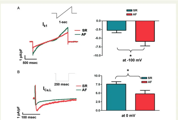
Atrial fibrillation-induced ionic current alterations were studied by whole-cell current recordings in cardiomyocytes isolated from LA tissue (SR and AF). In analogy to changes in protein expression of Kir2.1,IK1density recorded at100 mV increased in AF cells com- pared with SR (P< 0.05,Figure5A). Similarly, as suggested by reduced CaV1.2 protein expression,ICa,Ldensity in AF (P< 0.05,Figure5B).
Ex vivo tachypacing in human left atrial cardiomyocytes
We cultured atrial tissue slices obtained from SR patients and electri- cally stimulated them ex vivo(0.6 Hz vs. 4 Hz). Under these condi- tions, 0.6 Hz ought to mimic SR and 4 Hz simulated AF. Tachypacing led to a non-significant tendency in reduced miR-1 levels by26%
and mildly increased miR-328 by25% (N= 7/7;P¼NS;Figure6A).
These changes qualitatively replicated tendencies of changesin vivoal- though they only indicate a trend.
Age-dependent changes in ion channel expression
For the purpose of this analysis, samples of patients were dichoto- mized into two groups at the median age (51 years).
MicroRNA-328 expression was35% greater in the ‘old’ (mean age 59 ± 2 years) compared to the ‘young’ (mean age 44 ± 3 years) group (N= 8/7,P< 0.05,Figure7A), coinciding with a numerical trend towards decreased mRNA levels of CACNA1c, CACNB1, and Figure 4AF-induced remodelling in right and left atria. This figure illustrates differences in RNA and protein expression between SR and AF atria from patients undergoing MVR. (A) mRNA ion channel subunit expression levels in the RA from AF (n= 14) and SR (n= 8) patients illustrate a de- crease in CACNA1c expression and a tendency towards an increase in Kir2.1 mRNA expression. The right-sided panel is an analogous representa- tion of mRNA expression changes occurring in the LA from the same AF and SR patients as in panelA. A similar signature of remodelling can be appreciated. (B) Changes in microRNA expression in RA (top) and LA (bottom) tissue samples are illustrated. (C) Representative western blots and mean ± SEM data for L-type calcium and inward-rectifier channel subunits (CACNA1c, Kir 2.1) expression in AF relative to SR patients (n= 4 each).
Representative western blots and semiquantitative mean ± SEM data are shown for calcium (CACNA1c) andIK1channel subunits (Kir2.1 and Kir2.3). Membrane protein fractions were used, actin served as a loading control (not shown). Data are presented as mean ± SEM and RA levels were normalized to LA levels. Antibodies were applied at dilution of 1:1000. *P< 0.05. AF, atrial fibrillation; AU, arbitrary units; LA, left atrial; MVR, mitral valve repair; RA, right atrial; SEM, standard error of the mean; SR, sinus rhythm.
Downloaded from https://academic.oup.com/europace/article-abstract/21/8/1261/5498772 by University of Szeged user on 29 August 2019
Figure 5Ionic current changes in AF. Representative recordings and mean ± SEM current densities from LA cardiomyocytes of patients in SR and AF. (A)IK1was recorded with a ramp protocol (inset) in the presence of tertiapin-Q (100 nM) to avoid contamination byIKAch.IK1density was in- creased in LA cells in AF compared to SR (at100 mV:5.9±1.3 vs.2.7±0.7,n= 5 cells each, *P< 0.05). (B)ICa,Lwas recorded with a step proto- col (inset).ICa,Ldensity was decreased in AF compared to SR (at 0 mV:7.6±0.7 vs.4.8±1.0,n= 5 and 3 cells, respectively, *P< 0.05). AF, atrial fibrillation; AU, arbitrary units; LA, left atrial; MVR, mitral valve repair; RA, right atrial; SEM, standard error of the mean; SR, sinus rhythm.
Figure 6Ex vivotachypacing induced remodelling in human LA cardiomyocytes. Quantitative real-time PCR results from 4 Hz-paced control (pac- ing,n= 7) and 1 Hz-paced control (SR,n= 7) human atrial tissues slices mimics results obtained comparing SR and AF tissue samples. Data are pre- sented as mean ± SEM, relative to ‘SR’. (A) Changes in microRNA expression are illustrated. miR-1 was significantly reduced and miR-328 increased in tachypaced tissue samples. (B) Changes in calcium and inward-rectifier channel subunits demonstrate a tendency towards changes observedin vivo.
Representative western blots and semiquantitative mean ± SEM data are shown for calcium (CACNA1c) andIK1channel subunits (Kir2.1 and Kir2.3). Membrane protein fractions were used, actin served as a loading control (not shown). Data are presented as mean ± SEM and RA levels were normalized to LA levels.b-actin and U6 served as internal standards for mRNA and microRNA measurements, respectively. Antibodies were applied at dilution of 1:1000. *P< 0.05. AF, atrial fibrillation; AU, arbitrary units; LA, left atrial; PCR, polymerase chain reaction; SEM, standard error of the mean.
1266 P. Biliczkiet al.
Downloaded from https://academic.oup.com/europace/article-abstract/21/8/1261/5498772 by University of Szeged user on 29 August 2019
CACNB2 (Figure7B) expression. CaV1.2 protein levels were3.1- fold reduced (P< 0.05,Figure7C). Relative expression of miR-1 in- creased with age (56%,P< 0.05,Figure7A), expression of miR-26a also decreased with age (P< 0.05,Figure7A). MiR-26b showed a mild tendency towards less expression with age (P = NS, Figure 7A).
Protein levels for Kir2.1 and Kir2.3 showed a trend to decrease (Kir2.130%; Kir2.313%,P¼NS,Figure7BandC).
Discussion
This study sought to determine several aspects of AF remodelling: (i) to establish whether there is a difference in expression between right and left atria from each patient. (ii) To study if both atria are sub- jected to similar remodelling processes during human AF. (iii) To identify rapid heart rate as a determinant of the remodelling process in human atrial tissue. (iv) To evaluate changes related to age as a po- tential underlying modulator of the substrate for AF in left and right atria.
Main findings
Human atria exhibit regional heterogeneity regarding expression of calcium channel subunits but not inward-rectifier subunits. In healthy
RA there is70% greater abundance of CACNA1c mRNA and2- fold more CaV1.2 protein—underlying calcium currents—than in LA. This pattern is qualitatively similar in diseased hearts. In mitral valve disease patients, RA CACNA1c mRNA expression is 90%
greater than LA mRNA and protein is2.4fold more abundant.
We observed an age-dependent increase in expression of miR- 328, along with down-regulated CACNA1c mRNA and CaV1.2 pro- tein potentially underlying changes that may facilitate AF occurrence with age.
Regional ion channel distribution
The current knowledge about the cellular and molecular basis of AF in man is largely based on studies using RAAs, with only a few studies comparing cellular electrophysiology or gene expression in the RAAs and left atrial appendages (LAAs).13Even more, there are few studies examining changes in regions other than appendages from patients. If changes that have been described in RA could similarly be observed in LA remained largely unknown.
Greater IK1-densities in LAA than RA have been reported in patients with paroxysmal AF yielding a left to right gradient in action potential duration.12This finding is not readily consistent with our data showing similar remodelling processes in both atria during per- sistent/permanent AF. However, among the 95 patients in the Figure 7 Age-dependent changes in ion channel expression. This figure illustrates changes in relation to age in healthy donor heart tissue.
Quantitative real-time PCR results from healthy young (n= 8) and old (n= 7) human atrial tissue samples are represented. (A) MicroRNA expression is illustrated. (B) Ion channel coding mRNA expression is illustrated. Patient age was dichotomized at the mean and the ‘young’ group had an average age of young 44 ± 3 years vs. 59 ± 2 years for the ‘old’ group,P< 0.05. (C) Protein expression levels ofn= 6 each for CACNA1c,n= 6 each for Kir2.1, andn= 3 each for Kir2.3. Representative western blots and semiquantitative mean ± SEM data are shown for calcium (CACNA1c) andIK1
channel subunits (Kir2.1 and Kir2.3). Membrane protein fractions were used, actin served as a loading control (not shown). Data are presented as mean ± SEM and RA levels were normalized to LA levels.b-actin and U6 served as internal standards for mRNA and microRNA measurements, re- spectively. Antibodies were applied at dilution of 1:1000. *P< 0.05. AU, arbitrary units; n.s., not significant; PCR, polymerase chain reaction; SEM, stan- dard error of the mean.
Downloaded from https://academic.oup.com/europace/article-abstract/21/8/1261/5498772 by University of Szeged user on 29 August 2019
previous study, matched RA and LA samples were only available from four individual patients, while all our samples had an individual match from both atria. Furthermore, results obtained in patients with paroxysmal AF could not be replicated in persistent/permanent AF in the study by Voigtet al. Accordingly, our study and previous work might look at different stages of the remodelling process, which could explain discrepancies.
Regional expression differences in mRNA expression were studied in human tissue (RA, LA, ventricles, and Purkinje fibres).11This study used human donor heart tissue unsuitable for transplantation similar to our study. There was no statistically significant overall gene ex- pression difference between right- and left-sided chambers but tissue differences (i.e. atrium vs. ventricle vs. Purkinje fibres) were strong determinants of ion channel subunit expression pattern. In an earlier paper, mRNA transcripts of a set of ion channels and calcium modu- lating proteins were studied.14Right atrial appendage tissue samples of patients with valvular heart disease were compared these with car- diac tissue of patients with coronary disease.
Relevance for atrial fibrillation mechanisms
In this study, we have shown that previously identified key mechanis- tic features of electrical remodelling underlying AF15can be similarly observed in human LA and RA tissue.IK1augmentation is among the key mechanisms of action potential duration shortening, which criti- cally stabilizes atrial rotors maintaining AF.16,17According to our data up-regulation of this channel occurs in both atria with relatively greater changes in RA. This finding deserves further study.
We have shown, that bothICa,Land Ca2þchannel protein expres- sion are down-regulated in AF. We found elevated Kir2.1 mRNA and protein levels and greaterIK1density in AF samples. Changes in miR- 1, a regulator of Kir2.1, are coinciding with this pathology.6Notably, miR-1 has been shown to regulate multiple genes including the main cardiac connexin isoform Cx43, theIKschannel regulatory subunit minK encoded byKCNE1or calcium handling proteins, such as the Naþ/Ca2þexchanger NCX1,5which genes may also contribute to AF pathogenesis. According to our data in healthy human atrial tissue there is regional heterogeneity between LA and RA expression of calcium channel subunits with larger expression in RA. Less calcium channel and related current could lead to shorter action potential durations. This difference may contribute to explain why the LA is the natural habitat for AF.
Age-dependent changes
To date, no data regarding the influence of ageing on human electro- physiology exist. Work in animals found decreased Ca2þ current function and augmented potassium currents in aging dogs,4and indi- rect evidence for a reduction inICa,Las a potential contributor (be- sides increased fibrosis) to AF vulnerability in old canines.9
The results of our study confirm a reduction in calcium channel subunits in elderly humans and suggest that age-dependently in- creased expression of microRNAs (miR-1 and miR-328) and subse- quently down-regulated mRNA and protein levels of related genes could play a role in AF development. In particular, age-dependent down-regulation of calcium channel protein expression, as a conse- quence of increased miR-328 levels may lead to decreased L-type
Ca2þcurrent and could provide a contributory mechanistic explana- tion for the increased susceptibility for AF development with older age.
Limitations
As this study was performed in human atrial tissue, sample numbers are small and accordingly, statistical power is limited. Several changes were only documented as a trend owing to small sample numbers and large variability of measurements. This study could not address many nowadays all unanswered issues regarding human atrial electro- physiology and cellular aetiology of AF.
Conclusions
Healthy human LA exhibits less calcium channel subunit expression than RA—potentially owing to greater LA microRNA 328 expres- sion. Atrial fibrillation remodelling of inward-rectifier and calcium channel subunits occurs to a qualitatively similar extent in both atria, while RA has a relatively larger increase in inward rectifier subunits in response to AF. We found an age-dependent increase in microRNAs (miR-1 and miR-328) and down-regulated mRNA and protein levels of calcium channel genes that could play a role in AF development with older age.
Gene symbols
...
...
...
...
...
mRNA GenBank ID Function
CACNA1c NM_000719 Encodes CaV1.2, thea-subunit of the cardiac L-type calcium channel CACNB1 NM_000723 Encodesb1 regulatory subunit of cal-
cium channels
CACNB2 NM_000724 Encodesb2 regulatory subunit of cal- cium channels
KCNJ2 NM_000891 Encodes Kir2.1, the pore-forming subunit of cardiac inward-rectifier potassium channel
Protein GenBank GeneID
Function
CaV1.2 775 Thea-subunit of the cardiac L-type calcium channel
Kir2.1 3759 Pore-forming subunit of cardiac in- ward rectifier
Kir2.3 3761 Pore-forming subunit of cardiac in- ward rectifier
microRNA miRBase accession
miR-1 MI0000651
miR-26a MI0000083
miR-26b MI0000084
miR-328 MI0000804
1268 P. Biliczkiet al.
Downloaded from https://academic.oup.com/europace/article-abstract/21/8/1261/5498772 by University of Szeged user on 29 August 2019
Supplementary material
Supplementary materialis available atEuropaceonline.
Acknowledgements
The expert technical assistance of Christin Lo¨ßl is greatly appreciated.
Funding
J.R.E. received funding from Else Kro¨ner-Fresenius-Stiftung and Deutsche Stiftung fu¨r Herzforschung. Transport was supported by the grant NKFIH [GINOP-2.3.2-15-2016-00006].
Conflict of interest:none declared.
References
1. Heijman J, Algalarrondo V, Voigt N, Melka J, Wehrens XHT, Dobrev Det al. The value of basic research insights into atrial fibrillation mechanisms as a guide to therapeutic innovation: a critical analysis.Cardiovasc Res2016;109:467–79.
2. Goette A, Kalman JM, Aguinaga L, Akar J, Cabrera JA, Chen SAet al. EHRA/HRS/
APHRS/SOLAECE expert consensus on atrial cardiomyopathies: definition, char- acterization, and clinical implication.Europace2016;18:1455–90.
3. Heijman J, Voigt N, Nattel S, Dobrev D. Cellular and molecular electrophysiol- ogy of atrial fibrillation initiation, maintenance, and progression.Circ Res2014;
114:1483–99.
4. Dun W, Yagi T, Rosen MR, Boyden PA. Calcium and potassium currents in cells from adult and aged canine right atria.Cardiovasc Res2003;58:526–34.
5. Luo X, Yang B, Nattel S; Publishing Group. MicroRNAs and atrial fibrillation:
mechanisms and translational potential.Nat Rev Cardiol2015;12:80–90.
6. Girmatsion Z, Biliczki P, Bonauer A, Wimmer-Greinecker G, Scherer M, Moritz Aet al. Changes in microRNA-1 expression andIK1up-regulation in human atrial fibrillation.Heart Rhythm2009;6:1802–9.
7. Lu Y, Zhang Y, Wang N, Pan Z, Gao X, Zhang Fet al. MicroRNA-328 contrib- utes to adverse electrical remodeling in atrial fibrillation.Circulation2010;122:
2378–87.
8. Boon RA, Iekushi K, Lechner S, Seeger T, Fischer A, Heydt Set al. MicroRNA- 34a regulates cardiac ageing and function.Nature2013;495:107–10.
9. Anyukhovsky EP, Sosunov EA, Plotnikov A, Gainullin RZ, Jhang JS, Marboe CC et al. Cellular electrophysiologic properties of old canine atria provide a sub- strate for arrhythmogenesis.Cardiovasc Res2002;54:462–9.
10. Feng J, Yue L, Wang Z, Nattel S. Ionic mechanisms of regional action potential heterogeneity in the canine right atrium.Circ Res1998;83:541–51.
11. Gaborit N, Bouter SL, Szuts V, Varro A, Escande D, Nattel Set al. Regional and tissue specific transcript signatures of ion channel genes in the non-diseased hu- man heart.J Physiol2007;582:675–93.
12. Voigt N, Trausch A, Knaut M, Matschke K, Varro´ A, Wagoner DVet al. Left-to- right atrial inward rectifier potassium current gradients in patients with paroxysmal versus chronic atrial fibrillation.Circ Arrhythm Electrophysiol2010;3:
472–80.
13. Voigt N, Dobrev D. The biology of human pulmonary veins: does it help us to better understand AF pathophysiology in patients?Heart Rhythm2013;10:
392.
14. Gaborit N, Steenman M, Lamirault G, Meur NL, Bouter SL, Lande G et al.
Human atrial ion channel and transporter subunit gene-expression remodeling associated with valvular heart disease and atrial fibrillation.Circulation2005;112:
471–81.
15. Nattel S, Burstein B, Dobrev D. Atrial remodeling and atrial fibrillation: mecha- nisms and implications.Circ Arrhythm Electrophysiol2008;1:62–73.
16. Dhamoon AS, Jalife J. The inward rectifier current (IK1) controls cardiac excitabil- ity and is involved in arrhythmogenesis.Hear Rhythm2005;2:316–24.
17. Ehrlich JR. Inward rectifier potassium currents as a target for atrial fibrillation therapy.J Cardiovasc Pharmacol2008;52:129–35.
Downloaded from https://academic.oup.com/europace/article-abstract/21/8/1261/5498772 by University of Szeged user on 29 August 2019