R E S E A R C H A R T I C L E Open Access
SHMT1 1420 and MTHFR 677 variants are associated with rectal but not colon cancer
Viktor Komlósi1, Erika Hitre2, Éva Pap2, Vilmos Adleff2, Andrea Réti2, Éva Székely3, Anna Bíró4, Péter Rudnai5, Bernadette Schoket6, Judit Müller7, Béla Tóth8, Szabolcs Ottó2, Miklós Kásler2, Judit Kralovánszky2*, Barna Budai2
Abstract
Background:Association between rectal or colon cancer risk and serine hydroxymethyltransferase 1 (SHMT1) C1420T or methylenetetrahydrofolate reductase (MTHFR) C677T polymorphisms was assessed. The serum total homocysteine (HCY), marker of folate metabolism was also investigated.
Methods:TheSHMT1andMTHFRgenotypes were determined by real-time PCR and PCR-RFLP, respectively in 476 patients with rectal, 479 patients with colon cancer and in 461 and 478, respective controls matched for age and sex. Homocysteine levels were determined by HPLC kit. The association between polymorphisms and cancer risk was evaluated by logistic regression analysis adjusted for age, sex and body mass index. The population
stratification bias was also estimated.
Results:There was no association of genotypes or diplotypes with colon cancer. The rectal cancer risk was significantly lower forSHMT1 TT (OR = 0.57, 95% confidence interval (CI) 0.36-0.89) and higher forMTHFR CT genotypes (OR = 1.4, 95%CI 1.06-1.84). A gene-dosage effect was observed forSHMT1with progressively decreasing risk with increasing number of T allele (p = 0.014). The stratified analysis according to age and sex revealed that the association is mainly present in the younger (< 60 years) or male subgroup. As expected from genotype analysis, theSHMT1 T allele/MTHFRCC diplotype was associated with reduced rectal cancer risk (OR 0.56, 95%CI 0.42-0.77 vs all other diplotypes together). The above results are unlikely to suffer from population
stratification bias. In controls HCY was influenced bySHMT1polymorphism, while in patients it was affected only by Dukes’stage. In patients with Dukes’stage C or D HCY can be considered as a tumor marker only in case of SHMT11420CC genotypes.
Conclusions:A protective effect ofSHMT11420T allele orSHMT11420 T allele/MTHFR677 CC diplotype against rectal but not colon cancer risk was demonstrated. The presence ofSHMT11420 T allele significantly increases the HCY levels in controls but not in patients. Homocysteine could be considered as a tumor marker inSHMT11420 wild-type (CC) CRC patients in Dukes’ stage C and D. Further studies need to clarify whySHMT1andMTHFR polymorphisms are associated only with rectal and not colon cancer risk.
Background
Colorectal cancer (CRC) is the second leading cause of cancer morbidity in Hungary both in male and female populations, and is one of the most frequent cause of cancer-related deaths. The mean age of the patients at the diagnosis of CRC is about 60 years and yearly about 9,000 new cases occur including approximately 2,800 newly diagnosed rectal cancer cases. Numerous results
have been accumulated suggesting the role of folate and folate-related enzyme polymorphisms in the etiology of CRC. Low folate intake or low bioavailability of circulat- ing folate, key components of one-carbon metabolism, have been related to increased risk of CRC [1]. The results, however, are still not conclusive. In a recent report an opposite influence of folate on different stages of the adenoma-carcinoma sequence was documented [2]. On the other hand, there are studies providing evi- dence for the significance of genetic polymorphisms of folate-cycle enzymes (e.g. methylenetetrahydrofolate reductase (MTHFR)) in colorectal carcinogenesis [3].
* Correspondence: kralo@oncol.hu
2Department of Clinical Research, National Institute of Oncology, Budapest, Hungary
Full list of author information is available at the end of the article
© 2010 Komlósi et al; licensee BioMed Central Ltd. This is an Open Access article distributed under the terms of the Creative Commons Attribution License (http://creativecommons.org/licenses/by/2.0), which permits unrestricted use, distribution, and reproduction in any medium, provided the original work is properly cited.
The role of the MTHFRC677T polymorphism influen- cing CRC susceptibility is also inconsistent [4,5]. In a meta-analysis of 25 studies Hubner et al. reported that MTHFR677TT genotype is associated with a significant, but moderately reduced risk of CRC [6]. Another less widely studied enzyme of folate/one-carbon metabolism, the cytosolic serine hydroxymethyltransferase (SHMT1), has a frequent but functionally less characterized Leu- >
Phe polymorphism (SNP variant C1420T). This poly- morphism was associated with significantly reduced risk of acute lymphocytic leukemia [7] and malignant lym- phoma [8], but not colorectal adenoma [9]. However, in a CRC study no evidence of gene influence on the risk or association of this polymorphism with folate or homocysteine (HCY) levels were found [10]. The modi- fying effect ofSHMT1 C1420T polymorphism on CRC risk remained undecided [11,12].
Rectal and colon cancer differ in their histology, diag- nosis, sensitivity to radiotherapy and prognosis as well [13]. The treatment of rectal cancer usually includes preoperative radiotherapy, while colon cancer is non- radiosensitive. Both types of cancer have similar and also different risk factors [14,15]. Low folate and/or excessive alcohol intake are risk factors for both disease, but the physical activity have different effect on their risk [14]. In a large prospective US cohort body mass index (BMI) was related to increased risk of colon, but not rectal cancer [16]. There are few studies investigat- ing the genetic risk factors separately in rectal and colon cancer, showing net differences between rectal and colon cancer [17-19]. The inconsistent results of the previous studies examining the polymorphisms in CRC may arise from the fact that dissimilar proportions of rectal and colon cancer cases were included in the investigations. Based on this information ascertained above the separate investigation of rectal and colon can- cer cases is reasonable.
The SHMT1 is a vitamin B6 dependent enzyme that catalyzes the reversible conversion of serine and tetrahy- drofolate (THF) to glycine and 5,10-methylenetetrahy- drofolate (5,10-MTHF), a reaction which provides one- carbon units for S-adenosylmethionine (SAM), and for purine and pyrimidine synthesis.
Methylenetetrahydrofolate reductase irreversibly con- verts 5,10-MTHF to 5-methyltetrahydrofolate (5- methylTHF) using vitamin B2 as a cofactor. 5- methylTHF is the most stable and abundant form of folate metabolites, which in turn is the methyl donor in the conversion of HCY into methionine. Decrease in MTHFR reductase activity could lead to impaired HCY catabolism. There are two common functional SNPs on theMTHFRgene, which are in close linkage: the C677T and the A1298C substitutions. The C677T polymorph- ism on the exon 4 results in an Ala- > Val substitution
at codon 222. Compared to theMTHFR677C homozy- gotes the TT and CT genotypes have 70% and 30%
lower enzyme activity, respectively. The 677T allele has been associated with elevated plasma HCY levels [20].
In contrast, the exact functional consequence of the A1298C polymorphism is not well defined, and because of the linkage the extent of its independent contribution remains inconclusive [21,22].
Homocysteine is a sulphur-containing amino acid, produced mainly by S-adenosyl-methionine mediated methylation reactions. It is well-known that the total serum homocysteine (HCY) level among others is influ- enced by gender (higher in males), age (increasing with ageing), BMI (increased in obese individuals) and renal function (increased in renal impairment) [23-25]. More- over, its accumulation in the serum is related to a num- ber of disease conditions: vascular disease, cancer, neural tube defect, etc [26]. HCY reflects the combined pool of free, albumin bound, reduced and oxidized forms of HCY in the blood. Remethylation of homocys- teine by methionine synthase, a vitamin B12 dependent enzyme, is the major metabolic pathway. The HCY level is inversely associated with the folate level and was sug- gested to be a risk factor of cancers [27,28]. Moreover a study suggested the role of HCY as a tumor marker of CRC [27]. This finding led to the exclusion of HCY from our analysis of cancer risk to avoid an eventual bias.
The aim of our study was to separately analyze the risk of rectal and colon cancer in association with SHMT1 C1420T and MTHFRC677T polymorphisms.
The effect of the studied polymorphisms on the serum total HCY levels was also investigated.
Methods Study population
For this case-control study 955 patients with colorectal adenocarcinoma were included. Cases of colon and rectal cancers were 476 and 479, respectively. The 461 and 478 sex and age matched healthy individuals recruited all across Hungary were considered as controls. Consecutive series of patients from all regions of the country diag- nosed with rectal or colon tumors (Dukes’stage A, B, C and D) between 2001-2007 and who underwent surgery, radiotherapy and/or chemotherapy at the National Insti- tute of Oncology (NIO) were included. This institute is one of the largest radiotherapy centre in Hungary there- fore it treats higher number of rectal cancer patients than the average number treated elsewhere in Hungary. Out of the 24 patients with rectosigmoid lesion only 12, who ever received radiotherapy, were enrolled in the rectal cancer group. Patients with previous history of cancer or with synchronous cancer were not included in the study.
All patients with hyperhomocysteinemia (HCY > 35μM)
possibly due to any renal impairment were excluded. The cases with proven or suspected familial adenomatous polyposis or hereditary nonpolyposis colorectal cancer were also excluded. The control population comprised healthy blood donors, healthcare workers or non-cancer patients from different regions of the country. The body mass index (BMI) was recorded both for controls and patients.
Written informed consent was obtained from all patients and controls before enrolment or sample with- drawal. The study was approved by the Ethics Commit- tee of NIO.
Preparation of the samples
Blood samples from patients and controls were collected after overnight fasting into EDTA (15%) containing tubes. Peripheral blood mononuclear cells (PBMC) were isolated from the whole blood by Ficoll gradient. DNA from PBMC was extracted according to the manufac- turer’s instructions using Master Pure TM Genomic DNA Purification Kit (Epicentre Technologies, Madison, WI, USA). The aliquots of blood plasma were frozen within 10 minutes after blood sampling and stored at -84°C for HCY determination according to the instruc- tions of the HCY kit (Immundiagnostik AG, Bensheim, Germany).
Genotyping and serum total homocysteine (HCY) determination
TheMTHFRC677T (rs1801133) genotypes were deter- mined by PCR-RFLP in conformity with the method described by Frosstet al. [29]. About 10% of the sam- ples were re-examined by an investigator who had not attended the previous collection of data. There were no discrepancies in the results.
To determine the SHMT1 C1420T polymorphism (rs1979277) an allele discrimination method using fluorogenic 3’-minor groove binding (MGB) probes described by Skibola et al. was adapted [7]. The real- time PCR was performed in Rotorgene 2000 real-time cycler (Corbette Research, Mortlake, Australia). About 10% of the samples were parallel genotyped by real-time PCR and PCR-RFLP method usingEam1104I restriction enzyme (Fermentas Inc., Hanover, MD, USA). The dis- crepancy between the methods was below 1%.
The HCY level was determined by HPLC technique applying the HCY kit (Immundiagnostik AG, Bensheim, Germany). The interassay variability (overall coefficient of variation, %) of quality control samples was 4.4%. In patients the HCY levels were determined prior to surgi- cal intervention and/or (radio)chemotherapy in fasting state.
During the above processes the investigators were blinded from sample categories.
Statistical analysis
The means and proportions between cases and controls were compared by t test and c2test for goodness-of-fit, respectively, except for HCY where Mann-Whitney U test was used. The difference in the distribution of diplotypes was analysed by pooling the groups with var- iant allele(s). Beside sex, the median age of 60 years was used as threshold for stratification. The association between polymorphisms and cancer risk was evaluated by logistic regression analysis adjusted for age, sex and BMI. In case of stratified analysis according to sex, age or BMI the adjustment was made for age and BMI, sex and BMI or age and sex, respectively. The results are given as odds ratio (OR) and 95% confidence interval (95% CI). Trends in the OR (gene dosage effect) were calculated by assigning ordinal values to the genotypes.
The Hardy-Weinberg equilibrium (HWE) was tested using the Haploview 4.1 software (Daly Lab at the Broad Institute, Cambridge, MA, USA).
The assumable potential impacts of population stratifi- cation bias in the studied population were estimated with the formulas of Lee and Wang [30]. The used for- mula is: U = (GB)1/2 [(GB)1/2+1]2[(GB)1/2 +G]-1[(GB)1/2 +B]-1, where G = AHAL-1
(1-AL)(1-AH)-1 and B = DHDL-1
; AH, L- highest and lowest allele frequency; DH, L - highest and lowest disease rate. The effective OR should be higher than the calculated U in order to have a result, which cannot be explained away by population stratification bias alone.
The SHMT1 C1420T genotype frequencies of Eur- opean countries found in dbSNP [31], the MTHFR C677T genotype frequencies in Hungary presented in 12 independent publications and the colon and rectal can- cer incidences in Europe [32] and in Hungary (National Cancer Registry for 2001-2007, I. Gaudi, personal com- munication) were used.
The age, sex, BMI and Dukes’ stage adjusted HCY mean values in different geno- and diplotype groups were compared by Kruskal-Wallis ANOVA and Krus- kal-Wallis Z post hoc test. The same analysis for differ- ent Dukes’ stages was applied for HCY mean levels adjusted for age, sex and BMI. The linear trend was also tested for different Dukes’ stages. The patients with Dukes’ stage A, because of their low number, were included in the group with stage B.
All statistical tests were performed with NCSS soft- ware (Hintze, J. 2001. NCSS and PASS. Number Cruncher Statistical System, Kaysville, UT, http://www.
ncss.com). A level of 5% or if the 95% CI did not include unity was considered significant.
Results
Selected characteristics and the genotype distributions of patients and controls are summarized in Table 1.
The HCY levels were significantly higher in cancer patients (mean ± SE: 20.8 ± 0.30; p < 0.0001 and 19.1
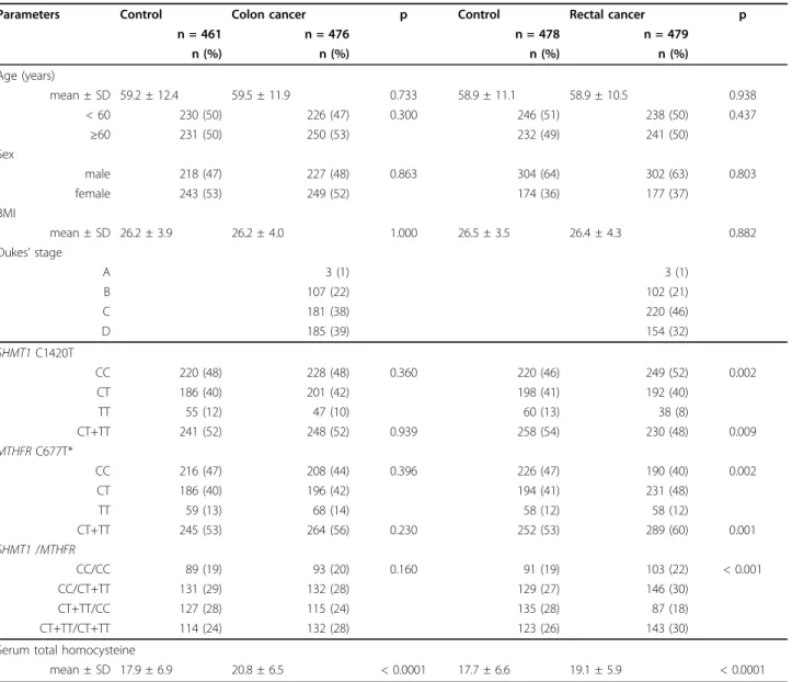
± 0.27; p < 0.0001 for colon and rectal cancer, respec- tively) than in controls (17.9 ± 0.32 and 17.7 ± 0.30) (Table 1). The distribution of SHMT1 C1420T and MTHFRC677T genotypes in controls and cases was in conformity with the HWE (SHMT1: controls p = 0.131 and colon cancer p = 0.843; controls p = 0.167 and rectal cancer p = 0.973; MTHFR: controls p = 0.074 and colon cancer p = 0.063; controls p = 0.126 and rectal cancer p = 0.403).
The univariate comparison of raw genotype distribu- tions of cases and controls revealed significant difference only in case of rectal cancer for bothSHMT1 C1420T and MTHFR C677T polymorphisms. An opposite
distribution shift from variant homozygotes toward wild type and from wild type to hetorozygotes was present in case of SHMT1 and MTHFR, respectively, hence the SHMT1 CT+TT/MTHFR CC diplotypes were signifi- cantly underrepresented among cases with rectal cancer (Table 1). The distribution of unpooled diplotype groups is provided in the Additional file 1.
No association was observed in colon cancer for SHMT1 and MTHFR CT or TT genotypes compared with the CC genotype (Table 2). In contrast with colon cancer, the adjusted risk ratio for rectal cancer was sig- nificantly lower for SHMT1TT and higher forMTHFR CT genotypes. A gene-dosage effect was observed only forSHMT1 with the progressively decreasing risk ratio with increasing number of T allele (p = 0.014).
Table 1 Selected characteristics and genotype frequencies ofSHMT1C1420T andMTHFRC677T polymorphisms of patients with colon cancer, rectal cancer and that of the respective controls
Parameters Control Colon cancer p Control Rectal cancer p
n = 461 n = 476 n = 478 n = 479
n (%) n (%) n (%) n (%)
Age (years)
mean ± SD 59.2 ± 12.4 59.5 ± 11.9 0.733 58.9 ± 11.1 58.9 ± 10.5 0.938
< 60 230 (50) 226 (47) 0.300 246 (51) 238 (50) 0.437
≥60 231 (50) 250 (53) 232 (49) 241 (50)
Sex
male 218 (47) 227 (48) 0.863 304 (64) 302 (63) 0.803
female 243 (53) 249 (52) 174 (36) 177 (37)
BMI
mean ± SD 26.2 ± 3.9 26.2 ± 4.0 1.000 26.5 ± 3.5 26.4 ± 4.3 0.882
Dukes’stage
A 3 (1) 3 (1)
B 107 (22) 102 (21)
C 181 (38) 220 (46)
D 185 (39) 154 (32)
SHMT1C1420T
CC 220 (48) 228 (48) 0.360 220 (46) 249 (52) 0.002
CT 186 (40) 201 (42) 198 (41) 192 (40)
TT 55 (12) 47 (10) 60 (13) 38 (8)
CT+TT 241 (52) 248 (52) 0.939 258 (54) 230 (48) 0.009
MTHFRC677T*
CC 216 (47) 208 (44) 0.396 226 (47) 190 (40) 0.002
CT 186 (40) 196 (42) 194 (41) 231 (48)
TT 59 (13) 68 (14) 58 (12) 58 (12)
CT+TT 245 (53) 264 (56) 0.230 252 (53) 289 (60) 0.001
SHMT1/MTHFR
CC/CC 89 (19) 93 (20) 0.160 91 (19) 103 (22) < 0.001
CC/CT+TT 131 (29) 132 (28) 129 (27) 146 (30)
CT+TT/CC 127 (28) 115 (24) 135 (28) 87 (18)
CT+TT/CT+TT 114 (24) 132 (28) 123 (26) 143 (30)
Serum total homocysteine
mean ± SD 17.9 ± 6.9 20.8 ± 6.5 < 0.0001 17.7 ± 6.6 19.1 ± 5.9 < 0.0001
* genotyping failed in 4 colon cancer patients
The stratified analysis according to age and sex revealed that the association of rectal cancer with these polymorphisms was present only in younger (< 60 year) or male subgroups (Table 2). Interestingly, in case of colon cancer an opposite effect of the presence of MTHFR variant allele was seen for younger (OR > 1) and older patients (OR < 1), however, the association was significant only in the former case. In the stratified analysis according to the BMI (< 25 vs≥25) there were non-significant differences only (data not shown).
Analyzing the association of cancer risk with diplo- types (Table 3) it was observed that the presence of SHMT1 T allele decreased the risk of rectal cancer only in case of wild type MTHFR. As had been expected from the results of genotype analysis the risk reducing effect of SHMT1 T allele was completely abolished when theMTHFRT allele was present. The ORs for all diplotypes is provided in the Additional file 1.
As the distribution of SHMT1C1420T genotypes are not available for the Hungarian population the European genotype frequencies and rectal cancer incidences were used instead to gauge any potential population stratifica- tion bias. The SHMT11420 CC+CT frequency in Eur- ope ranges from 0.833 to 0.913 [31], the rectal cancer incidence ranges from approximately 9 to 37 per 100 000 inhabitants in different countries in Europe [32].
Using the formula of Lee and Wang it was found that the upper bound for the bias is 1.29, which is less than Table 2 Overall, sex- and age-specific risk of colon and
rectal cancer according toSHMT1C1420T andMTHFR C677T polymorphisms
Colon cancer p Rectal cancer p
OR* (95% CI) p† OR* (95% CI) p†
SHMT11420
CC 1.00 (reference) 0.611 1.00 (reference) 0.014 CT 1.07 (0.81-1.41) 0.62 0.86 (0.66-1.13) 0.27 TT 0.86 (0.56-1.33) 0.49 0.57 (0.36-0.89) 0.013 CT+TT 1.02 (0.79-1.32) 0.90 0.80 (0.62-1.03) 0.08 MTHFR677
CC 1.00 (reference) 0.338 1.00 (reference) 0.083 CT 1.08 (0.81-1.42) 0.61 1.40 (1.06-1.84) 0.016 TT 1.19 (0.80-1.78) 0.39 1.14 (0.75-1.73) 0.53 CT+TT 1.11 (0.87-1.44) 0.43 1.35 (1.04-1.74) 0.024 Males
SHMT11420
CC 1.00 (reference) 0.334 1.00 (reference) 0.015 CT 0.93 (0.63-1.39) 0.73 0.92 (0.65-1.29) 0.63 TT 0.75 (0.40-1.39) 0.36 0.42 (0.23-0.75) 0.003 CT+TT 0.89 (0.61-1.30) 0.54 0.80 (0.58-1.10) 0.16 MTHFR677
CC 1.00 (reference) 0.121 1.00 (reference) 0.494 CT 1.36 (0.91-2.05) 0.14 1.45 (1.03-2.05) 0.034 TT 1.42 (0.80-2.50) 0.23 0.89 (0.52-1.51) 0.67 CT+TT 1.37 (0.94-2.00) 0.10 1.29 (0.94-1.79) 0.12 Females
SHMT11420
CC 1.00 (reference) 0.824 1.00 (reference) 0.390 CT 1.22 (0.83-1.80) 0.31 0.77 (0.49-1.22) 0.27 TT 0.94 (0.50-1.74) 0.83 0.97 (0.47-2.01) 0.93 CT+TT 1.13 (0.79-1.62) 0.50 0.80 (0.52-1.22) 0.29 MTHFR677
CC 1.00 (reference) 0.863 1.00 (reference) 0.052 CT 0.87 (0.59-1.28) 0.48 1.39 (0.88-2.19) 0.16 TT 1.07 (0.60-1.90) 0.82 1.81 (0.91-3.60) 0.09 CT+TT 0.93 (0.65-1.33) 0.68 1.45 (0.94-2.23) 0.09 Age < 60 years
SHMT11420
CC 1.00 (reference) 0.485 1.00 (reference) 0.002 CT 1.22 (0.82-1.82) 0.33 0.81 (0.55-1.19) 0.29 TT 0.67 (0.37-1.23) 0.19 0.32 (0.16-0.61) 0.0006 CT+TT 1.08 (0.74-1.57) 0.69 0.69 (0.48-0.99) 0.044 MTHFR677
CC 1.00 (reference) 0.047 1.00 (reference) 0.112 CT 1.72 (1.15-2.59) 0.008 1.97 (1.33-2.92) 0.0007 TT 1.37 (0.77-2.44) 0.29 0.98 (0.54-1.76) 0.94 CT+TT 1.65 (1.14-2.39) 0.008 1.67 (1.16-2.40) 0.005 Age≥60 years
SHMT11420
CC 1.00 (reference) 0.943 1.00 (reference) 0.763 CT 0.91 (0.62-1.34) 0.64 0.90 (0.61-1.32) 0.58 TT 1.13 (0.59-2.18) 0.71 1.04 (0.54-1.98) 0.91 CT+TT 0.92 (0.64-1.33) 0.67 0.92 (0.64-1.31) 0.63
Table 3 Colon and rectal cancer risk based onSHMT1 1420/MTHFR677 diplotypes
Diplotypes Colon cancer Rectal cancer
SHMT1/
MTHFR case/
control
OR* (95%
CI)
case/
control
OR* (95% CI)
CC/CC 93/89 1.00
(reference)
103/91 1.00
(reference) CC/CT+TT 132/131 0.95 (0.65-
1.39)
146/129 0.99 (0.69- 1.44) CT+TT/CC 115/127 0.87 (0.59-
1.27)
87/135 0.57 (0.39- 0.84)† CT+TT/CT+TT 132/114 1.10 (0.75-
1.62)
143/123 1.02 (0.71- 1.48)
* adjusted for age, sex and BMI;†different from reference (p = 0.005) or CT +TT/CT+TT (p = 0.002)
Table 2: Overall, sex- and age-specific risk of colon and rectal cancer according to SHMT1C1420T andMTHFR C677T polymorphisms(Continued)
MTHFR677
CC 1.00 (reference) 0.489 1.00 (reference) 0.432 CT 0.70 (0.47-1.03) 0.07 1.03 (0.70-1.52) 0.89 TT 0.99 (0.56-1.73) 0.96 1.33 (0.73-2.42) 0.35 CT+TT 0.75 (0.52-1.08) 0.13 1.08 (0.74-1.56) 0.70
* adjusted for BMI, sex and age for overall risk, BMI and age for sex-specific risk, BMI and sex for age-specific risk;†test of gene-dosage effect
1/0.55 = 1.82, the estimated odds ratio observed in our study for carriers of SHMT1 1420C allele in Hungary.
In the case ofMTHFR C677T, we have found 12 inde- pendent publications for the Hungarian population, thus the range of CT+TT frequencies were in the range of 0.412-0.7. The rectal cancer incidence in Hungary ranges from approximately 32 to 38 per 100,000 indivi- duals, thus the upper bound for the bias is 1.05, which is less than 1.38 found for Hungarian MTHFR 677T allele carriers.
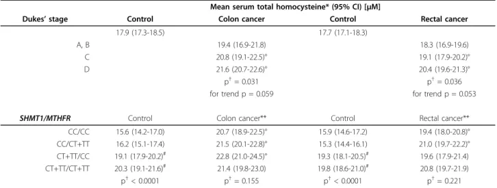
In order to investigate the effect ofSHMT1 C1420T andMTHFR C677T polymorphisms the age, sex and BMI adjusted mean HCY levels in different diplotypes were compared in controls and patients. The HCY levels of patients were also adjusted for Dukes’ stage, because the mean HCY levels were significantly higher in advanced stages of the disease (Table 4). In controls the presence of SHMT1 variant allele resulted in signifi- cantly higher HCY levels while this effect could not be observed in patients. The MTHFR T allele had no unequivocal effect on HCY levels. The adjusted mean HCY levels of all diplotypes are presented in the Addi- tional file 1.
Discussion
In our study both investigated polymorphisms,SHMT1 C1420T andMTHFRC677T, exert influence on the risk of rectal but not on that of colon cancer. Previous stu- dies underline the capacity of SHMT1 C1420T poly- morphism being directly related to cancer susceptibility.
Skibola et al. found risk reduction for the SHMT1 1420TT genotype in adult acute lymphocytic leukemia [7]. The same result was found by Hishida et al. in
malignant lymphomas [8]. Recently, it was observed that in a North Chinese population there was a reduced risk of esophageal squamous cell carcinoma and gastric car- dia adenocarcinoma in the case ofSHMT1 1420CT het- erozygotes compared to C homozygotes [33]. In the above mentioned studies the risk reduction influenced by the presence of SHMT11420T allele was in accor- dance with our findings regarding rectal cancer. The variant SHMT1 enzyme may result in decreased produc- tion of 5,10-MTHF and accumulation of tetrahydrofo- late, although the exact biological effect of this phenomenon or the complete mechanism leading to carcinogenesis is not yet known.
In a case-control study van den Donk et al. could not demonstrate association between colorectal adenoma risk andSHMT1 C1420T polymorphism. Unfortunately, the authors performed a stratified analysis according to sex and not to the adenoma localization [9].
Chenet al. could not prove any risk-reducing effect of the variant typeSHMT1C1420T polymorphism in case of CRC [10]. In his study only male cases and male hos- pital-based controls were used from Caucasian-Ameri- can populations, but the rectal and colon cancer patients were not separately analyzed, thus the risk modifying effect of theSHMT1 C1420T polymorphism might be obscured. Guerreiroet al. found an increased risk of CRC in case ofSHMT1 1420 C allele, although the strength of their observation is limited by the small number of variant homozygotes (n = 9) and moreover, the rectal and colon cancer patients were analyzed together [11]. Stecket al. found a borderline statistically significant decreased risk of colon cancer in whites, but not African Americans, with the SHMT 1420TT
Table 4 Mean serum total homocysteine levels according to Dukes’stage andSHMT11420/MTHFR677diplotypes in colon and rectal cancer patients and respective controls
Mean serum total homocysteine* (95% CI) [μM]
Dukes’stage Control Colon cancer Control Rectal cancer
17.9 (17.3-18.5) 17.7 (17.1-18.3)
A, B 19.4 (16.9-21.8) 18.3 (16.9-19.6)
C 20.8 (19.1-22.5)° 19.1 (17.9-20.2)°
D 21.6 (20.7-22.6)° 20.4 (19.6-21.3)°
p†= 0.031 p†= 0.036
for trend p = 0.059 for trend p = 0.053
SHMT1/MTHFR Control Colon cancer** Control Rectal cancer**
CC/CC 15.6 (14.2-17.0) 20.7 (18.9-22.5)° 15.9 (14.6-17.2) 19.4 (18.0-20.8)°
CC/CT+TT 16.2 (15.1-17.4) 21.5 (20.1-22.8)° 15.3 (14.4-16.1) 21.0 (19.7-22.2)°
CT+TT/CC 19.1 (17.9-20.2)# 22.8 (21.0-24.5)° 19.3 (18.1-20.5)# 19.6 (17.9-21.4)
CT+TT/CT+TT 20.3 (19.1-21.6)# 21.4 (19.8-23.0) 19.8 (18.6-21.0)# 20.8 (19.7-21.9)
p†< 0.0001 p†= 0.155 p†< 0.0001 p†= 0.221
* adjusted for age, sex and BMI; ** also adjusted for Dukes’stage;†Kruskal-Wallis one-way ANOVA;
#different from CC/CC or CC/CT+TT, Z-value test p < 0.05;° different from control, Mann-Whitney U-test p < 0.01
genotype as compared to the CC genotype. High folate intake reduced the risk of colon cancer in all genotypes.
In this study the control group for whites deviated strongly from HWE (p = 0.0042) [12].
The presence of theMTHFR677T allele represented a higher rectal, but not colon cancer risk compared to CC homozygotes. The relationship of MTHFRC677T poly- morphism and CRC risk based on the results of pre- vious meta- and pooled analyses remains unclarified [34,35]. It has to be mentioned that most of the studies investigating theMTHFRC677T polymorphisms did not separate rectal and colon cancer patients [35]. Recently, Caoet al. found that in males, among MTHFR677TT genotype carriers, the OR for colon cancer was 2.42, but that of for rectal cancer was 0.52 [19]. Their result was similar to ours regarding males older than 60 years with ORs 1.4 and 0.9 for colon and rectal cancer, respec- tively. The differing genotype distribution of MTHFR C677T in Chinese and Caucasian, the different lifestyle, etc. may account for the more modest difference.
In accordance with the explanations raised by Guer- reiroet al., - regarding the controversial results of pre- vious published studies about the CRC risk modified by the interaction of folate intake and MTHFR 677 poly- morphism -, we accept the following statement: the low folate intake and the presence ofMTHFR677T allele or TT genotype predicts if there is an increased risk of CRC, while on the contrary high/adequate folate intake results in a risk reduction if the variant allele is present [11]. In Hungary the folate intake is generally low [36], thus our result regarding the rectal cancer risk is in accordance with the previous statement, however, for colon cancer this relation can be found only in case of younger individuals (< 60 years). Iacopettaet al. demon- strated an increased proximal, but not distal CRC risk in the presence ofMTHFR 677T allele and low folate intake or older (≥65 years) individuals. Possible reasons for the discrepancy between their and our findings might include the classification of cancers as colon or rectal and the investigated Australian population con- sisted not only of Caucasians [37].
In the present study the combination of the SHMT1 1420CT+TT and MTHFR677CC genotypes was found to imply the lowest risk for rectal cancer. In a recent study an interactive influence of MTHFR C677T and SHMT1 C1420T polymorphisms in the risk of esopha- geal and gastric carcinomas was also observed [33].
Decreased activity of SHMT1 and the unaltered activity of MTHFR may result in a decreased amount of 5,10- MTHF available for pyrimidine synthesis. The combina- tion with the highest risk is characterized by high SHMT1 activity and decreased MTHFR activity, which resulted in an increased availability of folate for pyrimi- dine synthesis. The impact of the examined
polymorphisms on the DNA methylation is very com- plex. The decreased activity of SHMT1 may lead to a decreased DNA methylation through a negative feed- back chain accumulation of intermediers and decreased 5-methylTHF production.
Our study also suggests the role of gender differences in the etiology of rectal and colon cancer. Female hor- mones may influence the susceptibility for second pri- mary colorectal cancer [38]. Moreover, a statistically significant association was found between the DNA mis- match repair gene MSH2-118T > C polymorphism and a strong family history of CRC and this association was seen only in female but not male CRC patients [39].
The age of onset of CRC may also be influenced by polymorphisms [40]. An age-specific association was also observed for colon cancer but not for rectal cancer in case of apolipoprotein E (apoE) polymorphism [41].
The association between BMI and colon cancer described in a previous large cohort and a pooled study [16,42] was not demonstarted in our analysis, which could be due to the insufficient statistical power. The above studies also demonstrated sex-specific and age- specific associations.
Gauging the population stratification bias for both polymorphisms it can be concluded that our findings are unlikely to be biased. However, it should be consid- ered that in case ofSHMT1 the genotype distributions and CRC incidence used for calculations were not avail- able for Hungarian, but only for the European Caucasian population.
Homocysteine is produced during the methionine dependent DNA methylation. For its remethylation the product of the MTHFR enzyme, the 5-methylTHF, is essential. The less important metabolic transformation of homocystein implies cystathion beta-synthase or liver betain-homocysteine methyltransferase. Considering the important functional role of SHMT1 in the production of methyl group for multiple metabolic pathways any disturbance in the protein expression due to the poly- morphisms could result in the reduction of the available one-carbon units that is also necessary for the remethy- lation of homocysteine. Geiselet al. found non-signifi- cant elevated HCY levels in theSHMT11420TT group in senior healthy subjects [43]. Chen et al. reported sig- nificantly higher HCY levels in case of SHMT11420 CT genotypes in a study including healthy male subjects [10]. Similarly, in our study the presence of SHMT1420 T allele significantly increased the HCY levels in con- trols. In relative young healthy individuals (20-40 years) Pereiraet al. [21] and Bailyet al. [22] found significantly higher HCY levels in MTHFR 677 TT genotypes com- pared to the other genotypes. Almost the same result was found by Semmler et al. [44] with the exception that the above association could not be demonstrated
for older individuals (> 55 years). This latter data sup- port our results that in controls (mean age 59 years) there were no association between HCY levels and MTHFR677 polymorphism.
The influence of the investigated polymorphisms on HCY levels in case of patients may be obscured because of the much narrow distribution of HCY levels (coeffi- cient of variation lowered by ~20%) than in controls.
The effect is likely to be in a relationship with a yet unclarified HCY-increasing mechanism. Proliferating tumor cells appear to be the main cause of accumula- tion of HCY and thus, the development of hyperhomo- cysteinaemia in cancer patients [45]. The tumor marker character of the HCY, namely the elevated HCY levels in cancer patients [45-54], the elevated HCY levels in advanced clinical stages [49,53] and the usefulness of HCY to monitor therapeutic effects [48,49,54,55] has also been demonstrated. Moreover, HCY concentration followed CEA levels in CRC patients [55]. This effect might be strongly related to the malignant disease pro- gression as in our study the HCY gradually increased with the lymph node involvement (Dukes’stage C) and further on with the presence of distant metastases (Dukes’stage D).
In our study it seems at a first glance that HCY in Dukes’stage C and D is a tumor marker as it was sug- gested by Wuet al. [27], but if theSHMT11420 poly- morphism is taken into account then HCY can be considered as tumor marker only in case of wild (CC) genotypes. This fact remains valid even for patients of Dukes’D stage presenting the highest HCY levels (data not shown). Similarly, Battistelliet al. presented signifi- cantly elevated HCY levels compared to controls only in case of MTHFR 677 CC+CT genotypes [50].
An interesting question would be why the HCY levels of rectal cancer patients in case of the “lowest rectal cancer risk"-presenting diplotype are not significantly higher compared to that of the controls as it can be seen in case of colon cancers. Could an unknown HCY- lowering mechanism confer resistance in this context against rectal carcinogenesis? Based on present results, it might be supposed, that in case of the risk-reducing diplotypes the low HCY compared to controls is directly responsible for reduced risk. On the other hand the low HCY could be an ancillary result and other mechanisms may lead to the reduced risk. Recently it was demon- strated [56] that if the variantSHMT1is localized in the nucleus only an impairedde novothymidylate biosynth- esis is assured. The thymidylate biosynthesis is further diminished by the wild-type-MTHFR-transformed and thus depleted 5,10-MTHF levels. The impaired thymidy- late biosynthesis is unfavourable for cell proliferation.
These results suggest that the association between the elevated HCY levels and the cancer risk is not obvious
and first of all the “tumor marker”aspect of HCY needs to be underlined.
Conclusions
This is the first study presenting the protective effect of SHMT1 1420T allele orSHMT11420 T allele/MTHFR 677 CC diplotype against rectal cancer risk. SHMT1 1420 variant significantly increase HCY levels in con- trols but not patients. HCY could be considered tumor marker only in wild-type (CC) SHMT1 1420 CRC patients in Dukes’stage C and D. Higher HCY levels are characteristics of patients in advanced stages of the dis- ease. Further studies need to be conducted to reveal the complex role of SHMT1, MTHFR and other folate enzyme polymorphisms in colon and rectal carcinogen- esis. The importance of HCY level also need to be clarified.
Additional material
Additional file 1: Distribution, cancer risk and serum total homocysteine level of diplotypes. This file contains the distribution, cancer risk (OR and 95% CI) and mean serum total homocysteine (95%
CI) level of unpooledSHMT11420/MTHFR677 diplotypes.
Acknowledgements
This study was supported by the National Research and Development Programme (NKFP1-00024/2005) grant. Authors are grateful to I. Gaudi for statistical support and to the co-workers of the Department of Human and Experimental Pathology of the NIO, Budapest, Hungary for the
histopathological staging of the patients. The assistance of Dr. A. Hajnal, Serologic Laboratory of the National Blood Transfusion Service, Budapest, Hungary in providing control blood samples is acknowledged.
Author details
1School of PhD studies, Pathological Sciences, Semmelweis University, Budapest, Hungary.2Department of Clinical Research, National Institute of Oncology, Budapest, Hungary.3Medical Department,“Szent István és Szent László”Hospital, Budapest, Hungary.4Department of Cytogenetics and Immunology, National Institute of Chemical Safety, Budapest, Hungary.
5Department of Environmental Epidemiology, National Institute of Environmental Health, Budapest, Hungary.6Department of Molecular Environmental Epidemiology, National Institute of Environmental Health, Budapest, Hungary.7Second Department of Pediatrics, Semmelweis University, Budapest, Hungary.8Department of Dermatology, Venerology and Dermatooncology, Semmelweis University, Budapest, Hungary.
Authors’contributions
VK, VA and BB carried out the molecular genetic studies, ÉP and AR carried out the HPLC assays, VK, JK and BB participated in the design of the study and helped to draft the manuscript, EH, ÉS, AB, PR, BS and BT participated in the enrollment and conduct of the study, JM, SO and MK participated in study coordination and revised the manuscript, VK and BB conceived the study, participated in its design and statistical analyses. All authors read and approved the final manuscript.
Competing interests
The authors declare that they have no competing interests.
Received: 16 March 2010 Accepted: 4 October 2010 Published: 4 October 2010
References
1. Giovannucci E:Epidemiologic studies of folate and colorectal neoplasia: a review.J Nutr2002,132:2350S-2355S.
2. van Guelpen B, Hultdin J, Johansson I, Hallmans G, Stenling R, Riboli E, Winkvist A, Palmqvist R:Low folate levels may protect against colorectal cancer.Gut2006,55:1461-1466.
3. Sharp L, Little J:Polymorphisms in genes involved in folate metabolism and colorectal neoplasia: a HuGE review.Am J Epidemiol2004, 159:423-443.
4. Ulvik A, Vollset SE, Hansen S, Gislefoss R, Jellum E, Ueland PM:Colorectal cancer and the methylenetetrahydrofolate reductase 677C- > T and methionine synthase 2756A- > G polymorphisms: a study of 2,168 case- control pairs from the JANUS cohort.Cancer Epidemiol Biomarkers Prev 2004,13:2175-2180.
5. Chen K, Jiang QT, He HQ:Relationship between metabolic enzyme polymorphisms and colorectal cancer.World J Gastroenterol2005, 11:331-335.
6. Hubner RA, Houlston RS:MTHFR C677T and colorectal cancer risk: a meta-analysis of 25 populations.Int J Cancer2006,120:1027-1035.
7. Skibola CF, Smith MT, Hubbard A, Shane B, Roberts AC, Law GR, Rollinson S, Roman E, Cartwright RA, Morgan GJ:Polymorphisms in the thymidylate synthase and serine hydroxymethyltransferase genes and risk of adult acute lymphocytic leukemia.Blood2002,99:3786-3791.
8. Hishida A, Matsuo K, Hamajima N, Ito H, Ogura M, Kagami Y, Taji H, Morishima Y, Emi N, Tajima K:Associations between polymorphisms in the thymidylate synthase and serine hydroxymethyltransferase genes and susceptibility to malignant lymphoma.Haematologica2003, 88:159-166.
9. van den Donk M, Visker MH, Harryvan JL, Kok FJ, Kampman E:Dietary intake of B-vitamins, polymorphisms in thymidylate synthase and serine hydroxymethyltransferase 1, and colorectal adenoma risk: a Dutch case- control study.Cancer Lett2007,250:146-153.
10. Chen J, Kyte C, Valcin M, Chan W, Wetmur JG, Selhub J, Hunter DJ, Ma J:
Polymorphisms in the one-carbon metabolic pathway, plasma folate levels and colorectal cancer in a prospective study.Int J Cancer2004, 110:617-620.
11. Guerreiro CS, Cravo M, Costa AR, Miranda A, Tavares L, Moura-Santos P, MarquesVidal P, Nobre Leitão C:Risk of colorectal cancer associated with the C677T polymorphism in 5,10-methylenetetrahydrofolate reductase in Portuguese patients depends on the intake of methyl-donor nutrients.Am J Clin Nutr2008,88:1413-1418.
12. Steck SE, Keku T, Butler LM, Galanko J, Massa B, Millikan RC, Sandler RS:
Polymorphisms in methionine synthase, methionine synthase reductase and serine hydroxymethyltransferase, folate and alcohol intake, and colon cancer risk.J Nutrigenet Nutrigenomics2008,1:196-204.
13. Kapiteijn E, Liefers GJ, Los LC, Kranenbarg EK, Hermans J, Tollenaar RA, Moriya Y, van de Velde CJ, van Krieken JH:Mechanisms of oncogenesis in colon versus rectal cancer.J Pathol2001,195:171-178.
14. Howard RA, Freedman DM, Park Y, Hollenbeck A, Schatzkin A,
Leitzmann MF:Physical activity, sedentary behavior, and the risk of colon and rectal cancer in the NIH-AARP Diet and Health Study.Cancer Causes Controls2008,19:939-953.
15. Frattini M, Balestra D, Suardi S, Oggionni M, Alberici P, Radice P, Costa A, Daidone MG, Leo E, Pilotti S, Bertario L, Pierotti MA:Different genetic features associated with colon and rectal carcinogenesis.Clin Cancer Res 2004,10:4015-4021.
16. Adams KF, Leitzmann MF, Albanes D, Kipnis V, Mouw T, Hollenbeck A, Schatzkin A:Body mass and colorectal cancer risk in the NIH-AARP Cohort.Am J Epidemiol2007,166:36-45.
17. Le Marchand L, Wilkens LR, Kolonel LN, Henderson BE:The MTHFR C677T polymorphism and colorectal cancer: the multiethnic cohort study.
Cancer Epidemiol Biomarkers Prev2005,14:1198-1203.
18. Kim DH, Ahn YO, Lee BH, Tsuji E, Kiyohara C, Kono S:
Methylenetetrahydrofolate reductase polymorphism, alcohol intake, and risks of colon and rectal cancers in Korea.Cancer Lett2004,216:199-205.
19. Cao HX, Gao CM, Takezaki T, Wu JZ, Ding JH, Liu YT, Li SP, Su P, Cao J, Hamajima N, Tajima K:Genetic polymorphisms of
methylenetetrahydrofolate reductase and susceptibility to colorectal cancer.Asian Pac J Cancer Prev2008,9:203-208.
20. Gudnason V, Stansbie D, Scott J, Bowron A, Nicaud V, Humphries S:C677T (thermolabile alanine/valine) polymorphism in
methylenetetrahydrofolate reductase (MTHFR): its frequency and impact on plasma homocysteine concentration in different European populations. EARS group.Atherosclerosis1998,136:347-354.
21. Pereira AC, Schettert IT, Morandini AAF, Guerra-Shinohara EM, Krieger JE:
Methylenetetrahydrofolate reductase (MTHFR) c677t gene variant modulates the homocysteine folate correlation in a mild folate deficient population.Clin Chim Acta2004,340:99-105.
22. Bailey LB, Duhaney RL, Maneval DR, Kauwell GP, Quinlivan EP, Davis SR, Cuadras A, Hutson AD, Gregory JF:Vitamin B-12 status is inversely associated with plasma homocysteine in young women with C677T and/or A1298C methylenetetrahydrofolate reductase polymorphisms.J Nutr2002,132:1872-1878.
23. Sassi S, Cosmi B, Palareti G, Legnani C, Grossi G, Musolesi S, Coccheri S:
Influence of age, sex and vitamin status on fasting and post-methionine load plasma homocysteine levels.Haematologica2002,87:957-964.
24. Marchesini G, Manini R, Bianchi G, Sassi S, Natale S, Chierici S, Visani F, Baraldi L, Forlani G, Melchionda N:Homocysteine and psychological traits:
a study in obesity.Nutrition2002,18:403-407.
25. Parsons DS, Reaveley DA, Pavitt DV, Brown EA:Relationship of renal function to homocystein and lipoprotein(a) levels: The frequency of the combination of both risk factors in chronic renal impairment.Am J Kidney Dis2002,40:916-923.
26. Valik D, Radina M, Sterba J, Vojtesek B:Homocysteine: exploring its potential as a pharmacodynamic biomarker of antifolate chemotherapy.
Pharmacogenomics2004,5:1151-1162.
27. Wu LL, Wu JT:Hyperhomocysteinemia is a risk factor for cancer and a new potential tumor marker.Clin Chim Acta2002,322:21-28.
28. Oikawa S, Murakami K, Kawanishi S:Oxidative damage to cellular and isolated DNA by homocysteine: implications for carcinogenesis.
Oncogene2003,22:3530-3538.
29. Frosst P, Blom HJ, Milos R, Goyette P, Sheppard CA, Matthews RG, Boers GJH, den Heijer M, Kluijtmans LAJ, van den Heuve LP, Rozen R:A candidate genetic risk factor for vascular disease: a common mutation in methylenetetrahydrofolate reductase.Nat Genet1995,10:111-113.
30. Lee WC, Wang LY:Simple formulas for gauging the potential impacts of population stratification bias.Am J Epidemiol2007,167:86-89.
31. Database of Single Nucleotide Polymorphisms (dbSNP). Bethesda (MD):
National Center for Biotechnology Information, National Library of Medicine. dbSNP accession: {rs1979277 and rs1801133}, (dbSNP Build ID: 129) [http://www.ncbi.nlm.nih.gov/SNP/].
32. Ferlay J, Bray F, Pisani P, Parkin DM:GLOBOCAN 2002: Cancer incidences, mortality and prevalence worldwide. IARC Cancer Base No. 5 version 2.0. Lyon, France: IARC Press 2004.
33. Wang Y, Guo W, He Y, Chen Z, Wen D, Zhang X, Wang N, Li Y, Ge H, Zhang J:Association of MTHFR C677T and SHMT(1) C1420T with susceptibility to ESCC and GCA in a high incident region of Northern China.Cancer Causes Control2007,18:143-152.
34. Taioli E, Garza MA, Ahn YO, Bishop DT, Bost J, Budai B, Chen K, Gemignani F, Keku T, Lima CSP, Le Marchand L, Matsuo K, Moreno V, Plaschke J, Pufulete M, Thomas SB, Toffoli G, Wolf CR, Moore CG, Little J:
Meta- and pooled analyses of methylenetetrahydrofolate reductase (MTHFR) C677T polymorphism and colorectal cancer: a HuGE-GSEC review.Am J Epidemiol2009,170:1207-1221.
35. Huang Y, Han S, Li Y, Mao Y, Xie Y:Different roles of MTHFR C677T and A1298C polymorphisms in colorectal adenoma and colorectal cancer: a meta-analysis.J Hum Genet2007,52:73-85.
36. Zajkás G, Bíró L, Greiner E, Szórád I, Ágoston H, Balázs A, Vitrai J, Hermann D, Boros J, Németh R, Kéki Z, Martos É:Dietary survey in Hungary, 2003-2004. Micronutrients: vitamins.Orv Hetil2007, 148:1593-1600.
37. Iacopetta B, Heyworth J, Girschik J, Grieu F, Clayforth C, Fritschi L:The MTHFR C677T andΔDNMT3B C-149T polymorphism confer different risks for right- and left-sided colorectal cancer.Int J Cancer2009, 125:84-90.
38. Liang W:Age sex and the risk of grade-specific second primary colorectal cancer: Evidence for the protective effect of female hormone.
Eur J Cancer2007,43:1856-1861.
39. Mrkonjic M, Raptis S, Green RC, Monga N, Daftary D, Dicks E, Younghusband HB, Parfrey PS, Gallinger SS, McLaughlin JR, Knight JA, Bapat B:MSH2 118T > C and MSH6 159C > T promoter polymorphisms and the risk of colorectal cancer.Carcinogenesis2007,28:2575-2580.
40. Talseth BA, Ashton KA, Meldrum C, Suchy J, Kurzawski G, Lubinski J, Scott RJ:Aurora-A and Cyclin D1 polymorphisms and the age of onset of colorectal cancer in hereditery nonpoliposis colorectal cancer.Int J Cancer2007,122:1273-1277.
41. Slattery ML, Sweeney C, Murtaugh M, Ma KN, Potter JD, Levin TR, Samowitz W, Wolff R:Associations between apoE genotype and colon and rectal cancer.Carcinogenesis2005,26:1422-1429.
42. Jacobs ET, Ahnen DJ, Ashbeck EL, Baron JA, Greenberg ER, Lance P, Lieberman DA, McKeown-Eyssen G, Schatzkin A, Thompson PA, Martínez ME:Association between body mass index and colorectal neoplasia at follow-up colonoscopy: a pooling study.Am J Epidemiol 2009,169:657-666.
43. Geisel J, Hübner U, Bodis M, Schorr H, Knapp JP, Obeid R, Herrmann W:The role of genetic factors in the development of hyperhomocysteinemia.
Clin Chem Lab Med2003,41:1427-1434.
44. Semmler A, Moskau S, Stoffel-Wagner B, Weller M, Linnebank M:The effect of MTHFR c.677C > T on plasma homocysteine levels depends on health, age and smoking.Clin Invest Med2009,32:E310-E314.
45. Schroecksnadel K, Frick B, Fiegl M, Winkler C, Denz HA, Fuchs D:
Hyperhomocysteinaemia and immune activation in patients with cancer.
Clin Chem Lab Med2007,45:47-53.
46. Ferroni P, Palmirotta R, Martini F, Riondino S, Savonarola A, Spila A, Ciatti F, Sini V, Mariotti S, Del Monte G, Roselli M, Guadagni F:Determinants of homocysteine levels in colorectal and breast cancer patients.Anticancer Res2009,29:4131-4138.
47. Almadori G, Bussu F, Galli J, Cadoni G, Zappacosta B, Persichilli S, Minucci A, Giardina B:Serum folate and homocysteine levels in head and neck squamous cell carcinoma.Cancer2002,94:1006-1011.
48. Refsum H, Wesenberg F, Ueland PM:Plasma homocysteine in children with acute lymphoblastic leukemia: changes during a chemotherapeutic regimen including methotrexate.Cancer Res1991,51:828-835.
49. Ozkan Y, Yardim-Akaydin S, Firat H, Calişkan-Can E, Ardiç S, Simşek B:
Usefulness of homocysteine as a cancer marker: total thiol compounds and folate levels in untreated lung cancer patients.Anticancer Res2007, 27:1185-1189.
50. Battistelli S, Vittoria A, Stefanoni M, Bing C, Roviello F:Total plasma homocysteine and methylenetetrahydrofolate reductase C677T polymorphism in patients with colorectal carcinoma.World J Gastroenterol2006,12:6128-6132.
51. Eleftheriadou A, Chalastras T, Ferekidou E, Yiotakis I, Kyriou L, Tzagarakis M, Ferekidis E, Kandiloros D:Association between squamous cell carcinoma of the head and neck and serum folate and homocysteine.Anticancer Res2006,26:2345-2348.
52. Zacho J, Yazdanyar S, Bojesen SE, Tybjærg-Hansen A, Nordestgaard BG:
Hyperhomocysteinemia, methylenetetrahydrofolate reductase c.677C >
T polymorphism, and risk of cancer: cross-sectional and prospective studies and meta-analyses of 75,000 cases and 93,000 controls.Int J Cancer2010.
53. Gatt A, Makris A, Cladd H, Burcombe RJ, Smith JM, Cooper P, Thompson D, Makris M:Hyperhomocysteinemia in women with advanced breast cancer.Int J Lab Hematol2007,29:421-425.
54. Ruud E, Holmstrøm H, Brosstad F, Wesenberg F:Diagnostic value of family histories of thrombosis to identify children with thrombophilia.Pediatr Hematol Oncol2005,22:453-462.
55. Melichar B, Kalábová H, Krcmová L, Kasparová M, Malírová E, Melicharová K, Pecka M, Hyspler R, Solichová D:Serum homocysteine, cholesterol, retinol, alpha-tocopherol, glycosylated hemoglobin and inflammatory response during therapy with bevacizumab, oxaliplatin, 5-fluorouracil and leucovorin.Anticancer Res2009,29:4813-4820.
56. Anderson DD, Stover PJ:SHMT1 and SHMT2 are functionally redundant in nuclearde novothymidylate biosynthesis.PLoS ONE2009,4:e5839.
Pre-publication history
The pre-publication history for this paper can be accessed here:
http://www.biomedcentral.com/1471-2407/10/525/prepub
doi:10.1186/1471-2407-10-525
Cite this article as:Komlósiet al.:SHMT11420 andMTHFR677 variants are associated with rectal but not colon cancer.BMC Cancer201010:525.
Submit your next manuscript to BioMed Central and take full advantage of:
• Convenient online submission
• Thorough peer review
• No space constraints or color figure charges
• Immediate publication on acceptance
• Inclusion in PubMed, CAS, Scopus and Google Scholar
• Research which is freely available for redistribution Submit your manuscript at
www.biomedcentral.com/submit