72
Combined application of Raman spectroscopy and focused ion beam scanning electron microscopy on silicate melt inclusions to track metasomatism in spinel
peridotite xenoliths
Liptai, N.1,2*, Berkesi, M.1,3 .1,3, Griffin, W.L.4 4 & .1,3
1 2Oscope Research Group, Hungary; 2MTA CSFK Geodesic and Geophysical
Institute; 3Lithospher ; 4ARC Centre of Excellence for
Core to Crust Fluid Systems (CCFS) and GEMOC, Macquarie University, Australia; *liptai.nora@csfk.mta.hu Silicate melt inclusions (SMIs) play a key role in
mantle research as their composition represents characteristics of the melt present in the mantle at the time of entrapment, therefore they can preserve valuable information about metasomatic processes. There are several methods to study SMIs, but the combination of Raman spectroscopy with focused ion beam scanning electron microscopy (FIB-SEM) provides an exceptional opportunity to identify daughter phases, obtain their major element compositions and reconstruct a 3D model of the inclusion.
In this study we present analyses on partially crystallised primary SMIs hosted in clinopyroxene in a metasomatised spinel peridotite xenolith from
-
northern part of the Pannonian Basin in Central Europe. Xenoliths from this locality have been affected by several different metasomatic events prior to entrainment by the host basalt (Liptai et al., 2017). The youngest of these events is a melt-rock reaction which transformed the lherzolite wallrock d on the petrology of the xenolith featured in this study, the analysed SMIs have trapped the melt which can be linked to this metasomatism.
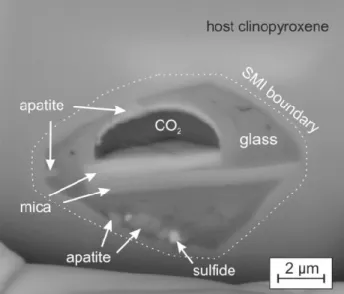
Raman spectroscopy and imaging revealed the presence of apatite, clinopyroxene, sulphates (anhydrite and barite) and amphibole within the inclusions. The volatile bubble is composed of CO2. Slicing with FIB-SEM revealed additional phases such as spinel, sulphide and mica (Fig. 1).
Geochemical analyses and 3D reconstruction of the inclusions allowed the calculation of the bulk major element composition of the SMIs, and LA- ICP-MS analyses on other inclusions of the same host clinopyroxene grain provided trace-element concentrations as well.
Based on the obtained bulk geochemistry, the melt trapped in the inclusions is enriched in Fe and has OIB-type characteristics. This is in agreement with the metasomatic process related to the formation of the wehrlites, which is inferred to have been caused by an intraplate mafic melt, similar to the host basalt of the xenoliths. Pre-entrapment evolution and reaction with the lherzolite wallrock resulted in intermedier melt composition.
Petrogenetic modeling showed that the melt was generated with a very small degree of partial melting of a garnet lherzolite source. Following the entrapment and partial crystallisation, a volatile bubble exsolved from the residual glass during
ascent at shallow depths, as suggested by homogeneous CO2-densities. Small crystals such as sulphates and mica, formed probably in a late stage at the boundary of the bubble and the glass, indicate the presence of S and possibly water in the original bubble composition.
Acknowledgement
This study was supported by a PhD scholarship of University (Hungary) to N. Lipta
Postdoctoral Research Fellowship of the Hungarian Pannon LitH2Oscope Research Group.
References
Liptai N. et al. (2017) J. Petrol. 58:1107-1144.
934.
Fig. 1. Backscattered electron image of an SMI slice obtained during FIB-SEM analyses.