DOI: 10.26430/CHUNGARICA.2021.51.2.109 Cardiologia Hungarica
2021; 51: 109–117.
$Np]LUDWiQpUNH]HWWDV]HUNHV]WĘVpJEHiQNHUOWHOIRJDGiVUD
Review
Genetic diagnosis in hypertrophic cardio-
myopathy: two steps forward, one step back
Lidia Hategan, Beáta Csányi, János Borbás, Eszter Dalma Pálinkás, Hedvig Takács, Viktória Nagy, Róbert Sepp
University of Szeged, Faculty of Medicine, Department of Internal Medicine and Cardiology Center, Szeged Levelezési cím:
Prof. dr. Sepp Róbert, Szegedi Tudományegyetem, Belgyógyászati Klinika, Non-Invazív Kardiológiai Részleg 6725 Szeged, Semmelweis u 8. E-mail: sepprobert@gmail.com
Hypertrophic cardiomyopathy (HCM) is the most common genetic cardiovascular disorder worldwide which ex- hibits considerable genetic heterogeneity. Widespread utilization of next-generation sequencing (NGS) in HCM has uncovered substantial genetic variation and highlighted the importance of a standardized approach to variant interpretation. According to this, accurate and consistent interpretation of sequence variants is essential for effec- tive clinical care for individuals and their families with HCM.
With this regard, the 2015 guidelines from the American College of Medical Genetics and Genomics and the As- sociation for Molecular Pathology (ACMG/AMP) were widely applicable, but several elements lacked specificity for given genes or diseases. The latter guideline was adapted for the most frequent causative HCM gene, the beta myosin heavy chain gene 0<+ by the ClinGen (Clinical Genome Resource) expert panel, the Inherited Cardi- omyopathy Expert Panel. Due to the adaptation, the guideline became gene-specific, with general considerations which are widely adaptable for most of the causative genes in HCM. Based on the modified guideline, web-based interpretation algorithms have been developed which integrate data from population databases and define pat- hogenicity of different variants independent of the observer, therefore aiding standardized clinical interpretation of genetic testing. The latter approach serves as a basis for recommendation for genetic testing in the recent ACC/
AHA HCM guideline published in 2020.
The current review is meant to compile the latest advances in HCM genetic testing in clinical practice, while brin- ging into focus some of the ongoing challenges clinical geneticists are still facing. Although nowadays the inter- pretation of genetic findings is two steps closer to a more accurate approach due to gene adaptation and auto- matization, the multitude of putative causative genes have been once again reduced to the 8 sarcomere genes, a backward step.
hypertrophic cardiomyopathy, genetic testing, variant interpretation, gene curation, automatic web tools .H\ZRUGV
Introduction
Hypertrophic cardiomyopathy (HCM) is the most com- mon genetic cardiovascular disorder worldwide with a prevalence of 1 in 500 in the general population (1). The disease is characterized by unexplained left ventricular hypertrophy (LVH) in the absence of other cardiac or non-cardiac conditions that could produce hypertrophy of similar proportions, with a wide array of clinical ma- nifestations and hemodynamic abnormalities (2). Typi- cal pathological features of the condition include hyper- trophy, fibrosis and cardiomyocyte disarray. However, HCM can be phenotypically heterogeneous and mani- fest with mild LVH, concentric LVH and without some of these typical characteristics, though retaining the inc- reased risk for sudden death (3).
HCM was first recognized over 50 years ago as a fami- lial myocardial disease with increased risk for sudden death, variable disease expressivity, and natural histo- ry. After more than 2 decades during which the cause of HCM remained unknown, the dawn of a molecular era arrived in 1989, when HCM was first mapped to a genetic locus on chromosome 14 by linkage analy- sis and mutations in the beta-myosin heavy chain gene 0<+ was strongly associated with disease for the first time (5). Since then, variants in several other genes
encoding proteins of the cardiac sarcomere0<%3&
71177300</0</711,and$&7& have been linked to HCM by multiple linkage studies in lar- ge pedigrees. Genetic testing throughout the years re- vealed 40–60% chance of identifying disease-causing variants in HCM patients diagnosed on clinical grounds, with a large predominance of pathogenic variants in these eight genes (6). The present paper compiles the latest updates on HCM genetic testing in clinical prac- tice, while shedding light on some of the ongoing chal- lenges clinical geneticists are still facing.
The advent of next-generation sequencing (NGS) fa- cilitated the genetic investigation of heterogeneous di- sorders such as HCM, leading to a rapid expansion in the number of non-sarcomeric genes included in a ty- pical diagnostic gene panel. At that time, whenever a protein-altering variant was observed in an individual, the absence of that particular variant in controls was often used as stand-alone evidence for pathogenicity, although little evidence could be provided from seg- regation and functional studies. If in those times that was sufficient, nowadays more substantial evidence is necessary to prove the association between variants in a given gene and disease. Consequently, a decade ago, the promise of identifying new genes to explain gene-negative HCM cases was the driver of nume-
*HQHWLNDLGLDJQy]LVKLSHUWUy¿iVFDUGLRP\RSDWKLiEDQNpWOpSpVWHOĘUHHJ\OpSpVWKiWUD
$NLIHMH]HWWJHQHWLNDLKHWHURJHQLWiVWPXWDWyKLSHUWUy¿iVFDUGLRP\RSDWKLD+&0D]HJ\LNOHJJ\DNRULEEPRQRJpQHVNDU- diovaszkuláris betegség a világon. A betegség genetikai hátterének feltérképezésében nemrégiben alkalmazni kezdett új generációs szekvenálás a korábbiakban ismertnél jóval kifejezettebb genetikai variábilitásra derített fényt. Utóbbi QpONO|]KHWHWOHQQpWHV]LD+&0HWIHOWpWHOH]KHWĘHQRNR]yJHQHWLNDLYDULiQVRNSRQWRVpVNRQ]LV]WHQVLQWHUSUHWiOiViWD JHQHWLNDLYDULiQVRNVWDQGDUGL]iOWPHJtWpOpVpWDPHO\DODSYHWĘIRQWRVViJ~D+&0EHQV]HQYHGĘEHWHJHNpVFVDOiGWDJ- jaik klinikai megítélése szempontjából.
Fenti célból az American College of Medical Genetics and Genomics és az Association for Molecular Pathology (ACMG/
AMP) által 2015-ben publikált variáns interpretálási ajánlás széles körben használható volt, de egyes elemei nélkülöz- WpNDNRQNUpWEHWHJVpJHNUHLOOHWYHJpQHNUHVSHFL¿NXVDONDOPD]KDWyViJRW8WyEELUDD+&0HWRNR]yHJ\LNOHJJ\DNRULEE kóroki gén, a béta-miozin nehézlánc-gén 0<+YRQDWNR]iViEDQD&OLQ*HQ&OLQLFDO*HQRPH5HVRXUFHV]DNpUWĘL panelje, az Inherited Cardiomyopathy Expert Panel adaptálta az eredeti ACMG/AMP-ajánlást. Az adaptáció következ- WpEHQD]DMiQOiVMyUpV]WJpQVSHFL¿NXVViYiOWRO\DQiOWDOiQRVV]HPSRQWUHQGV]HUNLGROJR]iViYDODPHO\J\DNRUODWLODJ az összes HCM-ben ismert kóroki génre alkalmazható. A módosított ajánlás alapján olyan webalapú variáns interpretá- lási algoritmusok kerültek kidolgozásra, amelyek nagy populációs adatbázisok integrálásával, a vizsgálótól függetlenül tWpOLN PHJ D] HJ\HV YDULiQVRN SDWRJHQLWiViW HOĘVHJtWYH H]]HO D JHQHWLNDL YL]VJiODWRN HUHGPpQ\HLQHN VWDQGDUGL]iOW klinikai interpretálhatóságát. Utóbbi alapján került megfogalmazásra a genetikai tesztelés algoritmusa a 2020-as ACC/
AHA HCM-ajánlásban.
Jelen összefoglaló közlemény a HCM genetikai tesztelésével kapcsolatos legújabb eredményeket foglalja össze, hang- súlyozva a még nyitott kérdéseket is. A HCM genetikája a variáns interpretáció standardizálásával és automatizá- OiViYDONpWOpSpVWWHWWHOĘUHGH~J\OiWV]LNKRJ\WRYiEELHJ\pUWHOPĦNyURNLJpQWDOHJ~MDEEWHFKQLNiNNDOVHPOHKHWPiU WDOiOQL6ĘWDEL]WRVDQNyURNLJpQHNV]iPDV]DNpUWĘLSDQHOYpOHPpQ\H]pVHDODSMiQ|VV]HVHQQ\ROFVDUFRPHUJpQEHQ határozható meg.
.XOFVV]DYDN KLSHUWUy¿iVFDUGLRP\RSDWKLDJHQHWLNDLWHV]WHOpVYDULiQVLQWHUSUHWiFLyDXWRPDWDZHEDONDOPD]iV
Cardiologia Hungarica Hategan et al. Genetic diagnosis in hypertrophic cardio myopathy:
two steps forward, one step back
rous candidate gene studies (7). Thus, a tremendous amount of candidate causative genes (more than 64) was proposed between 2000 and 2015 (8).
Clinical validity of HCM genes in genetic testing
The development of DNA-based testing of patients with HCM can aid in diagnosis and management of patients, and permit cascade screening of families. However, genetic testing for HCM is not as straightforward as it might at first appear, as several issues, particularly re- lated to the interpretation of findings, limit the useful- ness of this test (4).
Over time, due to joined efforts from population data- bases of whole-exome sequencing (WES) and partly the whole-genome sequencing (WGS) projects, many variants previously associated with cardiomyopathi- es were found to be rather likely benign on account of increased frequency in the population (9). Given the uncertain associations of non-sarcomeric genes with HCM, a novel approach has been developed to help deciding which genes to include in the diagnostic pa- nels in clinical practice. Diagnostic effectiveness (Deff), a gene- and disease-specific score, provides informa- tion on a gene’s potential for family screening and the effective likelihood of pathogenicity of its variation when found in affected patients (6). The conclusion would be that expanded panels offer limited additional sensiti- vity, although it helps with the systematic screening of HCM-mimic genes.
Based on the considerations detailed above, thorough di- sease-specific gene curation is essential in dealing with a highly heterogeneous Mendelian disease like HCM. In a systematic approach to assess the validity of reported gene-disease associations, 57 genes were selected for curation. Eventually the proposed genes were classifi- ed into different categories: 8 genes were designated as definitive causative genes (the core sarcomeric ge- nes previously considered as classic): 0<%3&0<+
71177300</0</711,, and $&7& 7DEOH 1.) and 3 genes were found to have moderate evidence for disease causation: &653711& and -3+. Six- teen other genes had limited evidence, and six genes had no evidence at all for disease causation (7).
Thus, current diagnostic gene panels should include genes considered to have a definitive or strong evi- dence of disease association to minimize the risk of in- conclusive findings. When it comes to moderately as- sociated genes, caution may be required as they may be considered causative if there is very clear suppor- tive evidence of a functional or damaging effect for the variant. The genes with limited or no evidence for di- sease causation can be included only in segregation studies when a large pedigree may elucidate the VUS variant in a proband (7).
HCM mimics
Several other genetic conditions that are not caused by cardiac sarcomere mutations have been associa- ted with severe LVH and its associated consequences.
These conditions are referred to as HCM phenocopies and include a variety of systemic or metabolic disor- ders that mimic HCM such as glycogen storage disor- ders, lysosomal storage disorders, mitochondrial cyto- pathies, cardiac amyloidosis and disorders of fatty acid metabolism. These conditions differ significantly from HCM due to sarcomeric mutations in terms not only of pathogenesis of hypertrophy but also of clinical featu- res, prognosis, and most importantly, specific treatment (10).
The inclusion of the phenocopy genes (GAA, GLA, LAMP2, PRKAG2 and 775 in diagnostic panels is re- commended since it can aid with the differential diag- nosis, treatment decision and more personalized ap- proach 7DEOH . When causative variants in the synd romic genes are identified, concordance with the extracardiac phenotype features is important 7DEOH (7).
The two extremes, either being more inclusive or less inclusive within the gene panels carry certain risks.
While in the first option the possibility of getting false positive results is higher, in the latter may lead to false negative genetic tests (8).
In addition to current genetic testing strategies, futu- re prospects encompass WGS (whole genome sequ- encing – complete sequencing of the entire genome in an individual) and WES (whole exome sequencing- sequen cing of the 1% of the protein-coding genome).
While they might not be cost-effective in clinical practi- ce, they offer the advantage, in comparison to a restri- cted gene panel, of screening specific genes known to yield interpretable variants. WGS can be particularly useful in detecting CNVs (copy number variants) and protein-truncating variants that are the leading cause of haploinsufficiency such as in 0<%3& gene. For now,
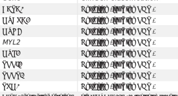
TABLE 1. Genes proven to have a definitive role for disease causation in isolated hypertrophic cardiomyopathy
Gene ClinGen classification
$&7& 'H¿QLWLYHLVRODWHG+&0 0<%3& 'H¿QLWLYHLVRODWHG+&0 0<+ 'H¿QLWLYHLVRODWHG+&0 MYL2 'H¿QLWLYHLVRODWHG+&0 0</ 'H¿QLWLYHLVRODWHG+&0 711, 'H¿QLWLYHLVRODWHG+&0 7117 'H¿QLWLYHLVRODWHG+&0
730 'H¿QLWLYHLVRODWHG+&0
$&7& actin alpha cardiac muscle 1; 0<%3& myosin binding protein C 3; 0<+ myosin heavy chain 7; 0</ myosin light chain 2; 0</ myosin light chain 3; 711, troponin I3, cardiac type; 7117 troponin T2, cardiac type; 730 tropomyosin 1
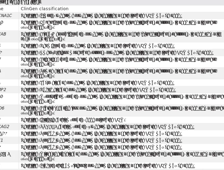
TABLE 2. Genes proven to have a definitive role for disease causation in hypertrophic cardiomyopathy associated with syndromic conditions
Gene ClinGen classification
CACNA1C 'H¿QLWLYH7LPRWK\V\QGURPH±V\QGURPLFFRQGLWLRQVZKHUHLVRODWHG+&0PD\EHVHHQ
&$9 'H¿QLWLYH&DYHROLQRSDWK\±V\QGURPLFFRQGLWLRQVZKHUHOHIWYHQWULFXODUK\SHUWURSK\LVVHHQRQO\ZLWKRYHUW systemic features)
CRYAB 'H¿QLWLYH$OSKD%FU\VWDOOLQRSDWK\±V\QGURPLFFRQGLWLRQVZKHUHOHIWYHQWULFXODUK\SHUWURSK\LVVHHQRQO\ZLWK overt systemic features)
DES 'H¿QLWLYH'HVPLQRSDWK\±V\QGURPLFFRQGLWLRQVZKHUHLVRODWHG+&0PD\EHVHHQ
)+/ 'H¿QLWLYH(PHU\'UHLIXVVPXVFXODUG\VWURSK\±V\QGURPLFFRQGLWLRQVZKHUHLVRODWHG+&0PD\EHVHHQ );1 'H¿QLWLYH)ULHGULFKDWD[LD±V\QGURPLFFRQGLWLRQVZKHUHOHIWYHQWULFXODUK\SHUWURSK\LVVHHQRQO\ZLWKRYHUW
systemic features)
GAA 'H¿QLWLYH3RPSHGLVHDVH±V\QGURPLFFRQGLWLRQVZKHUHOHIWYHQWULFXODUK\SHUWURSK\LVVHHQRQO\ZLWKRYHUW systemic features)
GLA 'H¿QLWLYH)DEU\GLVHDVH±V\QGURPLFFRQGLWLRQVZKHUHLVRODWHG+&0PD\EHVHHQ LAMP2 'H¿QLWLYH'DQRQGLVHDVH±V\QGURPLFFRQGLWLRQVZKHUHLVRODWHG+&0PD\EHVHHQ
/'% 'H¿QLWLYH0\R¿EULOODUP\RSDWK\±V\QGURPLFFRQGLWLRQVZKHUHOHIWYHQWULFXODUK\SHUWURSK\LVVHHQRQO\ZLWKRYHUW systemic features)
MYO6 'H¿QLWLYH%LODWHUDOKHDULQJORVV±V\QGURPLFFRQGLWLRQVZKHUHOHIWYHQWULFXODUK\SHUWURSK\LVVHHQRQO\ZLWKRYHUW systemic features)
PLN 'H¿QLWLYHLQWULQVLFFDUGLRP\RSDWK\JHQH±LVRODWHG+&0
PRKAG2 'H¿QLWLYH35.$*FDUGLRP\RSDWK\V\QGURPLFFRQGLWLRQVZKHUHLVRODWHG+&0PD\EHVHHQ 3731 'H¿QLWLYH1RRQDQV\QGURPH±V\QGURPLFFRQGLWLRQVZKHUHLVRODWHG+&0PD\EHVHHQ RAF1 'H¿QLWLYH1RRQDQV\QGURPH±V\QGURPLFFRQGLWLRQVZKHUHLVRODWHG+&0PD\EHVHHQ 5,7 'H¿QLWLYH1RRQDQV\QGURPH±V\QGURPLFFRQGLWLRQVZKHUHLVRODWHG+&0PD\EHVHHQ
6/&$ 'H¿QLWLYH0LWRFKRQGULDOGLVHDVH±V\QGURPLFFRQGLWLRQVZKHUHOHIWYHQWULFXODUK\SHUWURSK\LVVHHQRQO\ZLWKRYHUW systemic features)
775 'H¿QLWLYH7UDQVWK\UHWLQDP\ORLGRVLV±V\QGURPLFFRQGLWLRQVZKHUHLVRODWHG+&0PD\EHVHHQ
&$&1$& calcium voltage-gated channel subunit alpha 1 C; &$9caveolin 3; &5<$% crystallin alpha B; '(6 desmin;
)+/ four and a half LIM domains 1; );1 frataxin; *$$ alpha glucosidase; */$galactosidase alpha; /$03 lysosomal associated membrane protein 2; /'% LIM domain binding 3; 0<2 myosin VI; 3/1 phospholamban; 35.$* protein kinase AMP-activated non-catalytic subunit gamma 2; 3731 protein tyrosine phosphatase non-receptor type 11; 5$) Raf-1 proto-oncogene, serine/threonine kinase; 5,7 ras like without CAAX 1; 6/&$ solute carrier family 25 member 4;
775 transthyretin
the main role of these strategies lies in identification of putative causative variants outside of known HCM genes.
The bottom line is that analysis should be initially focu- sed on validated/curated HCM genes and if negative, it can be expanded to novel genes (with WGS and WES).
Interpretation of genetic findings
Accurate variant interpretation is a crucial and complex task in clinical practice, and it has been conducted, for almost two decades, by the American College of Medi- cal Genetics and the Association of Molecular Patholo- gists (ACMG/AMP) (4). The current guidelines released in 2015 can be applied to all variants in all Mendelian genes. According to these recommendations, the va- riant interpretation should contain the evidence sup- porting the variant classification including its predicted effect on the respective protein and whether any vari-
ant identified are likely to fully or partially explain the patient’s indication for testing (11).
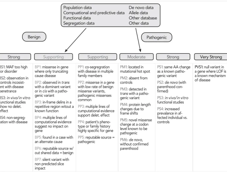
Based on certain essential criteria/databases such as population frequency data, computational and predic- tive (bioinformatics) data, segregation studies, de novo data, functional data, allelic data as well as other da- tabases, the variants can be classified into 5 catego- ries: pathogenic (P), likely pathogenic (LP), variant of un known significance (VUS), likely benign (LB) and be- nign (B) )LJXUH.
According to the latest guidelines, the fine-tuning of variant interpretation can be achieved by following 8 types of evidence, each including 2 sets of crite- ria with different strengths in favor of a pathogenic or a benign role. First, a set of criteria is checked inde- pendently for each variant. Each criterion assessed a particular supporting evidence information, such as its frequency in population databases, in silico predi- ction of a protein damaging effect or co-segregation in family members. Criteria are grouped by different
Cardiologia Hungarica Hategan et al. Genetic diagnosis in hypertrophic cardio myopathy:
two steps forward, one step back
levels of evidence and by pathogenic/benign classes.
Finally, a set of rules combines the evaluated criteria and classifies a variant accordingly to the final ACMG/
AMP 5-tier system.
Beta-myosin heavy chain gene (MYH7) standardized variant interpretation
The beta myosin heavy chain gene 0<+ is one of the most frequently tested genes in a clinical setting since it is a major contributor to several cardiomyopa- thies (HCM, DCM, RCM) (2). Although the ACMG/AMP guideline framework represents a major step forward for variant classification in the context of Mendelian di- sease, it needs constant improvement and refinement as the understanding of these diseases develops. The lack of standardization has led over time to numerous interpretation differences in ClinVar database, mostly
misclassifications, which can have serious consequen- ces in medically actionable variants.
In 2017, the ClinGen (Clinical Genome Resource) has established expert panels to adapt the ACMG/AMP for specific genes and diseases. The Inherited Cardiomyo- pathy Expert Panel (CMP-EP) selected 0<+ as a pilot gene to develop guidelines adaptation for its variants.
To achieve this goal, an expert task team of clinicians and medical molecular geneticists from 3 different ins- titutions reviewed the original ACMG/AMP framework and developed proposed changes to adapt them for 0<+ (11). Their assignment was as follows: to select 60 representative 0<+ variants, to test and apply as many rules as possible to them, to cover a range of classifications, and to include discrepant ClinVar asser- tions.
They realized that out of the original 28 ACMG/AMP ru- les, 9 were not applicable and another 12 required di- sease- and/or gene-specific adjustments. In 5 rules the Population data
Compuational and predictive data Functional data
Segregation data
De novo data Allele data Other database Other data
Benign
Supporting Supporting Moderate Strong Very Strong
Strong
Pathogenic
BS1: MAF too high for disorder BS2: observation in controls incosist- ent with disease penetrance BS3: in vivo/in vitro functional studies show no delet.
effect
BS4:non-segreg- ration with disease
BP1: missense in gene where only truncating cause disease BP2: observed in trans with a dominant variant or in cis with a patho- genic variant
BP3: in-frame delins in a repetitive region witout a known function BP4: multiple lines of computational evidence suggest no impact on gene
BP5: found in a case with an alternate cause BP6: reputable source w/
out shared data = benign BP7: silent variant with non predicted slice impact
PP1: co-segregration with disease in multiple family members PP2: missense in a gene with low rate of benign missense variants, pathogenic missenses common
PP3: multiple lines of cumputational evidence support delet. effect PP4: patient’s pheno- type or family history highly specific for gene PP5: reputable source = pathogenic
PM1: located in mutational hot spot PM2: absent from controls
PM3: detected in trans with a patho- genic variant PM4: protein length changes due to frame shifts PM5: novel missense change at a codon level known to be pathogenic PM6: de novo, without confirmed parenthood
PS1: same AA change as a known patho- genic variant PS2: de novo (with parenthood con- firmed)
PS3: in vivo/in vitro functional studies PS4: increased prevalance in af- fected individual vs.
controls
PVS1: null variant in a gene where LOF is a known mechanism of disease
FIGURE 1. Criteria for classifying varints by the type of evidence as well as the strength of the criteria for a benign (left side) or pathogenic (right side) assertion. Abbreviations: BS, benign strong; BP, benign supporting; LOF, loss-of-function; MAF, minor allele frequency; AA, aminoacid; PM, pathogenic moderate; PP, pathogenic supporting; PS, pathogenic strong; PVS, pathogenic very strong. Figure adapted from: Richards S, Aziz N, Bale S, et al. Standards and guidelines for the interpretation of sequence variants:
A joint consensus recommendation of the American College of Medical Genetics and Genomics and the Association for Molecular Pathology. Genet Med 2015; 17(5): 405–424. doi:10.1038/gim.2015.30
strength criteria were modified. Four rules of the patho- genic framework (PVS1, PM3, PP2, PP4, see in )LJXUH 1) and 3 rules of the benign framework (BS2, BP1, BP3, see in )LJXUH) were evaluated as not applicable either entirely or in the original strength level suggested. PVS1 (since null variants are not a known mechanism for di- sease, loss of function variants being very rare and their contribution to inherited cardiomyopathy is incompletely understood) has been downgraded to PVS1_Moderate evidence. Two additional rules are not applicable: PP5/
BP6 (reputable source reports variant as pathogenic/
benign, but evidence is not accessible) (15).
The main adaptations to the classic rules are concer- ning the minor allele frequency–driven rules (BA1, BS1, and PM2, see in )LJXUH). They were found to be overly conservative such as BA1 criterion (the allele frequency threshold above which a variant is considered benign).
For many Mendelian conditions, the default threshold of 5% is way higher than it needs to be.
Furthermore, regarding the segregation with disea- se (PP1, PP1_Moderate, PP1_Strong), the original framework contains several areas of vagueness (such as the absence of quantitative guidance for increasing the weight depending on the extent of “segregation with disease”). The CMP-EP specified three levels of evi- dence using autosomal dominant likelihood ratios of 10 (3 meioses), 30 (5 meioses), and 100 (7 meioses) to count as supporting, moderate, and strong evidence provided that PM2 (absent or rare in large population cohorts) is met. Finally, the CMP-EP waived the ACMG/
AMP recommendation for demonstrating segregation in more than one family given that 0<+ is a well-est- ablished cardiomyopathy gene.
For the rules addressing the increased prevalence of variant in probands versus controls (PS4, PS4_Mode- rate, PS4_Supporting) the conservative universal WKUHVKROGVZHUH VHWWR SUREDQGFRXQWVRI VXSSRU- WLQJ PRGHUDWH DQG VWURQJ IRU WKH VDNH RI simplification. To apply these rules, PM2 criterion (ab- sent/extremely rare, <0.004%, from large population studies) must be met.
PS2 rule (see in )LJXUH) concerning occurrence of de novo variants has also been modified by removing the necessity of maternity confirmation in a patient.
Also, when paternity has not been established, de novo occurrence receives moderate weight (PM6) but the CMP-EP allowed upgrading it to “strong” (PS2) when at least 3 de novo occurrences have been re- ported.
The ACMG/AMP framework assigns strong weight to well-established in vitro or in vivo functional studies that are supportive of a damaging effect on the gene or protein (PS3). However, in vivo studies are not realy- ly feasible for 0<+ gene while the in vitro ones are less conclusive and currently not considered strong evidence. Nevertheless, the CMP-EP recognized that as soon as in vitro models (that accurately predict the
effect in vivo) become available, their weight can be reconsidered.
The protein domain related rules were also affected.
Whereas the ACMG/AMP framework assigns suppor- ting evidence of pathogenicity to missense variants in a gene that has a low rate of missense variation and moderate evidence for variants located in a hotspot and/or critical domain without benign variation (PM1), it has been well established that missense variants in 0<+are the predominant class of pathogenic al- leles.
Consequently, by applying the original rules to the 60 pilot variants covering a wide range of scenarios, the experts found that 8 out of them have been misclassifi- ed, stressing on the importance of framework standar- dization and data sharing (15).
Automatic web tools for variant classification
The implementation of ACMG/AMP guidelines for vari- ant interpretation can be cumbersome for each individual patient because of the complexity of criteria that need to be evaluated over a large set of variants. Therefore, in- formatics tools (web calculators) have been developed to aid in the process in clinical settings. They allow the user to select the criteria verified by the variant of interest, and then they automatically compute the final classification (16). One example would be the ClinGen Pathogenicity Calculator that provides supporting data to reach more definitive conclusion. However, web calculators lack au- tomatization of the entire ACMG/AMP system and fail to interpret a large set of variants per patient.
ACMG/AMP guidelines was needed to solve comple- xity and reproducibility aspects over manual applica- tion (17). Several automatic tools have been developed in the last couple of years: InterVar, allowing the inter- pretation of multiple variants occurring in any Mendeli- an genes, (18) CardioClassifier that interprets variants occurring in 40 genes associated with cardiovascu- lar disease (19) and CardioVAI (20), a web tools that supports genomic variant classification according to ACMG/AMP rules.
CardioClassifier (https://www.cardioclassifier.org) was developed by the Cardiovascular Genetics and Gen- omics team of the Imperial College, London. The tool is designed to facilitate variant interpretation across a wide range of inherited cardiac conditions. It integ- rates data retrieved from multiple sources through an interactive interface to support variant interpretation.
Combining disease- and gene-specific knowledge with variant observation in large cohort of cases and controls, computational ACMG criteria have been refi- ned. This tool is transparent, generates fast, reprodu- cible and interactive variant pathogenicity reports. It displays all the information along with the final clas- sification.
Cardiologia Hungarica Hategan et al. Genetic diagnosis in hypertrophic cardio myopathy:
two steps forward, one step back
Developed by University of Pavia, Italy, CardioVAI (Car- dio Variant Interpreter) (http://cardiovai.engenom.com) is a web tool that automatically classify genomic va- riants in heritable cardiac disorders related genes ac- cording to ACMG/AMP 5-tier class, showing supporting evidence in terms of activated guidelines criteria. Rele- vant information was retrieved from public databases such as ClinVar, MedGen, ExAc, Disease Ontology and Orphanet (21). The tailored, specific information such as genotype-phenotype correlation and hotspot doma- ins are also incorporated. The generated pathogenicity score is assigned to highlight VUS variants as possible candidates for a further assessment. With a specificity of 97.08% and an average sensitivity of 74.8%, Cardio- VAI ranks the best among the other similar tools (Inter- Var and CardioClassifier), reducing the number of VUS to 70.9%. Compared to them, CardioVAI evaluates bet- ter criteria like PS1, PP5 and BP6 and incorporates recent refined data for 0<+ gene variants interpre- tation. It incorporates disease specific knowledge ga- thered from omics-resources and CMP-EP guidelines adaptation for 0<+ variants. Additional validation on 60 0<+ variants reports a classification concordance of 93.4%, showing the importance of population-data- base selection for the evaluation of criteria such as BS1 and PM2 (20).
Due to gene curation efforts from ClinGen and automa- ted variant interpretation webtools such as CardioClas- sifier and CardioVai great progress has been achie- ved, minimizing the risk of false-positive results. This, in turn, may increase the false negative result rate.
Recently, in HCM, since functional studies are limited and the disease is highly heterogeneous, around 8%
of HCM patients are genetically misclassified as VUS variant carriers when, in fact, they may have a rather pathogenic one. These issues can be overcome by data sharing within international centers that would be beneficial particularly in cases of encountering interest- ing founder, endemic variants. Such opportunity alre- ady exists (the ShaRe registry, https://theshareregistry.
org) with the main goal of sharing knowledge between different centers through ClinVar database. For examp- le, using shared data, understandings like the one in which patients with 0<+ variants had a higher risk of advanced heart failure and worse outcome comparing with 0<%3& gene cohort have become available (8).
Worth mentioning is also the fact that patients with complex genotypes carrying 2 pathogenic variants are at greater risk of adverse outcome and SCD, regard- less of the genes involved.
Regarding the genetic interpretation of novel vari- ants, recent study suggests that, when found in a patient confirmed to have disease, the novel rare variants in established HCM-related genes are em- pirically shown to have a sufficiently high probabil- ity of pathogenicity to support a “likely pathogenic”
classification, even without additional segregation or
functional data. This could increase the yield of high confidence actionable variants, consistent with the framework and recommendations of current guide- lines (12).
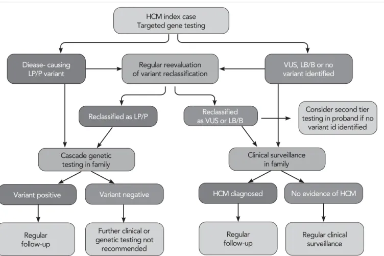
Targeted gene testing in HCM
Although current diagnosis of HCM is solely based on clinical findings, genetic diagnosis of HCM provides very important additional information. According to this, genetic testing is recommended in patients ful- filling diagnostic criteria for HCM, when it enables cascade genetic screening of their relatives. Genetic screening strategy in HCM is summarized in )LJXUH 2. Genetic testing for HCM is first initiated in an indi- vidual with a clear-cut diagnosis of HCM, usually in the index case. If a definitive likely pathogenic (LP) or pathogenic (P) variant is found, then cascade ge- netic testing in relatives can be initiated. Demonstra- tion of a VUS in a proband is not a clinically action- able result. In selected circumstances, family member testing may be undertaken, at either a clinical or re- search level, to further elicit the pathogenicity of the variant (e.g., through cosegregation analysis in family members, determine de novo status through parental testing, etc.). However, this is most appropriate in the setting of guidance from a cardiovascular genetics ex- pert. If genetic testing does not identify a pathogenic variant in a patient with HCM (i.e., only identifies be- nign/likely benign variants), there is no indication to do genetic testing in family members as the identification of such variants will not change clinical management, including the need for continued clinical screening. In genotype-negative relatives of individuals with geno- type-positive HCM, no further clinical follow-up is re- quired. Over time, as more knowledge is gained, some variants previously thought to be likely pathogenic or pathogenic may be downgraded to a VUS or benign category. In such instances, family relatives who were released from clinical surveillance on the basis of the previous gene result need to be notified and regular clinical screening recommenced (3).
Interpretation of genotype-negative HCM
In general, those patients lacking pathogenic variants in the 8 definitive sarcomeric genes have a better cli- nical outcome compared to the variant-positive ones.
Ingles et al described nonfamilial HCM occurring in 40% of studied cohort with distinct phenotype from genotype-positive cases. However, that should not discourage clinicians to regularly monitor the family members as well, since HCM is known for its high va- riability in terms of age of onset and phenotype seve- rity (13, 3).
Conclusion and future perspectives
Although nowadays the interpretation of genetic find- ings is two steps closer to a more accurate approach due to gene adaptation and automatization, the multi- tude of putative causative genes have been once again reduced to the 8 sarcomere genes, a backward step.
HCM remains basically a disease of the sarcomere, characterized by incomplete penetrance and highly va- riable phenotypic expressivity, with an increased rate (40-60%) of negative-genotype. This indicates that other genetic and epigenetic factors (like obesity and hypertension) may contribute to the development of di- sease. Other genetic factors may include common, low frequency, rare variants that can play a protective or modifier role (14). Such factors can be identified via lar- ge-scale genome-wide association studies and WGS, followed by integration of their genetic profile with the specific nongenetic ones.
Current genetic testing strategies rely on more restricted gene panels with proven, definitive association with HCM.
Studies have shown that new, additional putative genes are accounting for only a limited number of cases (8, 3).
The presence of multiple rare genetic variants in an in- dividual has a cumulative effect on clinical phenotype and prognosis, irrespective of variant pathogenicity, suggesting that variants that are non-damaging sepa- rately may play a modifying role in conjunction with ot- her variants.
Further, three challenges still remain: the lack of opti- mal accuracy of variant interpretation, the lack of clear genotype-phenotype correlations and the need of ex- panding research on genetic causes of HCM beyond the monogenic Mendelian inheritance model.
$FNQRZOHGJHPHQWV
7KLVZRUNZDVVXSSRUWHGE\JUDQWV*,123 Ä5LWNDEHWHJVpJHNSDWRJHQH]LVpQHNNXWD tása, új diagnosztikai és terápiás eljárásokat megalapo ]yIHMOHV]WpVHN´*,123ÄeOHWHW YH6]pO\H]7HWĘ $NXW PHJEHWHJHGpVHN V~O<RVViJL pV K$/iOR]iVLPXWDWyOQDNMD9tWiVDWUDQV]OiFLyVRUYRVWXGR PiQ\LP(JN|]HOtWpVEHQ±67$<$/,9(´DQGWKHÄ+HWp Q\L*p]D´5HVHDUFK)XQGRIWKH)DFXOW\RI0HGLFLQHRI the University of Szeged.
HCM index case Targeted gene testing
Consider second tier testing in proband if no
variant id identified
Regular follow-up
Regular follow-up Further clinical or
genetic testing not recommended
Regular clinical surveillance Diease- causing
LP/P variant
Reclassified as LP/P
Variant positive Variant negative HCM diagnosed
VUS, LB/B or no variant identified
Reclassified as VUS or LB/B Regular reevaluation
of variant reclassification
Cascade genetic testing in family
Clinical surveillance in family
No evidence of HCM
FIGURE 2. Flowchart for genetic testing process in HCM. HCM: hypertrophic cardiomyopathy; LB/B, likely benign/benign; LP/P, likely pathogenic or pathogenic; and VUS, variant of unknown significance. Figure adapted from: Ommen SR, Mital S, Burke MA, et al.
2020 AHA/ACC guideline for the diagnosis and treatment of patients with hypertrophic cardiomyopathy: a report of the American College of Cardiology/American Heart Association Joint Committee on Clinical Practice Guidelines. J Am Coll Cardiol 2020; 76:
e159–240.
Cardiologia Hungarica Hategan et al. Genetic diagnosis in hypertrophic cardio myopathy:
two steps forward, one step back
5()(5(1&(6
1.Maron BJ, Gardin JM, Flack JM, Gidding SS, Kurosaki TT, Bild DE. Prevalence of Hypertrophic Cardiomyopathy in a General Popu- lation of Young Adults. Circulation 1995; 92(4).
https://doi.org/10.1161/01.CIR.92.4.785
2.Marian AJ, Braunwald E. Hypertrophic Cardiomyopathy: Genet- ics, Pathogenesis, Clinical Manifestations, Diagnosis, and Therapy.
Circ Res 2017; 121(7): 749–770.
https://doi.org/10.1161/CIRCRESAHA.117.311059
3.Ommen SR, Mital S, Burke MA, et al. 2020 AHA/ACC Guideline for the Diagnosis and Treatment of Patients With Hypertrophic Car- diomyopathy. Vol 142; 2020.
https://doi.org/10.1161/cir.0000000000000937
4. Ackerman MJ, Priori SG, Willems S, et al. HRS/EHRA expert con- sensus statement on the state of genetic testing for the channelo- pathies and cardiomyopathies this document was developed as a partnership between the Heart Rhythm Society (HRS) and the Eu- ropean Heart Rhythm Association (EHRA). Hear Rhythm 2011; 8(8):
1308–1339. https://doi.org/10.1016/j.hrthm.2011.05.020
5. Jarcho JA, McKenna W, Pare JA, et al. Mapping a gene for familial hypertrophic cardiomyopathy to chromosome 14q1. N Engl J Med 1989;3 21(20): 1372–1378.
https://doi.org/10.1056/NEJM198911163212005
6.Mazzarotto F, Girolami F, Boschi B, et al. Defining the diagnostic effectiveness of genes for inclusion in panels: the experience of two decades of genetic testing for hypertrophic cardiomyopathy at a sin- gle center. https://doi.org/10.1038/s41436
7. Ingles J, Goldstein J, Thaxton C, et al. Evaluating the Clinical Valid- ity of Hypertrophic Cardiomyopathy Genes. Circ Genomic Precis Med 2019; 12(2): 57–64. https://doi.org/10.1161/CIRCGEN.119.002460 8.Mazzarotto F, Olivotto I, Boschi B, et al. Contemporary Insights Into the Genetics of Hypertrophic Cardiomyopathy: Toward a New Era in Clinical Testing? J Am Heart Assoc 2020; 9(8): e015473.
https://doi.org/10.1161/JAHA.119.015473
9.Golbus JR, Puckelwartz MJ, Fahrenbach JP, Dellefave-Castillo LM, Wolfgeher D, McNally EM. Population-based variation in cardio- myopathy genes. Circ Cardiovasc Genet 2012; 5(4): 391–399.
https://doi.org/10.1161/CIRCGENETICS.112.962928
10.Lopes LR, Zekavati A, Syrris P, et al. Genetic complexity in hy- pertrophic cardiomyopathy revealed by high-throughput sequenc- ing. J Med Genet 2013; 50(4): 228–239.
https://doi.org/10.1136/jmedgenet-2012-101270
11.Richards S, Aziz N, Bale S, et al. Standards and guidelines for the interpretation of sequence variants: A joint consensus rec- ommendation of the American College of Medical Genetics and
Genomics and the Association for Molecular Pathology. Genet Med 2015; 17(5): 405–424. https://doi.org/10.1038/gim.2015.30
12. Walsh R, Mazzarotto F, Whiffin N, et al. Quantitative approaches to variant classification increase the yield and precision of genetic testing in Mendelian diseases: The case of hypertrophic cardiomyo- pathy. Genome Med 2019; 11(1).
https://doi.org/10.1186/s13073-019-0616-z
13.Ingles J, Burns C, Bagnall RD, et al. Nonfamilial Hypertrophic Cardiomyopathy: Prevalence, Natural History, and Clinical Implica- tions. Circ Cardiovasc Genet 2017; 10(2).
https://doi.org/10.1161/CIRCGENETICS.116.001620
14.Harper AR, Goel A, Grace C, et al. Common genetic variants and modifiable risk factors underpin hypertrophic cardiomyopathy susceptibility and expressivity. Nat Genet 2021; 53(2): 135–142.
https://doi.org/10.1038/s41588-020-00764-0
15.Kelly MA, Caleshu C, Morales A, et al. Adaptation and validation of the ACMG/AMP variant classification framework for MYH7-asso- ciated inherited cardiomyopathies: Recommendations by ClinGen’s Inherited Cardiomyopathy Expert Panel. Genet Med 2018; 20(3):
351–359. https://doi.org/10.1038/gim.2017.218
16. Patel RY, Shah N, Jackson AR, et al. ClinGen Pathogenicity Cal- culator: a configurable system for assessing pathogenicity of genetic variants. Genome Med 2017; 9(1): 3.
https://doi.org/10.1186/s13073-016-0391-z
17.Amendola LM, Jarvik GP, Leo MC, et al. Performance of ACMG- AMP Variant-Interpretation Guidelines among Nine Laboratories in the Clinical Sequencing Exploratory Research Consortium. Am J Hum Genet 2016; 98(6): 1067–1076.
https://doi.org/10.1016/j.ajhg.2016.03.024
18. Li Q, Wang K. InterVar: Clinical Interpretation of Genetic Variants by the 2015 ACMG-AMP Guidelines. Am J Hum Genet 2017; 100(2):
267–280. https://doi.org/10.1016/j.ajhg.2017.01.004
19.Whiffin N, Walsh R, Govind R, et al. CardioClassifier: disease- and gene-specific computational decision support for clinical ge- nome interpretation. Genet Med 2018; 20(10): 1246–1254.
https://doi.org/10.1038/gim.2017.258
20. Nicora G, Limongelli I, Gambelli P, et al. CardioVAI: An automatic implementation of ACMG-AMP variant interpretation guidelines in the diagnosis of cardiovascular diseases. Hum Mutat 2018; 39(12):
1835–1846. https://doi.org/10.1002/humu.23665
21.Kibbe WA, Arze C, Felix V, et al. Disease Ontology 2015 up- date: an expanded and updated database of human diseases for linking biomedical knowledge through disease data. Nucleic Acids Res 2015; 43(Database issue): D1071-8.
https://doi.org/10.1093/nar/gku1011