DESCRIPTION OF NEW GENERA AND SPECIES IN THE TRIBE RHIZOECINI (HOMOPTERA,
COCCOIDEA, PSEUDOCOCCIDAE)
KOZÁR, F. and FOLDI, I.
Plant Protection Institute, Hungarian Academy of Sciences H-1525 Budapest P. O. Box 102, Hungary. E-mail: h2405koz@ella.hu
Dèpartement Systèmatique et Evolution, Entomologie CP 50, Musèum National d’Histoire Naturelle, 45 rue Buffon, 75005 Paris, France. E-mail: foldi@cimrs1.mnhn.fr
Two new genera and nine new species are described.Benedictycoccinagen. n. includes four species; three of them are new.Hambletonia gen. n. with one new species. The genus CoccidellaHAMBLETON, 1946 is re-established, including eight species, among them five are new. A new morphological character, the structure of female genital organ is described. A re- view for the tribe and a key for the genera, and species are given.
Key words: Homoptera, Coccoidea, Pseudococcidae, Rhizoecini, new genera and species, re- view, keys
INTRODUCTION
The Rhizoecinae is a distinct subfamily of the Pseudococcidae. The members of the subfamily were intensively studied by H
AMBLETON(1946a, 1976). The subfamily was also studied in detail by M
ATILEF
ERRERO(1976), T
ANG(1992), W
ILLIAMSand G
RANARA DAW
ILLINK(1992), W
ILLIAMS(1998, 2004), and B
EN-D
OV(1994), K
OZÁRand K
ONCZNÉB
ENEDICTY(2002, 2003) and others. In the Scalenet computer database (the Pseudococcidae family last updated on 26 May 2001) (M
ILLERet al. 2001) contains the most important information (taxon- omy, distribution, biology, etc). The world fauna of the subfamily was not well ex- plored, thus the distribution of the known species is poorly known.
In the past, the internal female genitalia were not used as character to separate
species. The authors studied them in some species. A tentative, to establish the dif-
ferent types of female reproductive system in Coccoidea, by referring to 28 spe-
cies, including one Rhizoecus species. In 11 families, eight basic types, and their
evolutionary trends were discussed by
DEM
ARZOet al. (1990). A recent descrip-
tion on the general organization of the reproductive system of a soft scale (Cocci-
dae) based on the scanning electron microscope observations was reported by
F
OLDI(1997), is serving the basis to recognize the cuticular structures of the KOH
treated genitalia, visible on microscopic slides.
The aim of the present work is the study of the subfamily Rhizoecinae on a world-wide scale, in more detail, the Rhizoecini tribe, and to prepare a revision, to learn more about the distribution patterns of each species. During this project sev- eral new genera and species were found, which need further study. We are describ- ing here two new genera, also, one genus is re-established, and nine new species of this tribe are described.
MATERIALS AND METHODS
This study presents the results of the analyses of about 5000 samples from many parts of the World. The descriptions follow the terminology of morphological characters as given in the works of HAMBLETON(1946a, 1976), and WILLIAMS(1998).
The insects were collected by visual survey from soil, from Berlese funnel and sifting samples (KOZÁR& MILLER2000). The sample collectors are mentioned in the species descriptions. Most of the studied insects are from the scale insect collection of the Plant Protection Institute, Hungarian Acad- emy of Sciences (PPI), Budapest, and from the Arachnoidea collection of the Hungarian Natural His- tory Museum, Budapest, Hungary (HNHM). Some other samples from the Natural History Museum (London), Smithsonian Institutions, and the Natural History Museum (Paris), have been also studied.
New morphological characters, the female internal genitalia, were also studied in some spe- cies. Usually, with the traditional mounting process the internal genitalia of females were not pre- served, and often even their cuticular parts were destroyed. However, in most of the Rhizoecini species, the ectodermic parts of internal genitalia are heavily sclerotized and could be observed on microscopic slides. The shape and size of vagina and locations of vaginal glands are variable, thus they can be useful complementary characters to separateRhizoecusspecies.
Most of the studied females, including some immature stages, were preserved on microscopic slides at the PPI.
RESULTS
In the studied samples 530 Rhizoecinae females and 139 immature stages were found. The analysis of our samples and other materials resulted the discovery several new species and two new genera described below, while one genus is being re-established in this paper. A key for the genera and species of the tribe Rhizoe- cini for the treated genera are also given.
Benedictycoccina K
OZÁRet F
OLDIgen. n.
Type species:Ripersiella ornataHAMBLETON, 1946a
Description: Body elongate, oval. Antennae 6 segmented, typical for the tribe. The sensory seta on the fifth segment of antennae short and blunt. Legs well developed. Dorsum and venter with
tritubular pores (cerores) surrounded by trilocular pores, which in some species appear as tritubular pore structures. Tubular ducts and multilocular pores absent or present. Anal ring with 6 setae, anal ring cells in small number, with spiculae on outer row. Ostioles present, with two circuli in the known species. Genitalia simple, as long as the of width one segment.
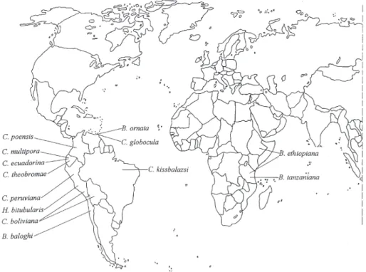
Distribution: The species of this genus are distributed in the Neotropical and Ethiopian re- gions (Fig. 1).
Etymology: The new genus is named after ZSUZSANNAKONCZNÉBENEDICTYin acknowledg- ing her help with the study of these species.
Comments: This genus is distinct from other genera, especially by the unique groups of trilocular pores surrounding the tritubular pores (cerores). Four species are included in the genus, three of them are new to science.
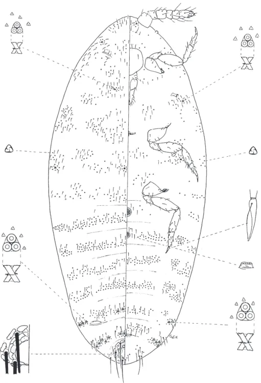
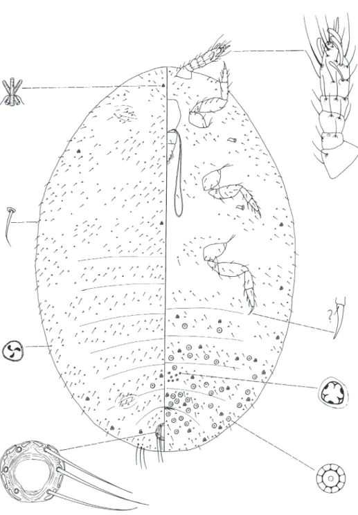
Benedictycoccina baloghi K
OZÁRet F
OLDIsp. n.
(Fig. 2)
Type material: The holotype female, (coll. J. BALOGH), D-Am. Chile, No. 201. Deposited in the Hungarian Natural History Museum (Budapest, Hungary).
Fig. 1.Distribution of the studied genera and species
Description: Body elongate oval. Slide-mounted specimen (Fig. 2) 1.08 mm long and 0.51 mm wide. Antenna 6 segmented, the length of the segments: 1st – 28 µm, 2nd – 15 µm, 3rd – 23 µm, fourth – 10 µm, fifth – 9 µm, sixth 34 µm long. There is one sensory pore on the 2nd segment of the an- tenna. The 3rd segment is almost parallel sided. The apical segment has four falcate sensory setae. On the fifth segment one, 12 µm long, blunt seta. Most segments of the antenna have a few hair-like setae; the longest one is 23 µm. Eyes well visible. Anal lobe is slightly developed with three long, hair-like setae.
Venter. Labium appears two-segmented, 64 µm long. Stylet loop twice longer than labium.
Cephalic plate not visible. Legs robust: coxa of anterior legs 30 µm, trochanter 24 µm, femur 60 µm, tibia 48 µm, tarsus 36 µm, and claw 19 µm. Coxa of middle legs 30 µm, trochanter 25 µm, femur 52 µm, tibia 45 µm, tarsus 31 µm, and claw 20 µm long. Coxa of posterior legs 32 µm, trochanter 28 µm, femur 55 µm, tibia 52 µm and tarsus 45 µm, and claw 17 µm, tarsal digitules absent, claw digitules short, 4 µm long. Trochanter with two pores on each side. Claw without denticle. Legs with few hair-like setae, tibia and tarsus with 13 µm long setae. On the ventral segments few tritubular duct present on margin surrounded by trilocular pores. Multilocular pores with 5–8 pores present in small number on all segments, 5–6 µm in diameter. The diameter of anterior spiracles 7 µm. Venter with a small number of scattered hair-like setae. Circulus two, 25–28 µm in diameter. Tubular ducts absent.
Internal genitalia longer than width of one segment; middle part of vulva enlarged bearing ductules of the vaginal glands.
Dorsum. Ostioles present, not sclerotized. Multilocular pores present. Anal ring oval, 67 µm wide. Anal ring with six, 62 µm long hair-like setae. Anal ring pores (cells) typical, in small number, with strong blunt spicules on pores of outer row. Tritubular pores of two sizes, the smaller are on tho- rax and head, the tubes 2–5 µm wide. Situated on the margin and in middle line Tubular ducts absent.
Hair-like setae 11 µm long, trilocular pores 3 µm wide, scattered on the dorsum, among them some unusual for the group, bilocular pores present also.
Distribution: Chile (Fig. 1).
Etymology.The species is named after late Prof. JÁNOSBALOGH, the collector of the species.
Affinities: This species similar to B. ethiopiana, but differs by the absence of tubular ducts.
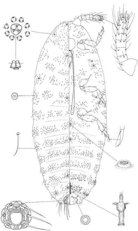
Benedictycoccina ethiopiana F
OLDIet K
OZÁRsp. n.
(Fig. 3)
Type material: The holotype female, in middle of the slide (marked), twoBenedictycoccina females on the same slide. Ethiopia, Addis Abeba, 28.09.1980 (leg. A. DEMETER), No. 329. One paratype female, the slide contains twoBenedictycoccinaimmatures, also, from the same collection as the holotype, No. 325. One paratype from Tanzania, Uluguru, 12.07.1972, No. 118 (leg. T. PÓCS).
Types are deposited in the Hungarian Natural History Museum (Budapest, Hungary).
Description: Body elongate oval. Slide-mounted specimen (Fig. 3) 0.96 mm long and 0.39 mm wide. Antenna 6 segmented, the length of the segments: 1st – 33 µm, 2nd – 15 µm, 3rd – 28 µm, fourth – 20 µm, fifth – 22 µm, sixth 50 µm long. There is one sensory pore on the 2nd segment of the antenna. The 3rd segment is almost parallel sided. The apical segment has four falcate sensory setae.
On the fifth segment one 13 µm long, strong, blunt seta present. Most segments of the antenna have a
Fig. 2.Benedictycoccina baloghiKOZÁRet FOLDIsp. n.
Fig. 3.Benedictycoccina ethiopianaFOLDIet KOZÁRsp. n.
few hair-like setae; the longest one is 25 µm. Eye well visible. Anal lobe is slightly developed with three long, hair-like setae.
Venter. Labium appears two-segmented, 67 µm long. Stylet loop twice longer than labium.
Cephalic plate not visible. Legs robust: coxa of anterior legs 43 µm, trochanter 30 µm, femur 95 µm, tibia 67 µm, tarsus 52 µm, and claw 22 µm. Coxa of middle legs 53 µm, trochanter 34 µm, femur 88 µm, tibia 67 µm, tarsus 48 µm, and claw 22 µm long. Coxa of posterior legs 55 µm, trochanter 37 µm, femur 104 µm, tibia 95 µm, tarsus 54 µm, and claw 26 µm, tarsal digitules absent, claw digitules short, 5 µm long. Trochanter with two pores on each side. Claw without denticle. Legs with few hair-like setae, tibia and tarsus with 16 µm long setae. On the ventral segments few tritubular pores surrounded by trilocular pores, which appear as tritubular pores, present on margin. Multilocular pores present on all segments. The diameter of anterior spiracles 13 µm. Venter with a small number of scattered hair-like setae. One circulus present, 19 µm in diameter. Tubular ducts present. Trilocular pores scat- tered on the venter, 3 µm in diameter. Internal genitalia longer than width of one segment; vaginal glands located at higher part of vagina.
Dorsum. Ostioles present, sclerotized. Multilocular pores present on all segments. Anal ring oval, 41 µm wide and 38 µm long. Anal ring with six, 56 µm long hair-like setae. Anal ring pores (cells) typical, in small number, with spicules on pores of outer row. Tritubular pores of one size, 3–5 on each segment, tubes 4 µm in diameter, 10 µm long, all surrounded by trilocular pores, which ap- pears as tritubular pores, 8 µm long. Tubular ducts present, 3 µm long. Hair-like setae 11 µm long.
Trilocular pores 3 µm wide, scattered on the dorsum.
Distribution: Ethiopia and Tanzania (Fig. 1).
Etymology: The species is named after the country of origin.
Affinities: This species differs from all other species by having tubular ducts.
Benedictycoccina ornata (H
AMBLETON, 1946a) (Fig. 4)
Comment: This species differs from other members of the genus by the ab- sence of multilocular pores, and by the presence of wide bands and groups of trilocular pores on the abdomen.
Distribution: Trinidad (Fig. 1).
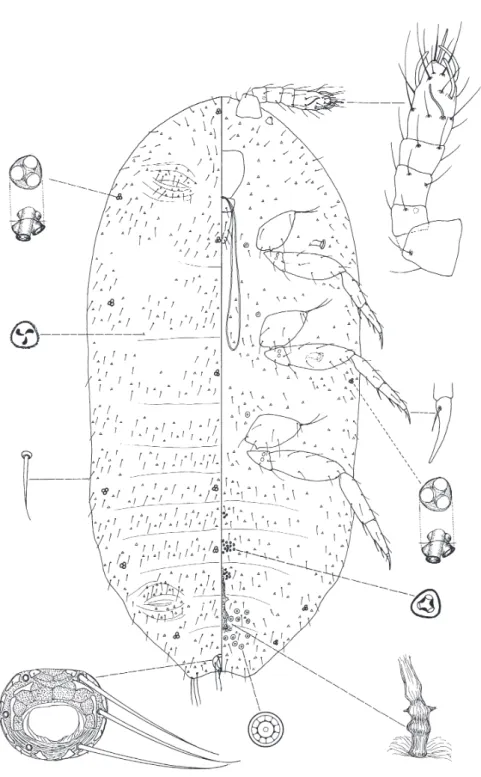
Benedictycoccina tanzaniana K
OZÁRet F
OLDIsp. n.
(Fig. 5)
Type material: The holotype female, in middle of the slide, Tanzania, 1987 (coll. S. MAHUN - KA), No. 3402 (103). Six paratype females, on five slides, from the same collection as the holotype, No. 3402 (103) (two slides), and No. 560 (three slides). Types are deposited in the Hungarian Natural History Museum (Budapest, Hungary).
Fig. 4.Benedictycoccina ornata(HAMBLETON, 1946) (after WILLIAMS& GRANARA DEWILLINK1992)
Fig. 5.Benedictycoccina tanzanianaKOZÁRet FOLDIsp. n.
Description: Body elongate oval. Slide-mounted specimen (Fig. 5) 0.71 mm long and 0.26 mm wide. Antenna 6 segmented, the length of the segments: 1st – 29 µm, 2nd – 12 µm, 3rd – 15 µm, fourth – 14 µm, fifth – 12 µm, sixth 29 µm long. There is one sensory pore on the 2nd segment. The 3rd segment is almost parallel-sided. The apical segment has four falcate sensory setae. On the fifth segment one, strong 13 µm long, blunt seta. Most segments of the antenna have a few hair-like setae;
the longest one is 19 µm. Eyes well visible. Anal lobe is slightly developed with three long, hair-like setae.
Venter. Labium appears two-segmented, 51 µm long. Stylet loop four times longer than la- bium. Cephalic plate not visible. Legs robust: coxa of anterior legs 25 µm, trochanter 24 µm, femur 55 µm, tibia 33 µm, tarsus 31 µm, and claw 18 µm. Coxa of middle legs 27 µm, trochanter 22 µm, femur 45 µm, tibia 35 µm, tarsus 26 µm, and claw 17 µm long. Coxa of posterior legs 29 µm, trochanter 23 µm, femur 50 µm, tibia 45 µm, tarsus 38 µm, and claw 20 µm, tarsal digitules absent, claw digitules short, as long as claw, blunt. Trochanter with two pores on each side. Claw without denticle. Legs with few hair-like setae, tibia and tarsus with 11 µm long setae. On the ventral segments few tritubular ducts present on margin surrounded by trilocular pores. Some multilocular pores present around of vulva, 6 µm in diameter. The diameter of anterior spiracles 9 µm. Venter with a small num- ber of scattered hair-like setae. Two circuli present, 24 µm in diameter. Tubular ducts absent. Internal genitalia as long as width of one segment; close to distal part, vagina is enlarged bearing vaginal glands.
Dorsum. Ostioles present, not sclerotized. Multilocular pores absent. Anal ring oval, 55 µm wide and 48 µm long. Anal ring with six, 51 µm long hair-like setae. Anal ring pores (cells) typical, few, with spicules on pores of outer row. Tritubular ducts of one size, in small number on last seg- ments, and scattered on thorax, the tube 4 µm long and 3 µm wide. Tubular ducts absent. Hair-like setae 11 µm long, trilocular pores scattered on the dorsum. Bilocular pores present on all segments.
Distribution: Tanzania (Fig. 1).
Etymology.The species is named after the country of origin.
Affinities: This species differs from all others in the genus, by having multi- locular pores only on the last ventral abdominal segments.
Key for species of Benedictycoccina
1 Multilocular pores absent B. ornata (H
AMBLETON, 1946a)
– Multilocular pores present 2
2 Tubular ducts present B. ethiopiana sp. n.
– Tubular ducts absent 3
3 Multilocular pores only on the last abdominal segments of venter
B. tanzaniana sp. n.
– Multilocular pores both on venter and dorsum B. baloghi sp. n.
Coccidella H
AMBLETON, 1946b
Type species: Morrisonella poensis HAMBLETON, 1946b
Description: Body elongate oval. Antennae 5 or 6 segmented, typical for the tribe, but the sen- sory seta on fifth segment is narrow and long, when the species has five segmented antennae it is situ- ated near to the base of the fifth segment. Legs well developed. Dorsum and venter with tritubular pores (cerores). Tubular ducts absent or present. Anal ring normal, with 6 setae, cells in small num- ber, outer row cells with spicula. Ostioles present, circulus absent. Internal genital organ heavily sclerotized, usually as long as width of one segment.
Distribution: The genus contains eight species, all are from the Neotropical Region (Fig. 1).
Comments: This genus was synonymized by F
ERRIS(1953), and latter H
AMBLETON(1946b) accepted this act. However, F
ERRIS(1953) was not entirely sure about his decision and noted “It may seem desirable to resurrect the genus Coccidella and to name even more.” The present re-establishment is based on sev- eral newly described species and the study of type material, which gave possibility to find some new characters, and describe more definitely the genus, which fol- lows H
AMBLETON’s (1946a) description, but adds several new and important characters, which were not considered by him.
Coccidella H
AMBLETON, 1946b, is a replacement name of Morrisonella H
AMBLETON, 1946a. This genus is distinct from other genera by having groups of trilocular pores with special structures on some of the ventral segments of the ab- domen.
Coccidella boliviana K
ONCZNÉB
ENEDICTYet K
OZÁRsp. n.
(Fig. 6)
Type material: The holotype female, in the middle of the slide, Bolivia, 1966, (coll. J.
BALOGH), D-Am No. 276. Two paratype females, from the same collection as the holotype, No. 275.
Three female on one slide similar to this species, but was not included into the paratype series, Peru, Iquitus, 01. 12. 1971 (coll. J. BALOGH) IQ No. B 9. Deposited in the Collection of Plant Protection In- stitute, Hungarian Academy of Sciences (Budapest, Hungary).
Description: Body elongate oval. Slide-mounted specimen (Fig. 6) 1.08 mm long and 0.50 mm wide. Antenna 5 segmented, the length of the segments: 1st – 40 µm, 2nd – 20 µm, 3rd – 31 µm, fourth – 24 µm, fifth – 65 µm long. There is one sensory pore on the 2nd segment of the antenna. The 3rd segment is almost parallel sided. The apical segment has four falcate sensory setae, one of them narrow, 31 µm long, blunt seta. Most segments of the antenna have a few hair-like setae; the longest one is 22 µm. Eyes well visible. Anal lobe is slightly developed with three long, hair-like setae.
Venter. Labium appears two-segmented, 76 µm long. Stylet loop three times longer than la- bium. Cephalic plate not visible. Legs robust: coxa of anterior legs 51 µm, trochanter 48 µm, femur
Fig. 6.Coccidella bolivianaKONCZNÉBENEDICTYet KOZÁRsp. n.
118 µm, tibia 77 µm, tarsus 67 µm, and claw 24 µm. Coxa of middle legs 65 µm, trochanter 50 µm, fe- mur 103 µm, tibia 70 µm, tarsus 58 µm, and claw 26 µm long. Coxa of posterior legs 72 µm, trochanter 50 µm, femur 122 µm, tibia 98 µm, tarsus 70 µm, and claw 29 µm, tarsal digitules absent, claw digitules short, 8 µm long. Trochanter with two pores on each side. Claw without denticle. Legs with few hair-like setae, tibia and tarsus with 13 µm long setae. On the ventral segments few tritubular pores, 7 µm in diameter present on margin, diameter of tubes 4 µm. Few multilocular pores with ten pores present around vulva, and some scattered in the midline of the thorax, 8 µm in diameter. The di- ameter of anterior spiracles 17 µm. Venter with a small number of scattered hair-like setae. Circulus absent. Tubular ducts absent. Trilocular pores widely distributed on venter. On the segments IV and V, and on the head a group of special trilocular pores present, 4 µm in diameter, the structure appears as tritubular. Internal genitalia, forming a characteristic, particularly elongate structure, as long as two segments; near the basal part the vagina enlarged; vaginal glands not seen.
Dorsum. Ostioles present, sclerotized. Multilocular pores absent. Anal ring oval, 41 µm wide and 37 µm long. Anal ring with six, 54 µm long hair-like setae. Anal ring pores (cells) typical, in small number, with small spicules on pores of outer row. Tritubular pores of one size, 1–3 on each segment, 10 µm in diameter, ducts short, diameter of tubes 4 µm. Tubular ducts absent. Hair-like setae 11 µm long, trilocular pores 3 µm wide, scattered on the dorsum.
Distribution: Bolivia and Peru (Fig. 1).
Etymology.The species is named after the country of origin.
Affinities: The species is similar to C. theobromae, but differs from it by hav- ing short and wide tritubular pores, and five segmented antennae.
Coccidella ecuadorina K
ONCZNÉB
ENEDICTYet F
OLDIsp. n.
(Fig. 7)
Type material: The holotype female, in the middle of the slide, Ecuador, Guayaguil-Riobamba trans., 1973 (coll. J. BALOGH), No. EC 37. Two paratype females, from the same collection as the holotype, No. EC 34. Deposited in the Collection of Plant Protection Institute, Hungarian Academy of Sciences (Budapest, Hungary).
Description: Body elongate oval. Slide-mounted specimen (Fig. 7) 1.55 mm long and 0.75 mm wide. Antenna 6 segmented, the length of the segments: 1st – 52 µm, 2nd – 22 µm, 3rd – 30 µm, fourth – 26 µm, fifth – 76 µm long. There is one sensory pore on the 2nd segment of the antenna. The 3rd segment is almost parallel sided. The apical segment has four falcate sensory setae. On fifth seg- ment one narrow, 35 µm long, blunt seta. Most segments of the antenna have a few hair-like setae; the longest one is 27 µm. Eyes well visible. Anal lobe is slightly developed with three long, hair-like setae.
Venter. Labium appears two-segmented, 105 µm long. Stylet loop twice longer than labium.
Cephalic plate visible. Legs robust: coxa of anterior legs 70 µm, trochanter 43 µm, femur 120 µm, tibia 86 µm, tarsus 77 µm, and claw 26 µm. Coxa of middle legs 74 µm, trochanter 50 µm, femur 125 µm, tibia 84 µm, tarsus 72 µm, and claw 26 µm long. Coxa of posterior legs 86 µm, trochanter 65 µm, femur 146 µm, tibia 118 µm and tarsus 84 µm, and claw 29 µm, tarsal digitules absent, claw digitules short, 9 µm long. Trochanter with two pores on each side. Claw without denticle. Legs with few hair-like setae, tibia and tarsus with 13 µm long setae. On the ventral segments few tritubular pores
Fig. 7.Coccidella ecuadorinaKONCZNÉBENEDICTYet FOLDIsp. n.
present, 2–3 µm in diameter. Multilocular pores present on all segments. The diameter of anterior spi- racles 18 µm. Venter with a few of scattered hair-like setae. Circulus and tubular ducts absent.
Trilocular pores widely present on the venter, 3 µm in diameter. On the segments III, IV and V, a group of trilocular pores, with special structure present, 4 µm in diameter. Internal genitalia, a little longer than width of one segment; basal part of vagina constricted just below of the vaginal glands.
Dorsum. Ostioles present, sclerotized. Multilocular pores present all over the dorsum. Anal ring oval, 50 µm wide and 46 µm long. Anal ring with six, 78 µm long hair-like setae. Anal ring pores (cells) typical, in small number, with spicules on pores of outer row. Tritubular ducts of one size, 2–4 on each segment, tubes short, 4 µm in diameter. Tubular ducts absent. Hair-like setae 15 µm long, trilocular pores 3 µm wide, scattered on the dorsum.
Distribution: Ecuador (Fig. 1).
Etymology.The species is named after the country of origin.
Affinities: This species is similar to C. poensis, but differs from it by the short, 4 µm wide tritubular pores.
Coccidella globocula (H
AMBLETON, 1946b) (Fig. 8)
Distribution: Trinidad (Fig. 1).
Comment: This species has only two small groups of trilocular pores, with special structure, on venter. Few multilocular pores with six–seven pores only on venter around vulva. Tritubular pores with narrow, long tubes (W
ILLIAMS& G
RA- NARA DEW
ILLINK1992).
Coccidella kissbalazsi K
ONCZNÉB
ENEDICTYet K
OZÁRsp. n.
(Fig. 9)
Type material: The holotype female, in middle of the slide, 1995 (coll. J. BALOGH), No. Br. B 81. One paratype female, from the same collection as the holotype, No Br. B82. Deposited in the Hungarian Natural History Museum (Budapest, Hungary).
Description: Body elongate oval. Slide-mounted specimen (Fig. 9) 1.22 mm long and 0.75 mm wide. Antenna 5 segmented, the length of the segments: 1st – 31 µm, 2nd – 12 µm, 3rd – 20 µm, fourth – 17 µm, fifth – 58 µm long. There is one sensory pore on the 2nd segment of the antenna. The 3rd segment rounded. apical segment with four falcate sensory setae. On this segment one narrow, 22 µm long, blunt seta near base of segment. Most segments of the antenna have a few hair-like setae, the longest one is 14 µm. Eyes not visible. Anal lobe not developed with three long, hair-like setae.
Venter. Labium appears two-segmented, 57 µm long. Stylet loop twice longer than labium.
Cephalic plate not visible. Legs robust: coxa of anterior legs 38 µm, trochanter 28 µm, femur 81 µm,
Fig. 8. Coccidella globocula(HAMBLETON, 1946) (modified after WILLIAMS& GRANARA DE WILLINK1992)
Fig. 9.Coccidella kissbalazsiKONCZNÉBENEDICTYet KOZÁRsp. n.
tibia 50 µm, tarsus 46 µm, and claw 23 µm. Coxa of middle legs 36 µm, trochanter 26 µm, femur 74 µm, tibia 48 µm, tarsus 45 µm, and claw 22 µm long. Coxa of posterior legs 47 µm, trochanter 29 µm, femur 90 µm, tibia 65 µm and tarsus 54 µm, and claw 26 µm, tarsal digitules absent, claw digitules not seen. Trochanter with two pores on each side. Claw without denticle. Legs with few hair-like setae, tibia and tarsus with 18 µm long setae. On the ventral segments few tritubular pores present.
Multilocular pores present on last segments of abdomen, with 8–10 pores. The diameter of anterior spiracles 11 µm. Venter with few scattered hair-like setae. Circulus and tubular ducts absent.
Trilocular pores widely present on the venter, 3 µm in diameter. On segment V, a group of special trilocular pores present, 3 µm in diameter. Internal genital organ not seen.
Dorsum. Ostioles present, not sclerotized. Only one multilocular pore found on the dorsum.
Anal ring oval, 41 µm wide and 38 µm long. Anal ring with six, 54 µm long hair-like setae. Anal ring pores (cells) typical, in small number, with spicules on pores of outer row. Tritubular ducts of one size, with long narrow tubes, 8 µm long and 1 µm wide, a few are situated on the last abdominal seg- ments and some on the thorax. Tubular ducts absent. Hair-like setae 9 µm long, trilocular pores 3 µm wide, scattered on the dorsum.
Distribution: Brazil (Fig. 1).
The species is named after Dr. BALÁZSKISSin acknowledging his help in our coccidological studies.
Affinities: The species is similar to C. globocula, but has only five-seg- mented antennae, more multilocular pores and tritubular ducts on venter than in C.
globocula.
Coccidella multipora K
OZÁRet F
OLDIsp. n.
(Fig. 10)
Type material: The holotype female, on the right side of the slide (marked), one paratype fe- male on the left side of the same slide, Ecuador, Quito (coll. J. BALOGH), No. QB B57. Deposited in the Hungarian Natural History Museum (Budapest, Hungary).
Description: Body elongate oval. Slide-mounted specimen (Fig. 10) 1.09 mm long and 0.64 mm wide. Antenna 5 segmented, the length of the segments: 1st – 53 µm, 2nd – 24 µm, 3rd – 46 µm, fourth – 24 µm, fifth – 82 µm long. There is one sensory pore on the 2nd segment of the antenna. The 3rd segment is almost parallel sided. The apical segment has four falcate sensory setae. One nar- rower, 38 µm long, blunt sensory seta situated near base of the segment. Most segments of the an- tenna have a few hair-like setae; the longest one is 29 µm. Eye well visible. Anal lobe is slightly developed with three long, hair-like setae.
Venter. Labium appears two-segmented, 96 µm long. Stylet loop three times longer than la- bium. Cephalic plate visible. Legs robust: coxa of anterior legs 72 µm, trochanter 48 µm femur 132 µm, tibia 84 µm, tarsus 79 µm, and claw 38 µm. Coxa of middle legs 82 µm, trochanter 54 µm, femur 122 µm, tibia 86 µm, tarsus 74 µm, and claw 31 µm long. Coxa of posterior legs 82 µm, trochanter 62 µm, femur 151 µm, tibia 108 µm and tarsus 86 µm, and claw 41 µm, tarsal digitules absent, claw digitules short, 8 µm long. Trochanter with two pores on each side. Claw without denticle. Legs with few hair-like setae, tibia and tarsus with 29 µm long setae. On the ventral segments few tritubular
Fig. 10.Coccidella multiporaKOZÁRet FOLDIsp. n.
pores. Multilocular pores with 8–12 pores, on all segments. The diameter of anterior spiracles 18 µm.
Venter with a small number of scattered hair-like setae. Circulus absent. Tubular ducts absent.
Trilocular pores scattered on venter, 3 µm in diameter. On the segments IV and V, a group of special trilocular pores, 3 µm in diameter. Internal genitalia, resembling an elongated cross-shaped structure, as long as the width of one segment; middle part of vagina enlarged bearing ductules of vaginal glands.
Dorsum. Ostioles present, sclerotized. Multilocular pores present on all segments. Anal ring oval, 58 µm wide and 53 µm long. Anal ring with six, 80 µm long hair-like setae. Anal ring pores (cells) typical, few, with spicules on pores of outer row. Tritubular pores of one size, 3–6 on each seg- ment, tubes 9 µm long and 4 µm wide. Tubular ducts present, 4 µm long and 3 µm wide. Hair-like setae 31 µm long, trilocular pores 3 µm wide, scattered on the dorsum.
Distribution: Ecuador (Fig. 1).
Etymology.The species is named after the high number of multilocular pores on both sides of the body.
Affinities: This species differs from all others having high number of thick and short tubular ducts on both surfaces.
Coccidella peruviana K
OZÁRet K
ONCZNÉB
ENEDICTYsp. n.
(Fig. 11)
Type material: The holotype female, in middle of the slide, Peru, Iquitos, 01. 12. 1971 (coll. J.
BALOGH), No.IQ B16. Four paratype females, on three slides, from the same collection as the holotype, No. IQ B2, B18, B28. Two paratype females, Peru, Iquitos, Locuna Quistocodia, No. IQ B8. Three paratype females, Peru, Iquitos, Sungarococha, No. IQ B10, B35. Types are deposited in the Hungarian Natural History Museum (Budapest, Hungary).
Description: Body elongate oval. Slide-mounted specimen (Fig. 11) 1.24 mm long and 0.98 mm wide. Antenna 5 segmented, the fifth segment with slight division line, the length of the seg- ments: 1st – 55 µm, 2nd – 17 µm, 3rd – 41 µm, fourth – 34 µm, fifth – 72 µm long. There is one sensory pore on the 2nd segment of the antenna. The 3rd segment is almost parallel sided. The apical segment has four falcate sensory setae, and one narrow, 43 µm long, blunt seta. Most segments of the antenna have a few hair-like setae; the longest one is 24 µm. Eyes well visible. Anal lobe is slightly developed with three long, hair-like setae.
Venter. Labium appears two-segmented, 86 µm long. Stylet loop three times longer than la- bium. Cephalic plate visible. Legs robust: coxa of anterior legs 60 µm, trochanter 48 µm, femur 144 µm, tibia 101 µm, tarsus 79 µm, and claw 30 µm. Coxa of middle legs 84 µm, trochanter 58 µm, femur 146 µm, tibia 101 µm, tarsus 77 µm, and claw 31 µm long. Coxa of posterior legs 77 µm, trochanter 53 µm, femur 156 µm, tibia 137 µm and tarsus 84 µm, and claw 29 µm, tarsal digitules absent, claw digitules short, 7 µm long. Trochanter with two pores on each side. Claw without denticle. Legs with few hair-like setae, tibia and tarsus with 14 µm long setae. On the ventral segments few tritubular ducts on margin, the tubes 3 µm in diameter. Multilocular pores absent. The diameter of anterior spi- racles 21 µm. Venter with few scattered hair-like setae. Circulus absent. Tubular ducts absent.
Trilocular pores distributed on all segments. On the abdominal segments, four groups of special trilocular pores present, 4 µm in diameter. Internal genitalia, forming an elongated cross-shaped
Fig. 11.Coccidella peruvianaKOZÁRet KONCZNÉBENEDICTYsp. n.
Fig. 12.Coccidella poensis(HAMBLETON, 1946) (after WILLIAMS& GRANARA DEWILLINK1992)
structure, as long as width of one segment; vagina at its middle part is weakly constricted just below of the vaginal glands.
Dorsum. Ostioles present, sclerotized. Multilocular pores absent. Anal ring oval, 42 µm wide and 38 µm long. Anal ring with six, 71 µm long hair-like setae. Anal ring pores (cells) typical, few, with spicules on pores of outer row. Tritubular ducts of one size, 2–3 on each segment, tubes 4 µm in diameter, 10 µm long. Tubular ducts absent. Hair-like setae 17 µm long, trilocular pores 3 µm wide, scattered on the dorsum.
Distribution: Peru (Fig. 1).
Etymology.The species is named after the country of origin.
Affinities: This species differs from all species by the absence of multilocular pores.
Coccidella poensis (H
AMBLETON, 1946b) (Fig. 12)
Comment: This species has only two small groups of trilocular pores, with special structure on venter. Numerous multilocular pores on venter and dorsum.
Tritubular pores with narrow, long tubes on dorsum, on venter in smaller size (W
ILLIAMS& G
RANARA DEW
ILLINK1992).
Distribution: Columbia (Fig. 1).
Coccidella theobromae (H
AMBLETON, 1946b) (Fig. 13)
Comment: This species has on venter only two small groups of trilocular pores, with special structure. Few multilocular pores on venter, on dorsum even fewer. Tritubular pores with narrow, long tubes on dorsum, on venter of smaller size. Few tubular ducts on venter (W
ILLIAMS& G
RANARA DEW
ILLINK1992)
Distribution: Ecuador (Fig. 1).
Key for species of Coccidella
1 Multilocular pores absent C. peruviana sp. n.
– Multilocular pores present 2
Fig. 13.Coccidella theobromae(HAMBLETON, 1946) (after WILLIAMS& GRANARA DEWILLINK1992)
2 Tubular ducts in high number on both surfaces C. multipora sp. n.
– Tubular ducts absent (C. theobromae has a few on venter) 3 3 Multilocular pores on dorsum absent or very few 4 – Multilocular pores on dorsum numerous on all segments 7 4 Multilocular pores only on the last ventral abdominal segments 5 – Multilocular pores present also on venter of thorax 6
5 Antennae five segmented C. kissbalazsi sp. n.
– Antennae six segmented C. globocula (H
AMBLETON, 1946b) 6 Tritubular ducts short, wide, antennae five segmented C. boliviana sp. n.
– Tritubular ducts long, narrow, antennae six segmented
C. theobromae (H
AMBLETON, 1946b) 7 Tritubular ducts short and wide, 4 µm in diameter C. ecuadorina sp. n.
– Tritubular ducts narrow and long C. poensis (H
AMBLETON, 1946b)
Hambletonia K
OZÁRet F
OLDIgen. n.
Type species:Hambletonia bitubularissp. n.
Description: Body elongate oval. Antennae 5 segmented, typical for the tribe. The sensory seta on the fifth segment of antennae long and blunt. Legs well developed. Dorsum and venter with bitu- bular pores (cerores). Tubular ducts absent. Instead of multilocular pores some five locular pores present on the venter of last abdominal segment. Unique trilocular pores form large groups in median area of two ventral abdominal segments, one group on the head. Trilocular pores on entire body also are unusual. Anal ring with 6 setae, anal ring, few, with spiculae on outer row. Ostioles present, no circulus. Internal genital organ sclerotized, has a special structure, as long as width of one segment.
Distribution: Peru (Fig. 1).
Etymology.The new genus is named in honour of the deceased expert of this subfamily E. J.
HAMBLETON.
Comments: This genus is distinct from the other genera, especially by the
groups of unique trilocular pores on abdominal venter and head. One new species
is included in the genus.
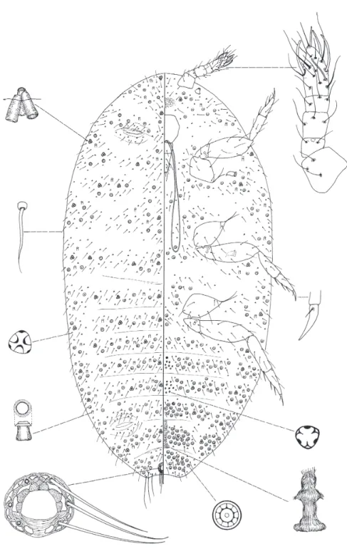
Fig. 14.Hambletonia bitubularisKOZÁRet FOLDIsp. n
Hambletonia bitubularis K
OZÁRet F
OLDIsp. n.
(Fig. 14)
Type material: The holotype female, in center of the slide (marked), one paratype female and an immature on the same slide, Peru, Oxypampa, 01.11.2000 (coll. G. SZÖVÉNYI), No. 6189, from moss. Three paratype females, on one slide, from the same collection as the holotype. One paratype female, Peru (coll. G. SZÖVÉNYI, YC 2400) No. 6190. One paratype female, Peru (coll. G. SZÖVÉNYI, YC 2500) No. 6194. Holotype deposited in the Hungarian Natural History Museum(Budapest, Hun- gary), paratypes deposited in Collection of Plant Protection Institute, Hungarian Academy of Sci- ences (Budapest, Hungary).
Description: Body elongate oval. Slide-mounted specimen (Fig. 14) 1.13 mm long and 0.67 mm wide. Antenna 5 segmented, the length of the segments: 1st – 53 µm, 2nd – 22 µm, 3rd – 38 µm, fourth – 24 µm, fifth – 76 µm long. One sensory pore on 2nd segment of the antenna. The 3rd segment is almost parallel sided. The apical segment has four falcate sensory setae, and one narrow, 38 µm long, blunt seta. Most segments of the antenna have a few hair-like setae, the longest one is 43 µm.
Eyes well visible. Anal lobe slightly developed with three long, hair-like setae.
Venter. Labium appears two-segmented, 101 µm long. Stylet loop twice longer than labium.
Cephalic plate visible. Legs robust: coxa of anterior legs 74 µm, trochanter 48 µm, femur 122 µm, tibia 84 µm, tarsus 84 µm, and claw 36 µm. Coxa of middle legs 70 µm, trochanter 55 µm, femur 127 µm, tibia 84 µm, tarsus 82 µm, and claw 34 µm long. Coxa of posterior legs 84 µm, trochanter 62 µm, femur 139 µm, tibia 101 µm and tarsus 89 µm, and claw 36 µm, tarsal digitules absent, claw digitules short, 9 µm long. Trochanter with two pores on each side. Claw without denticle. Legs with few hair-like setae, tibia and tarsus with 25 µm long setae. On segments few bitubular ducts present on margin, the tubes 5 µm in diameter, and 10 µm long. Instead of multilocular pores some five locular pores present on the last segment. The diameter of anterior spiracles 17 µm. With a small number of scattered hair-like setae. Circulus absent. Tubular ducts absent. Trilocular pores distributed on all segments. On median area of abdominal segments, two groups of unique trilocular pores, 4 µm in di- ameter.
Also one more unique trilocular pore group on the head. Internal genitalia have an unusual structure and as long as width of one segment. Internal genitalia with two lateral extensions appearing to be the well developed ductules of vaginal glands, situated at its middle part of vagina; between these glands and vulva, vagina is strongly enlarged.
Dorsum. Ostioles sclerotized. Multilocular pores absent. Anal ring oval, 50 µm wide and 43 µm long. Anal ring with six, 74 µm long hair-like setae. Anal ring pores (cells) typical, few, with spicules on pores of outer row. Bitubular ducts of one size, 4–6 on each segment, tubes 5 µm in diam- eter, 10 µm long. Tubular ducts absent. Hair-like setae 22 µm long, trilocular pores 3 µm wide, scat- tered on the dorsum, with a special structure, three central pores surrounded by nine small pores.
Distribution: Peru (Fig. 1).
Etymology.The species is named after the presence of bitubular pores.
CHECKLIST OF GENERA OF THE TRIBE RHIZOECINI Rhizoecus K
UNCKELD’H
ERCULAIS, 1878
Type species:Rhizoecus falciferKUNCKELD’HERCULAIS, 1878, by monotypy.
Ripersiella T
INSLEY, 1899
Type species:Ripersia rumicisMASKELL, 1892
Capitisetella H
AMBLETON, 1977
Type species:Pseudorhizoecus migransGREEN, 1933, by monotypy and original designation.
Pseudorhizoecus G
REEN, 1933
Type species:Pseudorhizoecus proximusGREEN, 1933, by original designation.
Pygmaeococcus M
CK
ENZIE, 1960
Type species:Pygmaeococcus morrisoniMCKENZIE, 1960, by monotypy and original designation.
Geococcus G
REEN, 1902
Type species:Geococcus radicumGREEN, 1902, by original designation.
Leptorhizoecus W
ILLIAMS, 1998
Type species:Leptorhizoecus deharvengiWILLIAMS, 1998
Brevicoccus H
AMBLETON, 1946a
Type species:Brevicoccus clavisetosusHAMBLETON, 1946a, by monotypy and original designation.
Prorhizoecus M
ILLERet M
CK
ENZIE, 1971
Type species:Prorhizoecus atopoporusMILLERet MCKENZIE, 1971, by monotypy and original designation.
Marottarhizoecus K
OZÁRet K
ONCZNÉB
ENEDICTY, 2002
Type species:Marottarhizoecus issisiKOZÁRet KONCZNÉBENEDICTY, 2002, by monotypy and original designation.
The genus Coccidella H
AMBLETON, 1946b was re-established, and two new genera added. By the new descriptions the number of genera in the Rhizoecini tribe increases to 13.
KEY TO THE GENERA OF THE TRIBE RHIZOECINI (after W
ILLIAMS(1998), T
ANG(1992), K
OZÁRand K
ONCZNÉB
ENEDICTY(2002) with changes and additions)
1 Anal ring with protuberances Pseudorhizoecus
– Anal ring without protuberances 2
2 Anal ring ventral in position Leptorhizoecus
– Anal ring dorsal in position 3
3 Bulbous tubular ducts present 4
– Bulbous tubular ducts absent 5
4 Cerarii present Prorhizoecus
– Cerarii absent Pygmaeococcus
5 All body setae knobbed 6
– All body setae flagellate 7
6 Antennae 3 segmented Capitisetella
– Antennae 4 segmented Brevicoccus
7 Antennae of 5 or 6 segments 8
8 Anal lobes well developed, with a stout spine-like seta Geococcus – Anal lobes not well-developed, without spine-like seta 9
9 Derm with tritubular pores 10
– Derm with bitubular pores 14
10 Body without groups of trilocular pores on venter, or around tritubular pores 13
– Body with groups of trilocular pores 11
11 With groups of trilocular pores on venter Coccidella
– Without groups of trilocular pores on venter 12
12 Tritubular pores surrounded by multilocular pores Marottarhizoecus – Tritubular pores surrounded by trilocular pores Benedictycoccina gen. n.
13 Body with tritubular cerores, without groups of trilocular pores Rhizoecus 14 Groups of trilocular pores on venter absent Ripersiella – Groups of trilocular pores on venter present Hambletonia gen. n.
*
Acknowledgements– The authors would like to thank the OTKA (Hungarian National Science Fund) (Grant No. T 034236), and Musèum National d’Histoire Naturelle, Paris, France, for financial support of this project. To Dr. S. MAHUNKAand late Prof. Dr. J. BALOGHwho made available for us to study the Arachnoidea collection of the Hungarian Natural History Museum (Budapest, Hungary), and to Dr. T. JERMYfor the grammatical corrections.
REFERENCES
BEN-DOV, Y. (1994)A systematic catalogue of the mealybugs of the World (Insecta: Homoptera:
Coccoidea: Pseudococccidae and Putoidae). Intercept Ltd., Andover, 686 pp.
DEMARZO, L., ROMANO, V. & TRANFAGLIA, A. (1990) Types of the female reproductive system in some scale insects (Homoptera: Coccoidea).Proceedings of ISSIS-VI, Krakow, pp. 41–46.
FERRIS, G. F. (1953)Atlas of the scale insects of North America. The Pseudococcidae (Part II).Stan- ford Univ. Press, California, pp. 279–506.
FOLDI, I. (1997) Internal anatomy of the adult female. Pp. 73–90.In:BEN-DOV, Y. & HODGSON, C.
J. (eds):Soft Scale Insects: Their Biology, Natural Enemies and Control. Elsevier, Amsterdam
& New York, 452 pp.
HAMBLETON, E. J. (1946a) Studies of Hypogeic Mealybugs.Rev. de Entomologia17: 1–77.
HAMBLETON, E. J. (1946b) A new name for a mealybug.Proc. Biol. Soc. Washington59: 177.
HAMBLETON, E. J. (1976) A revision of the New World mealybugs of the genus Rhizoecus (Homo- ptera: Pseudococcidae).USDA Techn. Bull.1522: 1–88.
KOZÁR, F. & KONCZNÉBENEDICTY, Z. (2002) Description of the Marottarhizoecus issisi gen. et sp.
nova (Homoptera, Coccoidea, Pseudococcidae, Rhizoecinae) from Africa with a review and key of the subfamily.Boll. Zool. Agr. Bachic.34: 213–218.
KOZÁR, F. & KONCZNÉBENEDICTY, Z. (2003) Description of four new species from Australian, Austro-Oriental, New Zealand and South Pacific regions (Homoptera, Coccoidea, Pseudo- coccidae, Rhizoecinae), with a review, and a key to the species Ripersiella.Boll. Zool. agr.
Bachic.35: 225–239.
KOZÁR, F. & MILLER, D. (2000) World revision of the genus Ortheziola Sulc, 1895 (Homoptera:
Coccoidea) with descriptions of eleven new species.Systematic Entomology25: 15–45.
MATILEFERRERO, D. (1976) La faune terrestre de l’Ile de Sainte-Helene. 7. Coccoidea. Annales.
Mus‚e Royal de l’Afrique Centrale. Tervuren. Serie in 8.Sciences Zoologiques215: 292–318.
MILLER, D. R., BEN-DOV, Y. & GIBSON, G. A. P. (2001) [1999]: Scalenet: a searchable information system on scale insects.Entomologica33: 37–46.
TANG, F. T. (1992)The Pseudococcidae of China.Chinese Agricultural Science Technology Press, Beijing, 767 pp.
WILLIAMS, D. J. (1998) Mealybugs of the genera Eumyrmococcus Silvestri associated with the ant genus Acropyga Roger and a review of the subfamily Rhizoecinae (Hemiptera, Coccoidea, Pseudococcidae).Bull. nat. Hist. Mus. Lond. (Ent.)67: 1–64.
WILLIAMS, D. J. (2004)Mealybugs of the Far East. Southdene Sdn. Bhd., Kuala Lumpur, Malaysia 904 pp. [in print]
WILLIAMS, D. J. & GRANARA DEWILLINK, M. C. (1992)Mealybugs of Central and South America.
C. A. B. International Institute of Entomology, London, 635 pp.
Revised version received November 10, 2004, accepted November 12, 2004, published November 24, 2004