Systematic & Applied Acarology 23(2): 367–379 (2018) http://doi.org/10.11158/saa.23.2.12
ISSN 1362-1971 (print) ISSN 2056-6069 (online) Article http://zoobank.org/urn:lsid:zoobank.org:pub:D15B2D49-6D27-40A5-B756-8BDD42D68B8D
New taxa of oribatid mites from Korup National Park (Cameroon).
The genus Pilizetes (Acari, Oribatida, Galumnidae)
SERGEY G. ERMILOV1*, JOSEF STARÝ2 & JENŐ KONTSCHÁN3
1Tyumen State University, Tyumen, Russia. E-mail: ermilovacari@yandex.ru
2Biology Centre v.v.i., Czech Academy of Sciences, Institute of Soil Biology, České Budějovice, Czech Republic. E-mail:
jstary@upb.cas.cz
3Center for Agricultural Research, Plant Protection Institute, Hungarian Academy of Sciences, Budapest, Hungary. E-mail:
jkontschan@gmail.com
*Corresponding author
Abstract
Two new species of oribatid mites of the genus Pilizetes (Oribatida, Galumnidae) are described from litter and soil in the Korup National Park (Cameroon). Pilizetes paradudichisp. nov. differs from Pilizetes dudichi Balogh, 1966 by the presence of long interlamellar setae and clearly longer notogastral setae. Pilizetes parasellnickisp. nov. differs from Pilizetes sellnicki Balogh, 1958 by the presence of thickened, heavily ciliated and long epimeral setae 3b,4a,4b and 4c.
Key words: galumnid mites, new species, morphology, systematics, Ethiopian region
Introduction
During taxonomic identification of oribatid mites (Acari, Oribatida) from the Korup National Park in Cameroon, we found two new species of the genus Pilizetes Sellnick, 1937 (Galumnidae), all from the nominative subgenus. The main goal of our paper is to describe and illustrate these new species.
The genus Pilizetes comprises two subgenera and 15 species (Ermilov & Klimov 2017; see Ermilov
& Koehler 2017 in addition), which are distributed in the Ethiopian region collectively (Subías 2004, updated 2017). The generic and subgeneric diagnoses were presented by Ermilov and Klimov (2017) and Ermilov and Koehler (2017). The identification keys to selective species of Pilizetes were presented by Balogh (1960, 1966), Mahunka (1984), Balogh and Balogh (2002) and Ermilov et al.
(2010).
At present, the Cameroonian oribatid mite fauna is poorly investigated, and only one species of the genus Pilizetes is known in this country: P.(Pseudopilizetes) camerunensis Ermilov, 2017 (see Ermilov & Koehler 2017).
Material and methods
Material examined.Pilizetes paradudichisp. nov. (holotype, male; seven paratypes, two females and five males) and Pilizetes parasellnickisp. nov. (holotype, male; 14 paratypes, three females and 11 males): Cameroon, South-West Province, Korup National Park, Rengo Camp, about 8 km NW of Mundemba, latitude 05°02'11.64"N, longitude 08°49'45.96"E, altitude 300 m, litter and soil sifting sample, 12–16.V.2006 (V.V. Grebennikov).
Methods. Specimens were mounted in lactic acid on temporary cavity slides for measurement and illustration. Body length was measured in lateral view, from the tip of the rostrum to the posterior edge of the ventral plate. Notogastral width refers to the maximum width of the notogaster in dorsal view (behind pteromorphs). Lengths of body setae were measured in lateral aspect. All body measurements are presented in micrometers. Formulas for leg setation are given in parentheses according to the sequence trochanter–femur–genu–tibia–tarsus (famulus included). Formulas for leg solenidia are given in square brackets according to the sequence genu-tibia-tarsus.
Drawings were made with a camera lucida using a Leica transmission light microscope “Leica DM 2500”.
Morphological terminology used in this paper follows that of F. Grandjean (see Ermilov &
Klimov 2017 for review and application).
The following abbreviations are used: L—lamellar line; S—sublamellar line; N—prodorsal leg niche; E, T—lateral ridges of prodorsum; r—ridge; f—furrow; ro, le, in, bs—rostral, lamellar, interlamellar and bothridial setae, respectively; D—dorsophragma; P—pleurophragma; c,la,lm,lp, h,p—notogastral setal alveoli; ia,ip—notogastral lyrifissures; gla—opisthonotal gland opening; h, m,a—subcapitular setae; or—adoral seta; v,l,d,cm,acm,ul,sul,vt,lt—palp setae; sac—axillary saccule; cha,chb—cheliceral setae; Tg—Trägårdh’s organ; Pd I,Pd II—pedotecta I, II, respectively;
1a,1c,3b,4a,4b,4c—epimeral setae; dis—discidium; cp—circumpedal carina; GP—genital plates;
AP—anal plates; g,ag,an,ad—genital, aggenital, anal and adanal setae, respectively; iad—adanal lyrifissure; p.o.—preanal organ; Tr, Fe, Ge, Ti, Ta—leg trochanter, femur, genu, tibia, tarsus, respectively; t—tooth; p.a.—leg porose area; ω,σ,φ—solenidia; ɛ—leg famulus; v,ev,bv,l,d,ft, tc,it,p,u,a,s,pv,pl—leg setae.
The following abbreviations of collections are used: SMNH—Senckenberg Museum of Natural History, Görlitz, Germany; TSUMZ—Tyumen State University Museum of Zoology, Tyumen, Russia.
Descriptions of new species
Pilizetes paradudichi sp. nov. (Figs 1–11)
Diagnosis. Body size: 332–356 × 232–257. Surface of notogaster and pteromorphs with system of depressions and ridges between them, forming a reticulate pattern. Prodorsum with system of ridges.
Rostral, lamellar and interlamellar setae well developed, le longest. Bothridial setae setiform, dilated in mediodistal part, ciliated. Notogastral porose areas and saccules absent. Notogastral setae of medium size, bacilliform, heavily barbed; pteromorphal setae not longer than others. Median pore absent. Epimeral setal formula: 1-0-1-3; 3b and 4a bacilliform, heavily barbed, others thin and setiform, slightly barbed. Genital setae setiform, slightly barbed; aggenital, anal and adanal setae erect, barbed.
Description.Measurements. Body length: 332 (holotype), 332–356 (7 paratypes); notogaster width: 240 (holotype), 232–257 (7 paratypes). No differences between females and males in body size.
Integument (Figs 1–4, 10, 11). Body brown to light yellow-brownish. Body surface densely microtuberculate (diameter of tubercles up to 2). Subcapitular mentum, genital and anal plates and margins of pteromorphs foveolate; foveolae on GP (up tp 8) larger than others (up to 2). Lateral sides of prodorsum slightly microgranulate.
Prodorsum (Figs 1–3). With well-developed system of ridges. Rostrum broadly rounded, with deep transverse furrow. Lamellar lines thickened, sublamellar lines thin, both parallel, curving
backwards at ventral ends. Prodorsal leg niches and lateral ridges of prodorsum well-developed.
Rostral (53–57), lamellar (77–82) and interlamellar (49–53) setae setiform, ro and in barbed, le densely ciliated; ro thinnest. Bothridial setae (110–118) setiform, but distinctly dilated in mediodistal part, ciliated. Exobothridial setae and sejugal porose areas absent. Dorsophragmata slightly visible.
Notogaster (Figs 1–4). Notogaster and pteromorphs with system of depressions and ridges, forming a reticulate pattern. Porose areas and saccules absent. With 10 pairs of bacilliform, heavily barbed setae, p1–p3 (41–45) shorter than others (53–61). Median pore absent. Lyrifissures (except distinct ia; and ip located between p1 and p2) not visible. Opisthonotal gland openings small, located lateral to h2and distanced from them.
Gnathosoma (Figs 5–7). Subcapitulum size: 102–110 × 90–98. Subcapitular setae setiform, smooth, a (16) longer than m and h(10–12). Adoral setae (10–12) setiform, barbed. Length of palps:
65–69. Postpalpal setae (6) spiniform, smooth. Length of chelicerae: 118–127. Cheliceral setae setiform, barbed, cha(41–49) longer than chb (26–32). Trägårdh’s organ of chelicerae long, elongate triangular.
Epimeral and lateral podosomal regions (Figs 2, 3). Anterior tectum of epimere I smooth. Pedotecta I and II rounded in ventral view. Discidia triangular, rounded distally. Epimeral setal formula: 1-0-1-3. Epimeral setae 3b (53–57) and 4a (45–49) bacilliform, heavily barbed, 1a,4b (14–16) and 4c (20) setiform, slightly barbed.
Circumpedal carinae long, reaching the level of pedotecta I.
FIGURE 1.Pilizetes paradudichisp. nov., adult: dorsal view (legs not shown). Scale bar 100 μm.
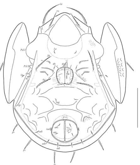
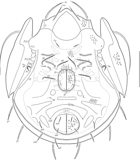
FIGURE 2.Pilizetes paradudichi sp. nov., adult: ventral view (gnathosoma and legs not shown). Scale bar 100 μm.
TABLE 1. Leg setation and solenidia of adult Pilizetes paradudichisp. nov. and P. parasellnickisp. nov.
Roman letters refer to normal setae, Greek letters to solenidia (except ɛ = famulus). Single prime (’) marks setae on anterior and double prime (”) setae on posterior side of the given leg segment. Parentheses refer to a pair of setae.
Anogenital region (Figs 2–4, 10, 11). With specific system of depressions, bordered by thick ridges (see Figs. 2, 10, 11). Six pairs of genital setae (g1, 24; g2, 20; g3, 16; g4–g6, 6–8) setiform, slightly barbed. One pair of aggenital (16), two pairs of anal (12) and three pairs of adanal (16) setae setiform, erect, barbed. Anterior edge of genital plates with three setae. Aggenital setae equal distanced from genital and anal plates. Adanal lyrifissures located parallel and close to anal plates.
Adanal setae ad1 posterior, ad2 posterolateral, ad3 lateral to anal aperture and located close to margin of the ventral plate. Distance ad1–ad2 equal to ad2–ad3.
Leg Tr Fe Ge Ti Ta
I v’ d, (l), bv” (l), v’,σ (l), (v),φ1,φ2 (ft), (tc), (it), (p), (u), (a), s, (pv), v’, (pl), l”, ɛ,ω1,ω2
II v’ d, (l), bv” (l), v’,σ (l), (v),φ (ft), (tc), (it), (p), (u), (a), s, (pv),ω1,ω2
III v’ d, ev’ l’,σ l’, (v),φ (ft), (tc), (it), (p), (u), (a), s, (pv)
IV v’ d, ev’ d, l’ l’, (v),φ ft'', (tc), (p), (u), (a), s, (pv)
FIGURES 3–4.Pilizetes paradudichisp. nov., adult: 3—lateral view (pteromorph, gnathosoma and legs not shown); 4—posterior view. Scale bar 100 μm.
FIGURES 5–7.Pilizetes paradudichisp. nov., adult: 5—subcapitulum, ventral view; 6—palp, right, antiaxial view; 7—chelicera, right, antiaxial view. Scale bars 17 μm (5, 7), 15 μm (6).
Legs (Figs 8, 9). Median claw distinctly thicker than laterals, all slightly barbed on dorsal side.
Genua IV with anterodorsal tooth. Porose areas on all femora and on trochanters III, IV poorly visible. Formulas of leg setation and solenidia: I (1-4-3-4-20) [1-2-2], II (1-4-3-4-15) [1-1-2], III (1- 2-1-3-15) [1-1-0], IV (1-2-2-3-12) [0-1-0]; homologies of setae and solenidia indicated in Table 1.
Famulus on tarsi I inserted posterolateral to solenidion ω1. Solenidion on tibiae IV inserted in anterior part of the segment.
Type deposition. The holotype (in ethanol with drop of glycerol) and three paratypes (in ethanol with drop of glycerol) are deposited in the collection of SMNH. Four paratypes (in ethanol with drop of glycerol) are deposited in the collection of TSUMZ.
Etymology. The specific name paradudichi refers to the similarity of the new species to the species Pilizetes dudichi Balogh, 1966.
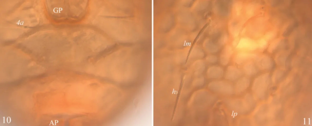
Remarks.In having notogaster and pteromorphs with system of depressions and ridges between them, forming a reticulate pattern, Pilizetes paradudichisp. nov. is morphologically most similar to Pilizetes dudichi Balogh, 1966 from Chad (see Balogh 1966), but differs by the presence of long interlamellar setae (vs. short in P. dudichi) and longer notogastral setae, lm reaching insertions of h3 (vs. shorter, lm clearly not reaching insertions of h3 in P. dudichi).
Pilizetes parasellnicki sp. nov. (Figs 12–22)
Diagnosis. Body size: 390–398 × 265–290. Body surface foveolate. Prodorsum with one pair of longitudinal ridges between lamellar setae. Rostral, lamellar and interlamellar setae well developed;
ro shortest. Bothridial setae setiform, dilated in mediodistal part, slightly barbed. Notogastral porose areas and saccules absent. Setae of notogaster of medium size, bacilliform, heavily barbed;
pteromorphal setae distinctly longer, setiform, slightly barbed. Median pore present. Epimeral setal formula: 2-0-1-3; 1a setiform, barbed, 3b,4a,4b and 4c thickened, heavily ciliated. Genital and aggenital setae setiform, erect, slightly barbed, anal and adanal setae bacilliform, barbed; ad longest.
FIGURES 8–9.Pilizetes paradudichisp. nov., adult: 8—leg I, without trochanter, right, antiaxial view; 9—leg IV, left, antiaxial view. Scale bar 50 μm.
Description.Measurements. Body length: 398 (holotype), 390–398 (14 paratypes); notogaster width: 282 (holotype), 265–290 (14 paratypes). No differences between females and males in body size.
FIGURES 10–11.Pilizetes paradudichisp. nov., adult, microscope images: 10—part of anogenital region;
11—part of notogaster.
FIGURE 12.Pilizetes parasellnickisp. nov., adult: dorsal view (legs not shown). Scale bar 100 μm.
FIGURE 13.Pilizetes parasellnicki sp. nov., adult: ventral view (gnathosoma and legs not shown). Scale bar 100 μm.
Integument (Figs 12–16, 21). Body brown to light yellow-brownish. Body surface (including subcapitular mentum and genital and anal plates) foveolate; foveolae on prodorsum and ventral side (up to 4) smaller than on notogaster and pteromorphs (up to 12). Lateral sides of prodorsum slightly microgranulate.
Prodorsum (Figs 12–14). Rostrum broadly rounded. With one pair of strong longitudinal ridges in the middle part and three small depressions between interlamellar setae. Lamellar lines thickened, sublamellar lines thin, both parallel, curving backwards at ventral ends. Prodorsal leg niches and lateral ridges of prodorsum well-developed. Rostral (53–65), lamellar (82–90) and interlamellar (82–
90) setae setiform, ro and in barbed, le densely ciliated; ro thinnest. Bothridial setae (118–135) setiform, but distinctly dilated in mediodistal part, slightly barbed. Exobothridial setae and sejugal porose areas absent. Dorsophragmata not visible.
Notogaster (Figs 12–15). Porose areas and saccules absent. With 10 pairs of setae, c very long (127–141), setiform, slightly barbed, others of medium size (57–65), bacilliform, heavily barbed.
Median pore present, located between insertions of setae h2. All lyrifissures (except distinct ia) and opisthonotal gland openings not visible.
Gnathosoma (Figs 16–18). Subcapitulum size: 102–110 × 90–98. Subcapitular setae setiform, smooth, a (14) longer than m and h(10). Adoral setae (10) setiform, barbed. Length of palps: 69–77.
Postpalpal setae (6) spiniform, smooth. Length of chelicerae: 123–135. Cheliceral setae setiform, barbed, cha (36–45) longer than chb (24–28). Trägårdh’s organ of chelicerae long, elongate triangular.
FIGURES 14–15.Pilizetes parasellnickisp. nov., adult: 14—lateral view (pteromorph, gnathosoma and legs not shown); 15—posterior view. Scale bar 100 μm.
FIGURES 16–18.Pilizetes parasellnicki sp. nov., adult: 16—subcapitulum, ventral view; 17—palp, right, antiaxial view; 18—chelicera, right, antiaxial view. Scale bars 17 μm (16, 18), 15 μm (17).
Epimeral and lateral podosomal regions (Figs 13, 14). Anterior tectum of epimere I smooth.
Pedotecta I and II rounded in ventral view. Discidia triangular, rounded distally. Epimeral setal formula: 2–0–1–3. Epimeral setae 1c represented by alveoli, 1a (12–16) setiform, erect, barbed, 3b (53–57), 4a,4b and 4c (32–41) thickened, heavily ciliated. Circumpedal carinae long, reaching the level of pedotecta I.
Anogenital region (Figs 13–15, 21, 22). With specific system of depressions, which are bordered by thick ridges (see Figs 13, 21, 22). Six pairs of genital (g1, g3–g6, 8–10; g2, 12–14) and one pair of aggenital (8–10) setae setiform, erect, slightly barbed. Anterior edge of genital plates with three setae. Aggenital setae located closer to genital plates than to anal plates. Two pairs of anal (20–28) and three pairs of adanal (ad1,ad2, 41–45; ad3, 28–36) setae bacilliform, barbed. Adanal lyrifissures located parallel to anal plates and slightly distanced from them. Adanal setae ad1 posterior, ad2 posterolateral, ad3 lateral to anal aperture and located close to margin of the ventral plate. Distance ad1–ad2 equal to ad2–ad3.
Legs (Figs 19, 20). Median claw distinctly thicker than laterals, all slightly barbed on dorsal side.
Genua IV with anterodorsal tooth. Porose areas on all femora and on trochanters III, IV poorly visible. Formulas of leg setation and solenidia: I (1-4-3-4-20) [1-2-2], II (1-4-3-4-15) [1-1-2], III (1- 2-1-3-15) [1-1-0], IV (1-2-2-3-12) [0-1-0]; homologies of setae and solenidia indicated in Table 1.
Famulus on tarsi I inserted posterolateral to solenidion ω1. Solenidion on tibiae IV inserted in anterior part of the segment.
Type deposition. The holotype (in ethanol with drop of glycerol) and three paratypes (in ethanol with drop of glycerol) are deposited in the collection of SMNH. Eleven paratypes (in ethanol with drop of glycerol) are deposited in the collection of TSUMZ.
Etymology. The specific name parasellnicki refers to the similarity of the new species to the species Pilizetes sellnicki Balogh, 1958.
Remarks.In general morphological traits (body surface foveolate; porose areas and saccules absent; bothridial setae slightly barbed; setae of notogaster of medium size, pteromorphal setae long, setiform; median pore present1; body of medium size), Pilizetes parasellnicki sp. nov. is morphologically most similar to Pilizetes sellnicki Balogh, 1958 from Angola (see Balogh 1958,
1960), but differs by the presence of thickened, heavily ciliated and comparatively long epimeral setae 3b,4a, 4b and 4c (vs. simple in P. sellnicki).
FIGURES 19–20.Pilizetes parasellnickisp. nov., adult: 19—leg I, without trochanter, right, antiaxial view;
20—leg IV, left, antiaxial view. Scale bar 50 μm.
1. Balogh (1958, 1960) did not describe and not draw a median pore in Pilizetes sellnicki. We studied the type material from the Hungarian National Museum (Budapest, Hungary) and note that a median pore is present in this species.
2018 ERMILOV ET AL.: NEW TAXA OF ORIBATID MITES FROM CAMEROON 379
FIGURES 21–22.Pilizetes parasellnickisp. nov., adult, microscope images: 21—part of epimeral region; 22—
part of anogenital region.
Acknowledgements
We cordially thank Dr. Marut Fuangarworn (Chulalongkorn University, Bangkok, Thailand) and two anonymous reviewers for the valuable comments. The reported study was funded by the Russian Foundation for Basic Research (RFBR) according to the research project № 18-04-00096 and by the Academy of Sciences of the Czech Republic, under Research Plan No. AV0Z606960521.
References
Balogh, J. (1958) Oribatides nouvelles de l'Afrique tropicale. Revue de Zoologie et de Botanique Africaines, 58(1–2), 1–34.
Balogh, J. (1960) Oribates (Acari) nouveaux d'Angola et du Congo Belge (2ème série). Companhia de Dia- mantes de Angola, Lisboa, 51, 15–40.
Balogh, J. (1966) On some oribatid mites from Tshad and East Africa collected by Prof. H. Franz, Vienna.
Opuscula Zoologica Budapest, 6(1), 69–77.
Balogh, J. & Balogh, P. (2002) Identification keys to the oribatid mites of the Extra-Holarctic regions. Vol. 1.
Miskolc, Well-Press Publishing Limited, 453 pp.
Ermilov, S.G. & Klimov, P.B. (2017) Generic revision of the large-winged mite superfamily Galumnoidea (Acari, Oribatida) of the world. Zootaxa, 4357(1), 1–72.
https://doi.org/10.11646/zootaxa.4357.1.1
Ermilov, S.G. & Koehler, H.H. (2017) New data on oribatid mites (Acari, Oribatida) of Cameroon: results of the Joint German-Cameroonian scientific expedition (April 2016). Systematic & Applied Acarology, 22(12), 2233–2244.
https://doi.org/10.11158/saa.22.12.13
Ermilov, S.G., Sidorchuk, E.A. & Rybalov, L.B. (2010) New species of oribatid mites of the superfamily Galumnoidea (Acari: Oribatida) from Ethiopia. Zootaxa, 2646, 43–62.
Mahunka, S. (1984) Oribatids of the Eastern part of the Ethiopian Region (Acari). V. Acta Zoologica Hun- garica, 30(1–2), 87–136.
Sellnick, M. (1937) Eine neue Milbe aus Ostafrika. Zoologischer Anzeiger, 117, 130–132.
Subías, L.S. (2004) Listado sistemático, sinonímico y biogeográfico de los ácaros oribátidos (Acariformes:
Oribatida) del mundo (excepto fósiles). Graellsia, 60 (número extraordinario), 3–305. Online updated version accessed in February 2017, 598 pp.; http://escalera.bio.ucm.es/usuarios/bba/cont/docs/RO_1.pdf
Submitted: 23 Nov. 2017; accepted by Marut Fuangarworn: 4 Feb. 2018; published: 20 Feb. 2018