_____________________________________________________________
Two new species of centipedes, Lithobius keelungensis sp. nov.
and Lithobius (Monotarsobius) qingquanensis sp. nov., from Taiwan (Chilopoda, Lithobiomorpha, Lithobiidae)
J.L.CHAO1*,K.S.LEE2&H.W.CHANG3
1Jui-Lung Chao, Invertebrate Section, Biology Department, National Museum of Natural Science (NMNS), 1 Guancian Road, Taichung 40453, Taiwan, R.O.C., E-mail: chaojuilung@gmail.com, *Corresponding author
2Kwen-Shen Lee, Invertebrate Section, Biology Department, National Museum of Natural Science (NMNS), 1 Guancian Road, Taichung 40453, Taiwan, R.O.C.
3Hsueh-Wen Chang, Department of Biological Sciences, National Sun Yat-Sen University, 70 Lien-Hai Road, Kaohsiung 804, Taiwan, R.O.C.
Abstract. Male secondary sexual characters are diverse in Taiwanese Lithobius. We describe two new species with their male secondary sexual characters, Lithobius (Ezembius) keelungensis sp. nov. and Lithobius (Monotarsobius) qingquanensis sp. nov.. In L. (E.) keelungensis, male 14–15th femora and tibiae are markedly thick, the femora have a deep furrow on each dorsal surfaces, and the tibiae are oval, with a wide shallow excavation on each dorsal surfaces.
In L. (M.) qingquanensis, a small wart-like outgrowth bearing about 15 slightly curved setae is present on the dorsoposterior surface of the male 15th femur.
Keywords. Keelung, male secondary sexual character, taxonomy.
INTRODUCTION
he centipede genus Lithobius Leach, 1814 is among the poorly studied taxa of Taiwan.
Takakuwa (1939, 1941a, 1941b) described and recorded eight species of Lithobius from Tai- wan. However, his specimens were destroyed in an air attack during the war in 1945. Wang (1955, 1956, 1957, 1959, 1963) recorded the localities of seven species of Lithobius from Taiwan without descriptions, and we could not locate Wang’s specimens in any collections. We studied new specimens of Lithobius from Tai- wan, and deposited them at the National Mu- seum of Natural Science (NMNS). Male sec- ondary sexual characters are important characters in the taxonomy of Lithobius: several several longitudinally arranged long setae were present on the ventral surface of male 15th tibia in Lithobius trichopus Takakuwa, 1939; a small tunnel at the top of a longitudinal excavation is present on the dorsal surface of 14th tibia in
male Lithobius ongi Takakuwa, 1939, and the tunnel and bottom of the excavation bear nu- merous small pores (Chao et al. 2018a); a large ventral swelling on the male 15th femur in Lithobius (Monotarsobius) meifengensis Chao, Lee & Chang, 2018, and the apical region of the swelling bearing numerous small pores (Chao et al. 2018b). We here describe two new species from Taiwan, Lithobius (Ezembius) keelungen- sis sp. nov. and Lithobius (Monotarsobius) qingquanensis sp. nov. using two other types of male secondary sexual characters.
MATERIAL AND METHODS One and fifteen specimens, respectively, of the two new species treated below were col- lected from Keelung City and Hsinchu County, Taiwan. The specimens were examined by light microscopy (Leica MZ16) and SEM (Hitachi SU-1510). Type specimens are preserved in 75% alcohol and deposited in the Department of
T
Biology, National Museum of Natural Science, Taichung, Taiwan. Terminology of the external anatomy follows Bonato et al. (2010). The fol- lowing abbreviations are used in the text and tables: a––anterior, C––coxa, F––femur, m––
median, p––posterior, P––prefemur, S/SS––
sternite/sternites; t––trochanter, T/TT––
tergite/tergites, Ti––tibia.
TAXONOMY
Order Lithobiomorpha Pocock, 1895 Family Lithobiidae Newport, 1844
Genus Lithobius Leach, 1814 Subgenus L. (Ezembius) Chamberlin, 1919
Lithobius keelungensis sp. nov.
(Figures 1–15)
Material examined. Holotype. ♂ (NMNS8103-001): forest floor, Hepin Island, Keelung City, Taiwan, 25°09.36'N, 121°45.94'E, 13 m in elevation, 12 Jan 2019, leg. Jui-Lung Chao. Paratypes, 1♂
(NMNS8103-002): same data as holotype.
Other material, 2♂♂, 2♀♀ (NMNS7843- 019, NMNS7843-020, NMNS7843-021, NM- NS7843-022), same locality as holotype, 14 Jan 2018, leg. Jui-Lung Chao; 1♀ (NMNS8103- 003), 12 Jan 2019 and 2♂♂, 4♀♀ (NMNS8103- 004), 08 Apr 2019, forest floor, Keelung City, 25°07.85'N, 121°43.72'E, 84 m, leg. Jui-Lung Chao; 2♂♂ (NMNS8103-005), forest floor, Keelung City, 25°08.90'N, 121°46.73'E, 33 m, 27 Jan 2018, leg. Jyh-Jong Cherng.
Etymology. Refers to the type locality.
Diagnosis. A Lithobius species, antennae with 20 articles; 7–9 ocelli arranged in three ir- regular rows [1 + 2, 3(4), 2(1)], posterior ocellus largest, two posterosuperior ocelli large, ventral seriate ocelli smallest; Tömösváry’s organ larger than adjacent ocelli; 2+2 coxosternal teeth;
porodonts posterolateral to outer tooth; all tergites lacking posterior triangular projections;
coxal pores 4–6, round; male secondary sexual characters on 14–15th: femora and tibiae mark-
edly thick, 14–15th femora with a furrow on each dorsal surface, male 14–15th tibiae oval, with a wide shallow excavation on each dorsal surfaces; male gonopods with three long setae;
female gonopods with 2+2 sharp coniform spurs, point of terminal claw undivided, a small sharp lateral denticle on base of terminal claw.
Description. Body length: 11–13.5 mm.
Body colour: dark brown (Figs. 1–2). Antennae with 20 articles; basal article width subequal to length, following articles markedly longer than wide; distal article much longer than wide, up to 3.4 times as long as wide; abundant setae on antennal surface, less so on basal articles, grad- ual increase in density to around fourth article, then more or less constant in number. Cephalic plate smooth, convex, width subequal to length, posterior marginal ridge moderately broader and weakly concave; setae scattered sparsely over whole surface (Fig. 7). 7–9 ocelli on each side [1 + 2, 3(4), 2(1)], one posterior, two dorsal, three or four middle and one or two ventral, ar- ranged in three irregular rows; posterior ocellus largest, two posterosuperior ocelli large, ventral seriate ocelli smallest; ocelli domed, translucent, usually darkly pigmented (Figs. 3–4).
Tömösváry’s organ nearly rounded, situated at the anterolateral margin of the cephalic plate, larger than adjoining ocelli (Fig. 4). Forcipular coxosternite sub-trapezoidal, anterior margin narrow, external side slightly longer than inter- nal side; median longitudinal cleft moderately deep; anterior border with 2+2 large triangular coxosternal teeth, inner tooth slightly larger than outer one; porodonts moderately slender, seti- form, posterolateral to the outermost tooth (Figs.
5–6); some scattered setae on the ventral side of coxosternite.
Tergites smooth, without wrinkles, backside slightly hunched; T1 generally trapeziform, pos- terior margin narrower than anterior margin, nar- rower than T3 and cephalic plate; T3 slightly nar- rower than cephalic plate; TT7, 8 and 10 broader than other tergites; T7 slightly rectangular, about 0.5 times as long as wide, posterior margin of T7 straight (Fig. 7); posterior margin of TT1, 3, 5 weakly concave, TT8, 10, 12 concave, T14 lateral deeply concave, middle straight (Figs. 8–9); TT1, 3 and 5 with continuous lateral and posterior
marginal ridges, other tergites with discontinuous posterior marginal ridges; posterior angles of all tergites lacking triangular projections (Fig. 8);
tiny setae scattered very sparsely over the surface.
Sternites narrower posteriorly, generally trapezi- form, comparatively smooth, setae emerging from pores scattered very sparsely over the sur- face (Figs. 10–12).
Figures 1–6. Lithobius (Ezembius) keelungensis sp. nov. 1 = habitus, male, legs 14–15 thick; 2 = habitus, female; 3 = lateral view of the head; 4 = eight ocelli and Tömösváry’s organ (To) on the left side; 5 = ventral view of the head; 6 = 2+2 coxosternal teeth
and porodonts (1 & 3–6: NMNS8103-001; 2: NMNS8103-004).
Figures 7–15. Lithobius (Ezembius) keelungensis sp. nov. 7 = male, habitus, dorsal view by SEM; 8 = posterior body on male, dorsal view; 9 = 14th tergite; 10 = posterior body on male, ventral view; 11 = 14th claw with short anterior (aas) and long posterior
accessory spines(pas); 12 = coxal pores of 12–15th leg; 13 = male first genital sternite and gonopods (arrow); 14 = female first genital sternite and gonopods; 15 = claw of female gonopod (7: NMNS8103-005; 8–13: NMNS8103-001;
14: NMNS8103-004; 15: NMNS8103-004).
Legs: tarsi well-defined on all legs; all legs with fairly long claws, curved ventrally; thick posterior accessory spines present on base of all claws; long and slender anterior accessory spines present on claws 1–13; very short ante- rior accessory spines on 14th claws (Fig. 11);
legs 15 lack anterior spine. Male secondary sex- ual characters (see below) present on thick 14th and 15th legs (Figs. 8–10); female without sec- ondary sexual characters. Leg plectrotaxy as in Table 1. Coxal pores 4–6, round, inner pores small, coxal pore field set in a relatively shallow
groove, margin of coxal pore-field with slightly eminence (Fig. 12).
Male secondary sexual characters: 14–15th femora and tibiae markedly thick (Figs. 8–10), and 14–15th femora with a deep furrow on each dorsal surface; male 14–15th tibiae oval, with a wide shallow excavation on each dorsal surfaces (Figs. 7–8). Male first genital sternite: wider than long, usually well chitinized; posterior margin quite deeply concave between the gono- pods, without a medial bulge; comparatively
long setae scattered evenly on the ventral sur- face; male gonopods short and small, as a semi- spherical bulge with 3 long setae, apically well chitinized (Fig. 13).
Female first genital sternite: wider than long; posterior margin of the genital sternite deeply concave, with a medial bulge (Fig. 14);
short to long setae sparsely scattered over the ventral surface of the genital segment. Female gonopod: first article fairly broad, bearing up to 23 long setae, arranged in four irregular rows;
2+2 sharp coniform spurs, inner spur smaller (Fig. 14); second article with 7–9 rather long setae arranged in two irregular rows on its ven- tral side; third article usually with 2–3 long se- tae on its ventral surface; point of terminal claw undivided, a small sharp lateral denticle on the base of the terminal claw (Fig. 15).
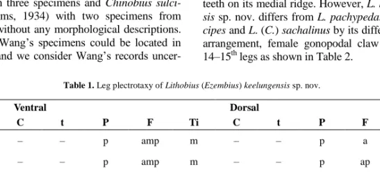
Remarks. Wang (1956, 1959) recorded Chinobius sachalinus Verhoeff, 1937, with six specimens, Chinobius pachypedatus Takakuwa, 1941 with three specimens and Chinobius sulci- pes (Attems, 1934) with two specimens from Taiwan, without any morphological descriptions.
None of Wang’s specimens could be located in Taiwan, and we consider Wang’s records uncer-
tain. According to the descriptions of Takakuwa (1941b), Matic (1973), Zalesskaja (1978) and Eason (1996), the male secondary sexual characters – markedly thick male 14–15th femora and tibiae – are present in L. (Chinobius) sachalinus Verhoeff, 1937, L. pachypedatus Takakuwa, 1938 and L. sulcipes (Attems, 1927) from Japan and Eastern Russia. Takakuwa (1941b) redescribed L. pachypedatus with 1+5 ocelli on each side, and male 15th femora without any furrows, L. sulcipes with seven ocelli in two rows on each side, and male 14–15th femora and 15th tibiae with a deep furrow, and L. (C.) sacha- linus with nine ocelli on each side, male 15th femora and tibiae with a furrow, and 14th femora with a longitudinal arched swelling with dense setae. The same character-set of L. (C.) sachali- nus is also found in Matic’s key (1973) and Zalesskaja’s description (1978). Furthermore, Zalesskaja (1978) and Eason (1996) added the female characters for L. (C.) sachalinus: 2+2 spurs on the female gonopod, and gonopodal claw without denticles but with two or three large teeth on its medial ridge. However, L. keelungen- sis sp. nov. differs from L. pachypedatus, L. sul- cipes and L. (C.) sachalinus by its different ocelli arrangement, female gonopodal claw and male 14–15th legs as shown in Table 2.
Table 1. Leg plectrotaxy of Lithobius (Ezembius) keelungensis sp. nov.
leg pair Ventral Dorsal
C t P F Ti C t P F Ti
1 – – p amp m – – p a a
2 – – p amp m – – p ap a
3 – – p amp am – – p ap a
4 – – mp amp am – – p ap ap
5–10 – – mp amp am – – ap ap ap
11–12 – – mp amp am – – amp ap ap
13 – – amp amp am – – amp ap ap
14 – m amp amp am – – amp p p
15 – m amp am a – – amp p –
Table 2. Main morphological characters of four species of Lithobius from East Asia.
Species L. (E.) keelungensis sp.
nov.
L. (C.) pachypedatus L. (C.) sulcipes L. (C.) sachalinus Description
from
This paper Takakuwa (1938, 1941b)
Takakuwa (1941b) Takakuwa (1941b) Matic (1973) Zalesskaja (1978)
Eason (1996)
Antennae 20 articles 20–21 articles 19–22 articles 19–20 articles
Ocelli 1+2,3,2; arranged in 3 rows
1+5; arranged in 2 rows
1+6; arranged in 2 rows
1+5~8; arranged in 2 rows Male 15th
femur
A deep dorsal furrow No furrow A deep dorsal furrow A dorsal furrow Male 15th
tibia
A wide dorsal furrow No furrow A deep dorsal furrow A dorsal furrow Male 14th
femur
A dorsal furrow; absence of dense setae on dorsoposterior surface
No furrow; absence of dense setae on dorsoposterior surface
A deep dorsal furrow;
absence of dense se- tae on dorsoposterior
surface
A dorsal furrow; sev- eral setae clustered on dorsoposterior surface
Coxal pore 4–6, round 3–6, round 4–5, round 4–6, round
Male gonopod
3 long setae Several setae No data 3 long setae
Female gonopod
2+2 spurs; claw with a large denticle on tip, a small sharp lateral
denticle on base
3+3 spurs; claw with a denticle on tip, and a small tooth on its
medial ridge
2+2 spurs; claw divided, biapiculate
2+2 spurs; claw without denticles, but
with two or three large teeth on its medial ridge
Genus Lithobius Leach, 1814 Subgenus L. (Monotarsobius) Verhoeff, 1905
Lithobius (Monotarsobius) qingquanensis sp. nov.
(Figures 16–25)
Material examined. Holotype, ♂ (NMNS81 03-006), garden, Qingquan, Hsinchu County, Taiwan, 24°34.36'N, 121°06.34'E, 570 m in ele- vation, 13 Mar. 2019, leg. Jui-Lung Chao.
Etymology. Refers to the type locality.
Diagnosis. A Lithobius (Monotarsobius) species with 17–18 elongate antennal articles;
body colour brown; 3 ocelli arranged in one row, middle ocellus largest; Tömösváry’s organ in front of ocelli, slightly smaller than anterior
ocellus; 2+2 coxosternal teeth; porodonts pos- terolateral to the outermost tooth; all tergites lack posterior triangular projections; TT1, 3 and 5 with continuous lateral and posterior ridges;
posterior margin of TT1, 3, 5, 8, 10 and 12 weakly concave, posterior margin of T14 con- cave; tarsi fused on legs 1–13, well-defined on legs 14–15; male secondary sexual characters on dorsal surface of 15th femur, a longitudinal ex- cavation on central surface, and a small wart- like outgrowth with about 15 slightly curved setae on posterointernal surface; both 15th tibia and 15th tarsus I oval in male, with a wide shal- low excavation on the dorsal surface; coxal pores 3433, round; male gonopods with two long setae.
Description. Body length: 8 mm. Body col- our: brown (Fig. 16). Antennae with 17–18 arti- cles; most articles markedly longer than wide;
distal article about 2.8 times as long as wide (Fig. 17); abundant setae on antennal surface,
less so on basal articles, gradual increase in den- sity to around fifth article, then more or less
Figures 16–25. Lithobius (Monotarsobius) qingquanensis sp. nov. 16 = habitus, male; 17 = antennae; 18 = three ocelli and Tömösváry’s organ (To) on the left side; 19 = anterior body, dorsal view; 20 = ventral view of the head; 21 = 2+2 coxosternal teeth
and porodonts; 22 = posterior body and 15th leg, dorsal view; 23–24 = 15th femur, lateral view;
25 = male first genital sternite and gonopods (16–25: NMNS8103-006).
constant in number. 3 ocelli on each side, ar- ranged in one row, middle ocellus largest (Fig.
18); ocelli domed, translucent, usually darkly pigmented. Tömösváry’s organ comparatively small, nearly rounded, in front of ocelli, slightly smaller than the anteriormost ocellus (Fig. 18).
Cephalic plate smooth, convex, width subequal to length, posterior marginal ridge moderately broader and weakly concave (Fig. 19); setae scattered sparsely over the whole surface. For- cipular coxosternite with 2+2 large triangular teeth, outer tooth slightly larger than inner one, the line of their apices recurved (Figs. 20–21);
porodonts moderately slender, setiform, poster- olateral to the outer tooth (Figs. 20–21); some scattered setae on ventral side of coxosternite.
Tergites smooth, without wrinkles, backside slightly hunched; T1 generally trapeziform, poste-
rior margin narrower than anterior margin, nar- rower than T3 and cephalic plate (Fig. 19); TT1, 3 and 5 with continuous lateral and posterior ridges;
posterior margin of TT1, 3, 5, 8, 10 and 12 weakly concave, posterior margin of T14 concave; all tergites lack posterior triangular projections; tiny setae scattered very sparsely over the surface.
Sternites narrower posteriorly, generally trapeziform, comparatively smooth, setae emerging from pores scattered very sparsely over the surface.
Legs: tarsi fused on legs 1–13, well-defined on legs 14–15; all legs with fairly long claws, curved ventrally; anterior accessory spine long and slender on legs 1–13, lacking on legs 14–15;
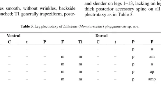
thick posterior accessory spine on all legs; leg plectrotaxy as in Table 3.
Table 3. Leg plectrotaxy of Lithobius (Monotarsobius) qingquanensis sp. nov.
leg pairs Ventral Dorsal
C t P F Ti C t P F Ti
1 – – – – – – – p a a
2 – – – m m – – p am a
3–4 – – – m m – – p a a
5 – – – m m – – p ap a
6 – – – m m – – p amp ap
7–9 – – – m m – – p ap ap
10 – – a m m – – p ap ap
11 – – a m m – – mp ap ap
12 – – mp m m – – mp ap ap
13 – – mp m m – – mp p ap
14 – m mp m – – – amp – –
15 – m mp m – – – amp – –
Male secondary sexual character on dorsal surface of 15th femur: a longitudinal excavation on central surface (Fig. 22), several long setae scattered sparsely over the surface, and a small wart-like outgrowth bearing about 15 slightly curved setae present on dorsoposterior surface (Figs. 23–24); both 15th tibia and tarsus I oval, with a wide shallow excavation on the dorsal surface (Fig. 22).
Coxal pores: 3433, round, inner pores small, coxal pore field set in a relatively shallow groove, margin of coxal pore-field with slightly eminence (Fig. 25).
Male sternite 15: trapeziform, posterolater- ally narrower than anterolaterally, posterior margin straight, long setae scattered sparsely over the surface (Fig. 25). Male first genital
sternite: wider than long, usually well chi- tinized; posterior margin quite deeply concave between gonopods, without a medial bulge;
comparatively long setae evenly scattered on ventral surface; male gonopods short and small, as a semi-spherical bulge with two long setae, apically slightly chitinized (Fig. 25).
Remarks. Murakami (1965) described a Lithobius (Monotarsobius) species from Japan, L. (M.) tuberculatus Murakami, 1965, with a male secondary sexual character, a wart-like outgrowth with several long curved setae on the dorsoposterior surface of male 15th tibiae. Matic (1970) described two Lithobius (Monotarsobius) species from Korea, L. (M.) dziadoszi Matic, 1970 and L. (M.) riedeli, Matic, 1970. A wart- like outgrowth with several short setae on the dorsoposterior surface of 15th femora is present in male L. (M.) dziadoszi, and a similar out- growth with several short setae on the dorsoposterior surface of 15th tibiae in male L.
(M.) riedeli. However, L. (M.) qingquanensis sp.
nov. differs from L. (M.) dziadoszi by its 3 ocelli arranged in one row, and Tömösváry’s organ slightly smaller than the adjoining ocellus, con- trasting with L. (M.) dziadoszi with 7 ocelli ar- ranged in two irregular rows, and Tömösváry’s organ larger than the adjoining ocellus. L. (M.) qingquanensis sp. nov. differs from L. (M.) tu- berculatus and L. (M.) riedeli by its wart-like outgrowth on 15th femora, contrasting with L.
(M.) tuberculatus and L. (M.) riedeli with their wart-like outgrowth on 15th tibiae.
Key to the known Taiwanese species of the genus Lithobius Leach, 1814
1. Posterior angles of TT9, 11 and 13with triangular project- tions ……….…2 – Posterior angles of TT9, 11 and 13 without triangular pro- jecttions .……...………..…...………...…4
2. Male secondary sexual characters present on leg 14 or 15 ...
………..3 – Male secondary sexual characters absent ………...……
………..………...…..L. bidivisa Takakuwa, 1939
3. Posterior angles of T7 with a triangular projection; a small tunnel at top of a longitudinal excavation on dorsal surface of male 14th tibia ……..………..…L. ongi Takakuwa, 1939 – Posterior angles of T7 without projections; about 40 long setae longitudinally arranged on ventral face of male 15th tibia, most setae concentrated in posterior part ……….
………..L.trichopus Takuwa, 1939
4. Tarsi fused on legs 1-13, well-defined on legs 14-15 …….
……….5, subgenus L. (Monotarsobius) – Tarsi well-defined on all legs …...9, subgenus L. (Ezembius)
5. Male secondary sexual characters present on leg 15 ….…..6 – Male secondary sexual characters absent ………...………..7
6. A large swelling on ventral surface of male 15th femur, apical region of swelling bearing numerous small pores …..
…...………..L.(M.) meifengensis Chao, Lee & Chang, 2018 – A small wart-like outgrowth bearing ca. 15 slightly curved setae present on dorsoposterior surface of male 15th femur ..
………...L. (M.) qingquanensis sp. nov.
7. 2222 coxal pores; terminal claw of female gonopod di- vided, biapiculate ……...L. (M.) obtusus Takakuwa, 1941 – 3-5 coxal pores; terminal claw of female gonopod undivided
………..8
8. 5555 coxal pores; a small sharp tooth on the base of termi- nal claw of female gonopod ………..
………...…L. (M.) ramulosus Takakuwa, 1941 – 3–4 coxal pores; terminal claw of female gonopod with irregular internal and external ridges ...
... L. (M.) holstii (Pocock, 1895)
9. Male secondary sexual characters present both on leg 14- 15:14-15th femora and tibiae thick, 14-15th femora with a furrow on each dorsal surfaces; female gonopods with 2+2 spurs ………...……. L. (E.) keelungensis sp. nov.
– Male secondary sexual characters absent; female gonopods with 3+3 spurs ………...10
10. Female gonopodal claw bipartite, a large denticle on tip, a small sharp lateral denticle on base of terminal claw
….………..L (E.) bidens Takakuwa, 1939 – Female gonopodal claw undivided, without a lateral denticle
………...L. (E.) lineatus Takakuwa, 1939
Acknowledgements – We are grateful to Dr. Gregory D.
Edgecombe for everlasting help during our study. Thanks also to Mr. Jyh-Jong Cherng for his specimens, to Mr. Shih-Chen Chang for translating the Japanese literatures, and to Ms.
Shao-Kang Hu for preparing the SEM micrographs. We are deeply obliged to Prof. Sergei Golovatch, Prof. Huiqin Ma, Dr. Carlos Alberto Martinez-Muñoz and Dr. Alexandr Evsyu- kov for their help with literature.
REFERENCES
BONATO, L., EDGECOMBE, G.D., LEWIS, J.G.E., MINELLI. A.,PEREIRA, L.A.,SHELLEY,R.M.&
ZAPPAROLI,M.(2010): A common terminology for the external anatomy of centipedes (Chilop- oda). ZooKeys, 69: 17–51.
doi: 10.3897/zookeys.69.737
CHAO,J.L.,LEE,K.S.&CHANG,H.W. (2018a): Neo- type designation and redescription of the centi- pede Lithobius ongi Takakuwa, 1939 (Chilopoda,
Lithobiomorpha, Lithobiidae). Collection and Re- search, 31: 63–69.
CHAO, J.L., LEE, K.S. & CHANG, H.W. (2018b):
Lithobius (Monotarsobius) meifengensis, a new species of centipede from high altitude forest in central Taiwan (Chilopoda, Lithobiomorpha, Lithobiidae). ZooKeys, 741: 181–192.
doi: 10.3897/zookeys.741.21036
EASON,E.H. (1996): Lithobiomorpha from Sakhalin Island, the Kamchatka Peninsula and the Kurile Islands. Arthropoda Selecta, 5(3/4): 117–123.
MATIC,Z. (1970): Contribution à la connaissance des Lithobiides (Chilopoda, Lithobiomorpha) de Corée. Annales Zoologici Warszawa, 28: 55–63.
MATIC, Z. (1973): Revision du genre Chinobius Verhoeff, avee des descriptions de trios espèces nouvelles (Chilopoda, Lithobiidae). Annales Zo- ologici Warszawa, 30: 33–47.
MURAKAMI,Y. (1965): Postembryonic development of the common Myriapoda of Japan. XIX. Two new species of Monotarsobius (Chilopoda:
Lithobiidae). Zoological Magazine, 74: 69–75.
TAKAKUWA,Y. (1938): Über neue Art von Monotar- sobius und von Lithobius aus Japan. Zoological Magazine, 50(11): 456–461.
TAKAKUWA,Y. (1939): Über Japanische Lithobius- Arten. Transactions of the Sapporo Natural His- tory Society, 16(1): 29–30, 36–37.
TAKAKUWA, Y. (1941a): Über Einige Japanische Lithobiiden. Transactions of the Natural History Society of Formosa, 31(213): 292–297.
TAKAKUWA,Y. (1941b): Class Chilopoda, Epimor- pha, Lithobiomorpha. Fauna Nipponica, Vol. 9 Fas. 8 No. (3). Sanseido Book Store, Tokyo, 104 pp..
WANG,Y.H.M. (1955): Serica 1a: Records of myria- pods on Formosa with description of new species.
Quarterly Journal of the Taiwan Museum, 8: 13–
16.
WANG,Y.H.M. (1956): Serica 1e: Records of myria- pods on Formosa with description of new species (2). Quarterly Journal of the Taiwan Museum, 9:
155–159.
WANG,Y.H.M. (1957): Serica 1f: Records of myria- pods on Taiwan Islands (3) - Pescadore Islets, Kao-Yung, Pingtung, Changhua and Taipei.
Quarterly Journal of the Taiwan Museum, 10:
23–29.
WANG,Y.H.M. (1959): Serica 1j: On Chilopoda from Taiwan with a new Lithobid. Quarterly Journal of the Taiwan Museum, 12: 195–199.
WANG, Y.H.M. (1963): Serica 1q: Millipedes and Chilopoda of Quemoy, Fukien Province and Tai- wan Island, Botel Tobago (Lan Yu), Taiwan province and of Singapore. Quarterly Journal of the Taiwan Museum, 16: 89–96.
ZALESSKAJA,N.T. (1978): Identification book of the lithobiomorph centipedes of the USSR (Chilop- oda: Lithobiomorpha). Moscow, Nauka Publ.
House, 212 pp.