1
© The Author(s) 2018. Published by Oxford University Press on behalf of Entomological Society of America.
This is an Open Access article distributed under the terms of the Creative Commons Attribution Non-Commercial License (http://creativecommons.org/
licenses/by-nc/4.0/), which permits non-commercial re-use, distribution, and reproduction in any medium, provided the original work is properly cited.
For commercial re-use, please contact journals.permissions@oup.com
The Only Blue Mimeresia (Lepidoptera: Lycaenidae:
Lipteninae) Uses a Color-Generating Mechanism Widely Applied by Butterflies
Zsolt Bálint,
1,5Szabolcs Sáfián,
2Adrian Hoskins,
3Krisztián Kertész,
4Antal Adolf Koós,
4Zsolt Endre Horváth,
4Gábor Piszter,
4and László Péter Biró
41Hungarian Natural History Museum, Budapest, Hungary, 2Faculty of Forestry, University of West Hungary, Sopron, Hungary, 3Royal Entomological Society, London, United Kingdom, 4Institute of Technical Physics and Materials Science, Centre for Energy Research, Budapest, Hungary, and 5Corresponding author, e-mail: balint.zsolt@nhmus.hu
Subject Editor: Konrad Fiedler
Received 21 February 2018; Editorial decision 25 April 2018
Abstract
The butterfly Mimeresia neavei (Joicey & Talbot, 1921) is the only species in the exclusively African subtribal clade Mimacraeina (Lipteninae: Lycaenidae: Lepidoptera) having sexual dimorphism expressed by structurally blue- colored male and pigmentary colored orange–red female phenotypes. We investigated the optical mechanism generating the male blue color by various microscopic and experimental methods. It was found that the blue color is produced by the lower lamina of the scale acting as a thin film. This kind of color production is not rare in day-flying Lepidoptera, or in other insect orders. The biological role of the blue color of M. neavei is not yet well understood, as all the other species in the clade lack structural coloration, and have less pronounced sexual dimorphism, and are involved in mimicry-rings.
Key words: Africa, Lycaenidae, mimicry, thin film, wing scale
The late John Nevill Eliot in his fundamental work on Lycaenidae classification subdivided the family into sections, tribes, and sub- families (Eliot 1973, 1990). Subsequently, in co-authorship with the British microscopist John Tilley, he examined the color-generating scales of various subfamilies, investigating whether the microstruc- ture characteristics reflect the phylogeny of Lycaenidae (Tilley and Eliot 2002). The authors extensively sampled the family and sug- gested that the color-producing structures found in the majority of examined species represent the Urania (pepper-pot)-type. They also discovered that some color-generating scales are ‘undifferentiated’
and some species have Morpho-type scales.
Tilley and Eliot did not sample species from the Afrotropical region, where the Lycaenidae fauna is distinctive, especially regard- ing the endemic subfamily Lipteninae (alternatively classified as a tribe of Poritiinae by Vane-Wright 2003) and the optically highly interesting African taxa of the Old-World groups Liphyrinae and Miletinae. Among the representatives of these assemblages, there are many species obviously possessing structural coloration (D’Abrera 2009). Subsequent studies elaborating the structural coloration of Lycaenidae also lack information on the African taxa (Ingram and Parker 2008, Wilts et al. 2009). Among the African mimeresine lycaenids, Mimeresia neavei (Joicey and Talbot 1921) is the only one that displays strong sexual dimorphism, having a male with unusual
blue dorsal wing surface, whilst the female with its bright orange appearance is a typical mimeresine. This Lycaenidae clade mimicks the poisonous African members of the subfamily Acraeinae (Larsen 2005, D’Abrera 2009). Even their denomination ‘mimacraeina’
reflects this mimicry. This paper is dedicated to explore the blue col- oration of this outstanding species with the following aims: 1) to document and describe the wing scales, 2) to study and model the optical properties related to the wing coloration, and 3) to briefly annotate the results in the light of our present knowledge regarding the structural coloration of Lycaenidae.
Materials and Methods
SpeciesClassification: M. neavei: Mimacraeina: Liptenini: Lipteninae:
Lycaenidae. The genus Mimeresia Stempffer, 1961 (type species:
Liptena libentina Hewitson, 1866) has been documented with a detailed morphological description by Larsen (2005) and was cata- loged by D’Abrera (2009).
The species M. neavei (Joicey and Talbot 1921) (Pseuderesia neavei holotype male, Uganda: ‘West Semliki river, near Lesse’) belongs to the subtribe Mimacraeina (forewing veins 6 and 7 stalked, male genitalia without brachia and brush organ, imagines
doi: 10.1093/jisesa/iey046 Research
Downloaded from https://academic.oup.com/jinsectscience/article-abstract/18/3/6/5001952 by MTA Wigner Research Centre for Physics user on 17 September 2018
mimicking other lepidopterans), which comprises 13 recognized genera with ~120 species (Eliot 1973: Mimacraea section). In this subtribe, the only species with blue dorsal coloration is M. neavei (D’Abrera 2009). As control species, we examined the congeneric Mimeresia libentina (Hewitson, 1866). Both species occur in the Guineo–Congolian forest zone but with different distribution pat- terns. Caterpillars of all the species of the subtribe are strongly sus- pected to feed on blue–green algae (Bampton 1995).
Specimens
The specimens were curated in the Hungarian Natural History Museum, Budapest with the following data: M. neavei: ‘Uganda, radio Hill, Mabira Forest, 2010.IV.2–15, leg. Sáfián Sz. and Ward P.’ (male) (Fig. 1), ‘Mpanga Forest, Uganda, Sz. Sáfián’ (female).
M. libentina: ‘Ghana, Eastern Region, Asuom Amanfrom, Amanfrom Forest, Kade district, 2005.III.20–24, leg Sáfián Sz., Csontos G. and Kormos B. (male)’, ‘Ghana, New-Debiso, Bia NP, 2001. I. 21–23, leg.
Sáfián Sz’. (female).
Optical Microscopy
Optical imaging of the wing scales was carried out using a Zeiss (Jena, Germany) Axio Imager A1 microscope in reflected light. The wing scales stand at an angle of approximately 15° respective to the wing membrane, so for better visibility we used focus stacking to compensate for the narrow depth of field of the high-resolution microscope objectives. Single scale images were inspected in reflected and transmitted light.
Scanning and Transmission Electron Microscopy (SEM and TEM)
SEM images were taken using an LEO (Jena, Germany) 1540 XB.
Wing pieces were cut and mounted on the sample holder with dou- ble-sided conductive tape; also, single scales were placed on con- ductive tape. To ensure that the original structure of the wing scales was preserved, no other treatment was applied. For TEM, a few millimeter size wing pieces were embedded in a special resin and 70 nm thick slices were cut with an ultramicrotome. For better con- trast, the sections were stained, and then inspected, using a Philips (Hillsboro, OR) CM20 TEM apparatus operated at 200 keV accel- erating voltage.
Atomic Force Microscopy (AFM)
AFM lithography was performed with a Veeco Multimode Nanoscope V SPM (Billerica, MA), in contact mode and under ambi- ent conditions. Silicon scanning tips from Nanosensors had a force constant of 50 N/m and radius less than 25 nm. The tip was moved
at a constant height with a tip velocity of 20 μm/s over the blue wing scale. Decreasing the distance between the tip and surface, we were able to remove the ridges and cross ribs from the scale making visible the lower lamina.
Spectroscopy
We measured reflectance spectra of the wings with a modular Avantes (Apeldoorn, Netherlands) fiber optic spectrophotometer (HS 1024*122TEC) and a light source (AvaLight DH-S-BAL). All spectra were recorded against a white reference standard WS-2 (Avantes). For the measurement of the reflected specular component, we used a bifurcated probe oriented normally (perpendicular) to the wing.
Modeling
The lower laminae of the wing scales were modeled as chitin thin films using a transfer matrix method (Pendry and MacKinnon 1992, Yeh 2005). The thickness of the lower lamina of the scales was meas- ured on the cross-sectional TEM images in multiple points. These values were averaged and used as input in the simulations. We sim- ulated reflectance spectra for normally incident light and compared the results with the measured spectra. The calculated and measured results were transformed into the CIE 1931 chromaticity diagram, which represents the human color vision and can be used for the comparison of reflectance spectra and for comparing the visual impression of color experienced by a human observer.
Results
MicroscopyThe scales of M. neavei are typical of lycaenids: both of the wing surfaces are densely covered by two layers of scales. There is one layer of cover scales and another one of ground scales, both scale- types being shovel-shaped. The wing margins possess narrower and longer scales called fringes (Bálint et al. 2006). However, there are some notable differences or specificities.
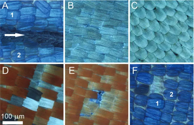
Androconia, as the name suggests, are restricted to male individ- uals. It is a well-documented characteristic of the Mimeresini clade (Eliot 1973) that the dorsal forewing veins are swollen and sparsely covered by minute splinter-like scales with ~50 µm in length and 1 µm in width (Fig. 2A). These scales are not present on the hindwing dorsum, or on the ventral surface of any of the wings.
In both sexes, along the inner margin of the forewing ventral surface, both layers of the scales are grey colored. They differ only slightly in shape and size, the cover scales being somewhat longer and wider than the ground scales. The arrangement of these scales
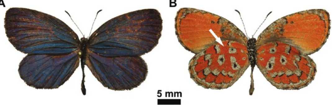
Fig. 1. Mimeresia neavei imago: male in dorsal view (A) and ditto, in ventral view (B); the overlapping wings hides the area (indicated by arrow) where the highly ordered scales are found (Fig. 2C).
2 Journal of Insect Science, 2018, Vol. 18, No. 3
Downloaded from https://academic.oup.com/jinsectscience/article-abstract/18/3/6/5001952 by MTA Wigner Research Centre for Physics user on 17 September 2018
seems to be tighter but less ordered compared to the color-generating scales covering the other parts of the wing surfaces (Fig. 2B). In the hindwing dorsal surface along the costa, which is covered by the forewing anal margin (Fig. 1), there is only one type of scale in a sin- gle layer, but the scales are highly ordered (Fig. 2C). The individual scale is shovel-shaped with slightly curved margins and a rounded apex with approximate ~90 µm in length and ~40 µm in width.
Comparing cover and ground scales, the ground scales seem to be wider and shorter, measuring ~60 µm and ~50 µm respectively (Fig. 2A and F). The black, grey, brown, orange, and white color generating scales on male ventral wing surface and female dorsal and ventral wing surfaces are arranged in two layers: cover and ground- scales. Cover scales are shovel-shaped with parallel margins and a slightly dentate apex, measuring ~100 µm in length and ~40 µm in width. The ground scales are similarly shaped but slightly shorter and wider, measuring ~60 µm and ~50 µm (Fig. 2D). The light micrographs of individual scales are shown in Fig. 3. As these cover scales show very clear differences in transmitted light, one may infer that the orange and brown cover scales contain a higher amount of broadband-absorbing pigment, most likely ommochrome and melanin, respectively (Stavenga et al. 2014). Regarding micro- and nanostructures, these scales are identical, as their dorsal surfaces are structured by longitudinal ridges and cross ribs separated by cca.
1 µm and 0.5 µm free spaces, respectively. The scale lower lamina, visible in the windows formed by the longitudinal ridges and the cross ribs, is decorated with conical protrusions (Fig. 4B). The pat- tern formed by the coloration of these scales resembles that generally
seen in the ventral wing surface pattern of many Lycaenidae species (Schwanwitsch 1949).
The scales of the male dorsal wing surface generate the blue color, unique in the clade. There are two layers of scales; they are different in shape: the cover scales have no dentate apex but are slightly waved, and the ground scales are wider and shorter than the cover scales (Fig. 2F). The surfaces of the scales are also structured by longitudinal ridges and cross ribs, but the notable difference com- pared to the scales of the ventral wing surface is that in the windows there are no conical protrusions (Fig. 4A), and only the unstructured scale lower lamina is visible. Both scales generate the peculiar blue coloration of the male wing surfaces (Fig. 1A, Fig. 2F).
TEM inspection shows that the scales on both the dorsal and ventral wing surfaces are without a complex internal nanostructure, and consist of a lower and upper scale lamina (Fig. 5A and B).
Optical Properties
As we can observe in Fig. 4A, the blue wing scale has a flat lower lamina and that the upper lamina consists of ridges and cross ribs.
This structure did not meet our expectations since the blue colors of Lycaenidae are usually associated with a photonic crystal type nanostructure filling the lumen of the wing scales (Biró and Vigneron 2010) and a dark pigment (broadband absorber, usually melanin) in the chitin (Wilts et al. 2009). It is also known from earlier studies of Lyceanidae, that due to the relatively large distance between the ridges and cross ribs they do not contribute to the reflected light spectra in the visible (Biró and Vigneron 2010).
Fig. 2. Optical microscopic images of Mimeresia neavei scales: male forewing dorsal surface showing basal region of the cubital vein area with androconia on the vein (indicated by white arrow) and the layers of blue-colored cover scales (1) and ground scales (2) (A); anal margin area of ventral forewing surface (B);
costal margin area of dorsal forewing surface hidden by overlapping wing surfaces (C); hindwing ventral wing surface cover scales forming one light spot in the medial area and showing different chemical colors according to the presence of pigment in their lumen, the white scales have no pigments (D); hindwing ventral surface showing orange-colored cover scales and lighter, less pigmented ground scales, which are somewhat shorter and wider than the cover scales; the central part of the image is blue, because of the transparency of the wing membrane the dorsal blue structural coloration can be seen (E); male dorsal forewing with structurally blue-colored cover scales and ground scales, the latter one is wider and shorter (2), the former is longer and narrower (1) (F).
Downloaded from https://academic.oup.com/jinsectscience/article-abstract/18/3/6/5001952 by MTA Wigner Research Centre for Physics user on 17 September 2018
Searching for the origin of the blue coloration we purposefully removed the grating formed by the ridges and cross ribs. A single blue scale fixed onto conductive tape was placed under the tip of an AFM operating in contact mode. The scanning of the AFM tip was set perpendicular to the ridges. While continuously scanning, we approached the AFM tip in small increments until we observed on a real-time optical microscope the change in the scale surface.
The AFM tip was able to remove the ridge-cross rib network in the scanned area. Fig. 6A shows how the scanned window is light-blue, but the ridges and cross ribs appear darker blue. The latter is the consequence of the additional light scattering produced by the ridge- cross rib network in the regions where it was not removed. Where in the lower lamina of the scale a complete hole is opened, the color of the conductive tape is visible. In the SEM image (Fig. 6B) one can observe the broken parts (left side of the scale) swept off the scale by the AFM tip and the remaining supporting points of the cross ribs. These two images clearly show that the blue coloration is not affected by the presence of the upper part of the scale. This means that the source of the blue coloration is the lower lamina of the cover scales, which acts as an optical thin film, by reflecting the blue region of the incident white light. The reflectance band peak at
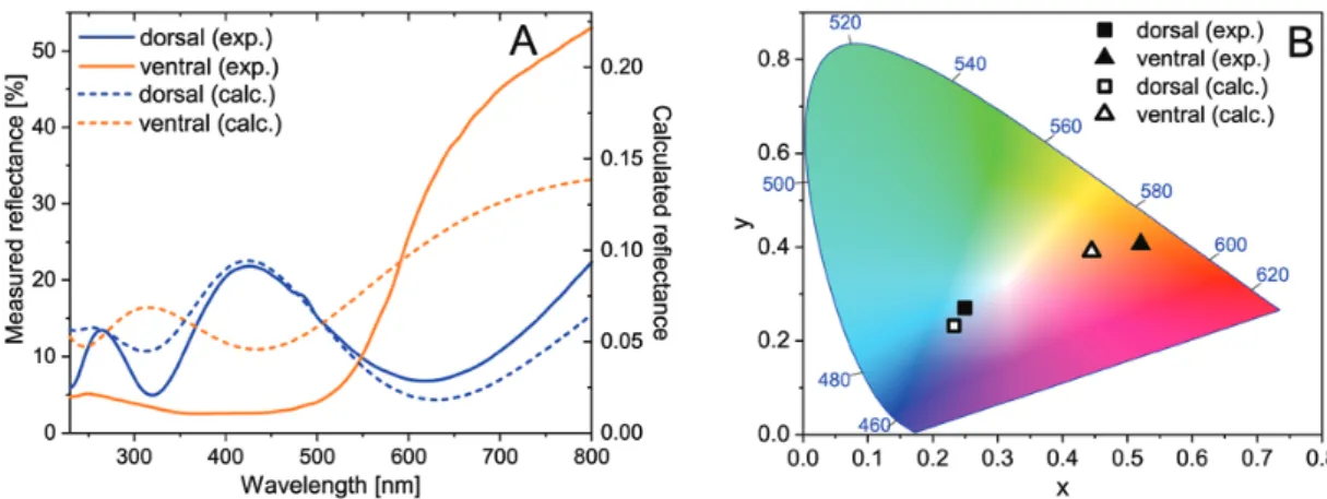
425 nm causes the dorsal blue color, while the increasing reflectance at wavelengths >550 nm yields an orange wing coloration. (Fig. 7A).
The brown and orange wing scales have conical protrusions in ran- dom positions on the top surface of the scale lower lamina, markedly different from the blue scales (Fig. 4B).
If regarded in an effective medium approximation—which takes into account only the averaged refractive index of a given layer—the presence of the conical protrusions is equivalent to a so-called ‘index matching layer’, which has the role of reducing reflection when light enters from a medium with a lower index of refraction (for air n = 1) into one with higher index of refraction (for chitin n = 1.56), thus increasing the absorption of light in the scale lower lamina, which in the brown scales contains melanin pigment (Lohmueller et al. 2010), and likely in the orange scales ommochrome pigment (Stavenga et al.
2014). In transmitted light the blue scales are more transparent, indi- cating a reduced pigment content (Fig. 3).
Modeling
To show that the lamina of the wing scales can act as a thin film, generating the structural coloration, the reflectance of a thin film was calculated based on the measured data of the TEM images. We used
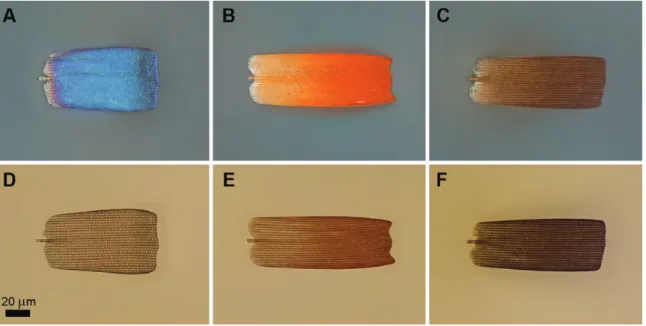
Fig. 3. Optical microscopic images of blue-, orange-, and brown-colored male wing scales of Mimeresia neavei in reflected (A, B, C) and in transmitted light (D, E, F).
Fig. 4. Scanning electron micrographs of male Mimeresia neavei in dorsal view. The lower lamina of the scale is flat, blue scale (A). In the windows formed by ridges and cross ribs one can see at disordered positions conical protrusions of the scale lower lamina, brown scale (B).
4 Journal of Insect Science, 2018, Vol. 18, No. 3
Downloaded from https://academic.oup.com/jinsectscience/article-abstract/18/3/6/5001952 by MTA Wigner Research Centre for Physics user on 17 September 2018
the measured lamina thicknesses of both the blue and the orange scales, to investigate the possible color generation by thin film inter- ference. The reflectance of a thin film can be computed analytically using the transfer matrix method.
The TEM images (Fig. 5A and B) show that if we remove the ridge-cross rib grating, only a thin membrane remains. This single layer of chitin produces a structural color. Measuring at multiple points the thickness of the layer in TEM images, an average value of 201 ± 20 nm in the blue case and 131 ± 10 nm in the orange case was found. Considering the material of the scales as chitin, its com- plex refractive index is 1.56 + 0.1i (Yoshioka and Kinoshita 2007).
The calculated reflectance spectra for normally incident light are presented in Fig. 7A. They are in good agreement with the meas- ured spectra of both scales. The simulated spectra were represented using the CIE 1931 chromaticity diagram. Both the measured and the simulated spectra were transformed and were plotted in Fig. 7B.
One can see the good separation of the differently colored scales, while the simulated and measured results are almost identical. In the case of the orange wing scales, the simulated reflectance spectra are less saturated than the measured spectra. This shows that additional pigment content (ommochromes) can be found in the wing scales, which increases the saturation of the orange structural color. Further evidence can be observed in Fig. 3 too, where the blue wing scale is
almost fully transparent (low amount of pigment), the brown ventral scale has low transmission (highly pigmented, no structural color) while the transmission of the orange scale falls between the two, showing that the orange structural coloration is supplemented with orange pigment coloration (Stavenga et al. 2014).
Discussion
ScalesThe scales of M. neavei are commonplace in Lycaenidae and our observations regarding the clade Mimacraeina microstructures are in accordance with those of the previous workers (Tilley and Eliot 2002): androconia are present and are restricted to the male dorsal forewing surface (Fig. 1A). The scales that give the color and pattern is composed of cover and ground scales. The androconia presuma- bly disseminate aphrodisiacs (Nieberding et al. 2008), which is an important way of communication during courtship for recognition and possibly to aid stimulation of females for copulation. This aphro- disiac is most probably species specific, but Lycaenidae studies in this direction are exceedingly rare (Ômura et al. 2013). The morphology of androconial scale also merits interest as there is growing evidence that it is also indicative of taxonomy (Courvoisier 1916, Eliot and Kawazoé 1983, Ômura et al. 2015). As we already mentioned in the
Fig. 6. Male Mimeresia neavei blue scale with partially removed longitudinal ridges and cross ribs: optical microscope image (A), scanning electron micrograph (rotated by 90°) (B). It is obvious that the scale lower lamina is responsible for the blue coloration and not the structures, what have been removed. The opened hole in the lamina is notated by black arrow.
Fig. 5. Transmission electron micrographs of male Mimeresia neavei wing cover scales: dorsal side (A), ventral side (B). The arrows show the position of the ridges.
Downloaded from https://academic.oup.com/jinsectscience/article-abstract/18/3/6/5001952 by MTA Wigner Research Centre for Physics user on 17 September 2018
introduction in the case of the Lipteninae, it is an important charac- teristic the way in which the androconia are arranged on the male wing surface (Eliot 1973).
The cover and ground scales are involved in forming the pat- tern and color displayed by wing surfaces. The structurally-colored male dorsal wing surface and the pigmentary-colored ventral wing surface have presumably different biological functions. The ventral male and the female wing surface patterns are identical, and this pattern is the one generally found in the other members of the clade (D’Abrera 2009). The same color-generating mechanism is used for the dorsal wing surfaces of the male and female phenotypes of the other members of this clade; only the M. neavei males exhibit a blue structural color.
Mimicry Versus Sexual Dimorphism
When color and pattern is used for sexual communication, there is a need for more complicated structures, because the signals have to be species-specific (Bálint et al. 2012, Giraldo et al. 2016). This is why numerous day-flying Lepidoptera families apply highly sophisticated nanoarchitectures (Ingram and Parker 2008, Biró and Vigneron 2010). The female M. neavei phenotype, but not the male, share the dorsal wing pattern and coloration of the congeners which mimic other Lepidoptera orders (Larsen 2005).
However, field observations of M. neavei have not provided any evidence of mimicry-related behavioral traits. It is a rainforest butterfly sharing habitats with several Mimeresia species breed- ing in the close proximity of Crematogaster-infested ‘ant tree’
(e.g., M. debora barnsi (Hawker-Smith, 1933) and M. dinora dis- cirubra (Talbot, 1937) in Uganda, often sharing the same tree).
According to Congdon and Collins (1998), and also observed by Sáfián (unpublished observations), both sexes circle slowly round semi-shaded tree trunks, occasionally settling on them. They usu- ally fly from 3 to 10 meters above the ground. Imagines flutter slowly, and individuals rest with closed wings on branches or tree trunks hiding their dorsal wing surfaces (Fig. 8A and B). At this stage, our knowledge is insufficient to decide if the blue of the M. neavei males have either a defensive or sexual role (Fig. 8C and D). Specifically designed field work has to be conducted for answering this question. On the other hand, taking into account that M. neavei is the only member of the clade possessing this coloration and that the coloration appears only on the dorsal wing surfaces of the males, it seems plausible that the blue color is involved in sexual communication.
The Coloration of Male M. neavei
We have shown that a thin film nanostructure is responsible for the male M. neavei dorsal blue coloration. The ventral wing sur- face orange coloration is mainly originated from pigments, which is supplemented with the thin film reflectance. The other structural building blocks, like the ridges and the cross ribs, only scatter the incident and the reflected light to generate wing reflection with higher angular visibility. The structural background of this nano- structure has been recorded by Tilley and Eliot (2002) as ‘undif- ferentiated colored scales’ for the blue female Rapala pheretima (Hewitson, 1863) (Theclinae), the pale blue female Allotinus subvi- olaceus C. & R. Felder, 1865 (Miletinae) and for the metallic cop- per-orange reflection of Curetis stigmata Moore, 1879 (Curatinae), Lycaena virgaurae (1758) (Lycaeninae) and Poecilmitis chrysaor (Trimen, 1864) (Aphaneinae). Although it was suggested that some of the scales documented could be responsible for the recorded color, they remarked that ‘the origin of the color of all these scales merits further investigations’.
None of these species or their close relatives has been ever addressed by any experimental studies in relation to structural col- oration. The undifferentiated scale, which provides the basic organ- ization of all Lepidoptera scales, is an assembly of two laminae (Ghiradella 1989, Kristensen and Simonsen 2003). The lower lamina is a flat and thin layer, while the upper lamina consists of an array of ridges connected by cross ribs. The lower lamina acts as an optical thin film and can be the main contributor of the color as we have demonstrated in the case of M. neavei, but when the lower lamina is loaded with pigments or the upper lamina structurally is more com- plex it loses the main role in color generation. Ingram and Parker (2008) and Wilts et al. (2009) examined Lycaenidae scales with per- forated multilayers, working as photonic crystals, but not the rela- tively simple structure we examined in the present paper, as all the examined taxa showed a remarkable complexity of 3D structures.
As we demonstrated, M. neavei males manipulate light with a seemingly less sophisticated mechanism. This color-producing system with a simple-structured scale has been widely applied in other butterfly groups (Satyrinae: Ingram et al. 2008; Papilionidae:
Stavenga et al. 2012, 2014b, 2015; Nymphalinae and Morphinae:
Stavenga et al. 2014a; Giraldo et al. 2016; Giraldo and Stavenga 2016; Heliconinae: Wilts et al. 2017) and a similar mechanism has been recorded in other insect groups (Shevtsova 2011, Stavenga 2014). Therefore we suspect, that color generation by the scale lower lamina is not a rare phenomenon neither among lycaenids, but hitherto it has remained unnoticed. Future research should be
Fig. 7. Perpendicular reflectance spectra measured on Mimeresia neavei blue (dorsal) and orange (ventral) wing surfaces and the calculated spectra (A), the measured and calculated spectra represented in CIE chromaticity diagram (B).
6 Journal of Insect Science, 2018, Vol. 18, No. 3
Downloaded from https://academic.oup.com/jinsectscience/article-abstract/18/3/6/5001952 by MTA Wigner Research Centre for Physics user on 17 September 2018
also focused on exploring clades closely related to Mimacraea and to map the color-generating mechanism, especially in the sections with distinctive imagines as Iridana and Epitola (Libert 1999, Collins and Sáfián 2014).
Phylogeny and Structural Coloration
Many questions were posed for wing scale microstructures in relation with phylogeny by Tilley and Eliot (2002). They concluded that the ancestral color-generating structure in Lycaenidae was the Urania- type with its multi-layered nature, and that later the Morpho-type appeared several times independently.
In a recent paper Giraldo et al. (2016) carried optical investi- gations on the monophyly of the subtribe Morphina (Morphini:
Nymphalinae: Nymphalidae) and suggested via experimental results that most probably the first color-generating scale in the subtribe was a simple one-layer architecture, which could serve as a ground plan for further directions to develop the Morpho- and the Urania-type scales. We think this might have been the evolutionary route in the case of Lycaenidae, too. The structure we described here can be still observed in the hypothetically older taxa as Aphnaeinae, Curetinae and other Lipteninae (Bálint et al. 2017), but it is no longer serving as the most common color-generating architecture in the family, as supposedly new multi-layered developments replaced it (Wilts et al.
2009).
Classical workers involved in Lycaenid classification and sys- tematics had a consensus that the Afrotropical Lipteninae and the Old-world tropical Liphyrinae and Miletinae represented the older taxa (Aurivillius 1925, Stempffer 1967, Eliot 1973, Larsen 2005).
Although no recent papers address Lycaenidae higher systematics based on modern molecular approaches, in the light of a recent study dedicated to Papilionoidea phylogeny this classical insight of relationships stands (Wahlberg et al. 2005, Espenald et al. 2018).
However, because a significant proportion of taxa have not yet been sampled, this insight needs more support from evidence-based studies.
The scale morphology and its color-generating mechanism can- not reveal exclusively the age of the taxon with certainty, but can offer clues to the environment and the life history of the taxa. For example, recent findings of Triassic-Jurassic data revealed that lep- idopteran scales contained nanostructures similar to the ones we recorded in recent species (Bálint et al. 2006), and some of them could work as an optical device (Bálint et al. 2006, van Edjik et al.
2018). This underlines the supposition that direct light had been already intensively manipulated by early lepidopterans.
The blue color of M. neavei males illustrates that color-generating architectures are important features for light-manipulating organ- isms. By combining results gained from the interdisciplinary meth- ods of investigating materials science, molecular and environmental biologists will certainly reveal how life can benefit from applying such light-manipulating devices. This may also prove highly relevant for human applications.
Acknowledgments
The work is supported by the Hungarian OTKA K 111741 and K 115724.
Useful discussions with Prof. D. G. Stavenga are thankfully acknowledged. We express our thanks to Peter Bygate (United Kingdom) and Raimund Schutte Fig. 8. Mimeresia libentina with entirely closed wings hanging on a twig showing the cryptic and/or aposematic ventral wing surface pattern (photo by:
Adrian Hoskins) (A), Mimeresia neavei resting on tree-trunk during displaying similar behavior (photo courtesy: Raimund Schutte) (B), M. libentina, one of the congeneric species sharing habitat with M. neavei, resting on leaf surface and displaying warning coloration; (photo courtesy: Peter Bygate) (C), and M. neavei perching on a tree-trunk covered by mosses and lichens exposing the dorsal wing surface structural blue coloration (photo courtesy: Raimund Schutte) (D).
Downloaded from https://academic.oup.com/jinsectscience/article-abstract/18/3/6/5001952 by MTA Wigner Research Centre for Physics user on 17 September 2018
(SA) for allowing the use of the images of M. libentina and M. neavei, respec- tively. We have no conflicts of interest.
References Cited
Aurivillius, C. 1925. Lycaenidae, Die Großschmetterlinge der Erde, Band 13: Abteilung 2, Die exotischen Großschmetterlinge, Die afrikanischen Tagfalter. Alfred Kernen Verlag, Stuttgart, Germany.
Bálint, Zs., Z. Vértesy, and L. P. Biró. 2006. Microstructures and nanostruc- tures of high Andean Penaincisalia lycaenid butterfly scales (Lepidoptera:
Lycaenidae): descriptions and interpretations. J. Nat. Hist. 39: 2935–2952.
Bálint, Zs., K. Kertész, G. Piszter, Z. Vértesy, and L. P. Biró. 2012. The well- tuned blues: the role of structural colours as optical signals in the spe- cies recognition of a local butterfly fauna (Lepidoptera: Lycaenidae:
Polyommatinae). J. Roy. Soc. Interface. 9: 1745–1756.
Bálint, Zs., A. Heath, G. Katona, K. Kertész, and Sz. Sáfián. 2017. Male sec- ondary sexual characters in Aphnaeinae wings (Lepidoptera: Lycaenidae).
Opusc. Zool. 48: 27–34.
Bampton, I. 1995. A discussion on the larval food of the subfamily Lipteninae (Lepidoptera: Lycaenidae). Metamorphosis. 6: 162–166.
Biró, L. P., and J. P. Vigneron. 2010. Photonic nanoarchitectures in butterflies and beetles: valuable sources for bioinspiration. Laser Photonics Rev. 5: 27–51.
Collins, S., and S. Z. Sáfián. 2014. Notes on the Iridana obscura species group with the description of a new species from western Cameroon (Lycaenidae:
Lipteninae: Epitolini). Metamorphosis. 25: 141–146.
Congdon, C., and S. Collins. 1998. Kielland’s butterflies of Tanzania. ABRI/
Lambillionea, Nairobi/Tervuren, Kenya/Belgium.
Courvoisier, L. G. 1916. Über Männchenschuppen bei Lycaeniden. Verhandl.
Nat. Ges. Basel. 26: 11–48.
D’Abrera, B. 2009. Butterflies of the afrotropical region. New and revised edi- tion part III, lycaenidae and riodinidae. Hill House Publishers, London, United Kingdom.
van Edjik, T. J. B., T. Wappler, P. K. Strother, C. M. H. van der Weijst, H. Rajaei, H. Visscher, and B. van de Schootbrugge. 2018. A Triassic- Jurassic window into the evolution of Lepidoptera. Sci. Adv. 4: 1701568.
Eliot, J. N. 1973. The higher classification of the Lycaenidae (Lepidoptera): a tentative arrangement. Bull. Br. Mus. Nat. Hist. 28: 371–505.
Eliot, J. N. 1990. Notes on the genus Curetis Hübner (Lepidoptera, Lycaenidae). Tyô ta Ga. 41: 201–225.
Eliot, J. N., and A. Kawazoé. 1983. Blue butterflies of the lycaenopsis group.
British Museum (Natural History), London, United Kingdom.
Espenald, M., J. Breinholt, K. R. Willmott, A. D. Warren, R. Vila, E. F. A Toussaint, S. C. Maunsell, K. Aduse-Poku, G. Talavera, R. Eastwood, et al. 2018. A comprehensive and dated phylogenomic ana- lysis of butterflies. Curr. Biol. Rep. 28: 1–9.
Giraldo M. A., and D. G. Stavenga. 2016. Brilliant iridescence of Morpho butterfly wing scales is due to both a thin film lower lamina and a multi- layered upper lamina. J. Comp. Physiol. A 202: 381–388.
Giraldo, M. A., S. Yoshioka, C. Liu, and D. G. Stavenga. 2016. Coloration mech- anisms and phylogeny of Morpho butterflies. J. Exp. Biol. 219: 3936–3944.
Ghiradella, H. 1989. Structure and development of iridescent butterfly scales:
lattices and laminae. J. Morphol. 202: 69–88.
Ingram, A. L., and A. R. Parker. 2008. A review of the diversity and evolu- tion of photonic structures in butterflies, incorporating the work of John Huxley (The Natural History Museum, London from 1961 to 1990).
Philos. T. Roy. Soc. B. 363: 2465–2480.
Ingram, A. L., V. Lousse, A. R. Parker, and J. P. Vigneron. 2008. Dual gratings interspred on a single butterfly scale. J. Roy. Soc. Interface. 5: 1387–1390.
Kristensen, N. P., and Th.J. Simonsen. 2003. ‘Hairs’ and scales, pp. 9–22. In N. P. Kristensen (ed.), Lepidoptera, moths and butterflies. Volume 2: mor- phology, physiology and development. Walter e Gruyter, Berlin/New York, Germany/NY.
Larsen, T. B. 2005. Butterflies of West Africa. Apollo Books, Stenstrup, Denmark.
Libert, M. 1999. Révision des Epitolia (l. s.): révision des genera Epitola Westwood, Hypophytala Clench et Stempfferia Jackson, et description de trois nouveaux genres (Lepidoptera Lycaendiae). ABRI/ Lambillionea, Nairobi/Tervuren, Kenya/Belgium.
Lohmueller, T., R. Brunner, and J. P. Spatz. 2010. Improved properties of opti- cal surfaces by following the example of the “moth eye”, pp. 451–467.
In A. Mukherjee (eds.), Biomimetics learning from nature. IntechOpen, London, United Kingdom.
Nieberding, C. M, H. de Vos, M. V. Schneider, J. M. Lassance, N. Estramil, J. Andersson, J. Bång, E. Hedenström, C. Löfstedt, and P. M. Brakefield.
2008. The male sex pheromone of the butterfly Bicyclus anynana: towards an evolutionary analysis. PLoS One 3: 2751.
Ômura, H., K. Yakumaru, K. Honda, and T. Itoh. 2013. Two lactones in the androconial scent of the lycaenid butterfly Celastrina argiolus ladonides.
Naturwissenschaften. 100: 373–377.
Ômura, H., T. Itoh, M. D. Wright, H. Pavulaan, and S. Schröder. 2015.
Morphological study of alar androconia in Celastrina butterflies. Entomol.
Sci. 18: 353–359.
Pendry, J. B., and A. MacKinnon. 1992. Calculation of photon dispersion rela- tions. Phys. Rev. Lett. 69: 2772–2775.
Schwanwitsch, B. N. 1949. Evolution of the wing-pattern in the Lycaenid Lepidoptera. J. Zool. 119: 189–263.
Shevtsova, E., and C. Hansson. 2011. Species recognition through wing interference patterns (WIPs) in Achrysocharoides Girault (Hymenoptera, Eulophidae) including two new species. Zookeys. 154: 9–30.
Stavenga, D. G. 2014. Thin film and multilayer optics cause structural colors of many insects and Birds. Mater. Today. 1 (Suppl 1): 109–121.
Stavenga, D. G., A. Matsushita, K. Arikawa, H. L. Leertouwer, and B. D. Wilts. 2012. Glass scales on the wing of the swordtail butterfly Graphium sarpedon act as thin film polarizing reflectors. J. Exp. Biol.
215: 657–662.
Stavenga, D. G., H. L. Leertouwer, and B. D. Wilts. 2014a. Coloration prin- ciples of nymphaline butterflies - thin films, melanin, ommochromes and wing scale stacking. J. Exp. Biol. 217: 2171–2180.
Stavenga, D. G., H. L. Leertouwer, and B. D. Wilts. 2014b. The colouration toolkit of the Pipevine Swallowtail butterfly, Battus philenor: thin films, papiliochromes, and melanin. J. Comp. Physiol. 200: 547–561.
Stavenga, D. G., A. Matsushita, and K. Arikawa. 2015. Combined pigmentary and structural effects tune wing scale coloration to color vision in the swallowtail butterfly Papilio xuthus. Zoological Lett. 1: 14.
Stempffer, H. 1967. The genera of the African Lycaenidae (Lepidoptera:
Rhopalocera). Bull. Br. Mus. Nat. Hist. 22 (Suppl 10): 1–322.
Tilley, R. J. D., and J. N. Eliot. 2002. Scale microstructure and its phyloge- netic implications in lycaenid butterflies (Lepidoptera, Lycaenidae). Trans.
Lepid. Soc. Japan 53: 153–180.
Vane-Wright, D. 2003. Evidence and identity in butterfly systematics, pp. 477–512. In C. E. Boggs, W. B. Watt, and P. R. Ehrlich (eds.), Butterflies: ecology and evolution taking flight. University of Chicago Press, Chicago, IL.
Wahlberg, N., M. F. Braby, A. V. Brower, R. de Jong, M. M. Lee, S. Nylin, N. E. Pierce, F. A. H Sperling, R. Vila, A. D Warren, et al. 2005. Synergistic effects of combining morphological and molecular data in resolving the phylogeny of butterflies and skippers. P. Roy. Soc. B-Biol. Sci. 272:
1577–1586.
Wilts, B. D., H. L. Leertouwer, and D. G. Stavenga. 2009. Imaging scattomer- try and microspectrophotomerty of lycaenid butterfly wing scales with perforated multilayers. J. Roy. Soc. Interface. 6: 185–192.
Wilts, B. D., A. J. M. Vey, A. D. Briscoe, and D. G. Stavenga. 2017. Longwing (Heliconius) butterflies combine a restricted set of pigmentary and struc- tural coloration mechanisms. BMC Evol. Biol. 17: 226.
Yeh, P. 2005. Optical waves in layered media. Wiley, New York, NY.
Yoshioka, S., and S. Kinoshita. 2007. Polarization-sensitive color mix- ing in the wing of the Madagascan sunset moth. Opt. Express. 15:
2691–2701.
8 Journal of Insect Science, 2018, Vol. 18, No. 3
Downloaded from https://academic.oup.com/jinsectscience/article-abstract/18/3/6/5001952 by MTA Wigner Research Centre for Physics user on 17 September 2018