Scale granules and colours: Sexual dimorphism in Trichonis (Lepidoptera: Lycaenidae, Theclinae)
Zsolt B alint
a,*, Andrew Parker
b, Abigail Ingram
c, Kriszti an Kert esz
d, G abor Piszter
d, Zsolt E. Horv ath
d, Levente Ill es
d, L aszl o P eter Bir o
daHungarian Natural History Museum, Baross utca 13, Budapest, H-1088, Hungary
bGreen Templeton College, University of Oxford, 43 Woodstock Road, Oxford, OX2 6HG, United Kingdom
cDepartment of Science, West Kent College, Brook Street, Tonbridge, Kent, TN9 2PW, United Kingdom
dInstitute of Technical Physics and Materials Science, Centre for Energy Research, P.O. Box 49, Budapest, H-1525, Hungary
a r t i c l e i n f o
Article history:
Received 23 May 2021 Accepted 12 September 2021 Available online xxx
Keywords:
Androconia Nanoarchitecture Scale layer Scale lumen Structural colour Wing pattern
a b s t r a c t
A large fraction of dorsal wing surface ground scales show an unusual granulated nature, composed of material apparently extruded from the scale lumen in male individuals of bothTrichonisHewitson, 1865 species in the tribe Eumaeini, a rare GuyanianeAmazonian genus. Only a few not-granulated male specimens are known, females are not granulated. The granulated scales are investigated by various microscopic (optical, scanning and transmission electron microscopy, focused ion beam lamella cutting) and spectroscopic (optical reflectance, energy-dispersive X-ray (EDS), Raman) techniques. The charac- teristic blue colour unique in the South American representatives of the tribe is documented and ana- lysed. EDS spectra show that the granules contain additional calcium and oxygen as compared with the un-granulated regions of the same scale. Electron diffraction (inside the TEM) did not reveal any crys- talline component in the granules. The granulated wing surfaces of the males exhibit a UV absorption band at 280 nm, characteristic for biogenic CaCO3; therefore, the material of the granules is tentatively identified as CaCO3. It is shown that the granules influence the optical properties of the dorsal wing surface resulting in a characteristic spectrum.
©2021 The Author(s). Published by Elsevier Ltd. This is an open access article under the CC BY license (http://creativecommons.org/licenses/by/4.0/).
1. Introduction
Papilionoid lepidopterans, especially members of the family Lycaenidae, are well known because of frequently encountered sexual dimorphism. This is expressed in various ways from wing shape and venation, via colouration and pattern, to the scale macro- and micromorphology (Vertesy et al., 2006). Regarding this last aspect, the most obvious one is the presence or absence of sexual secondary characters composed of highly specialised scales for thermal regulation and scent dissemination in the tribe Eumaeini (Faynel and Balint, 2012).
The sexual dimorphism of the very rare Neotropical eumaeine lycaenid butterfly genusTrichonis Hewitson, 1865 (type species:
Papilio theanusCramer, 1777, by original designation) is expressed in all the above-mentioned ways. These are so remarkable that the opposite sexes of the Trichonis type species were described as
representing different species by the same classical worker Pieter Cramer (1721e1776) (Cramer, 1775,1777). The male of the other species, discriminated later byLathy (1930), was consideredfirst as the female ofTrichonis's type species by William Chapman Hewit- son (1806e1878), who established the genus (Hewitson, 1865).
Later it was pointed out that this „female” was an undescribed species' male, which also belonged to the genusTrichonis(Lathy, 1930). Robbins (1986) monographing Trichonis pointed out and briefly described the remarkable male secondary sexual characters expressed by highly specialized scales found on the ventral wing surface of the forewings and dorsal wing surface of the hindwing as large scent pads.Robbins (1986) documentedfirst the female of Trichonis immaculataLathy (1930), a very rare jewel of the lowland Lycaenidae fauna in the Amazonian basin and the Guyana shield.
The maleTrichonishas an unusual structural blue colour which is unique amongst the representatives of its Neotropical relatives, which was why it came to our attention (Balint et al., 2008).
Working on the scale nanomorphology and optics ofTrichoniswe discovered a remarkable trait on male wing scales: in their lumen
*Corresponding author.
E-mail address:balint.zsolt@nhmus.hu(Z. Balint).
Contents lists available atScienceDirect
Arthropod Structure & Development
j o u r n a l h o m e p a g e :w w w . e l s e v i e r . c o m / l o c a t e / a s d
https://doi.org/10.1016/j.asd.2021.101113
1467-8039/©2021 The Author(s). Published by Elsevier Ltd. This is an open access article under the CC BY license (http://creativecommons.org/licenses/by/4.0/).
under optical microscope they seemed to be „granulated”. Although there are extensive literature on Lycaenidae scale micro- and nanonomorphology (Eliot, 1973;Tilley and Eliot, 2002;Wilts et al., 2008), a similar phenomenon has not yet been observed in this or the other butterfly families (Ingram and Parker, 2008). The aim of the present paper is to report on ourfindings via (1) doc- umenting colour generating nanoarchitectures and the presence of granules and their properties by various microscopic techniques, (2) analysing the colour and the phenomenon by several materials science methods and (3) to discuss our results in the light of physiological and biological perspectives.
2. Materials and methods 2.1. Species investigated
BothTrichonisspecies,Trichonis hyacinthus(Cramer, 1775) and T. immaculata Lathy (1930), are rare lowland rain-forest species with Amazonian-Guyanan distribution (Robbins, 1986) (Fig. 1). The genus is classified as follows: sectionHypostrymon: tribe Eumaeini:
subfamily Theclinae: family Lycaenidae (Robbins, 2004).
2.2. Specimens
For non-destructive macroscopic and microscopic in- vestigations 15 male and 5 female museum specimens of T. hyacinthusand 8 male and one female specimens ofT. immaculata were used. For destructive investigations (optical spectroscopy, SEM, TEM and EDS, etc. see below) one male and one female specimen of T. hyacinthus were sacrificed as indicated (see Appendix 1).
2.3. Optical microscopy
Each specimen examined was mounted on a piece of Styrofoam beneath a Zeiss Stemi Apo II (Oberkochen, Germany) (at Natural History Museum, London) or an Olympus SZX12 (Shinyuku, Japan) (at Hungarian Natural History Museum, Budapest) microscope, in such manner that the left dorsal wing extended beyond the edge of the mount and could be illuminated, observed and documented in reflected (Fig. 2) and transmitted, or combined light (Fig. 3). Mi- crographs were taken using a digital camera attached to the mi- croscope. Initially e when checking that the granule-density occurred on several specimenseobservations were made of the entire left dorsal wing surface to reveal the presence or absence of granules. In those individuals with granulated scales, an average
granule frequency was calculated from all ground scales visible in 0.01 mm2. Based on these means, four categories of granule-density were established for comparison between individuals: (1) none: no granulated scales; (2) low: number of granulated scales 1e5; (3) medium: number of granulated scales 6e10; and (4) high: number of granulated scales 11 or more.
2.4. Transmission electron microscopy (TEM)
After preparation according to standard techniques (Balint et al., 2012) the 70 nm thin layers from wing cross-section and the
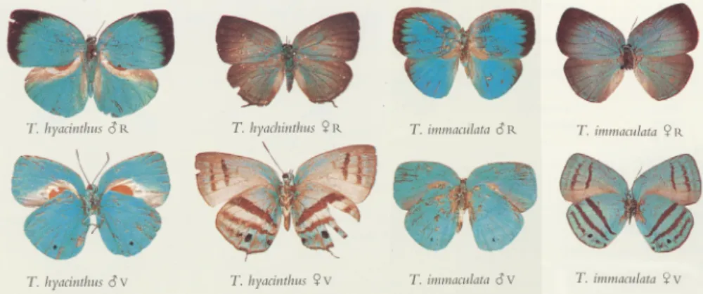
Fig. 1. Trichonismale and female phenotypes documented byD’Abrera (1995). The male secondary characters are especially well visible inT. hyacinthushaving a large scent pad in the basal areas of the dorsal hindwing and the ventral forewing surfaces (all in same magnification:“T. hyacinthus_R”forewing costa length¼18 mm).
Fig. 2.Dorsal wing surface discal area ofTrichonis hyacinthus(Hewitson, 1865) under optical microscope (arrows indicate ground scales). A¼male. B¼female.
alint, A. Parker, A. Ingram et al. Arthropod Structure & Development 65 (2021) 101113
lamella extracted using Focused ion Beam (FIB) sculpting from a granule located inside the combined SEM/FIB microscope (Zeiss LEO 1540 XB (Jena, Germany)) were inspected with a Philips CM20 (Eindhoven, The Netherlands) at 200 kV. After imaging, the electron-diffraction patterns were checked on the granules to decide if they were crystalline or amorphous.
2.5. Scanning electron microscopy (SEM) and energy-dispersive X- ray spectroscopy (EDS)
Scanning electron microscopy examination of wing scales was carried out as reported earlier (Balint et al., 2012). Wing pieces and single scales were attached to stubs via conductive tape. In order to ensure the imaging of the original surface, no metallic coating was applied. All SEM images were captured by a Zeiss LEO 1540 XB (Jena, Germany) microscope. Inside this apparatus the EDS inves- tigation and the preparation of TEM lamella were carried out using FIB. In FIB cutting after the deposition inside the SEM/FIB micro- scope of a protective layer of metallic Pt, a narrow beam of Ga ions was used to locally sputter away the sample, so that a TEM lamella of suitable thickness was cut from the sample. Then this lamella was broken away and placed on a conventional TEM grid. In this way it was ensured that the TEM lamella was cut precisely from the feature previously identified during the detailed SEM examination.
2.6. Optical spectroscopy
Spectral measurements were carried out as reported earlier (Kertesz et al., 2020) using an Avantes (Apeldoorn, The Netherlands) fibre-optic system consisting of an AvaSpec-HERO spectrometer, an AvaLight-DH-S-BAL light source, an integrating
sphere (AvaSphere-50-REFL) and a Avantes WS-2 diffuse reference tile. Opposite to the normal incidence probe, the integrating sphere picked up all light reflected under any angle in the upper hemisphere.
2.7. Raman spectroscopy
Raman measurements were carried out using a Witec 300RSAþconfocal Raman system with 633 nm excitation laser.
Laser power was set to 2 mW for the line cuts and 0.5 mW for single spectrum measurements.
3. Results
3.1. Optical microscopy
We observed thatTrichonisis commonplace in respect of scale layers and scale shapes (Tilley and Eliot, 2002;Vertesy et al., 2006).
On the wing surfaces of both sexes, there were two layers composed by the upper cover and the lower ground scales. The lumen of the cover scales contained amorphous gyroid photonic nanoarchitectures similar to Pseudolycaena marsyas (Linnaeus, 1758) (Vertesy et al., 2006;Seller et al., 2017) generating a char- acteristic blue colour, but the morphologies and the dimensions in the sexes were different. The typical length/width/thickness dimension of the male cover scales was 150/50/1mm with straight or slightly undulated apices and outer margins slightly narrowing towards the wing membrane. In females the cover scale dimensions was 250/50/1 mm with heavily dentate apices and parallel outer margins. The ground scales were similar in both sexes with their wide shovel shape having the dimension 100/80/1mm, but the male scales had the granulated nature in the dorsal wing surface (Fig. 2).
In the male dorsal wing surface in bothTrichonisspecies around the main veins the ground scales were granulated, particularly in worn areas where there were no colour generating cover scales.
Very rarely, the blue cover scales showed granules similar to the ground scales. There were no granulated scales in the ventral wing surfaces. None of the females investigated (n ¼6) showed any granules. Three of the males investigated (3/23¼13%) also had no granulated scales. Fifteen specimens of T. hyacinthus males and eight specimens T. immaculata males were investigated. From these, low granule-density was recorded in the cases of four T. hyacinthus(4/15¼26.6%) and in fourT. immaculata(4/8¼50%) specimens respectively. Medium granule-density was recorded in the cases of ten T. hyacinthus (10/15 ¼ 66.6%) and in three T. immaculata(3/8¼37.5%) specimens respectively. None of them were highly granulated (Fig. 3).
3.2. SEM and TEM
Based on the scanning and cross-sectional transmission electron microscopy, the lumen of theTrichonis hyacinthuscover scales of both sexes shows periodical quasiordered photonic nano- architecture (Fig. 4), which is responsible for the generation of the characteristic colour solely found inTrichonisamongst the repre- sentatives of the tribe presented by more than 1200 species in South America (D'Abrera, 1995;Robbins, 2004). This blue colour observable with the naked eye is very similar in the different sexes, but can be precisely characterized by spectral measurements.
When comparing TEM images (Fig. 5) of the structure of the ground and the cover scales, one can clearly distinguish them. The ground scales contain in their lumen a dark granular filling which is disordered in distribution. The cover scales contain a quasiordered filling, showing some similarity with the photonic nanostructures of other blue lycaenids (Balint, 2012;Kertesz et al., 2017), however Fig. 3.A slightly worn area of maleTrichonis hyacinthus(Hewitson, 1865) dorsal wing
surface in the discal cell under various illuminations and magnifications. The granu- lated ground scales are obvious. A¼in reflected light, B¼in transmitted light.
alint, A. Parker, A. Ingram et al. Arthropod Structure & Development 65 (2021) 101113
no continuous layers can be identified in the high-resolution TEM image in Fig. 5C. As the thickness of the ultramicrotomed TEM section is constant (70 nm), the layers exhibiting different contrast indicate changing pigment loading within the cover scales. The lower layer, closer to the wing membrane, is more pigmented than the region below the ridges.
The lumen of the ground scales does not contain any regular or quasiregular nanoarchitectures, only the discontinuous traces of such a nanoarchitecture can be seen both in the SEM and the TEM images. Therefore the homogenous, pale blue colour may be generated by the scale lamina, which is a widely distributed phe- nomenon, also in lycaenids (Balint et al., 2018, but other butterflies see e.g.,Stavenga, 2014;Thayer et al., 2020) (see later,Fig. 7A). It is worth pointing out that, as seen for example inFig. 5, the ground scales give a much stronger contrast in the TEM when compared with the cover scales. This is an indication that the pigment loading of the ground scales is higher. When granules are present within the TEM section, they can be observed easily on the TEM images of the scale as large voids, because the material building the granules falls out during sample preparation (Fig. 5). The TEM sections are 70 nm thick, and the cutting is done by a very sharp diamond knife.
If the mechanical properties of the scale and of the material building up the granules are different, due to the high forces during the cutting, the thin section of the granule can be fractured and will fall out of the TEM lamella. As can be seen on the SEM images the material of the granule completelyfills the lumen of the ground scales, protruding through the top (Fig. 4DeF). The arrangement and the size of the protrusions clearly show that the pressure causing the protruding was highest in the central regions of the granules (Fig. 4F).
3.3. Optical spectroscopy
The reflectance measurements carried out using the integrating sphere reveal that the blue colour generated by the cover scales is almost identical in both male fore and hind wing surfaces (Fig. 6A).
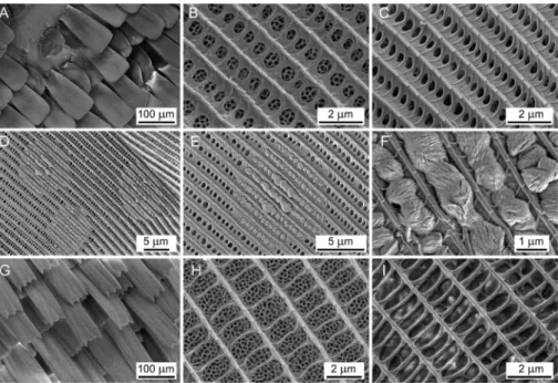
The dorsal blue colour of the males and the females is similar, with the highest reflectivity around 510 nm, however,fine differences can be revealed (Fig. 6B). The blue colour generated by the dorsal wing surface of the males measured with the integrating sphere is different in the near-UV wavelength region as the reflectance spectrum has a different shape between 250 and 400 nm. This may Fig. 4.Scanning electron micrographs ofTrichonis hyacinthus(Hewitson, 1865), dorsal wing surface discal area, male (AeF) and female (GeH). A¼showing small part where there are no cover scales, and the granulated ground scales can be seen. B¼male cover scale surface with photonic nanoarchitecture in its lumen visible via windows. C¼normal male ground scale surface without photonic nanoarchitecture and granule. D¼male ground scale surface with three granules. E¼one granule under larger magnification. F¼pellet-like nanostructure of the granule extruded from the scale lumen via the windows. G¼female cover scales with characteristic dentate morphology. H¼female cover scale surface with photonic nanoarchitecture. I¼female ground scale.
Fig. 5. Transmission electron micrographs of maleTrichonis hyacinthus(Hewitson, 1865) scales. A¼ground and cover scales (arrow indicates the place of the granule in one ground scale); B¼ground scale in higher magnification with pigment granules. C¼cover scale in higher magnification with colour generating nanoarchitectures, the lower stratum contains pigments.
alint, A. Parker, A. Ingram et al. Arthropod Structure & Development 65 (2021) 101113
be attributed to the granulated nature of the ground scales as all the other spectra of the wing surfaces without granules do not contain this type of feature. The three measurements carried out on the male wing surface in different points all show the typical features attributed to the granules, while neither the ventral surface on the
male wing nor the dorsal, or ventral surface of the female wings showed this type of feature (Fig. 6C).
The spectra measured in the range of 600e800 nm show different reflectance between male and female and fore and hindwing. This is due to the less pigmented nature of the ground scales which results in a brighter colour of the ventral wing surface.
Male dorsal colour is outstanding because a higher reflectance peak associated with also higher pigment concentration yielding the lowest reflectance in the range of 600e800 nm.
3.4. Materials composition of the granules
To reveal the nature and properties of the granules found in the lumen of the dorsal wing surface ground scales of males, we carried out a series of experiments, including Raman spectroscopy, FIB sectioning, and energy-dispersive X-ray spectroscopy (EDS).
Using the light source of the scanning Raman microscope (Witec 300RSAþ), a 2 mW red (633 nm) laser beam was scanned in a line across the object on a ground scale. We produced a cut easily discernible in the optical or SEM image (Fig. 7). The laser created a clear separation of the scale ridges and cross ribs, but there was no difference in the granule area, suggesting higher heat capacitance and material compactness in that circular area. According to our earlier studies, the pepper-pot type nanoarchitecture, or any other solid chitinous material in the scale has 100e150 nm thickness, which was cut through with laser. The Raman spectroscopy did not find any characteristic differences between the investigated areas of the ground scales due to the high photoluminescence of this biological sample.
Transmission electron microscopy, and electron bean diffraction on the edges of a single granule was carried out in the TEM. For this purpose, a single detached scale wasfixed between two copper TEM grids with square windows. Placing this sandwich of grid- scale-grid in the microscope, we obtained an image (Fig. 7C) where it was easy to observe the parallel ridges, cross-ribs and the structure in the lumen of the ground scale. The granule appeared black in this image, meaning high opacity for the electron beam of 200 kV. We were able to obtain a diffraction image only near the edge of the circular area, but there were no signs of any crystalline component.
Focused ion beam was used to cut a thin section from the ground scale by eliminating the material under two rectangular areas, leaving only a 50 nm thick slice (Fig. 7). After being sculpted, the lamella was broken away and placed on a standard TEM grid and examined. It was possible to observe (Fig. 8) the homogeneous chitinous material of the scale and a spongy materialfilling the air voids. In this volume, a section of the granule can be found which gives a clear contrast respective to the chitin and air, but again, we were not able to detect any crystalline component with electron beam diffraction.
The material composition of the granules was investigated with EDS. A single wing scale was placed on conductive carbon tape to ensure the imaging without charging of the sample. The false colour mapping of the elements (Fig. 9) shows contrast in the granulated area and the rest of the scale. The granules contained calcium (Ca) and show excess in Oxyen (O), as compared to the non-granulated region of the scale. As a reference, in top right corner of the image the carbon tape is visible, delineated in broken red lines. In the carbon map the granules appear darker, this means that the compact body of the granules transmits a smaller fraction of the signal coming from the carbon tape on which the scale is fixed than the holey, notfilled regions of the chitinous scale. But this does not mean that the granules do not contain carbon. It only means that the carbon content of the granules is below the com- bined C signal from the carbon tape on which the scale wasfixed Fig. 6.Reflectance spectra of male and femaleTrichonis hyacinthus(Hewitson, 1865)
measured with the integrating sphere. Spectra of (A) dorsal and (B) ventral wing surfaces are shown. In (C), the measured reflectance of male and female specimen can be compared. In (A) and (C) arrow indicates the difference caused by granules.
alint, A. Parker, A. Ingram et al. Arthropod Structure & Development 65 (2021) 101113
Fig. 7. Experiments onTrichonis hyacinthus(Hewitson, 1865) ground scale. (A) Optical and (B) SEM images showing the effect of a linear scan with the laser beam of the Raman microscope carries on sample. (C) TEM image of a single (another) ground scale near the granule area. The electron beam was perpendicular to the plane of the scale in this measurement.
Fig. 8. SEM image showing from one side the lamella cut crossingTrichonis hyacinthus(Hewitson, 1865) wing scale containing a granule (A) and TEM image of the same section (B).
The numbers indicate the chitin of the scale (1), the granule substance (2) and the protective layer (3).
Fig. 9.Analysis ofTrichonis hyacinthus(Hewiton, 1865) ground scale granules. SEM and EDS false colour maps of the elemental composition of ground scale with granules.
A¼normal (in the left upper corner a region of the carbon tape is seen delineated in red broken lines. This gives the highest Carbon signal seen in the carbon map). B¼Carbon, C¼ Calcium, D¼Oxygen. The granules being more compact than the holey chitin scale are seen as dark spots on the Carbon map, while they appear brighter on the Calcium and Oxygen maps. (For interpretation of the references to color in thisfigure legend, the reader is referred to the Web version of this article.)
alint, A. Parker, A. Ingram et al. Arthropod Structure & Development 65 (2021) 101113
and the signal generated by the scale itself. As the scale material is mostly chitin, it must contain carbon, too. Therefore, the quanti- tative evaluation of the carbon content of the scale and of the granules is made difficult by conductive carbon tape.
4. Discussion 4.1. Androconia
In general, it is a consensus that Lycaenidae wing surfaces have two layers of scales responsible of the characteristic colouration and pattern (Schwanwitsch, 1949; Eliot, 1973; Tilley and Eliot, 2002). The scales of the lower layer lack photonic nano- architectures. The cover scale lumen of the upper layer contains the photonic nanoarchitecture responsible for colour generation.
However it is a rather simplified view as there are many cases when beside these two kinds of scales further specialised scales occur, primarily on males. Therefore they are called androconia (Eliot, 1973). Although many of these scales are often arranged in eumaeine lycaenids as patches or pads (Faynel and Balint, 2012), there are examples where androconia are dispersed amongst the normal colour-generating scales, the most remarkable example is the tribe Polyommatini (Courvoisier, 1916).
InTrichonis, there is a complex system of scent patch and scent pad (seeFig. 1) that can be observed in many papilionoid families, as well as in representatives of eumaeines. But in the tribe Eumaeini, it is also recorded that in certain areas of the wing androconia may occur mixed with the colour-generating scales (Balint, 2009;Faynel and Balint, 2012;Martins et al., 2019). In the bordering areas of scent patches, the androconia are often dispersed further and have been gradually replaced by normal colour generating scales. Scent patches containing androconia are mostly present in wing areas bordering by the principal veins, radius and cubitus. This is obligatory from a physiological point of view, as the wing membrane itself is not dead matter as the cells in the veins are living and used to sense the environment (Salcedo and Socha, 2020). Most recently, it was experimentally shown that not only in the tube-like veins was there circulating haemolymph but also in the wing membrane discal area bordered by the principal veins (Tsai et al., 2020). Hence, we can presume that living cells in the veins and wing membrane are responsible for scent dissemination.
The granulated areas in Trichonis males are restricted to the dorsal wing surfaces around the main veins. In the ventral wing surface there are no granules. This may suggest that the dorsal wing surface ground scales may function as scent disseminators, some- how homologous with the polyommatine androconia. However the material composition of the granules may contradict this hypoth- esis, knowing that Lycaenidae scent aphrodites are composed of very different chemical compounds (Omara et al., 2013). Other^ contradicting evidence is, thatTrichonismales have a sophisticated system of androconia, as mentioned above in the Introduction.
4.2. The granules
Ground scales with a granulated nature were not recorded anywhere, although we checked representatives of generaBalin- tus d’Abrera, 2001; CelmiaJohnson, 1991; Dicya Johnson, 1991;
Erora Scudder, 1872 and Iaspis Kaye, 1904 placed in the Hypo- strymonsection of Robbins in his phylogenetically oriented check- list, indicated as closely related groups (Robbins, 2004). We did notfind granulated scales in specimens representing further rel- atives of the same or other families randomly selected on the basis of a superficially similar androconial system:AntirrrheaHübner, 1822; (Morphinae, Nymphalidae; Neotropical); Aslauga Kirby,
1890 (Miletinae, Lycaenidae; Afrotropical); Dismorphia Hübner, 1816 (Dismorphiinae, Pieridae; Neotropical), Euploea Fabricius, 1807 (Danainae, Nymphalidae; Austral and Oriental). Hence ac- cording to our best knowledge granulated scales occur exclusively in maleTrichonis.
On the basis of our EDS analysis the granules seem to contain excess calcium and oxygen. Calcium oxide is usually made by the thermal decomposition of materials, that contain calcium carbon- ate (CaCO3). Theoretically this is accomplished by heating the material to above 825 C, a process called calcination or lime- burning, to liberate a molecule of carbon dioxide (CO2). This is certainly impossible for a living organism, so there must be another mechanism during the biosynthesis process. Furthermore, CaO is unstable in air and under humid conditions. On the other hand, CaCO3is frequently synthetized on biogenic route, for example in pearls, and nacre and coral (Reef et al., 2009;Yan et al., 2017,2021).
As the EDS elemental maps show that the granules contain carbon too, although less carbon than the carbon tape on which the scale wasfixed, it is reasonable to investigate if the presence of CaCO3
can be ruled out or supported.
The characteristic of this biogenic CaCO3is an UV absorption band at 280 nm (Reef et al., 2009;Yan et al., 2017,2021) (see also in Fig. 6), which is associated with the biotemplated growth of CaCO3. This characteristic band can be identified only on the optical spectra of the dorsal wing surface of the male (Fig. 6). Calcium is known to have an important role in the scale synthesis process during metamorphosis (Otaki, 2012). The careful examination of thefine structure of the granules as revealed by SEM images (Fig. 4C and E) indicates that the system of ridges and cross-ribs must be in place before the formation of the granules, as the material of the granules is“extruded”through the grating formed by the ridges and cross-ribs. The FIB sectioning and the examination of the sculpted lamella in the TEM shows that the material of the scale and the material of the granule are interpenetrated.
The curious feature in the reflectance spectra of the male dorsal wing surfaces in the range of 250e400 nm taken by the integration sphere can be attributed to the increased light scattering due to the presence of the granules and the contribution of the UV absorption of the biogenic CaCO3. This is supported by the 280 nm UV ab- sorption band characteristic for biogenic CaCO3.
4.3. Colouration and pattern
The colouration and pattern of male and the femaleTrichonisare basically different; only the characteristic blue colour of the male is shared by some female wing areas. The male and femaleTrichonis phenotypes express distinct behaviour and lifestyle. We hypothe- size thatTrichonismales are lekking in sunny forest glades or in the canopy. Presumably, males are territorial as most of the forest dwelling theclines, and theirflight is rapid and furious, which for the human eye is difficult to follow (Takeuchi and Imafuku, 2005;
Prieto and Dahners, 2006). What is curious in theTrichonismale phenotype is the presence of the same structural colour in both of the almost patternless wing surfaces (seeFig. 1).
Female Trichonis adopt a phenotype which is well presented amongst the Neotropical papilionoid butterflies. Not only genera of the tribe Eumaeini but other families apply this pattern of trans- verse bands displayed by the hindwing ventral wing surfaces. This phenotype mainly typifies butterfly imagines which live deep un- der the forest canopy or forest understory which reveals a rhythmic pattern of dappled light. Most probably in these circumstances it is difficult for sexes to locate each other and females dependfirst on optical signals provided by males.
According to our knowledge regarding male structural colour- ation, the only tribes possessing a comparable appearance in the
alint, A. Parker, A. Ingram et al. Arthropod Structure & Development 65 (2021) 101113
Neotropical region are the satyrid Eupthyciini (cf.D'Abrera, 1988:
766e769;Nakahara et al., 2018) and the riodinid Mesosemiini (cf.
D'Abrera, 1994: 901e902). Both are also primarily Amazonian.
Males of some species of these enigmatic butterflies have wing surfaces with identical structural colouration (Fig. 10). But in contrast toTrichonis, both male and female phenotype ventral wing surfaces are striped and granules were not found in scale lumina.
This phenotypic appearance may indicate that both sexes of these riodinid and satyrid butterflies spend their lives in an environment optically similar to that ofTrichonisfemales. However because male Trichonis are characteristically patternless, they may produce a unique signal that can be easily spotted by their females.
5. Concluding notes
We showed that the sexual dimorphism ofTrichonisis remark- able, as beside the commonplace grossly-differing phenotype expressed by wing shape, pattern, colouration and male secondary characters (androconia), the sexes differ in an additional character:
the layer of ground scales with granulated physiology. Although we elaborated on the morphology and material composition of the granules, its development remains unknown; only various hy- potheses could be formulated: (1) the granules may start to develop sometime after the hatching of the male individual proceeding interactions with the environment or (2) it is a male individual aberration. Further, the function of the granules is not understood, but we have shown that it has an influence on the spectral prop- erties of the wing surfaces, and as such may have an effect on mate selection. It would be interesting to determine whether the female is able to distinguish between the male's colouration with and without granules.
It is interesting to note that physical changes of butterfly imagines usually caused by external physical factors are becoming more and more intensive with time. This in general may result in the loss of scales, which cause reduced attractiveness in males (Kemp, 2007), but in females it may bring benefits (Pyrcz et al., 2018). If it has been confirmed that the maleTrichonisindividuals produce the granules after hatching in the scales that would be supporting evidence for the idea that the external physiology of butterfly imagines is sometimes incomplete.
In the future, it would be interesting to study in thefield how mesosemine riodinids, euptychine satyrids andTrichonismales and females share their habitat in temporal, spatial and
microgeographical terms. Although such questions are worth pur- suing, the rarity ofTrichonisand the deforestation taking place in the Amazonian ecosystem together suggest that these questions will remain unanswered.
Author contributions
Conceptualization: Zs.B.; Methodology: Zs.B., L.P.B., K.K., G.P.;
Investigation: A.I., Zs.B., K.K., G.P.; Z.E.H; L.I.; Resources: Zs.B.; Data curation: Zs.B., A.I.; Writingeoriginal draft: Zs.B.; Writingereview and editing: Zs.B., L.P.B., A. I., K.K., A.P., P.G.; Visualization: K.K., G.P.;
Funding acquisition: Zs.B., L.P.B., K.K.,G.P.
Funding
The research was partly conducted during the EU6 program
“Specific Targeted Research”Nest/Pathfinder/Biophot-012915, and recently covered by the joint scientific agreement between the Hungarian Natural History Museum and Institute of Technical Physics and Materials Science.
Acknowledgements
Thanks are due to Lucilla d’Abrera (Victoria, Australia) for allowing us to reproduce images from the volumes ofButterflies of the Neotropical Region, to Christoph Faynel (Montaud, France) for specimens and documentations, and to Gergely Katona (HNHM) for composing plates ofFigs. 1 and 10. G.P. acknowledges the support of the Janos Bolyai Research Scholarship of the Hungarian Academy of Sciences.
Appendix
Museum specimens ofTrichonisexamined. Aeserial number;
B e scientific binomen; C e sex (* ¼ specimen examined by SEM and TEM); De collecting locality; collector or collection;
Eedegree of granule-density (0¼none, 1¼low, 2¼medium);
F e condition of the specimen; (*) ¼documented in D'Abrera (1995, see Fig. 1); specimen examined by SEM and TEM; Ge depositor (BMNH¼Natural History Museum, London (formerly:
British Museum of Natural History); HNHM¼Hungarian Natural History Museum, Budapest).
Fig. 10.Male and female satyrid and riodinid butterfly phenotypes from the Amazonian fauna superficially resemblingTrichonisdocumented byD'Abrera (1988and1994). The satyridCepheuptyhcia cephus(Fabricius, 1775) male has a striped wingsurface verso. The riodinidSemomesia marisa(Hewitson, 1858) male wingsrufcae recto is striped but has androconial system somewhat similar toTrichonis. The female wingsurface versos are striped (all in same magnification:“E. cephus_R”forewing costa length¼21 mm).
alint, A. Parker, A. Ingram et al. Arthropod Structure & Development 65 (2021) 101113
References
Balint, Zs., 2009. Five chapters onAnnamaria columbiawith the description of a new genus (Lepidoptera: Lycaenidae: Eumaeini). Bol. Cient. Mus. Hist. Nat. Univ.
Caldas 13 (1), 75e82.
Balint, Zs., Ingram, A.L., Parker, A.R., Kertesz, K., Vertesy, Z., Biro, L.P., 2008. A journey amongst male wing scale micro- and naonarchitectures of the butterfyTrichonis hyacinthus (Lepidoptera: Lycaenidae). In: Berthier, S. (Ed.), Seventh Biophot Workshop. Institut des NanoSciences, Paris, pp. 17e18, 30.
Balint, Zs., Kertesz, K., Piszter, G., Vertesy, Z., Biro, L.P., 2012. The well-tuned Blues:
the role of structural colours as optical signals in the species recognition of a local butterfly fauna (Lepidoptera: Lycaenidae: Polyommatinae). J. R. Soc.
Interface 9 (73), 1745e1756.https://doi.org/10.1098/rsif.2011.0854.
Balint, Zs., Safian, Sz., Hoskins, A., Kertesz, K., Koos, A.A., Horvath, Zs.E., Piszter, G., Biro, L.P., 2018. The only blueMimeresia(Lepidoptera: Lycaenidae: Lipteninae) uses a color-generating mechanism widely applied by butterflies. J. Insect Sci. 18 (3), 1e8.https://doi.org/10.1093/jisesa/iey046, 18, 6.
Courvoisier, L.G., 1916. Über M€annchenschuppen bei Lycaeniden. Verh. Naturforsch.
Ges. 26, 11e48, 2 pls].
Cramer, P., 1775. De uitlandsche Kapellen voorkomende in de drie Waereld-Deelen Asia, Africa en America. Papillons exotiques des trois parties du monde l'Asie, l'Afrique et l'Amerique. Amsteldam, S. J. Baalde; Utrecht, Barthelemy Wild and.
J. Van Schoonhoven Comp. 1 (1/7), iexxx, 1-16, 1-132, pls. 1-84.
Cramer, P., 1777. De uitlandsche Kapellen voorkomende in de drie Waereld-Deelen Asia, Africa en America. Papillons exotiques des trois parties du monde l'Asie, l'Afrique et l'Amerique. Amsteldam, S. J. Baalde; Utrecht, Barthelemy Wild and.
J. Van Schoonhoven Comp. 2 (9/16), 1e151 pls. 97-192.
D'Abrera, B., 1988. Butterflies of the Neotropical Region. Part V. Nymphalidae.
(Conc.) and Satyridae. Victoria, Black Rock. Hill House [viii]þ679-877,figs.
D'Abrera, B., 1994. Butterflies of the Neotropical Region. Part VI. Riodinidae. Victoria, Black Rock. Hill House viiiþ879-1096,figs.
D’Abrera, B., 1995. Butterflies of the Neotropical Region. Part VII. Lycaenidae. Vic- toria, Black Rock. vols. i-xi. Hill House, pp. 1098-1270, (figs).
Eliot, J.N., 1973. The higher classification of the Lycaenidae (Lepidoptera): a tentative arrangement. Bull. Br. Mus. (Nat. Hist.) Entomol. 28 (6), 373e506, 6 pls., 162 figs., 4 tabs.
Faynel, C., Balint, Zs., 2012. An overview of alar organs in French Guiana hairstreaks (Lepidoptera: Lycaenidae: Theclinae, Eumaeini). Lepidopteres de Guyane 5, 46e54.
Hewitson, W.Ch., 1865. Illustrations of Diurnal Lepidoptera, vol. 2. Lycaenidae, London, John Van Voorst, pp. 37e76, 17-30.
Ingram, A.L., Paker, A.R., 2008. A review of the diversity and evolution of photonic structures in butterflies, incorporating the work of John Huxley (The Natural History Museum, London from 1961 to 1990). Phil. Trans. R. Soc. B 363, 2465e2480.https://doi.org/10.1098/rstb.2007.2258(2008).
Kemp, D., 2007. Female butterflies prefer males bearing bright iridescent orna- mentation. Proc. Biol. Sci. 274 (1613), 1043e1047. https://doi.org/10.1098/
rspb.2006.0043.
Kertesz, K., Piszter, G., Horvath, Z.E., Balint, Zs., Biro, L.P., 2017. Changes in structural and pigmentary colours in response to cold stress inPolyommatus icarusbut- terflies. Sci. Rep. 7, 1118.https://doi.org/10.1038/s41598-017-01273-7.
Kertesz, K., Balint, Zs., Piszter, G., Horvath, Zs.E., Biro, L.P., 2020. Multi-instrumental techniques for evaluating butterfly structural colors: a case study onPoly- ommatus bellargus (Rottemburg, 1775) (Lepidoptera: Lycaenidae: Poly- ommatinae). Arthropod Struct. Dev. 61 (101010), 1e13.https://doi.org/10.1016/
j.asd.2020.101010.
Lathy, P.I., 1930. Notes on South American Lycaenidae, with descriptions of new species. Trans. Ethnol. Soc. Lond. 78 (1), 133e137 pl. 9.
Martins, A.R., Duarte, M., Robbins, R.K., 2019. Phylogenetic classification of the Atlidessection of the Eumaeini (Lepidoptera, Lycaenidae). Zootaxa 4563 (1), 119e134.https://doi.org/10.11646/zootaxa.4563.1.6.
Nakahara, S., Zacca, Th., Huertas, B., Neild, A.F.E., Hall, J.P.W., Lamas, G., Holian, L.A., Espeland, M., Willmott, K.W., 2018. Remarkable sexual dimorphism, rarity and cryptic species: a revision of the‘aegrota species group’of the Neotropical butterfly genusCaeruleuptychiawith the description of three new species (Lepidoptera, Nymphalidae, Satyrinae). Insect Systemat. Evol. 49 (2), 130e182.
https://doi.org/10.1163/1876312X-00002167.
Omara, H., Yakumaru, K., Hona, K., Itoh, T., 2013. Two lactones in the androconial^ scent of the lycaenid butterflyCelastrina argiolus. Naturwissenschaften 100, 373e377.https://doi.org/10.1007/s00114-013-1030-9.
Otaki, J.M., 2012. Structural analysis of eyespots: dynamics of morphogenic signals that govern elemental positions in butterfly wings. BMC Syst. Biol. 6, 17.https://
doi.org/10.1186/1752-0509-6-17.
Prieto, C.H., Dahners, H.W., 2006. Eumaeini (Lepidoptera: Lycaenidae) del cerro San Antonio: dinamica de la riqueza y comportamiento de "hilltopping. Rev.
Colomb. Entomol. 32 (2), 179e190, 9figs., 3 tabs.
Pyrcz, T.W., Garlacz, R., Kertesz, K., Biro, L.P., Balint, Zs., 2018. An imperfect imago?
Post-mating loss of iridescent scales inCheimasbutterflies may change female from attractive to cryptic (Lepidoptera: Nymphalidae, Satyrinae). J. Nat. Hist. 52 (19e20), 1333e1350.https://doi.org/10.1080/00222933.2018.1461945.
Reef, R., Kaniewska, P., Hoegh-Guldberg, O., 2009. Coral skeletons defend against ultraviolet radiation. PloS One 4, e7995. https://doi.org/10.1371/
journal.pone.0007995.
Robbins, R.K., 1986. Evolution and identification of the new world hairstreak but- terflies (Lycaenidae: Eumaeini): eliot'sTrichonissection andTrichonisHewitson.
J. Lepidopterists' Soc. 40 (3), 138e157, 8figs.
Robbins, R.K., 2004. Lycaenidae. Theclinae. Tribe Eumaeini. In: Lamas, G. (Ed.), Checklist: Part 4A. Hesperioideae Papilionoidea. Atlas of Neotropical Lepi- doptera. Volume 5A. Gainesville, Association for Tropical Lepidoptera; Scientific Publishers, pp. 118e137. Heppner, J. B.
A B C D E F G
1 Trichonis hyacinthus _ French Guyana: St Jean du Maroni; Le Moult 2 poor BMNH
2 Trichonis hyacinthus _ French Guyana; Bar 0 good BMNH
3 Trichonis hyacinthus _ British Guyana; Joicey 2 fair BMNH
4 Trichonis hyacinthus _ British Guyana; Kaden 1 fair BMNH
5 Trichonis hyacinthus _ British Guyana; Kaden 1 good BMNH
6 Trichonis hyacinthus _ French Guyana: Cayenne; Hewitson 2 good BMNH
7 Trichonis hyacinthus _ French Guyana: Cayenne; Hewitson 1 fair BMNH
8 Trichonis hyacinthus _ British Guyana 1 fair (*) BMNH
9 Trichonis hyacinthus _ British Guyana; Druce 0 poor BMNH
10 Trichonis hyacinthus _ French Guyana: Cayenne; Oberthür 1 good BMNH
11 Trichonis hyacinthus _ French Guyana: St. Laurent, Maroni; Joicey 2 poor BMNH
12 Trichonis hyacinthus _ Brazil: Para; Moss 2 poor BMNH
13 Trichonis hyacinthus _ Brazil: Para; Moss 2 fair (*) BMNH
14 Trichonis hyacinthus \ French Guyana; Oberthür 0 good BMNH
15 Trichonis hyacinthus \ Brazil: Maranham; Belt 0 poor (*) BMNH
16 Trichonis hyacinthus \ French Guyana: Maroni; Joicey 0 fair (*) BMNH
17 Trichonis hyacinthus \ French Guyana; Bar 0 good BMNH
18 Trichonis immaculata \ Peru: Iquitos, Rio Cachiyaca; Stuart 0 good (*) BMNH
18 Trichonis immaculata _ French Guyana: Gourdonville; Le Moult 1 poor BMNH
19 Trichonis immaculata _ Surinam; Fruh 1 fair (*) BMNH
20 Trichonis immaculata _ Peru: Iquitos, Rio Cachiyacu; Stuart 1 good BMNH
21 Trichonis immaculata _ No patria; Boisduval 0 poor BMNH
22 Trichonis immaculata _ No patria; Felder 2 poor BMNH
23 Trichonis immaculata _ No patria; Felder 1 poor BMNH
24 Trichonis immaculata _ Surinam; Fruhstorfer 2 fair BMNH
25 Trichonis immaculata _ Surinam; Fruhstorfer 2 fair BMNH
27 Trichonis hyacinthus _ French Guyana: Cacao; Faynel 2 good HNHM
28 Trichonis hyacinthus _(*) French Guyana: Cacao; Faynel 2 good HNHM
29 Trichonis hyacinthus \(*) French Guyana: Matouri; Faynel 0 fair HNHM
alint, A. Parker, A. Ingram et al. Arthropod Structure & Development 65 (2021) 101113
Salcedo, M.K., Socha, J.J., 2020. Circulation in insect wings: a review on the necessity of hemodynamics in wing functionality. Integr. Comp. Biol. 60 (5), 1e13.https://
doi.org/10.1093/icb/icaa124.
Schwanwitsch, B.N., 1949. Evolution of the wing-pattern in the lycaenid Lepidop- tera. Proc. Zool. Soc. Lond. 119 (1), 189e263, 337figs.
Seller, S.R., Man, W., Sahba, S., Florescu, M., 2017. Local self-uniformity in photonic networks. Nat. Commun. 8, 14439.https://doi.org/10.1038/ncomms14439.
Stavenga, D.G., 2014. Thinfilm and multilayer optics cause structural colors of many insects and birds. Mater. Today Proc. (Suppl. 1) 109e121. https://doi.org/
10.1016/j.matpr.2014.09.007.
Takeuchi, T., Imafuku, M., 2005. Territorial behavior of a green hairstreakChrys- ozephyrus smaragdinus (Lepidoptera: Lycaenidae): site tenacity and wars of Attrition. Zool. Sci. 22 (9), 989e994.https://doi.org/10.2108/zsj.22.989.
Thayer, R.C., Allen, F.I., Patel, N.H., 2020. Structural color in Junonia butterflies evolves by tuning scale lamina thickness. eLife 9 (52187), 1e21.
Tilley, R.J.D., Eliot, J.N., 2002. Scale microstructure and its phylogenetic implications in lycaenid butterflies (Lepidoptera: Lycaenidae). Trans. Lepidopterol. Soc. Jpn.
53 (3), 153e180, 14figs., 2 tabs.
Tsai, C.C., Childers, R.A., Nan Shi, N., Ren, C., Pelaez, J.N., Bernard, G.D., Pierce, N.E., 2020. Physical and behavioral adaptations to prevent overheating of the living wings of butterflies. Nat. Commun. 11, 551.https://doi.org/10.1038/s41467-020- 14408-8.
Vertesy, Z., Balint, Zs., Kertesz, K., Vigneron, J.P., Lousse, V., Biro, L.P., 2006. Wing scale microstructures and nanostructures in butterfliese natural photonic crystals. J. Microsc. 224 (1), 108e110. https://doi.org/10.1111/j.1365- 2818.2006.01678.x, 1fig.
Wilts, B.D., Leertouwer, H.L., Stavenga, D.G., 2008. Imaging scatterometry and microspectrophotometry of lycaenid butterfly wing scales with perforated multilayers. J. R. Soc. Interface 6 (Suppl_2), S185eS192.https://doi.org/10.1098/
rsif.2008.0299.focus.
Yan, J., Zhang, J., Tao, J., Hu, D., Peng, Q., 2017. Origin of the common UV absorption feature in cultured pearls and shells. J. Mater. Sci. 52, 8362e8369.https://
doi.org/10.1007/s10853-017-1111-9.
Yan, J., Zhang, J., Sun, Q., Yan, X., Sheng, J., 2021. The unique UVeVis reflection features of the nacre ofHyriopsis cumingiishells, and its formation mechanisms.
Dyes Pigments 184, 108753.https://doi.org/10.1016/j.dyepig.2020.108753.
alint, A. Parker, A. Ingram et al. Arthropod Structure & Development 65 (2021) 101113