SHORT REPORT
Description of the male and the larva
of Ixodes collaris Hornok, 2016 with drawings of all stages
Sándor Hornok1* , Dávid Murányi2, Jenő Kontschán2 and Vuong Tan Tu3,4
Abstract
Background: Ixodes collaris Hornok, 2016 is a recently discovered tick species associated with bats in Asia. This study provides the description of the male and the larva, as well as high quality drawings of all stages.
Methods: Ticks were collected from cave walls and bats in Phia Oac (Vietnam). DNA was extracted from one indi- vidual of each stage/sex, while another was morphometrically analysed. Based on two genetic markers, all ticks were identified as I. collaris.
Results: The male of I. collaris has long legs (i.e. the length of Haller’s organ exceeds the maximum diameter of tarsus I), unlike the male of I. simplex Neumann, 1906, but similarly to males of I. vespertilionis Koch, 1844 and I. ariadnae Hornok, 2014. The lateral and medial edges of the palpi of male I. collaris are both convexly curved, unlike in I. ariad- nae and I. simplex, but similarly to I. vespertilionis. The male of I. collaris has long palpal setae (up to 210 µm), unlike the males of I. ariadnae (30–100 µm) and I. simplex (20–80 µm), but similarly to I. vespertilionis (100–200 µm). Males of I.
collaris have sparse distribution of long palpal setae (vs dense in I. vespertilionis) and posteriorly diverging, sclerotized trapezoid ridge dorsally on the basis capituli (posteriorly convergent, U-shaped and less evident in I. vespertilionis).
The larva of I. collaris has long legs (unlike the larva of I. simplex, but similarly to I. vespertilionis and I. ariadnae), elon- gated club-shaped palpi (240 × 70 vs 200 × 90 µm in I. ariadnae, 200 × 70 µm in I. vespertilionis; and 140 × 60 µm in I. simplex:), pentagonal scutum, which is longer than broad (different from I. ariadnae and I. simplex, but similar to that of I. vespertilionis). The larva of I. collaris has strongly concave caudolateral margin of ventral basis with perpendicular angle (vs slightly concave, with obtuse angle in I. vespertilionis) and a prominent, dark sclerotized edge, “collar” (absent in I. vespertilionis).
Conclusion: Several features allow to distinguish the male and the larva of I. collaris morphologically from those of other bat-associated ixodid tick species.
Keywords: Ixodes collaris, Bat tick, Cave, Male, Larva, Description
© The Author(s) 2019. This article is distributed under the terms of the Creative Commons Attribution 4.0 International License (http://creat iveco mmons .org/licen ses/by/4.0/), which permits unrestricted use, distribution, and reproduction in any medium, provided you give appropriate credit to the original author(s) and the source, provide a link to the Creative Commons license, and indicate if changes were made. The Creative Commons Public Domain Dedication waiver (http://creat iveco mmons .org/
publi cdoma in/zero/1.0/) applies to the data made available in this article, unless otherwise stated.
Background
In a recent Eurasian survey on ixodid ticks infesting bats, high degree of mitochondrial gene heterogeneity of Ixodes vespertilionis Koch, 1844 was reported, postu- lating that it is actually a species complex, within which hitherto unknown or formerly not distinguished bat tick species might exist [1]. Accordingly, I. collaris Hornok,
2016, has been described, based on material from the intermediate horseshoe bats (Rhinolophus affinis) in Vietnam. However, the description of this new species was based only on the female and the nymph [2], because at that time male bat ticks were not seen on cave walls and larvae could not be collected from the only known host species.
This study provides the description of the male and the larva of I. collaris, in order to complete the description of the species. In addition, because detailed drawings of the female and the nymph of I. collaris have not been
Open Access
*Correspondence: hornok.sandor@univet.hu
1 Department of Parasitology and Zoology, University of Veterinary Medicine, Budapest, Hungary
Full list of author information is available at the end of the article
available, high quality drawings of its all stages are also provided here.
Methods Sample collection
Ixodes sp. ticks (two individuals per stage or sex) were collected at a cave located in the buffer zone of Phia Oac, Phia Den National Park (22.563611N, 105.874167E), Cao Bang Province, Vietnam (i.e. the type-locality of I. colla- ris) on June 1, 2016 and November 17, 2017. Male ticks were removed from cave walls, whereas females, nymphs and larvae were collected from R. affinis. All specimens were stored in 96% ethanol. After microscopical evalua- tion of their conspecificity, DNA was extracted from one individual of each stage/sex, while the other was mor- phometrically analysed and described. Specimens of I.
vespertilionis (used for comparison) were collected in Leány cave, Hungary.
Sample analyses
From the DNA extracts, molecular analyses of two mito- chondrial markers were performed as reported [1] by two PCRs: the first amplifying an approximately 710 bp long
fragment of the cytochrome c oxidase subunit 1 (cox1) gene with the primers HCO2198 (5′-TAA ACT TCA GGG TGA CCA AAA AAT CA-3′) and LCO1490 (5′- GGT CAA CAA ATC ATA AAG ATA TTG G-3′), and the second targeting an approximately 460 bp fragment of the 16S rRNA gene of Ixodidae with the primers 16S + 1 (5′-CTG CTC AAT GAT TTT TTA AAT TGC TGT GG-3′) and 16S-1 (5′-CCG GTC TGA ACT CAG ATC AAG T-3′). PCR products were visualized in a 1.5% aga- rose gel. Purification and sequencing was done by Biomi Inc. (Gödöllő, Hungary). Sequences were compared to those already deposited in GenBank by nucleotide BLASTN program (https ://blast .ncbi.nlm.nih.gov).
Pictures and measurements were made with a VHX- 5000 (Keyence Co., Osaka, Japan) digital microscope.
Results
Based on the amplified fragment of their cox1 and 16S rRNA genes, all ticks described here were identi- fied as I. collaris, having 100% sequence identity to for- merly reported female and nymph specimens of this tick species (cox1 gene: KR902756, 16S rRNA gene:
KR902771). The newly generated sequences were sub- mitted to the GenBank database under the accession
a c
b d
1000µm 1000µm
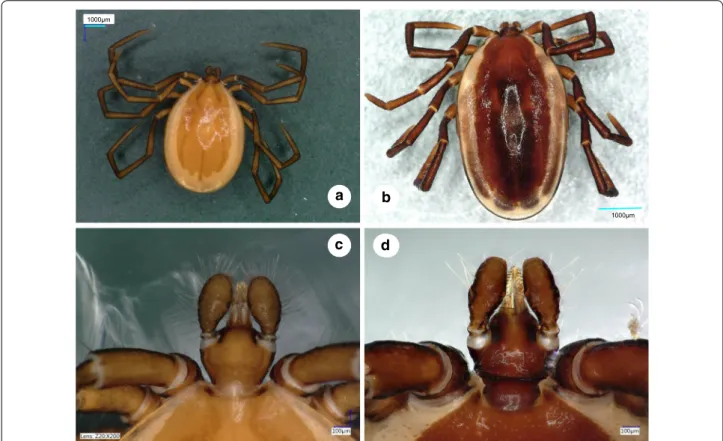
Fig. 1 Dorsal views of male Ixodes vespertilionis (a, habitus; c, gnathosoma) and I. collaris (b, habitus; d, gnathosoma)
numbers MK450318-MK450319 (cox1) and MK450320- MK450321 (16S rRNA). The measurements in the descriptions below are provided in millimetres.
Ixodes collaris Hornok, 2016 Description
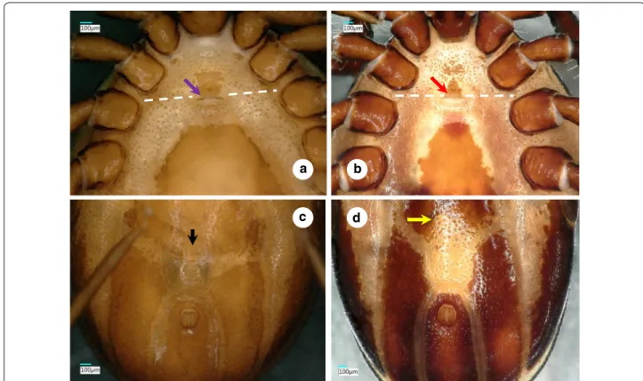
Male. [See Figs. 1–3.] Length of idiosoma (from half point between scapular apices to posterior margin) 4.49, breadth 2.78, ratio of idiosomal length/breadth 1.61. Conscutum centrally dark brown, laterally lightly colored, elongated, elliptical, broadest slightly anteri- orly to its mid-length (Fig. 1b). Length of scutum 4.2, breadth 2.45, ratio length/breadth 1.71. Cervical grooves anteriorly well-defined, paramedian grooves posteriorly shallow; scattered punctuations present. Genital aper- ture at level of second intercoxal space, surrounded by ivory color and scattered punctuations (Fig. 2b). Spirac- ular plates slightly oval (length/breadth ratio 1.23), with eccentric opening. Ivory coloration anterior to anus oval, elongated. Anal plate oval, elongated, broadest at mid- length. Adanal plates 1.5 times longer than anal plate.
Median plate anteriorly rounded, longer than broad (Figs. 2b, d, 3b).
Gnathosoma: length from palpal apices to posterior margin of basis 0.78, width of basis dorsally 0.48, ratio of length to width 1.63. Basis capituli dorsally broadest at base of palpi, with laterally and posteriorly elevated, backwardly diverging (trapezoid), dark, sclerotized ridge (Fig. 1d). Basis capituli ventrally trapezoidal, posteriorly tapering (Fig. 2b). Palpi relatively short, length (dorsally) 0.53, breadth 0.21, ratio length/breadth 2.52. Palpal seg- ment I. 0.04, II-III (with indistinct separation) 0.48 long.
Segments II-III curved both medially and laterally, with uneven surface (Fig. 1d). Palpal setae long anteriorly, showing sparse distribution laterally (less than 10 in number), on segments II-III up to 0.21 long (i.e. equal to width of palpi (Fig. 1d). Hypostome conical, length 0.26, breadth at basis 0.15, ratio length/breadth 1.73. Teeth poorly defined, with dental formula 3/3.
Legs long. Hallerʼs organ elongated, longer than maxi- mum breadth (diameter) of tarsus I (length: 1.37). Coxae medially rounded, without spines or spurs, but with short (0.07) setae (Figs. 2b, 3b).
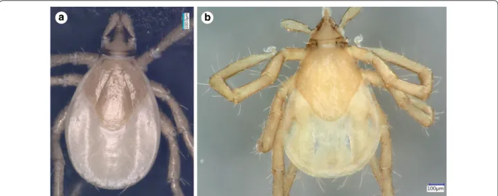
Larva. [See Figs. 4–6.] Length of idiosoma 0.96, breadth 0.76, ratio idiosomal length/breadth 1.26. Scutum pen- tagonal, posteriorly rounded, broadest slightly anteriorly a
c
b d
Fig. 2 Ventral views of male Ixodes vespertilionis (a, anterior part of the idiosoma; c, posterior part of the idiosoma) and I. collaris (b, anterior part of the idiosoma; d, posterior part of the idiosoma). The white dashed line indicates the second intercoxal space. The purple and red arrows show the genital aperture. The black and yellow arrows point to differences in the anterior part of the ivory coloration
to its mid-length (Fig. 4b), with rough surface. Length of scutum 0.52, breadth 0.49, ratio length/breadth 1.06.
Cervical grooves long, ending at deepest point of con- cavely curved posterolateral margin of scutum (Fig. 4b).
Scutal setae absent. Alloscutal setae longest caudally;
central dorsal setae (Cd1: 0.045, Cd2: 0.047) shorter than marginal dorsal setae (Md1-Md8: 0.088, 0.096, 0.105, 0.112, 0.125, 0.108, 0.133 and 0.148, respectively). Sternal setae ventrally (St1: 0.056, St2: 0.051; St3: 0.062) shorter than premarginal setae (Pm1-Pm4: 0.072, 0.075, 0.076 and 0.071, respectively) and preanal setae (Pa1: 0.064;
Pa2: 0.078); marginal ventral setae longest (Mv1: 0.092;
Mv2: 0.118; Mv3: 0.128) (Fig. 5c, d).
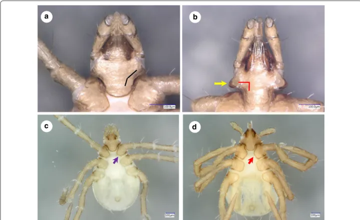
Gnathosoma: length from palpal apices to posterior margin of basis 0.34, width of basis capituli dorsally 0.25, ratio of length to width 1.36. Basis capituli dorsally trian- gular, with straight posterior margin. Ventral basis with 2 pairs of short (0.015) posthypostomal setae. Strongly concave caudolateral margin of ventral basis with per- pendicular angle and dark sclerotized edge (“collar”) at and behind maximum width of basis (Fig. 6b). Posterior margin of ventral basis rounded (Fig. 6b). Palpi elongated, Fig. 3 Drawings of Ixodes collaris male (a, dorsal view; b, ventral view) and female (c, dorsal view; d, ventral view)
club-shaped, curved both medially and laterally. Palpal length dorsally 0.24, breadth 0.07, ratio length/breadth 3.4. Segments I-III measure 0.03, 0.13 and 0.08, respec- tively. Palpal setae longest (0.06) near junction of seg- ments II-III. Hypostome conical, short (0.11), with dental formula 2/2.
Legs long. Hallerʼs organ elongated, longer than maximum breadth (diameter) of tarsus I (length: 0.48) (Figs. 4b, 5c, d, 6d,). Coxae medially rounded, without spines or spurs. Medial margins of coxae I-II straight, nearly equal in length; those of coxae III rounded (Fig. 6d). Coxae I-II with short (0.04) setae (2 and 1, respectively: Fig. 5d).
Differential diagnosis
Prior to its discovery, I. collaris may have been misiden- tified as I. vespertilionis in Southeast Asia (e.g. in [3]).
Therefore, the differential diagnosis (see Figs. 1, 2, 4 and 6) focuses on the latter species, as redescribed in [4] and [5]. Apart from data taken from the latter source on lar- vae of I. simplex Neumann, 1906, the redescription of the male of I. simplex was also taken into account [6], together with measurements (including those of I. ariad- nae Hornok, 2014) in [7].
The male of I. collaris has long legs (i.e. the length of Haller’s organ exceeds the maximum diameter of tar- sus I), unlike the male of I. simplex. The palpi of I. col- laris male have convexly curved lateral and medial edges, unlike the males of I. ariadnae (where the lateral edge is straight) and I. simplex (where the lateral edge is bent at an angle). The palpal setae are long (up to 210 µm), unlike in the males of I. ariadnae (30–100 µm) and I. simplex (20–80 µm). The most important features to distinguish
males of I. collaris from those of I. vespertilionis are on the gnathosoma: (i) more elongated palpi (length to breadth ratio 2.52 vs 2.0–2.1) (Fig. 1d vs 1c); (ii) the sparse distribution and low number (< 10) of long ante- rior and lateral palpal setae, which show dense distribu- tion and higher number (> 20) in I. vespertilionis (Fig. 1c);
and (iii) the posteriorly diverging (trapezoid), sclerotized ridge dorsally on the basis capituli, which is much less conspicuous and posteriorly converging (U-shaped) in I.
vespertilionis (Fig. 1c). In addition, (iv) the idiosoma of male I. collaris is more elongated than the idiosoma of male I. vespertilionis (length to breadth ratio 1.61 vs 1.35, Fig. 1b vs 1a) and (v) ventrally the ivory coloration ante- rior to the anus is oval, elongated in I. collaris, whereas this is less apparent in I. vespertilionis (Fig. 2c).
The larva of I. collaris has long legs, unlike the larva of I. simplex (in which the legs are short, i.e. the length of Haller’s organ does not exceed the maximum diameter of tarsus I). The palpi of I. collaris larva are elongated, club-shaped, with a length to breadth ratio above three (length × width: 240 × 70 µm), unlike in I. vespertilionis (Fig. 6a: club shaped, but shorter, 200 × 70 µm), I. ariad- nae (shorter and broader, 200 × 90 µm, laterally straight) and I. simplex (shorter and narrower, 140 × 60 µm). The scutum of I. collaris larva is longer than broad, pen- tagonal, with long cervical grooves, ending at the deep- est point of the concavely curved posterolateral margin of scutum (unlike in case of I. ariadnae and I. simplex).
The most important features to distinguish the larva of I. collaris from that of I. vespertilionis can be observed ventrally on the basis capituli: (i) its caudolateral edge is strongly concave in case of I. collaris, showing perpen- dicular angle at the concavity, whereas it is only slightly
a b
Fig. 4 Dorsal view of Ixodes vespertilionis (a) and I. collaris (b) larvae
concave, with an obtuse angle, in I. vespertilionis (Fig. 6a);
and (ii) at/behind the maximum breadth of ventral basis, the presence of a conspicuous, long, dark sclerotized edge (“collar”) also makes I. collaris larvae different from I. vespertilionis larvae, where this structure is less appar- ent (Fig. 6b vs 6a). In addition, (iii) caudal setae on the idiosoma are considerably longer than 100 µm in I. col- laris larvae, unlike in I. vespertilionis larvae (≤ 100 µm, see Figs. 4a and 6c); and (iv) the medial edge of coxa I is much shorter than that of coxa II in I. vespertilionis lar- vae (Fig. 6c), as contrasted to I. collaris larvae.
Discussion
With the morphological characters, high resolution pictures and drawings provided in the present study, descriptions of all stages of I. collaris are now complete.
Adding to already reported genetic differences between I. vespertilionis, I. collaris, I. ariadnae and I. simplex [1], these ixodid bat tick species show different morphology in all stages, as shown formerly for females and nymphs [2] and here for males and larvae.
In summary, short legs distinguish all stages of I. simplex from I. collaris. Ixodes ariadnae has laterally straight and Fig. 5 Drawings of Ixodes collaris nymph (a, dorsal view; b, ventral view) and larva (c, dorsal view; d, ventral view)
short palpi in all stages, unlike I. collaris. The male of I. ves- pertilionis has dense long palpal setae (sparse in I. collaris), whereas its females, nymphs and larvae lack the ventrolat- eral collar of gnathosoma (characteristic of I. collaris).
Conclusions
Based on the descriptions above, several features allow to distinguish the male and the larva of I. collaris morpho- logically from those of other bat-associated ixodid tick species.
Acknowledgements
The authors are grateful to Mr Nguyen Vu Khoi (director of Wildlife at Risk) and the Management Board of the IEBR for their support in the sample collection.
Special thanks to Ms Nóra Takács for performing PCRs.
Funding
Financial support was provided by NKFIH 115854 and by Rufford Foundation (RGS 18889-B). The publication of this research was supported by the 17896- 4/2018/FEKUTSTRAT grant of the Hungarian Ministry of Human Capacities.
Availability of data and materials
The sequences generated in this study are available in GenBank database under accession numbers MK450318-MK450321. Ticks morphologically investigated in this study are deposited at the Institute of Ecology and Biologi- cal Resources, Vietnam Academy of Science and Technology, Hanoi, Vietnam under the accession numbers VN16-053, VN16-060, VN16-064, VN16-068 and VN17-669). All other relevant data are included in the manuscript.
Authors’ contributions
SH initiated and supervised the study, extracted DNA, described the ticks and wrote the manuscript. JK made the microscopic pictures and DM prepared the drawings. VTT collected the specimens. All authors read and approved the final manuscript.
Ethics approval and consent to participate
Permissions for bat capture were provided by the Vietnamese Ministry of Agri- culture and Rural Development (Vietnam Administration of Forestry), People’s Committees of Cao Bang Province and the directorate of Phia Oac - Phia Den National Park (Vietnam), under decree numbers 378/TCLN-BTTN and 1399/
UBND-NC.
Consent for publication Not applicable.
Competing interests
The authors declare that they have no competing interests.
Publisher’s Note
Springer Nature remains neutral with regard to jurisdictional claims in pub- lished maps and institutional affiliations.
Author details
1 Department of Parasitology and Zoology, University of Veterinary Medi- cine, Budapest, Hungary. 2 Plant Protection Institute, Centre for Agricultural Research, Hungarian Academy of Sciences, Budapest, Hungary. 3 Institute of Ecology and Biological Resources, Vietnam Academy of Science and Tech- nology, Hanoi, Vietnam. 4 Graduate University of Science and Technology, Vietnam Academy of Science and Technology, Hanoi, Vietnam.
c d
a b
Fig. 6 Ventral views of larvae of Ixodes vespertilionis (a, gnathosoma; c, habitus) and I. collaris (b, gnathosoma; d, habitus). The black and red broken lines illustrate the angle of the caudolateral edge of the gnathosoma. The yellow arrow marks the “collar”. The purple and red arrows mark the medial edge of coxa I
•fast, convenient online submission
•
thorough peer review by experienced researchers in your field
• rapid publication on acceptance
• support for research data, including large and complex data types
•
gold Open Access which fosters wider collaboration and increased citations maximum visibility for your research: over 100M website views per year
•
At BMC, research is always in progress.
Learn more biomedcentral.com/submissions
Ready to submit your research? Choose BMC and benefit from:
References
1. Hornok S, Estrada-Peña A, Kontschán J, Plantard O, Kunz B, Mihalca AD, et al. High degree of mitochondrial gene heterogeneity in the bat tick species Ixodes vespertilionis, I. ariadnae and I. simplex from Eurasia. Parasit Vectors. 2015;8:457.
2. Hornok S, Görföl T, Estók P, Tu VT, Kontschán J. Description of a new tick species, Ixodes collaris n. sp. (Acari: Ixodidae), from bats (Chiroptera: Hip- posideridae, Rhinolophidae) in Vietnam. Parasit Vectors. 2016;9:332.
Pacific (Metastigmata: Argasidae, Ixodidae). Oriental Insects. 1970;4:37–46.
4. Arthur DR. The Ixodes ticks of Chiroptera (Ixodoidea, Ixodidae). J Parasitol.
1956;42:180–96.
5. Feider Z. Ixodoidea. In: Fauna of the Popular Republic of Romania, volume 5/2. Bucharest: Academiei Republicii Populare Romane; 1965. pp.
154–173 (In Romanian).
6. Sándor AD, Kontschán J, Plantard O, Péter Á, Hornok S. Illustrated rede- scription of the male of Ixodes simplex Neumann, 1906. Ticks Tick Borne Dis. 2018;9:1328–30.
7. Hornok S, Kováts D, Angyal D, Dányi L, Kovács R, Kontschán J. Descrip- tion of the male and the larva of Ixodes ariadnae Hornok, 2014. Ticks Tick Borne Dis. 2016;7:1252–5.