Specificity of Antibodies
ALFRED NISONOFF AND F. P. INMAN
Department of Microbiology, University of Illinois, Urbana, Illinois
Introduction
Investigations initiated by the work of Edelman (1959) have shown that antibody molecules are made up of multiple polypeptide subunits linked by disulfide bonds and weak noncovalent interactions (Edelman and Poulik, 1961; Fleischman et al, 1962, 1963; Olins and Edelman, 1962).
Methods have been devised for separating the component chains by reduction of disulfide bonds and subsequent exposure to conditions known to disrupt noncovalent interactions in proteins. T h e dissociation into subunits is reversible under appropriate conditions, and molecules similar to the native protein in configuration and molecular weight can be reconstituted in vitro. If the immunoglobulin preparation has anti- body activity, a significant proportion of the activity may be restored during the reassociation of subunits. Such investigations have permitted certain conclusions regarding the basis of antibody specificity, and one purpose of this paper is to summarize pertinent data. We will also discuss some recent structural studies and briefly review the accumulating evi- dence indicating that differences in antibody specificity are based on variation in amino acid sequence.
Antibody activity is associated with molecules collectively designated immunoglobulins. Table I summarizes some of the properties of three major classes common to many vertebrate species. In the guinea pig and in the mouse there are two 7 S immunoglobulins, distinct from γΑ, which have been designated γχ and γ2 (Benacerraf et al., 1963; Nussenzweig et al., 1964; Fahey et al., 1964). T h e former, which migrates more rapidly toward the anode during electrophoresis at neutral pH, mediates cuta- neous anaphylactic reactions within the same species but does not fix guinea pig complement. The opposite is true, with respect to both proper- ties, of the y2-globulin. Guinea pig yr and y2-globulins are similar with respect to carbohydrate content (Oettgen et al., 1965), as well as sedimenta-
39
40 ALFRED N I S O N O F F AND F . P . I N M A N
tion coefficient, and it is therefore difficult at present to decide which, if either, is the counterpart of the yG-globulins of other species.
Each of the three classes of molecule appears to comprise two types of polypeptide chains; the "light" chain has a molecular weight of approxi- mately 20,000 (Edelman and Poulik, 1961; Pain, 1963; Small et al, 1963) and is common to all three classes (Cohen, 1963; Carbonara and Here- mans, 1963); the classes differ with respect to their "heavy" chains which have molecular weights of 50,000-55,000 in yG-globulin (Pain, 1963;
Small et al, 1963). A molecule of yG-globulin consists of two light and two heavy chains (Fleischman et al, 1962, 1963; Pain, 1963). Bence Jones
TABLE I
PROPERTIES OF THE MAJOR CLASSES OF IMMUNOGLOBULIN«. &
Immune globulin
YG = y2c γΑ
γΜ<ζ
Sedimentation coefficient
6.5 S 6.5 S + polymers
19 S
Molecular weight 150,000 150,000 + polymers
800,000
Carbohydrate content
2 - 3 % 10%
10%
Dimensions 35 X 280 A
?
?
β T h e three classes have antigenic determinants in common present on "light"
polypeptide chains (molecular weight, 20,000).
& Antibody activity is associated with all three classes.
o Two classes similar to yG, designated y1 and γ2, have been demonstrated in guinea pigs and mice (see text).
d γΜ is depolymerized from 19 S to 7.5 S by mild reduction.
proteins, which are secreted in the urine of patients with multiple mye- loma, are monomers or dimers of light polypeptide chains (Edelman and Gaily, 1962). Incomplete heavy chains1 have been found in the urine of a few persons with lymphatic malignancies (Franklin et ah, 1964; Osser- man and Takatsuki, 1964). Such incomplete naturally occurring mole- cules are ordinarily included in the category of immunoglobulins.
A given type of polypeptide chain is characterized by marked hetero- geneity, even within a single immunoglobulin class of a given species. It seems likely, for example, that antibody specificity is determined by the amino acid sequence of both light and heavy chains. If this is correct, the variety of sequences in at least a portion of each type of polypeptide chain is obviously very large. Another basis of heterogeneity has been elucidated through studies of the antigenic determinants of the poly-
i T h e protein secreted corresponds closely in structure to the inactive fragment (Fc) of a papain digest.
peptide chains of immunoglobulins. For example, the light and heavy chains of rabbit yG-immunoglobulin are under the control of at least two genetic loci, with three alleles at each locus. The phenotypic expres- sion is an antigenic determinant, or group of determinants on the chain, which can evoke antibody formation on injection into a rabbit lacking the determinants. The classes of antigenic determinants are called allotypes (Oudin, 1956). An individual rabbit thus may possess light chains with one or two different allotypic specificities encompassed within the light chains of its yG-globulin, and one or two within the heavy chains. A single molecule, however, possesses only one type of light chain allotypic determinant and one type of heavy chain determinant (Oudin, 1961; Dray and Nisonoff, 1963). The inheritance of allotypic specificities is Mendelian and is not sex-linked. Each genotype is expressed phenotypically.
Variability of polypeptide chains similarly under genetic control and similarly characterized phenotypically by antigenic determinants on the chains, is also present in human immunoglobulins. The determinants on the heavy and light chains are known as the Gm and Inv groups, re- spectively (Grubb and Laurell, 1956; Ropartz et al., 1961). The Gm groups in particular are complex (Fudenberg, 1963) and will not be discussed here.
Another source of variability among polypeptide chains is exemplified by the existence of two subgroups, distinct from Inv groups, of the light chains of human immunoglobulins (Korngold and Lipari, 1956a,b; Burtin et al., 1956). Again, these subgroups are characterized by antigenic deter- minants present in each of the three major classes of immunoglobulin.
Appropriate antisera can be prepared in rabbits. In contrast to rabbit allotypes, and to human Gm and Inv groups, both of these subgroups are represented in the sera of all normal individuals (Franklin, 1962; Mannik and Kunkel, 1963; Migita and Putnam, 1963; Fahey, 1963). Group K (or I) comprises about 60% and Group L (or II), 30% of the population of normal yG-immunoglobulin molecules (Mannik and Kunkel, 1963).
Subgroups of normal populations of human heavy chains, characterized by their antigenic properties, have also been described (Dray, 1960;
Lichter and Dray, 1964; Grey and Kunkel, 1964; Terry and Fahey, 1964;
Ballieux et al., 1964).
In view of these manifold sources of heterogeneity, one may reasonably ask what characteristics are common to the light chains and heavy chains of a given class of immunoglobulin in a particular species. A definitive answer to this question will require a large amount of data on amino acid sequences in the various subgroups. At present, one can say that light
42 ALFRED N I S O N O F F AND F . P . I N M A N
chains of a particular class of immunoglobulin have in common the ability of combine stoichiometrically and spontaneously with heavy chains to form a complete molecule of the correct molecular size. This implies there is at least one region on each light polypeptide chain complementary to a region on a heavy chain (see Fig. 1), and indicates one type of struc- tural invariance. In the rabbit, light chains of different allotypic speci- ficity have some antigenic determinants in common, and average amino acid compositions as well as pep tide maps (fingerprints) are very similar although not identical (Reisfeld et al, 1965; Small et al., 1965). (Finger- prints may fail to demonstrate the presence of variable segments of molecules or subunits because of the low yield of peptides derived from such segments.) Heavy chains of different allotypic specificity, as well as light chains, possess one or more similar antigenic determinants.
Thus there appear to be invariant as well as variable regions in the anti- body molecule. Variations within a given class of polypeptide chain may reflect antibody specificity, heritable differences in sequence not associated with specificity, and the presence of multiple structural genes present in all individuals. The light chain groups, K and L, in the human immuno- globulins are in a somewhat special category since they share no antigenic determinants and yield fingerprints which appear to be unrelated (Put- nam, 1962). They are, however, similar in molecular weight and in their capacity to form active antibody molecules in combination with heavy chains. The fact that the peptide maps are completely different does not exclude the possibility that many corresponding positions in the sequences of the two polypeptides may be occupied by the same amino acid.
Of the three major classes of complete immunoglobulin molecules (yG, γΑ, γΜ), the structure of the yG type is known in the greatest detail.
A model incorporating many of the known structural features is shown in Fig. 1; it is essentially equivalent to the one proposed by Fougereau and Edelman (1965) and applies to rabbit and human yG-globulins. Con- siderable evidence indicates that the yG-globulins of many other species have similar structures.
The presence of multiple polypeptide chains in the yG-globulin mole- cule was established by the work of Edelman and his collaborators (Edel- man, 1959; Edelman and Poulik, 1961). Subsequent to the reduction of interchain disulfide bonds and alkylation to prevent reoxidation, the polypeptide subunits can be separated by treatment with urea, 1 M propionic acid, guanidine hydrochloride, or detergent (Edelman and Poulik, 1961; Fleischman et al, 1962; Small et al, 1963; Marier et al, 1964; Utsumi and Karush, 1964; Criddle, 1964). The light and heavy
chains can be isolated for analytical purposes on starch gel (Edelman and Poulik, 1961) or acrylamide gel (Cohen and Porter, 1964) in a dissociating solvent such as aqueous urea. On a preparative scale, the chains may be separated by gel nitration on Sephadex equilibrated with 1 M propionic acid (Fleischman et al., 1962) or, at neutral pH, with sodium decyl sulfate (Utsumi and Karush, 1964). T h e heavy and light chains are eluted as successive peaks in approximately a 5:2 weight ratio, which is also the
Combining f \\
site J - i
j
V W
Papain jt \ Pepsin
Fab
\Q
ΊΓ5Γ Ö
Fc
o°o
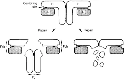
FIG. 1. Structural model of rabbit or human 7 S yG-immunoglobulin molecule, slightly modified from Fougereau and Edelman (1965). H and L signify "heavy" and
"light" polypeptide chains of molecular weight 55,000 and 20,000, respectively. Each small dark rectangular area between chains represents an interchain disulfide bond.
(In addition, there are about 17 intrachain disulfide bonds.) The H and L chains and the two halves of the Fc fragments are held together by noncovalent interactions as well as by a disulfide bond. Molecular weights of the Fab and Fc fragments are ap- proximately 43,000 and 55,000, respectively. The size of a combining site is exaggerated, since it actually represents roughly 1-2% of the surface area of the molecule.
ratio of the molecular weights of the chains. Gel filtration in 1 M propionic acid has been used in most experiments in which biological activity is restored by reassociation of separated light and heavy chains, and these investigations are discussed in some detail later. This method of separation has also permitted accurate estimation of the molecular weights of the polypeptide subunits (Pain, 1963).
Comparison of antigenic determinants and amino acid compositions of polypeptide chains isolated by this method with those of the fragments
44 ALFRED NISONOFF AND F. P. INMAN
liberated by papain, has established the relationship between polypeptide chain structure and the fragments formed by enzymatic digestion shown in Fig. 1 (Fleischman et al., 1962, 1963; Olins and Edelman, 1962).
Concurrently with the isolation and characterization of the subunits, useful information was derived from studies of the proteolysis of yG- globulin by enzymes such as papain and pepsin. The action of proteolytic enzymes on antibodies has been discussed in several reviews (e.g., Porter and Press, 1962; Eisen and Pearce, 1962; Nisonoff and Thorbecke, 1964) and will be considered very briefly. Porter (1958, 1959) demonstrated that the action of papain on rabbit yG-globulin, or on the specific antibody molecules contained in this fraction, resulted in release of three large fragments in good yield, together with some dialyzable material. Two of the fragments (designated Fab) are very similar to one another and are univalent. As such they are incapable of forming a large lattice or frame- work in combination with antigen; thus they cannot cause precipitation or agglutination of antigens. The fragments can combine with the anti- gen, however, and prevent subsequent interaction and precipitation with untreated antibody; i.e., the two Fab fragments have specific blocking ac- tivity. The third fragment, originally called fragment III and now desig- nated Fc (Fig. 1) does not combine with antigen but is crystallizable, in contrast to the original yG-globulin molecules. This suggests a consider- able degree of homogeneity of the Fc fragments, and also that much of the heterogeneity of rabbit yG-globulin resides in the Fab fragments. This is not surprising since the latter contain the combining sites which are neces- sarily variable. Certain biological activities, such as fixation to skin in passive cutaneous anaphylaxis and ability to cross the placenta in certain species, are associated with the inactive fragment. It may also be directly involved in complement fixation although this is controversial. In some instances, a large proportion of the antibodies formed on injection of yG-globulin into a heterologous species is directed against the Fc fragment (Porter, 1958, 1959). These subjects are considered in detail in the reviews cited above.
The action of pepsin on rabbit yG-globulin results in removal of a fragment that corresponds roughly to papain fragment Fc but is evidently somewhat smaller (Nisonoff et al., 1960; Nisonoff and Hong, 1964;
Nisonoff and Dixon, 1964; Jaquet and Cebra, 1965). After treatment with pepsin, the two fragments containing the active sites are linked by a disulfide bond. Thus, the major product of peptic digestion is still bivalent and capable of forming specific precipitates. It has a molecular weight approximately two-thirds that of the undegraded molecule. The
bivalent fragment formed by peptic digestion can be dissociated into two univalent fragments approximately equal in size, by reduction of one disulfide bond (Nisonoff et al., 1961); see Fig. 1. There appear to be no additional noncovalent interactions linking the univalent fragments at this juncture, since separation of the two fragments occurs at neutral pH after reduction.
A considerable degree of recombination occurs through reoxidation, if the reducing agent is rapidly removed and the protein solution allowed to stand at neutral pH (Mandy et al., 1961). Thus, the transformation from bivalent precipitating fragments of molecular weight 100,000 to univalent blocking fragments of molecular weight 50,000 is largely reversi- ble. If, after reduction, antibodies of two different specificities are mixed, the product contains a large proportion of individual molecules with mixed specificity (Nisonoff and Rivers, 1961; Nisonoff and Mandy, 1962;
Fudenberg et al., 1964). In the case of the yG-globulins, such molecules do not appear to occur naturally. A more complete description of the effects of pepsin and reducing agent on rabbit yG-globulin is given by Nisonoff and Hong (1964).
Dissociation of Rabbit yG-Globulin into Half-Molecules Another type of dissociation of rabbit yG-globulin, into half-molecules, occurs after mild reduction followed by acidification to pH 2.5 in dilute salt solution (Palmer et al., 1963). Two factors favor the formation of half-molecules rather than separate chains. First is the greater lability of the disulfide bond linking the heavy chains (cf., Fig. 1) in comparison to that of the bonds joining light and heavy chains (Palmer et al., 1963;
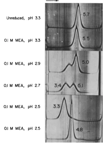
Palmer and Nisonoff, 1964). Second, at low pH the noncovalent inter- actions joining the heavy chains are less stable than those between light and heavy chains. After reduction of all the interchain disulfide bonds, dissociation into half-molecules takes place under milder conditions than are required for separation of light from heavy chains (Hong and Nisonoff, 1965). Figure 2 compares the effects of pH on the sedimentation patterns of unreduced and reduced rabbit yG-globulin. At pH 3.3, the sedimentation coefficients are approximately the same; in contrast, at pH 2.5 that of reduced yG-globulin is only 70% as great (s2o,w = 3.3 versus 4.8). At the intermediate pH values of 2.9 and 2.7, two components are present in the reduced preparation; the slower component increases in amount as the pH is lowered. Unreduced yG-globulin exhibited a single peak at each pH investigated; the decrease in sedimentation coeffi- cient is probably attributable to expansion of the molecule at low pH.
46 ALFRED NISONOFF AND F . P. INMAN
The 3.3 S component observed in Fig. 2 was found to have weight- average and Z-average molecular weights of 81,800 and 85,500, respectively (Palmer et al., 1963). These values are consistent with a molecular weight of 75,000 and the presence of about 10% of undissociated yG-globulin (molecular weight, 150,000). A small amount of faster-sedimenting ma- terial was present in the preparation examined even after passage through Sephadex G-200. In later studies it was found that dissociation at pH 2.5
Unreduced, pH 3.3
0.1 M MEA, pH 3.3
0.1 M MEA, pH 2.9
0.1 M MEA, pH 2.7
0.1 M MEA, pH 2.5
0.1 M MEA, pH 2.5
FIG. 2. Schlieren patterns of reduced and unreduced rabbit YG-globulin photo- graphed after ultracentrifugation for 80 minutes at 59,780 rpm at 20° C (except G, 64 minutes). MEA is an abbreviation for 2-mercaptoethylamine, the reducing agent employed. The slower-moving, 3.3 S component consists of half-molecules of yG- globulin and appears in increasing amounts as the pH of the reduced protein is lowered from 3.3 to 2.5; the numerals are $20 w values; sedimentation is from left to right. (Palmer et al., 1963.)
is more nearly complete in 0.025 M NaCl than in 0.1 M NaCl (Palmer and Nisonoff, 1964).
The data on molecular weights are therefore consistent with dissocia- tion into half-molecules. Further evidence includes the symmetry of the schlieren peak at low pH and the fact that essentially complete dissocia- tion occurs after a mild reduction that permits the separation of only small amounts of light chains in 1 M propionic acid. In addition, an experiment was carried out in which the extent of reduction was such that dissociation occurred in only a fraction of the population of mole- cules. After separation of the dissociable 3 S molecules from the undisso- ciable (unreduced) molecules by gel filtration at pH 2.4, it was found that their amino acid compositions were the same within experimental error. This result is again consistent with a separation into symmetrical half-molecules.
Finally, a large proportion of the yG-globulin can be dissociated into half-molecules after reduction of slightly more than one disulfide bond for each molecule rendered dissociable (Palmer and Nisonoff, 1964). This supports the view that two subunits are separated and, together with the data on molecular weights, provides strong evidence for symmetrical cleavage into half-molecules.
Spontaneous Reassociation of Half-Molecule Subunits at Neutral pH
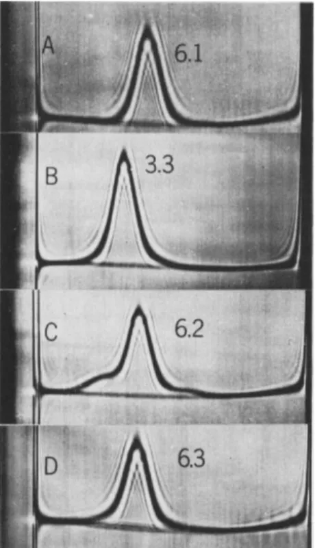
After dissociation into half-molecules at low pH, a large proportion of the yG-globulin spontaneously recombines at neutrality to form mole- cules having the same sedimentation coefficient as unreduced yG-globulin (~6 S). The recombination involves noncovalent interactions since it takes place even after sulfhydryl groups released on reduction have been inactivated by treatment with a reagent such as iodoacetate to prevent re- oxidation. Schlieren patterns of dissociated and recombined preparations are shown in Fig. 3.
Physical chemical studies were carried out on a recombined preparation that had been purified by gel filtration (Fig. 3D). The product migrated as a symmetrical 6.3 S peak in the ultracentrifuge. Its molecular weight and diffusion constant agreed closely with corresponding values for un- reduced yG-globulin. In another similar experiment the specific viscosities of native and reconstituted molecules were also found to agree within experimental error (Nisonoff and Hong, 1964).
48 ALFRED NISONOFF AND F. P. INMAN
FIG. 3. Schlieren patterns obtained in the ultracentrifuge; an illustration of disso- ciation of reduced rabbit yG-globulin into half-molecules at low pH and reassociation at neutrality. Sedimentation is from left to right. A, Normal yG-globulin; B, yG-globu- lin reduced at pH 5 with 2-mercaptoethylamine, and then acidified to pH 2.4 in 0.035 M NaCl; C, above sample after neutralization to pH 8 by dialysis; D, above neutralized sample after filtration through Sephadex G-200 for isolation of the major component. Centrifugation at 59,780 rpm at 20°C (except B, 10°C). Photographs were taken after 48 minutes (except B, 80 minutes) at full speed. The numerals are $20 w
values; Solvent for A, C, and D was saline-borate buffer, pH 8, ionic strength 0.16.
(Nisonoff and Hong, 1964.)
Evidence for Univalence of Half-Molecules of Rabbit yG-Globulin
Since a half-molecule comprises a light and a heavy chain one would predict that it is univalent. A direct demonstration of this property is complicated by the spontaneous recombination of half-molecules at neu-
tral pH at which antibody assays are carried out. An indirect approach took advantage of the finding that recombination of half-molecules of
purified antiovalbumin with the half-molecules of nonspecific yG-globulin is essentially random (Nisonoff and Palmer, 1964). Half-molecules of purified antiovalbumin were therefore recombined in the presence of an elevenfold excess of half-molecules of normal yG-globulin; the mixture was made at low pH and then neutralized (Hong et al., 1965). On the assumption of random recombination, 11/12 of the half-molecules derived from antibody should have recombined with half-molecules of yG-globu- lin to form univalent 6 S molecules. It was found that the recombined mixed molecules were capable of specifically inhibiting the precipitation of untreated antiovalbumin with ovalbumin. This is consistent with univalence of the mixed 6 S molecules, and therefore of half-molecules of antiovalbumin. As a control in this experiment, half-molecules of purified antiovalbumin were allowed to recombine in the absence of normal yG-globulin. The product formed specific precipitates; 81% of the protein was precipitable by an optimal concentration of the antigen (Hong et al., 1965).
Reversible Dissociation of Fragment Fc
As indicated earlier, the fact that two univalent (Fab') fragments are linked only by a disulfide bond after peptic digestion suggests that most of the noncovalent interactions between heavy chains are present in fragment Fc and therefore involve the portions of the two heavy chains comprising this fragment (Fig. 1). One would thus expect that reduced fragment Fc might dissociate into half-fragments under conditions similar to those resulting in dissociation of reduced yG-globulin into half-mole- cules. Prior to our work on this problem, Marier et al. (1964) had shown that reduced fragment Fc dissociates in guanidine hydrochloride and that the molecular weight of the product (26,000) is half that of undissociated Fc. Our work (Inman and Nisonoff, 1965a,b) indicates that dissociation of reduced Fc can also take place under conditions very similar to those required for dissociation of reduced yG-globulin into half-molecules.
Schlieren patterns illustrating the effect of pH on reduced Fc in 0.05 M NaCl are shown in Fig. 4; molecular weights are given in Table II. Disso- ciation occurs between pH 3.5 and 2.7, and increasing extents of dissocia- tion are observable at intermediate pH values. The molecular weight of the dissociation products is approximately half that of undissociated Fc (Table II).
As in the case of reduced yG-globulin, dissociation of Fc is reversible at neutral pH; the neutralized product has a sedimentation coefficient and
50 ALFRED NISONOFF AND F. P. INMAN
molecular weight very close to those of undissociated Fc (Inman and Nisonoff, 1965a,b).
Other factors relevant to this dissociation may be summarized as fol- lows: (a) Crystallized Fc prepared by digestion with papain in the presence of 0.001 M L-cysteine is only slightly dissociable at pH 2.4, whereas crystallized Fc prepared by digestion in the presence of 0.05 M L-cysteine
FIG. 4. The effect of pH on sedimentation of recrystallized Fc prepared by digestion with papain in the presence of 0.05 M L-cysteine. A-F correspond to pH values of 3.5, 3.2, 3.1, 3.0, 2.7, and 2.4, respectively; NaCl concentration, 0.05 M. Sedimentation (from left to right) for 80 minutes (photographs on the left) and 160 minutes (photo- graphs on the right) at 59,780 rpm at 20°C. Numerals are s2 0 w values. The 1.8 S peak represents a half-fragment of Fc.
is about 90% dissociable; (b) Fc prepared with the lower concentration of L-cysteine is rendered dissociable by reduction with 0.05 M L-cysteine, thus indicating the presence of an interchain disulfide bond; (c) reduction of this interchain bond is reversible. The reduced Fc fragment loses its capacity for dissociation at low pH if reoxidation is permitted to occur after removal of the reducing agent. These results provide support for the molecular model in Fig. 1 and are consistent with the view that most
of the noncovalent interactions linking the heavy chains are present in fragment Fc. They also suggest that papain and pepsin cleave on opposite
"sides" of the interchain disulfide bond joining the heavy chains (Fig. 1).
After peptic digestion this bond links the two Fab' fragments, whereas it
TABLE II
MOLECULAR WEIGHTS OF PURIFIED FC<*
Concentration of cysteine present during
digestion (M) 0.001 0.001 0.05 0.05
Molecular weight**
48,800 41,500 52,900 24,100
pHc 3.5 2.4 3.5 2.4
β Prepared from rabbit yG-globulin by digestion with 2 % by weight of papain in the presence of L-cysteine (Inman and Nisonoff, 1965a,b). Fragment Fc was purified by recrystallization or gel filtration.
& Molecular weights were determined by the method of sedimentation-diffusion.
c Refers to the p H during molecular weight determination.
is localized in Fc after papain digestion in the presence of a very low con- centration of reducing agent.
In Vitro Complementation of Polypeptide Subunits of yG-Globulins
There is some uncertainty at present about the extent to which light and heavy polypeptide chains contribute to the active site of an antibody molecule. T h e problem is complicated by the difficulty (Feinstein et al., 1963; Stemke, 1964) of preparing heavy chain fractions that are com- pletely free of light chains. Certain tentative conclusions can be drawn from the limited data available:
(a) Isolated heavy chains usually have some antibody activity.
(b) Isolated light chains in general have little if any activity.
(c) Recombination of antibody heavy chains with antibody light chains results in an increase in activity over that of the heavy chains alone; such recombination occurs spontaneously through noncovalent interactions.
(d) Recombination of heavy chains of antibody with light chains de- rived from antibody of a different specificity, or from nonspecific yG- globulin, gives little enhancement, although molecules similar to native antibody in molecular weight are formed in good yield.
52 ALFRED NISONOFF AND F. P. INMAN
(e) Complementation is most effective when the light and heavy chains of a particular specificity are derived from the same animal.
(f) T h e presence of hap ten stabilizes the noncovalent linkage of heavy and light chains. This results in the preference of heavy chains for homologous rather than heterologous light chains when competition is carried out in vitro in the presence of hapten.2
(g) The results of a number of investigations are characterized by con- siderable variation in the amounts of antibody activity associated with isolated heavy chains. The reasons for this variability have not been established. Factors that might be relevant are: (1) the specificity of the antibody; i.e., heavy chains of different antibodies might contribute to specificity to a variable extent; (2) the tendency of isolated heavy chains to aggregate; (3) differences in the extent of denaturation of chains during the isolation procedures; or, (4) the difficulty of completely separating heavy chains from light chains. In experiments in which the activity of isolated heavy chains was only a few per cent of that of the native anti- body (cf. Table III), the possibility that this residual activity was actually attributable to contamination by light chains capable of complementation with the heavy chains has not been completely excluded.
Evidence for Antibody Activity in Isolated Heavy Chains
At least two investigations have indicated the association of a substan- tial amount of antibody activity with separated heavy chains. Fleischman et al. (1963), found that heavy chains of horse yG-globulin, containing antibody against rabbit γ-globulin, specifically coprecipitated when added to a mixture of the antigen and untreated antibody. T h e degree of coprecipitation of heavy chains was quantitatively similar to that observed on the addition of comparable amounts of the untreated, anti- body-containing yG-globulin fraction to the same precipitating system.
Specificity was indicated by the fact that the heavy chains did not co- precipitate when added to another horse antigen-antibody system. In addition, they found that heavy chains derived from a different horse antibody, antidiphtheria toxoid, specifically inhibited the homologous antigen-antibody reaction presumably through combination with antigen.
The degree of inhibition per unit weight of added heavy chains was similar to that observed on the addition of untreated antibody globulin
2 Such an experiment is performed by mixing a deficiency of heavy chains with the two types of light chain in 1 M propionic acid and bringing the mixture to neutral pH.
The amount of each type of light chain bound to heavy chains is then determined.
to the precipitating mixture. Heavy chains derived from a different horse antibody had no effect.
Additional evidence for activity in the heavy chain fraction of yG- globulin comes from the studies of Utsumi and Karush (1964) who em- ployed rabbit antibody specific for the azophenyl-ß-lactoside hapten group. They dissociated the light and heavy chains of the reduced puri- fied yG-antibody with 0.05 M sodium decyl sulfate at neutral pH; the chains were separated on Sephadex G-200 equilibrated with the detergent which was then removed on an ion-exchange column. Antigenic analysis indicated that the separation was essentially complete. Equilibrium dialysis was used to measure the capacity of the separated chains for binding hapten. More than half of the heavy chains were found to have active combining sites; the average binding constant, however, was about one-eighth that of reduced undissociated antibody. Expressed as unitary free energy of combination, the decrease in binding affinity was 13%. In view of the possibility that loss of activity was actually attributable to denaturation rather than to the removal of light chains, the investigators tentatively concluded that the binding activity was exclusively associated with heavy chains.
Antibody Activity of Recombined Heavy and Light Chains
Considerable evidence now indicates that the specific recombination of light and heavy chains may yield a product with more activity than that resulting from the combination of heavy chains of antibody with nonspecific light chains, or of antibody light chains with nonspecific heavy chains.3 Suggestive evidence was first obtained by Franek and Nezlin (1963). After recombination of heavy and light chains derived from purified horse diphtheria antitoxin, about 3% of the protein was active, as judged by adsorption to an antigen covalently linked to cellu- lose which served as an inert matrix. Less than 1% of the protein was adsorbed when the heavy chains of diphtheria antitoxin were combined with light chains of a different antibody, or if light chains of the diph- theria antitoxin were combined with the heavy chains derived from a different horse antibody. Approximately 65% of the undegraded diph- theria antitoxin was specifically adsorbed. In the case of horse anti- tetanus toxin, however, there was little difference between the results obtained when the light chains were derived from the homologous anti-
3 In the experiments to be discussed, mixtures of chains were made at low pH, usually in 1 M propionic acid, and then dialyzed against neutral buffer.
V-rt
TABLE III
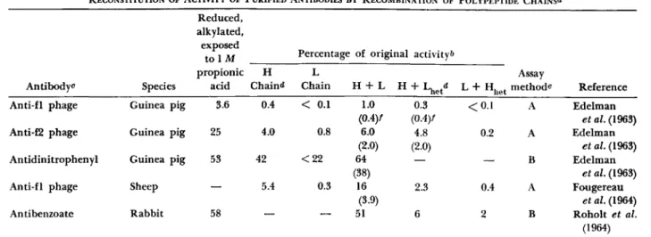
RECONSTITUTION OF ACTIVITY OF PURIFIED ANTIBODIES BY RECOMBINATION OF POLYPEPTIDE CHAINS«
Antibody^
Anti-fl phage Anti-f2 phage Antidinitrophenyl Anti-fl phage Antibenzoate
Species Guinea pig Guinea pig Guinea pig Sheep Rabbit
Reduced, alkylated, exposed
t o i M propionic
acid 3.6 25 53
——■
58
H Chain«*
0.4 4.0 42
5.4
Percentage L Chain
< 0.1 0.8
< 2 2 0.3
: of original activity*»
H + L 1.0 (0.4)/
6.0 (2.0) 64 (38)
16 (3.9) 51
H + Lhetd 0.3 (0.4)/
4.8 (2.0)
—
2.3 6
het
< 0 . 1 0.2
0.4 2
Assay methods
A A B A B
Reference Edelman
et al. (1963) Edelman
et al. (1963) Edelman
et al. (1963) Fougereau
et al. (1964) Roholt et al.
(1964) a In each experiment mixtures of light and heavy chains in approximately equimolar amounts were prepared at low pH, and then brought to neutrality.
δ Values in the table are percentages of the activity of the untreated, purified antibody.
c Antibodies were purified from the serum of an individual animal, except in the case of antibenzoate which was derived from a pool of sera.
<* Abbreviations: H = heavy chains of the antibody; L = light chains of the antibody; Lfaet = light chains from a heter- ologous source, either nonspecific yG-globulin or a different antibody; Hh e t = heavy chains from a heterologous source.
e Assay methods: A, phage neutralization; B, hapten-binding capacity measured by equilibrium dialysis.
/ Numbers in parentheses indicate activity expected if activities of the separate chains were additive.
>
r
M Ö
o O
•si
>
Ö
z
>
body or from a different horse antibody. In either case the amount of protein adsorbed was less than 1%.
Thus with either antibody at least 97% of the activity was lost, pos- sibly as a result of the fact that 6 M urea was used as solvent during the separation of chains on Sephadex. There would also appear to be some possibility of variation in the degree of nonspecific adsorption of the different preparations to cellulose.
Table III summarizes data which support the hypothesis that a specific combination of light and heavy chains from the same antibody is more effective in restoration of activity than a combination of specific heavy chains with nonspecific light chains or specific light chains with nonspecific heavy chains. The sixth column in Table III, labeled
"H -f- L," compares the degree of activity observed on combination of specific heavy and light chains with that predicted from the activity of either type of chain measured separately; the predicted value is given in parentheses. In each case there is a definite enhancement attributable to interaction of the chains.
A comparison of the sixth and seventh columns indicates that light chains derived from the antibody are more effective in complementation of specific heavy chains than are light chains derived from a different anti- body source or from normal yG-globulin.
The results in the eighth column (specific light chains -f- nonspecific heavy chains) indicate that the light chains of the antibodies investigated are relatively ineffective when combined with heterologous heavy chains.
The most striking results, obtained with rabbit antibenzoate antibody (Roholt et al., 1964), are shown in the fifth row of Table III. Approxi- mately 51% of the original activity, measured by the capacity for binding hapten, was restored on mixing the specific heavy and light chains. When chains from nonspecific yG-globulin were substituted for either specific heavy or specific light chains, nearly all of the activity was lost. The possi- bility was not eliminated that part of the binding by mixtures of specific and nonspecific chains, 6% and 2%, respectively, might have been due to the presence of a slight amount of residual heavy chain in the light chain fraction and vice versa.
An interesting effect of the presence of hapten during specific recombi- nation was noted by Metzger and Singer (1963) who worked with rabbit antibody specific for the dinitrophenyl hapten group. This antibody re- tains the capacity for binding hapten at low pH. It was found that the yield of free light chains obtained on gel filtration of the reduced anti- body in 1 M propionic acid was reduced from about 25% to 16% of the
56 ALFRED NISONOFF AND F . P . I N M A N
total protein, when 6 χ 10~6 M hapten, dinitrophenyllysine, was added to the reduced antibody preparation and to the buffer used for elution.
This result shows that hapten stabilizes the noncovalent linkage of heavy and light chains thus decreasing the extent of dissociation in 1 M propionic acid.
In a second investigation (Metzger and Mannik, 1964), competition of specific and nonspecific light chains for specific heavy chains derived from rabbit antidinitrophenyl antibody was measured. Equal amounts of the two types of light chains were used, and the molar ratio of light to heavy chains was 2:1; i.e., light chains were present in excess. When the com- bination of light and heavy chains was carried out in the absence of hapten, the nonspecific and specific chains competed about equally well for combination with the specific heavy chains. However, when the mixture was made in the presence of hapten, 3 χ 1 0- 5 M dinitrophenylamino- caproic acid, the ratio of specific to nonspecific light chains in the final product, after neutralization, was about 2:1. This again indicates a specific stabilizing effect of hapten on the interaction of heavy and light chains of antibody. It was also noted that the amount of antibody activity restored on combination of specific light and heavy chains was increased when hapten was present during the association.
Recently, Roholt et al. (1965) reported that the mixing of heavy and light chains of antibenzoate antibody from the same rabbit resulted in good recovery of antibody activity. In contrast, when heavy chains of antibenzoate antibody of one rabbit were recombined with light chains of antibenzoate antibody from another rabbit there was little restoration of activity. A possible mechanistic explanation for this result might involve the geometry of the site; i.e., a site with a particular configuration may be formed by a variety of chain pairs, but the configuration of the heavy chains would dictate or at least restrict the range of structures of effective complementary light chains. Alternatively, interaction with the light chain might influence specificity by altering the configuration of a heavy chain with the interaction between chains taking place outside the active site. This possibility is based on the assumption that the heavy chain contains the site. Again, a variety of light and heavy chain combina- tions might produce an active site, but the light chains from one antibody molecule might not correctly influence the structure of the heavy chain from a different antibody molecule of the same specificity.
Evidence for Direct Participation of Light Chains in the Active Site of an Antibody
The enhancement of specific antibody activity by light chains is con- sistent with the possibility that a portion of the light chain is physically present in the active site of an antibody. As indicated, however, an alternative explanation is conceivable. The experiments of Metzger and Singer, cited above, demonstrating stabilization of the noncovalent inter- action of heavy and light chains by hapten, are similarly consistent with direct participation of the light chain in the active site; stabilization by hapten could well result from a three-way interaction among hapten and the two types of polypeptide chain in the active site. As these workers indicated, however, the data do not exclude the possibility that hapten induces an alteration of configuration of the heavy chain which permits a stronger interaction with the light chain outside the active site.
Direct evidence in support of the physical participation of both types of chain in the active site was obtained by Metzger et al. (1964). Their previous work had shown that the active site of an antibody can be labeled by using specific hapten capable of interacting covalently with the side chains of proteins. For example, rabbit antibody against the dinitro- phenyl hapten group can be specifically labeled with p-nitrophenyldia- zonium fluoborate. The nitrophenyl portion of the molecule causes specific combination of the hapten with the antibody site. The diazonium group then reacts preferentially with a side chain in or very near the active site, by forming a covalent bond. Several lines of evidence have demonstrated the validity of this conclusion.
When antidinitrophenyl antibody was allowed to react with sufficient diazonium reagent so that 0.4 moles per mole of protein were incor- porated, it was found that two-thirds of the covalently bound hapten was attached to heavy chains and one-third to light chains. This experiment provides strong evidence for the direct participation of both types of chain in the active site of at least part of the population of antibody molecules.
In the same paper similar results were reported for another rabbit anti- body specific for the azophenylarsonate hapten group.
Evidence That Differences in Specificity Are Associated with Differences in Amino Acid Sequence
Perhaps the most striking characteristic of antibodies that distinguishes them from other proteins is that an enormous variety of specificities is associated with relatively small differences in structure and amino acid
58 ALFRED NISONOFF AND F . P. INMAN
composition. Thus, there must be large sequences common to the various antibody molecules. Pauling's (1940) template theory took cognizance of this fact by proposing that the variety of specificities is associated with a single primary sequence and that specificity is determined during the fold- ing of the polypeptide chain. Folding was assumed to occur in the pres- ence of antigen which acts as a template and leaves an imprint on the completed antibody molecule.
The work of White (1961) and Anfinsen et al. (1961), and subsequent similar work with a number of enzymes, has demonstrated that after reduction of disulfide bonds and unfolding many proteins spontaneously regain their native structure if permitted to refold and reoxidize under appropriate conditions. This indicates that the final tertiary structure must be determined by the amino acid sequence. Such experiments have made Pauling's theory untenable to most protein chemists. Recently, the active (Fab) fragments of purified antibody produced by digestion with papain were exposed to guanidine hydrochloride which caused extensive unfolding (Buckley et al., 1963; Noelken and Tanford, 1964). After removal of the denaturing agent a substantial fraction of the initial anti- body activity was restored. Later it was found possible to restore activity even after essentially complete reduction of disulfide bonds (Haber, 1964;
Whitney and Tanford, 1965). Reoxidation of the disulfide bonds as well as unfolding of the protein was allowed to occur. If, as suggested by phys- ical measurements, the polypeptide chains were completely unfolded dur- ing these procedures, the fact that activity was restored represents strong evidence in support of the concept that the final tertiary structure, and hence the antibody specificity, is determined by the primary amino acid sequence.
Further evidence that differences in specificity are related to differences in sequence comes from the work of M. E. Koshland and collaborators who have found small but significant differences among the amino acid compositions of carefully purified rabbit antihapten antibodies of differ- ent specificity (Koshland and Englberger, 1963; Koshland et al., 1964).
Of particular interest was the finding that a purified antibody directed against a negatively charged hapten contained more amino acids with positively charged side chains than an antibody directed against a positively charged hapten and vice versa. This observation is consistent with the possibility that the observed differences might actually reside in the active sites, especially since other studies have indicated the presence of oppositely charged side chains in the active sites of antibodies directed
against charged haptens. Direct localization of these differences in com- position, however, has not yet been accomplished.
Studies with Myeloma and Bence Jones Proteins
One of the most promising avenues of approach to the relationship between specificity and amino acid sequence of antibodies utilizes the products of malignant lymphoid cells. T h e patient with multiple mye- loma characteristically produces large amounts of a protein similar to normal yG- or normal γΑ-immunoglobulin, but much more homogeneous (Fahey, 1962). Many lines of evidence suggest that isolated myeloma pro- tein is homogeneous with respect to amino acid composition and se- quence. In a similar disease, Waldenstrom's macroglobulinemia, large amounts of a homogeneous γΜ-immunoglobulin (macroglobulin) are synthesized.
As already indicated, the Bence Jones proteins excreted in the urine are homogeneous light chain monomers or dimers derived from myeloma protein or macroglobulin. Homogeneity is of course a prerequisite for meaningful studies of amino acid sequence and attempts to determine the complete sequence of individual Bence Jones proteins are currently in progress in the laboratories of L. C. Craig and F. W. Putnam.
A basic premise of this approach is that the myeloma or Bence Jones protein is a typical, single representative of the heterogeneous γ-globulin population and is not really "abnormal/' Presumably, it represents the product of a clone of malignant lymphoid cells derived from a single precursor. In contrast, many clones contribute to the synthesis of normal immunoglobulins of a given class and even to antibody of one specificity derived from a single animal. There is little if any evidence in contra- diction to this view and many kinds of experimental data support it. If this theory is correct, studies of amino acid sequence in such "parapro- teins" will yield information regarding the extent of variability of se- quences among antibodies and should lead to the identification of the variable and invariant regions of the polypeptide chains.
One limitation is that, so far, antibody activity has not been demon- strated in a myeloma protein. This might reflect the difficulty of finding the antigen associated with such a protein. Alternatively, it is conceivable that many or even most γ-globulin molecules are synthesized without any instruction from an antigen and that specificity is an accident of associa- tion of a particular pair of light and heavy chains; i.e., many immuno-
60 ALFRED NISONOFF AND FRANKLIN P. INMAN
globulin molecules do not have a corresponding antigen. In any event, the study of myeloma and Bence Jones proteins may yield, for the first time, precise information as to the degree of heterogeneity of γ-globulins and may result in localization of the active regions of the light and heavy chains.
ACKNOWLEDGMENTS
This work was supported by grants from the National Institute of Allergy and In- fectious Diseases (AI-06281) and the National Science Foundation (GB-1563). Dr.
Nisonoff is a recipient of a Research Career Award of the National Institutes of Health.
REFERENCES
ANFINSEN, C. B., HABER, E., SELA, M., AND W H I T E , F. H., J R . (1961). T h e kinetics of
formation of native ribonuclease during oxidation of the reduced polypeptide chain. Proc. Natl. Acad. Sei. U.S. 47, 1309-1314.
BALLIEUX, R. E., BERNIER, G. M., TOMINAGA, K., AND PUTNAM, F. W. (1964). Gamma
globulin antigenic types defined by heavy chain determinants. Science 145, 168-170.
BENACERRAF, B., OVARY, Z., BLOCH, K. J., AND FRANKLIN, E. C. (1963). Properties of
guinea pig 7 S antibodies. / . Exptl. Med. 117, 937-949.
BUCKLEY, C. E., I l l , WHITNEY, P. L., AND TANFORD, C. (1963). T h e unfolding and re-
naturation of a specific univalent antibody fragment. Proc. Natl. Acad. Sei. U.S.
50, 827-834.
BURTIN, P., HARTMANN, L., FAUVERT, R., AND GRABAR, P. (1956). Etudes sur les protéines du myelome. I. Etude critique des techniques d'identification de la protéine de Bence-Jones et de leur values diagnostique. Rev. Franc. Etudes Clin. Biol. 1, 17-28.
CARBONARA, A. O., AND HEREMANS, J. F. (1963). Subunits of normal and pathological γΙΑ-globulins (ß2A-globulins). Arch. Biochem. Biophys. 102, 137-143.
COHEN, S. (1963). Properties of the separated chains of human γ-globulin. Nature 197, 253-255.
COHEN, S., AND PORTER, R. R. (1964). Heterogeneity of the peptide chains of γ-globulin.
Biochem. J. 90, 278-284.
CRIDDLE, R. S. (1964). Dissociation and separation of gamma globulin into subunits.
Arch. Biochem. Biophys. 106, 101-111.
DRAY, S. (1960). Three γ-globulins in normal human serum revealed by monkey precipi- tins. Science 132, 1313-1314.
DRAY, S., AND NISONOFF, A. (1963). Contribution of allelic genes A^ and A^ to forma- tion of rabbit 7 S γ-globulins. Proc. Soc. Exptl. Biol. Med. 113, 20-26.
EDELMAN, G. M. (1959). Dissociation of γ-globulin. / . Am. Chem. Soc. 81, 3155-3156.
EDELMAN, G. M., AND GALLY, J. A. (1962). T h e nature of Bence Jones proteins. Chemical similarities to polypeptide chains of myeloma globulins and normal γ-globulins.
/ . Exptl. Med. 116, 207-227.
EDELMAN, G. M., AND POULIK, M. D. (1961). Studies on structural units of the γ-globulins.
/ . Exptl. Med. 113, 861-884.
EDELMAN, G. M„ OLINS, D. E., GALLY, J. A., AND ZINDER, N . D. (1963). Reconstitution of
immunologie activity by interaction of polypeptide chains of antibodies. Proc.
Natl. Acad. Sei. U.S. 50, 753-761.
EISEN, H. N., AND PEARCE, J. H . (1962). T h e nature of antibodies and antigens. Ann. Rev.
Microbiol. 16, 101-126.
FAHEY, J. L. (1962). Heterogeneity of γ-globulins. Advan. Immunol. 2, 42-109.
FAHEY, J. L. (1963). Two types of 6.6 S γ-globulins, ß2A-globulins, and 18 S y^macro- globulins in normal human serum and γ-microglobulins in normal urine. / . Im- munol. 91, 438-447.
FAHEY, J. L., WUNDERLICH, J., AND MISHELL, R. (1964). T h e immunoglobulins of mice.
I. Four major classes of immunoglobulins: 7Sy9-, ISy^, γ1 Α, and 18SYm-globulins.
J. Exptl. Med. 120, 223-242.
FEINSTEIN, A., GELL, P. G. H., AND KELUS, A. S. (1963). Immunochemical analysis of rabbit gamma-globulin allotypes. Nature 200, 653-654.
FLEISCHMAN, J. B., PAIN, R. H., AND PORTER, R. R. (1962). Reduction of γ-globulins.
Arch. Biochem. Biophys. Suppl. 1, 174-180.
FLEISCHMAN, J. B., PORTER, R. R., AND PRESS, E. M. (1963). T h e arrangement of the
peptide chains in γ-globulin. Biochem. J. 88, 220-228.
FOUGEREAU, D. V. M., AND EDELMAN, G. M. (1965). Corroboration of recent models of the VG immunoglobulin molecule. / . Exptl. Med. 121, 373-393.
FOUGEREAU, D. V. M., OLINS, D. E., AND EDELMAN, G. M. (1964). Reconstitution of anti- phage antibodies from L and H polypeptide chains and the formation of inter- species molecular hybrids. / . Exptl. Med. 120, 349-358.
FRANEK, F., AND NEZLIN, R. S. (1963). Recovery of antibody combining activity by inter- action of different peptide chains isolated from purified horse antitoxins. Folia Microbiol. (Prague) 8, 128-130.
FRANKLIN, E. C. (1962). Two types of y1 A-globulin in sera from normals and patients with multiple myeloma. Nature 195, 393-394.
FRANKLIN, E. C , LOWENSTEIN, J., BIGELOW, B., AND MELTZER, M. (1964). Heavy chain disease—A new disorder of serum γ-globulins. Am. J. Med. 37, 332-350.
FUDENBERG, H . H. (1963). T h e hereditary human gamma globulin (Gm) groups: inter- pretations and extensions. Progr. Allergy 7, 1-31.
FUDENBERG, H. H., DREWS, G., AND NISONOFF, A. (1964). Serologie demonstration of dual specificity of rabbit bivalent hybrid antibody. / . Exptl. Med. 119, 151-166.
GREY, H . M., AND KUNKEL, H. G. (1964). H chain subgroups of myeloma proteins and normal 7 S γ-globulin. / . Exptl. Med. 120, 253-266.
GRUBB, R., AND LAURELL, A. B. (1956). Hereditary serological human serum groups. Ada Pathol. Microbiol. Scand. 39, 390-398.
HABER, E. (1964). Recovery of antigenic specificity after denaturation and complete re- duction of disulfides in a papain fragment of antibody. Proc. Natl. Acad. Set. U.S.
52, 1099-1106.
HONG, R., AND NISONOFF, A. (1965). Relative labilities of two types of interchain disulfide bond of rabbit yG-immunoglobulin. / . Biol. Chem. 240, 3883-3891.
HONG, R., PALMER, J. L., AND NISONOFF, A. (1965). Univalence of half-molecules of rabbit antibody. / . Immunol. 94, 603-610.
INMAN, F. P., AND NISONOFF, A. (1965a). Reversible acid dissociation of fragment III (Fc) of rabbit yC-globulin. Federation Proc. 24, 200.
INMAN, F. P., AND NISONOFF, A. (1965b). / . Biol. Chem., in press.
JAQUET, H., AND CEBRA, J. J. (1965). Comparison of two precipitating derivatives of rabbit antibody: fragment I dimer and the product of pepsin digestion. Bio- chemistry 4, 954-963.
62 ALFRED NISONOFF AND F. P. ΙΝΜΑΝ
KORNGOLD, L., AND LIPARI, R. (1956a). Multiple-myeloma proteins. I. Immunological studies. Cancer 9, 183-192.
KORNGOLD, L., AND LIPARI, R. (1956b). Multiple-myeloma proteins. III. T h e antigenic relationship of Bence Jones proteins to normal gamma-globulin and multiple- myeloma serum proteins. Cancer 9, 262-272.
KOSHLAND, M. E., AND ENGLBERGER, F. M. (1963). Differences in the amino acid composi- tion of two purified antibodies from the same rabbit. Proc. Natl. Acad. Sei. U.S.
50, 61-68.
KOSHLAND, M. E., ENGLBERGER, F. M., AND SHAPANKA, R. (1964). Differences in the amino acid composition of a third rabbit antibody. Science 143, 1330-1331.
LICHTER, E. A., AND DRAY, S. (1964). Immunoelectrophoretic characterization of human serum proteins with primate antisera. / . Immunol. 92, 91-99.
MANDY, W. J., RIVERS, M. M., AND NISONOFF, A. (1961). Recombination of univalent subunits derived from rabbit antibody. / . Biol. Chem. 236, 3221-3226.
MANNIK, M., AND KUNKEL, H . G. (1963). Two major types of normal 7 S γ-globulin. / . Exptl. Med. 117, 213-230.
MARLER, E., NELSON, C. A., AND TANFORD, C. (1964). T h e polypeptide chains of rabbit γ-globulin and its papain-cleaved fragments. Biochemistry 3, 279-284.
METZGER, H., AND MANNIK, M. (1964). Recombination of antibody polypeptide chains in the presence of antigen. / . Exptl. Med. 120, 765-782.
METZGER, H., AND SINGER, S. J. (1963). Binding capacity of reductively fragmented anti- bodies to the 2,4-dinitrophenyl group. Science 142, 674-676.
METZGER, H., WOFSY, L., AND SINGER, S. J. (1964). T h e participation of A and B poly- peptide chains in the active sites of antibody molecules. Proc. Natl. Acad. Sei. U.S.
51, 612-618.
MIGITA, S., AND PUTNAM, F. W. (1963). Antigenic relationship of Bence Jones proteins, myeloma globulins, and normal h u m a n γ-globulin. / . Exptl. Med. 117, 81-104.
NISONOFF, A., AND DIXON, D. J. (1964). Evidence for linkage of univalent fragments or half-molecules of rabbit γ-globulin by the same disulfide bond. Biochemistry 3, 1338-1342.
NISONOFF, A., AND HONG, R. (1964). Subunits of rabbit γ-globulin. Brookhaven Symp.
Biol. 17, 204-221.
NISONOFF, A., AND MANDY, W. J. (1962). Quantitative estimation of the hybridization of rabbit antibodies. Nature 194, 355-359.
NISONOFF, A., AND PALMER, J. L. (1964). Hybridization of half molecules of rabbit gamma globulin. Science 143, 376-379.
NISONOFF, A., AND RIVERS, M. M. (1961). Recombination of a mixture of univalent antibody fragments of different specificity. Arch. Biochem. Biophys. 93, 460-462.
NISONOFF, A., AND THORBECKE, G. J. (1964). Immunochemistry. Ann. Rev. Biochem. 33, 355-402.
NISONOFF, A., WISSLER, F. C , LIPMAN, L. N., AND WOERNLEY, D. L. (1960). Separation
of univalent fragments from the bivalent rabbit antibody molecule by reduction of disulfide bonds. Arch. Biochem. Biophys. 89, 230-244.
NISONOFF, A., MARKUS, G., AND WISSLER, F. C. (1961). Separation of univalent fragments of rabbit antibody by reduction of a single labile disulfide bond. Nature 189, 293- 295.
NOELKEN, Μ. E., AND TANFORD, G. (1964). Unfolding and renaturation of a univalent antihapten antibody fragment. / . Biol. Chem. 239, 1828-1832.
NUSSENZWEIG, R. S., MERRYMAN, C , AND BENACERRAF, B. (1964). Electrophoretic separa- tion and properties of mouse antihapten antibodies involved in passive cutaneous anaphylaxis and passive hemolysis. / . Exptl. Med. 120, 315-328.
OETTGEN, H. F., BINAGHI, R. A., AND BENACERRAF, B. (1965). Hexose content of guinea pig γ1 and Y2-immunoglobulins. Proc. Soc. Exptl. Biol. Med. 118, 336-342.
OLINS, D. E., AND EDELMAN, G. M. (1962). T h e antigenic structure of the polypeptide chains of human γ-globulin. / . Exptl. Med. 116, 635-651.
OSSERMAN, E. F., AND TAKATSUKI, K. (1964). Clinical and immunochemical studies of four cases of heavy chain disease. Am. J. Med. 37, 351-373.
OUDIN, J. (1956). L'allotypie de certains antigènes proteidiques de serum. Compt. Rend.
242, 2606-2608.
OUDIN, J. (1961). On the associated state of rabbit allotypes, the existence of rabbit antibody molecules against two allotypes, and the dissociation of human γ-globulin antigens into smaller molecules. Biochem. Biophys. Res. Commun. 5, 358-361.
PAIN, R. H. (1963). T h e molecular weights of t h e peptide chains of γ-globulin. Biochem.
J. 88, 234-239.
PALMER, J. L., AND NISONOFF, A. (1964). Dissociation of rabbit γ-globulin into half- molecules after reduction of one labile disulfide bond. Biochemistry 3, 863-869.
PALMER, J. L., NISONOFF, A., AND VAN HOLDE, K. E. (1963). Dissociation of rabbit gamma globulin into subunits by reduction and acidification. Proc. Natl. Acad. Sei. U.S.
50, 314-321.
PAULING, L. (1940). A theory of the structure and process of formation of antibodies.
/ . Am. Chem. Soc. 62, 2643-2657.
PORTER, R. R. (1958). Separation and isolation of fractions of rabbit γ-globulin contain- ing the antibody and antigenic combining sites. Nature 182, 670-671.
PORTER, R. R. (1959). T h e hydrolysis of rabbit γ-globulin and antibodies with crystalline papain. Biochem. J. 73, 119-126.
PORTER, R. R., AND PRESS, E. M. (1962). Immunochemistry. Ann. Rev. Biochem. 31, 625- 652.
PUTNAM, F. W. (1962). Structural relationships among normal human γ-globulins, myeloma globulins, and Bence Jones proteins. Biochim. Biophys. Acta 63, 539-541.
REISFELD, R. A., DRAY, S., AND NISONOFF, A. (1965). Differences in amino acid composi- tion of rabbit yG-immunoglobulin light polypeptide chains controlled by allelic genes. Immunochemistry 2, 155-167.
ROHOLT, O. A., ONOUE, K., AND PRESSMAN, D. (1964). Specific combination of H and L chains of rabbit γ-globulins. Proc. Natl. Acad. Sei. U.S. 51, 173-178.
ROHOLT, O. A., RADZIMSKI, G., AND PRESSMAN, D. (1965). Polypeptide chains of anti- body: Effective binding sites require specificity in combination. Science 147, 613- 615.
ROPARTZ, C , LENOIR, J., AND RIVÂT, L. (1961). A new inheritable property of human sera: The InV factor. Nature 189, 586.
SMALL, P. A., KEHN, J. E., AND LAMM, M. E. (1963). Polypeptide chains of rabbit γ- globulin. Science 142, 393-394.
SMALL, P. A., REISFELD, R. A., AND DRAY, S. (1965). Peptide maps of light and heavy polypeptide chains of yG-immunoglobulin from rabbits of different genotype.
Federation Proc. 24, 201.
STEMKE, G. W. (1964). Allotypic specificities of A- and B-chains of rabbit γ-globulin.
Science 145, 403-405.
64 ALFRED NISONOFF AND F. P. INMAN
TERRY, W. D., AND FAHEY, J. L. (1964). Subclasses of human y2-globulin based on dif- ferences in the heavy polypeptide chains. Science 146, 400-401.
UTSUMI, S., AND KARUSH, F. (1964). The subunits of purified rabbit antibody. Bio- chemistry 3, 1329-1338.
WHITE, F. H., JR. (1961). Regeneration of native secondary and tertiary structures by air oxidation of reduced ribonuclease. / . Biol. Chem. 236, 1353-1360.
WHITNEY, P. L., AND TANFORD, C. (1965). Recovery of specific activity after complete unfolding and reduction of an antibody fragment. Proc. Natl. Acad. Sei. U.S. 53, 524-532.