CRITICAL ILLNESS ASSOCIATED NEUROMUSCULAR DISORDERS – KEEP THEM IN MIND
Réka NEMES1, Levente MOLNÁR1, Zoltán FÜLEP1, Klára FEKETE2, Mariann BERHÉS1, Béla FÜLESDI1
1Debreceni Egyetem, Orvos- és Egészségtudományi Centrum, Aneszteziológiai és Intenzív Terápiás Tanszék, Debrecen
2Debreceni Egyetem, Orvos- és Egészségtudományi Centrum, Neurológiai Klinika, Debrecen
KRITIKUS ÁLLAPOTOKKAL ÖSSZEFÜGGÔ
NEUROMUSCULARIS ZAVAROK – FIGYELJÜNK RÁ!
Nemes R, MD; Molnár L, MD; Fülep Z, MD; Fekete K, MD;
Berhés M, MD; Fülesdi B, MD PhD DSci Ideggyogy Sz 2014;67(11–12):364–375.
A szepszishez és egyéb súlyos, kritikus állapotokhoz társuló neuromuscularis tünetek nem ritka és újonnan felismert jelenségek, ennek ellenére a mindennapos klinikai gyakor- latban kevés jelentôséget tulajdonítanak nekik. A kritikus állapothoz társuló polyneuropathia (CIP) és myopathia (CIM) a szeptikus betegek közel felét érinti. Ezeket a betegeket nehezebb leszoktatni a lélegeztetôgéprôl, ezáltal megnyúlik az intenzív osztályos és a kórházi tartózkodásuk ideje, ami mind a beteg, mind az egészségügyi ellátórendszer szem- pontjából kedvezôtlen.
A közlemény célja, hogy összefoglaljuk a CIP/CIM pato - fiziológiai hátterét, a diagnosztikai lehetôségeket, áttekintést nyújtsunk a preventív és terápiás lehetôségekrôl és felhívjuk a figyelmet ezekre a kórképekre, valamint a korán megkezdett kezelés fontosságára.
Kulcsszavak: kritikus állapothoz társuló polyneuropathia, kritikus állapothoz társuló myopathia, szepszis, patofiziológia, fizikoterápia
Neuromuscular disorders complicating sepsis and critical ill- ness are not new and scarce phenomena yet they receive lit- tle attention in daily clinical practice. Critical illness polyneu- ropathy and myopathy affect nearly half of the patients with sepsis. The difficult weaning from the ventilator, the pro- longed intensive care unit and hospital stay, the larger com- plication and mortality rate these disorders predispose to, put a large burden on the patient and the health care sys- tem.
The aim of this review is to give an insight into the patho- physiological background, diagnostic possibilities and potential preventive and therapeutic measures in connection with these disorders to draw attention to their significance and underline the importance of preventive approach.
Keywords: critical illness polyneuropathy, critical illness myopathy, sepsis, pathophysiology, physiotherapy
Correspondent: Prof. dr. Béla FÜLESDI, Debreceni Egyetem, Orvos- és Egészségtudományi Centrum, Aneszteziológiai és Intenzív Terápiás Tanszék; H-4032 Debrecen, Nagyerdei krt. 98. Telefon: (06-52) 255-347,
fax: (06-52) 255-347. E-mail: fulesdi@med.unideb.hu Érkezett: 2013. szeptember 11. Elfogadva: 2013. december 5.
www.elitmed.hu
e
n first described in the 1980s1–3, critical illness polyneuropathy (CIP) and myopathy (CIM) were thought to be scarce complications of critical illness but in line with the development of intensive care and decreasing in-hospital mortality, thirty years’research has proven that they affect nearly half of the critically ill patients. The generalized muscle weakness affecting primarily the limb and respira- tory muscles presents in difficult weaning from the ventilator and prolonged immobilization with con- secutive complications. Beside short term compli-
cations, critical illness neuromuscular disorders predispose to protracted physical disability, increase hospital costs and set a severe social bur- den. With extensive research we are starting to find out more about the pathophysiological background and the more we know the more complicated it seems. Probably the complexity of pathophysiolo- gy explains why no specific treatment could be developed so far and this underlines the significance of certain preventive measures, such as tight glu- cose control and early rehabilitation which have
been shown to be effective in decreasing the inci- dence and severity of CIP and CIM. The aim of the present work is to summarize the present knowl- edge on the pathophysiology and possible diagnos- tic and therapeutic options and to draw attention to the clinical importance of this clinical syndrome.
Historical background
Muscle wasting in connection with critical illness had already been recognised by Hippocratesand several great physicians of the last centuries such as William Oslerwho wrote about the “rapid loss of flesh and strength” in life threatening infections in 18924–6. Hence until the middle of the 20thcentury the early mortality rate of infectious diseases and critical conditions was so high that there was no time for neuromuscular complications to develop.
With the evolution of modern intensive care the (early) mortality of sepsis has decreased significant- ly but parallel to this, ICU-acquired complications and comorbidities have become apparent.
In 1977 MacFarlaneand Rosenthalreported on electrophysiologically verified severe myopathy presenting in the form of quadriplegia in a patient with status asthmaticus7. In the ‘80s Boltonet al.
were the pioneers to report patients with weaning difficulty, acute onset paresis and primary distal, axonal degeneration of motor and sensory nerve fibres1. The 1990s was the decade of etiological debates. It took time to differentiate the newly dis- covered neuromuscular disorder from Guillain- Barré syndrome (GBS)3, and to clarify the role of neuromuscular blocking agents (NMBAs) as well as steroids in its pathogenesis. The millennium brought the spread of portable electrophysiological devices which made early electrophysiological examinations possible without imposing the risk of intrahospital transport. However, extensive re - search has been set back by the lack of commonly applied nomenclature. Authors used different terms and criteria to describe nearly the same conditions.
Finally in 2009, a round table conference was held in Brussels where the term intensive care unit- acquired weakness (ICUAW) was introduced and within this critical illness polyneuropathy, critical illness myopathy and in case of coexistence critical illness neuromyopathy (CINM) were defined6.
Incidence
Due to the numerous terms used and the differences in diagnostic criteria the incidence of CIP and CIM
is still unknown. In the beginning, mainly polyneu- ropathy was in the focus of research but recent stud- ies suggest that pure CIM might be more frequent than pure CIP8, 9 and their coexistence is seen in many cases8–10. Overall estimates have ranged between 13-89%. In the 2011 review by Latronico and Bolton11the incidence was estimated in differ- ent patient populations as follows: in patients with mechanical ventilation of 4-7 days duration or with increased risk of developing multi-organ failure (MOF) it was 25-33% based on clinical assess-
ABBREVIATIONS
AKT: v-akt murine thymoma oncogene homologue 1 protein kinase B
AMPK: 5’ adenosine monophosphate-activated protein kinase
ARDS: adult respiratory distress syndrome ATP: adenosine-triphosphate
CIM: critical illness myopathy CINM: critical illness neuromyopathy CIP: critical illness polyneuropathy CMAP: compound muscle action potential DMS: direct muscle stimulation
GBS: Guillain-Barré snydrome GLUT-4: glucose transporter 4 ENG: electroneurography EMG: electromyography
EMS: electrical muscle stimulation FOX-O: forkhead transcriptional factor ICU: intensive care unit
ICUAW: intensive care unit-acquired weakness IGF-1: insulin-like growth factor 1
IgM: immunoglobulin M IL: interleukin
IVIG: intravenous immunoglobulin LAS: lysosomal-autophagy system
MAFbx: atrogin-1, muscle atrophy F-box protein MOF: multi organ failure
MRC: medical research council mRNA: messenger ribonucleic acid mTOR: mammalian target of rapamycine MUP: motor unit potential
MURF-1: muscle ring finger ubiquitin ligase NMBA: neuromuscular blocking agent PDK-4: pyruvate-dehydrogenase kinase 4 PI3-k: phosphatidylinositide 3-kinase
SIRS: systemic inflammatory response syndrome SNAP: sensory nerve action potential
TNF-α: tumor necrosis factor alpha TOF: train-of-four stimulation UPS: ubiquitine-proteasome system US: ultrasound
4E-BP1: eukaryotic translation initiation factor 4E-binding protein 1
ment12–14and 30-85% based on electrophysiologi- cal examination8, 15, 16. Incidence was reported between 34-60% in patients with adult respiratory distress syndrome (ARDS)17, 18, 24-77% in those with longer (>1week) ICU stay19–22, 56-80% in those with MOF with or without sepsis or systemic inflammatory response syndrome (SIRS)16, 23–25, and 100% in those with septic shock26, severe sepsis and coma27. The 2009 Brussels criteria might help to have a clear picture on the incidence6 which is important in the scope of the worse prognostic and outcome characteristics of polyneuropathy8, 9.
Diagnostic methods
CLINICAL ASSESSMENT
Both CIP and CIM are primary direct consequences of SIRS and sepsis, hence it is crucial to exclude other pre-existing neuromuscular disorders before the final diagnosis. For this reason, a thorough review of medical records, time course of symp- toms and confounders is inevitable.
Both CIP and CIM present in the form of gener- al weakness predominantly affecting the limbs and respiratory muscles and the diaphragm. Facial mus- cles innervated by the cranial nerves are usually spared, if affected those are rather signs of myopa- thy not neuropathy28. The weakness is symmetrical and generally more pronounced in the lower extremities (length-dependent pattern in CIP29, 30) but CIM might present proximal as well28. Deep tendon reflexes are usually decreased or absent but can be preserved. In pure CIP hypaesthesia may be present but paraesthesia or allodynia are not typical like in other sensory-motor axonal type polyneu- ropathies. Typically the patient reacts on exerted pain stimulus administered on the nail with facial grimacing but no movement in the limbs is seen. In CIM sensation is usually spared.
For quantification of muscle strength the Medical Research Council (MRC) scoring system is used widely due to its simplicity and its fair inter- observer reliability31. It grades the strength of 3 pre- defined muscle groups in each extremity from 0 to 5 that gives a total of 60 points. A sum of <48 or an average score of <4 in each muscle group indicates ICUAW.
For objective assessment of maximum voluntary muscle contraction force two devices are used6, 32. The standard hand dynamometerevaluates hand grip strength on a calibrated continuous scale while the genioglossus myometermeasures the maximum
tongue protrusion force. The limitation of all clini- cal tests is that they require an alert and cooperative patient which is hard to achieve even with the latest sedation protocols favouring light sedation with daily interruption of sedatives33. MRC scoring is applicable for a crude assessment of muscle strength, the used scale is nonlinear. It does not measure distal muscle function like hand grip strength, which is usually affected first in the course of critical illness. Dynamometers and myometers are far more precise. Dynamometer measures distal muscles which show a good correlation with the MRC scores34, but they are less suitable for assess- ing the function of very weak muscles (MRC 1-3) and require costly equipment.
Objective techniques to assess muscle force through evoking contractions via electrical or mag- netic stimulation32are not widely applied at present due to their limited regular availability at the ICUs.
ELECTROPHYSIOLOGICAL TESTING
Electrophysiology is the gold standard method to diagnose critical illness neuromuscular disorders.
Beside electroneurography (ENG), needle elec- tromyography (EMG) and neuromuscular junction testing, direct muscle stimulation (DMS) is a recently developed method that enables the electro- physiologist to examine an unconscious, uncooper- ative patient, unable to perform voluntary muscle contraction. Beside the need for cooperation, there are several other limiting factors of electrophysio- logical testing. One is the extensive interstitial oedema, typically present in critically ill patients. It develops as a result of the hyperkatabolic state, hypoalbuminemia, transcapillary leak and acts as an electric seal35, which should make the examiner more cautious when interpreting low sensory nerve action potentials (SNAP). Attention should also be paid to keep proper skin temperature, electrical arte- facts and electrophysiological alterations in the medical history.
The clinical and electrophysiological onset time of the disorders is still a matter of debate, but there is growing evidence that clinical signs and weak- ness are well preceded by electrophysiological alterations. The latter may appear within days after the onset of SIRS and sepsis8–10, 36. They indicate temporary functional lesions and turn to definite morphological lesions in the later phase. These early alterations are good indicators of the impact of SIRS and sepsis and good predictors of higher mortality and to the development of CIP and CIM10, 37.
ELECTRONEUROGRAPHY
As CIP is an axonal type sensory-motor neuropathy it is characterized by amplitude reduction and nor- mal or near-normal conduction velocity. The distal latency is within the normal range. The same applies to CIM that is the reduction of amplitude – a result of the muscle fibre atrophy – with intact conduction velocity and distal latency. The charac- teristic feature of CIM is a longer duration of the elicited potentials, explained by the varying con- duction properties of the degenerated muscle fibres and reduced excitability38, 39. The other difference between the two disorders is that while in CIP both motor and sensory parameters are pathological, in CIM sensory fibres are usually spared.
ELECTROMYOGRAPHY
EMG is performed with a concentric needle electrode in three stages of muscle contraction: at rest, at mild and at full voluntary contraction. It is important to underline, that critically ill patients under mechani- cal ventilation and sedation or with septic encephalopathy usually cannot perform proper con- traction only in a later stage of the disease when they regain their consciousness. Therefore recruitment cannot be evaluated in the acute / subacute phase of the disease. At rest positive sharp waves and fibrilla- tion potentials characterise both CIP and CIM, repre- senting muscle denervation and necrosis. The degree of alterations can vary in different muscles. If the patient is capable of muscle contraction, motor unit action potentials (MUAP) can be recorded which show a myopathic character in CIM with short dura- tion and low amplitude MUAPs sometimes firing in short bursts. In CIP MUAP characteristics vary with time. Acutely normal morphology turns to short duration, low amplitude, polyphasic MUAPs in the next weeks30, 40. Classical long duration, high ampli- tude reinnervating MUAPs can be found no sooner than the third week after the onset of the disease15, 40. In neuropathy the interference pattern shows a reduc- tion while in myopathy early recruitment of MUAPs appears and the “envelope” amplitude of the maxi- mal contraction is reduced28, 29.
Phrenic nerve conduction study and diaphragm myography
On phrenic nerve ENG bilaterally reduced CMAP amplitudes with normal conduction velocity can be found2. The inconvenience caused by forced diaphragm contraction and local irritation by the stimulating electrode on the neck limits the use of
this technique in the daily clinical routine. Dia - phragm EMG can be performed from three app - roaches: a) using transcutaneous surface electrodes.
This is a non invasive technique but positioning of the electrodes is not easy and other electrical devices of the ICU may cause artefacts. b) The classical nee- dle electrode testing of the diaphragmbears the risk of several complications, like pneumothorax, liver puncture, etc. and it is difficult to interpret but this is the most precise technique. Short duration, low amplitude MUPs can be expected here as well. c) Diaphragm MUPs can be also collected with a set of ring electrodes placed on a special nasogastric or oesophageal tube. The key point of this non-invasive inner surface electrode testing is also positioning41. Examination of neuromuscular transmission
Repetitive nerve stimulation, single fibre EMG and TOF (train-of-four) testing play a role in the exclu- sion of other diseases (myasthenia gravis) or drug effects (eg. neuromuscular blocking agents) caus- ing weakness through the blocking of neuromuscu- lar transmission.
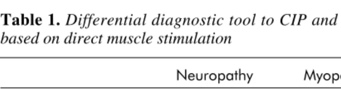
Direct muscle stimulation
The technique of direct muscle stimulation was developed by Richet al. to overcome the problem of eliciting voluntary muscle contraction in a non- cooperative patient42, 43. It compares the CMAP amplitudes elicited through stimulating the nerve (nCMAP) and the muscle (mCMAP) itself.
Neuropathy is suspect when the CMAPs elicited by stimulating the nerve are decreased though they are preserved when stimulating the muscle. In this case the ratio of nerve and muscle elicited CMAPs is
<0.5. In myopathy CMAPs are reduced or absent (<3mV) both by stimulating the nerve or the muscle signing the decreased excitability of muscle mem- brane. The nerve/muscle ratio is >0.5 (Table 1.).
NERVE AND MUSCLE BIOPSY
Histology helps to decide in questionable cases but it is no longer obligatory for diagnosis, or rather the
Table 1.Differential diagnostic tool to CIP and CIM based on direct muscle stimulation
Neuropathy Myopathy
nCMAP ↓ ↓
mCMAP n ↓
nCMAP / mCMAP <0.5 >0.5
indication and prognostic value is not clarified yet.
While electrophysiological alterations appear early in the course of critical illness, histological alter- ations turn up in the later phase. In case of CIP nerve histology shows primarily distal axonal degeneration involving both motor and sensory fibres without sign of demyelination or inflamma- tion1. Central nervous system histology revealed the chromatolysis of anterior horn cells that is the dis- integration of the chromophil substance, indicating the exhaustion of the cell or the damage to the axon30. Muscle histology shows denervation dam- age of type I and II muscle fibres and also myopath- ic characteristics27, 30. Later in the course of CIP, while recovery is ongoing, muscle biopsy will show grouped atrophy of the muscle fibres.
In CIM a mixed histological picture of acute necrosis with regeneration, selective loss of thick filaments (myosin) and atrophy of type II (fast twitch) muscle fibres can be seen27, 44. The selective loss of myosin signed by the loss of myofibrillar adenosine triphosphate on immunohistology is so characteristic that it led to the term “thick filament myopathy”45.
ULTRASONOGRAPHY
In recent years attention has turned to assessing muscle mass loss in ICU patients46. Presently B- mode ultrasonography (US) of rectus femoris (RF) muscle seems to be the most suitable technique. RF is the largest muscle of the body and was shown to have good correlation with lean body mass47. US was proven to be as accurate as magnetic resonance imaging beside being portable, non-invasive, easy to use and cost effective48. However it is yet to be determined if cross sectional area46, 49 or muscle layer thickness47, 50of rectus femoris muscle is the more reliable parameter for monitoring, which is less affected by interstitial oedema.
LABORATORY TESTING
Elevated serum creatine kinase levels are reported especially in the necrotizing type of CIM51, 52but the kinetics of serum level and the sensitivity is poorly studied.
Differential diagnosis
As mentioned before, the exclusion of other dis- eases resulting in acute onset tetraparesis is crucial to achieve diagnosis. Impairment of neuromuscular transmission can be excluded with simple electro-
physiological testing (eg. TOF stimulation or repet- itive nerve stimulation) and the review of patient history concerning NMBAs, chemotherapy, anti- retroviral drug administration. Apart from critical illness, several central nervous system disorders may also result in generalized muscle weakness.
Myopathy may be caused – among others – by elec- trolyte disturbances (hypokalemia, hypophos- phatemia), drugs (statins and fibrates). The so called propofol infusion syndrome presents with severe metabolic acidosis, cardiac failure, hypertriglyceri- daemia and rhabdomyolysis along with a conse- quent renal failure. It develops in patients receiving high doses of propofol (5 ml/kg/h) for more than two days53.
The classical differential diagnostic entity was the Guillain-Barré syndrome which presents also with acute onset tetraparesis, but certain clinical, laboratory and electrophysiological features help differentiation. These are the ascending characteris- tic, involvement of cranial and autonomic nerves, delayed onset time, elevated cerebrospinal fluid protein content and demyelinating characteristics on ENG (except for the axonal subtypes of GBS)54.
The common diabetic and alcoholic polyneu- ropathies may mimic the electrophysiological char- acteristics of CIP and give false positive diagnosis especially when there is no documentation in the previous history.
Pathophysiology
The pathophysiology of critical illness associated neuromuscular disorders is complex. They are no longer handled as isolated events, rather they are an integral part of the process leading to multiorgan dysfunction and failure and this way, the mutual and additive role of microcirculatory, cellular and metabolic pathophysiological mechanisms is pre- sumable11.
It was previously described in 1996 that electro- physiological alterations precede morphological deterioration27and emerging evidence suggests that definitive morphological lesions are established by functional failure. Yet, it is still undiscovered how the peripheral nerve and muscle exhibit a rapid onset of breaking down and this functional problem can turn out to be reversible55.
One explanation is the concept of sepsis-induced bioenergetic failure introduced by Boltonet al. in the 1990s which is still relevant today and grossly describes the pathophysiological process. The key point is that excitable tissues such as the nerves and muscles spend much of their energy on sustaining
excitability and function hence they are prone to develop dysfunction early in case of reduced ener- gy supply and use29.
Microcirculatory dysfunction is known to be a major pathophysiological factor in the development of sepsis-associated multiorgan failure56–58. The decrease in the number of perfused capillaries and the heterogeneity of microvascular circulation lead to alterations in oxygen extraction and tissue hypoxia in sepsis59, 60. These microcirculatory changes can resolve rapidly in response to adequate therapy but if they persist they predispose to higher mortality11, 57, 61, 62.
Mitochondrial dysfunction provoked by stress hormones, inflammatory cytokines, insulin resist- ance, reactive oxygen species or nitrous oxide also plays a pivotal role in the pathogenesis of cellular and organ failure through reduced adenosine- triphosphate (ATP) biosynthesis, energy generation and use63, 64. Brealey et al. found a correlation between muscle ATP concentration, mitochondrial dysfunction and the severity of septic shock, sug- gesting that bioenergetic failure is an important pathophysiological mechanism for muscle and multi organ dysfunction65. Furthermore, restoration of mitochondrial biogenesis, which maintains nor- mal mitochondrial number, structure and function was found to be an important factor favouring sur- vival66, 67.
Tumor necrosis factor alpha (TNF-α) and inter- leukin-6 (IL-6) are known to suppress the insulin effect through the direct inhibition of the Akt sig- nalling process culminating in peripheral insulin resistance and cellular energy depletion. Constatin et al. found a significant upregulation of muscle pyruvate dehydrogenase kinase 4 (PDK4) mRNA and protein expression in critically ill patients, pointing to the inhibition of muscle carbohydrate oxidation in these patients68, 69. Weber-Carstenset al. described faulty glucose transporter-4 (GLUT 4) disposition and diminished glucose utilization in the muscle of critically ill patients70. They explained the impaired transposition of the GLUT 4 transporter from perinuclear space to sarcolemma with the impairment of the insulin signalling pathway and the dysfunction of an AMPK energy sensor as a result of the lack of metabolic stimulation caused by the absence of muscle contraction.
Epi- and endoneurial vessel and concomitant leukocyte activation shown by increased E-selectin expression on the endothelial surface lead to the cessation of vascular autoregulation, endoneurial oedema and the accumulation of further cytopathic factors in the perineurial spaces71. Hyperglycemia and hypoalbuminaemia can further enhance these
processes which finally lead to ischemic hypoxia of the peripheral nerves even if oxygen supply is ade- quate8, 11, 23, 29. Beside the onkotic effects of the high serum glucose level, advanced glycation end-prod- ucts induce basement membrane hypertrophy in endoneurial microvessels and disrupt the blood- nerve-barrier by stimulating the release of trans- forming growth factor beta and vascular endothe- lial growth factor by pericytes72.
The next step in the pathophysiological cascade to be blamed for early electrophysiological distur- bances is “channelopathy”. Energy depletion leads to the insufficiency of membrane voltage-depend- ent sodium channels, that results in a shift of the voltage dependence of sodium channel fast inacti- vation towards more negative potentials73–75. The depolarization of resting membrane potential due to endoneurial hyperkalaemia or hypoxia can also be accounted for the hypo-, inexcitability of peripheral nerves and muscles43, 76. This similar appearance of channel dysfunction further supports the unifying hypothesis of CIP and CIM as different manifesta- tions of one disorder11but in vivo findings are yet to be verified in humans.
The prominent muscle wasting in septic patients is the result of the alterations in the balance of mus- cle protein synthesis and muscle protein break- down.
In healthy volunteers bed rest immobilization itself caused rapid muscle loss within a week, most prominently in the lower limbs77. In septic patients immobilization due to bed rest is aggravated by sedation and accompanied by insulin resistance and release of septic cytokines which are also inhibitory factors of muscle protein synthesis. Consequently the loss of lean body mass in critically ill patients was 1-1.6% per day, 7% per week in three weeks observational studies78, 79and 16-20% on ultrasound measurement in the first week in patients with sep- tic shock80.
Muscle protein synthesis is promoted via the PI3-K/AKT/mTOR pathway which is activated by local IGF-1 and nutrients and act through the initia- tion of several translational factors. Amino acids, especially branched-chain leucin, are known to stimulate muscle protein synthesis via both insulin- dependent (through PI3-K receptor) and insulin- independent mechanisms81. Yet in sepsis a leucin- resistance through the defect in leucin induced translation initiation has been observed82. Septic cytokine production, namely IL-1, IL-6, TNF-α directly inhibit the PI3-K/AKT/mTOR pathway with the deactivation of AKT protein kinase B resulting in decreased muscle protein synthesis69. Decreased mTOR kinase activity was evidenced by
reduced phosphorylation of both eukaryotic initia- tion factor (eIF)4E-binding protein (BP)-1 and ribo- somal S6 kinase (S6K)183.
The physiological role of the shift toward marked degradation of muscle fibres in the septic patient is to liberate energy and substrates for glu- coneogenesis and synthesis of acute phase proteins.
There are three main proteolytic ways acting in the process of critical illness associated muscle wasting (calpain-caspase system - CCS, ubiquitin- proteasome system - UPS, lysosomal autophagy system - LAS). The CCS is responsible for the initi- ation of breakdown of muscle fibres through releas- ing actin and myosin fibres from myofilaments thus exposing them to further cleavage by the protea- some system. Calpains are cystein proteases which are activated by the elevation of cytosolic Ca2+
level, in this case promoted by inflammatory cytokines84. The ATP-dependent UPS is responsi- ble for the large-scale degradation of damaged myofibrilla. Proteins ligated with the polypeptide ubiquitin are destined to breakdown by the 20S core of the 26S proteasome system. The key elements of the ligation process are the muscle specific MAFbx and MURF-1 E3 ubiquitin ligases which are induced by the FOX-O transcription factor just like the cathepsin B, D, and L enzymes in the LAS69, 85. The role of the LAS system in the critically ill has not been clarified yet: autophagy either protects against the collection of toxic myofibrillar aggre- gates, which initiate muscle dysfunction, or it exac- erbates the loss of the myofibrillar apparatus to induce atrophy86.
At the moment, these molecular pathways seem to be responsible for septic muscle protein synthe- sis and breakdown but to what extent and in what temporal dispersion that is yet to be elucidated.
After the standardized environment and results of animal research, human research produces diver- gent and sometimes contradictory results69, 70, 85, 87
concerning gene expressions, protein activities and the clinical relevance of the different biochemical mechanisms. In a comprehensive molecular study Constantinet al. showed the simultaneous down- regulation of signalling proteins thought to increase muscle protein synthesis (Akt1, mTOR, 4E-BP1, p70s6k) and a parallel activation of molecular path- ways driving muscle protein breakdown (MAFbx, MuRF1, 20S proteasome, cathepsin-L). Yet, the catabolic changes were paralleled by initiation of a cellular program of anabolic restoration at the tran- scriptional level, as suggested by a significant increase in mRNA of anabolic factors69. Compared to this Jespersenet al. found decreased MuRF1/
MAFbx levels and a higher Akt/mTOR/S6k/4E-
BP1 anabolic signalling expression and in septic patients what they explained with the exogenous administration of insulin which is an activator of this anabolic pathway through receptor PI3-K87.
Beside the loss of contractile elements (over- whelmingly myosin loss), mitochondrial dysfunc- tion and muscle membrane inexcitability, oxidative stress, impaired excitation-contraction coupling also contribute to the impaired force-generating capacity of muscle in critically ill patients88.
Risk factors
Over the years several factors have been suspected to be responsible for the development of CIP and CIM, the role of some factors has still remained debatable. The severity and duration of SIRS and sepsis12, 14, 22, 25, 89–91, number of organ failures25, 89, 90, duration of vasopressor and catecholamine sup- port20, duration of ICU stay20, 23, renal failure14, hyperglycaemia17, 20–23, 89, 90, low serum albumin23, 89,
92, hyperosmolality93, parenteral nutrition88, 93, neu- rological failure94, immobility89, 95, female sex12, 89 presently seem to be evident risk factors.
Three prospective observational studies conclud- ed that corticosteroids had a negative effect con- cerning the development of CINM12, 96, 97, yet sever- al others reported that they had no effect14, 20, 93, 98–100
especially if tight glucose control was kept. One study showed that they were protective probably though decreasing the cathecholamin and vaso- pressr need21.
Aminoglycoside antibiotics were also identified as risk factors in some studies15, 22, 37but not in oth- ers11, 12, 17, 20, 21, 23, 93, 101.
In the 1980s and 1990s prolonged application of neuromuscular blocking agents was considered as a causative factor in the development of ICUAW93,
102–105. In line with the introduction of intermittent administration NMBA rptpcols, and Hoffman-elim- inating short acting agents the direct causative con- nection has become questionable although immo- bility caused by NMBAs cannot be disregarded.
Renal replacement therapy was also found to be a protective93and causative factor106–108at the same time, but it is still a question whether renal replace- ment therapy or renal insufficiency itself is to be blamed.
Therapy
Up to the present day no specific, evidence-based effective therapy has been developed for the man-
agement of critical illness-associated neuromuscu- lar disorders. No hormonal therapies, nutritional or antioxidant supplements could prove to be decisive, yet several supportive and preventive measures have been found to be effective.
Since the large-scale prospective, randomized, controlled trial of Van den Berghe et al, where intensive insulin therapy reduced the risk of CIP and the duration on mechanical ventilation beside many other ICU complications20and knowing the pathological effects of hyperglycaemia there is no need to emphasize the significance of strict glucose control in the critically ill. Only the original Van den Berghe target range (4.4-6.1 mmol/l) has been modified to <10 mmol/l108since the former proto- col had turned out to increase the number of hypo- glycaemic episodes and ICU mortality109.
One of the major advances in rehabilitation of these patients is the shift toward preventive app - roach. Rehabilitation started early in the ICU after reaching hemodynamic stability with repeated daily passive mobilization, early physical and occupa- tional therapy up to the patient’s physical and men- tal status is associated with better outcomes110, 111. In connection with this we have to emphasize the role of daily interruption of sedation and the spon- taneous breathing trials. These measures help to preserve muscle mass, improve functional inde- pendence, shorten ventilator dependency, delirium, ICU and hospital stay33, 112, 113.
A promising advancement of the last two years was the introduction of electric muscle stimulation (EMS) in the ICU114. EMS was proved to improve muscle strength and muscle mass in chronic obstructive pulmonary disease and chronic heart failure patients115–119. In the critically ill its use is not circumscribed yet69, 120–123. So far it seems to stimulate biochemical mechanisms which are responsible for muscle protein synthesis and to stimulate muscle glucose uptake through the facili- tation of GLUT-4 transposition to the sarcolem- ma70, this way it can improve insulin resistance.
Gerovasiliet al. found that EMS might affect bene- ficially local and systemic microcirculation based on near-infrared spectroscopy measurement124and might help to preserve muscle mass according to ultrasound measurement121. Routsiet al. found that daily EMS sessions prevented critical illness neu- romyopathy120. Several trials are ongoing to clarify its affectivity and feasibility in preventing muscle wasting, to elucidate its biochemical effect and to
determine the optimal treatment parameters125, 126. Presently EMS seems more potent in preserving muscle strength compared to muscle mass113.
The role of immunoglobulins (IVIG) in the pre- vention of CIP and CIM is quite contradictory. In a retrospective chart analysis by Mohr et al. the administration of high dose immunoglobulin M (IgM) enriched IVIG was found to be protective101. Brunneret al. could find no beneficial effect of IgM enriched IVIG in their prospective randomised con- trolled double blind trial, however they adminis- tered IVIG only after the evolution of electrophysi- ological alterations and in smaller dose than in the Mohr study127.
Other therapeutic or preventive measures, like the administration of nutritional (leucin or gluta- mine supplementation), hormonal (growth hor- mone, testosterone or oxandrolone administration), antioxidant supplements have not proved to be effective so far.
Conclusions
The aim of this review was to give an insight into these long known but during daily clinical work often neglected disorders. Breathing insufficiency and movement inability as consequences of ICUAW are major contributors to ICU and hospi- tal mortality. Along with the development of med- ical care more lives can be saved but the number of physically disabled survivors is rising also set- ting severe social, health care and economical problems.
The author cannot stress the importance of pre- ventive approach and early intervention. Early mobilization and physiotherapy, the application of liberal sedation protocol and strict glucose control are proven to be beneficial not only from the aspect of neuromuscular complications. They favourably influence the course of critical illness interfering with septic cascade at multiple sites and thus help to preserve and/or restore organ function. Rehabi - litation should start already in the ICU.
ACKNOWLEDGEMENTS
The first and second authors were supported by the European Union and the State of Hungary, co- financed by the European Social Fund in the fra- mework of TÁMOP 4.2.4. A/2-11-1-2012-0001
‘National Excellence Program’.
REFERENCES
1. Bolton CF, Gilbert JJ, Hahn AF, Sibbald WJ. Polyneu - ropathy in critically ill patients. J Neurol Neurosurg Psychiatry 1984;47:1223-31.
2.Bolton CF, Laverty DA, Brown JD, Witt NJ, Hahn AF, Sibbald WJ.Critically ill polyneuropathy: electrophysio- logical studies and differentiation from Guillain-Barré syn- drome. J Neurol Neurosurg Psychiatry 1986;49:563-73.
3. Bolton CF.Electrophysiologic studies of critically ill pa - tients. Muscle Nerve 1987;10:129-35.
4.Hippocrates, Kelly EC. The Theory and Practice of Me - dicine. New York, Philosophical Library, 1964.
5.Osler W.The principles and practice of medicine. New York: D. Appleton and Company; 1892.
6.Stevens RD, Marshall SA, Cornblath DR, Hoke A, Need - ham DM, de Jonghe B, et al. A framework for diagnosing and classifying intensive care unit- acquired weakness. Crit Care Med 2009;37(10Suppl):S299-308.
7.MacFarlane IA, Rosenthal FD. Severe myopathy after sta- tus asthmaticus. Lancet 1977;2:615.
8.Latronico N, Bertolini G, Guarneri B, Peli E, Andreoletti S, Bera P, et al.Simplified electrophysiological evaluation of peripheral nerves in critically ill patients: the Italian multi-centre CRIMYNE study. Crit Care 2007;11:R11.
9.Koch S, Spuler S, Deja M, Bierbrauer J, Dimroth A, Behse F, et al.Critical illness myopathy is frequent: accompany- ing neuropathy protracts ICU discharge. J Neurol Neurosurg Psychiatry 2011;82:287-93.
10.Khan J, Harrison TB, Rich MM, Moss M.Early develop- ment of critical illness myopathy and neuropathy in patients with severe sepsis. Neurology 2006;67:1421-25.
11.Latronico N, Bolton CF.Critical illness polyneuropathy and myopathy: a major cause of muscle weakness and paralysis. Lancet Neurol 2011;10:931-41.
12.De Jonghe B, Sharshar T, Lefaucheur JP, Authier FJ, Durand-Zaleski I, Boussarsar M, et al.Paresis acquired in the intensive care unit: a prospective multicenter study.
JAMA 2002;288:2859-67.
13.De Jonghe B, Bastuji-Garin S, Sharshar T, Outin H, Broc - hard L.Does ICU-acquired paresis lengthen weaning from mechanical ventilation? Intensive Care Med 2004;30:1117- 21.
14.de Letter MA, Schmitz PI, Visser LH, Verheul FA, Schellens RL, Op de Coul DA, et al.Risk factors for the development of polyneuropathy and myopathy in critically ill patients.
Crit Care Med 2001;29:2281-6.
15.Leijten FS, Harinck-de Weerd JE, Poortvliet DC, de Weerd AW.The role of polyneuropathy in motor convalescence after prolonged mechanical ventilation. JAMA 1995;274:
1221-5.
16.Garnacho-Montero J, Madrazo-Osuna J, Garcia-Garmen - dia JL, Madrazo-Osuna J, Ortiz-Leyba C.Critical illness polyneuropathy: risk factors and clinical consequences. A cohort study in septic patients. Intensive Care Med 2001;
27:1288-96.
17.Bercker S, Weber-Carstens S, Deja M, Grimm C, Wolf S, Behse F, et al.Critical illness polyneuropathy and myopa- thy in patients with acute respiratory distress syndrome.
Crit Care Med 2005;33:711-5.
18.Hough CL, Steinberg KP, Taylor Thompson B, Rubenfeld GD, Hudson LD.Intensive care unit-acquired neuromyopa- thy and corticosteroids in survivors of persistent ARDS.
Intensive Care Med 2009;35:63-8.
19.Coakley JH, Nagendran K, Yarwood GD, Honavar M, Hinds CJ. Patterns of neurophysiological abnormality in prolonged critical illness. Intensive Care Med 1998;24:
801-7.
20.Van den Berghe G, Schoonheydt K, Becx P, Bruyninckx F, Wouters PJ.Insulin therapy protects the central and periph- eral nervous system of intensive care patients. Neurology 2005;64:1348-53.
21.Hermans G, Wilmer A, Meersseman W, Milants I, Wouters PJ, Bobbaers H, et al.Impact of intensive insulin therapy on neuromuscular complications and ventilator dependen- cy in the medical intensive care unit. Am J Respir Crit Care Med 2007;175:480-9.
22.Nanas S, Kritikos K, Angelopoulos E, Siafaka A, Tsikriki S, Poriazi M, et al. Predisposing factors for critical illness polyneuromyopathy in a multidisciplinary intensive care unit. Acta Neurol Scand 2008;118:175-81.
23.Witt NJ, Zochodne DW, Bolton CF, Grand’Maison F, Wells G, Young GB, et al.Peripheral nerve function in sepsis and multiple organ failure. Chest 1991;99:176-84.
24.Bednarik J, Lukas Z, Vondracek P.Critical illness polyneu- romyopathy: the electrophysiological components of a complex entity. Intensive Care Med 2003;29:1505-14.
25.Bednarik J, Vondracek P, Dusek L, Moravcova E, Cundrle I.Risk factors for critical illness polyneuromyopathy. J Neurol 2005;252:343-51.
26. Tennilä A, Salmi T, Pettilä V, Roine RO, Varpula T, Takkunen O.Early signs of critical illness polyneuropathy in ICU patients with systemic inflammatory response syn- drome or sepsis. Intensive Care Med 2000;26:1360-3.
27.Latronico N, Fenzi F, Recupero D, Guarneri B, Tomelleri G, Tonin P, et al.Critical illness myopathy and neuropa- thy. Lancet 1996;347:1579-82.
28.Lacomis D.Electrophysiology of neuromuscular disorders in critical illness. Muscle Nerve 2013;47:452-63.
29.Bolton CF.Neuromuscular manifestations of critical ill- ness. Muscle Nerve 2005;32:140-63.
30. Zochodne DW, Bolton CF, Wells GA, Gilbert JJ, Hahn AF, Brown JD, et al.Critical illness polyneuropathy: a compli- cation of sepsis and multiple organ failure. Brain 1987;
110:819-41.
31.Kleyweg RP, van der Meche FG, Schmitz PI.Interobserver agreement in the assessment of muscle strength and func- tional abilities in Guillain-Barré syndrome. Muscle Nerve 1991;14:1103-9.
32.Bittner EA, Martyn JA, George E, Frontera WR, Eiker - mann M.Measurements of muscle strength in the intensive care unit. Crit Care Med 2009;37(10Suppl):S321-30.
33.Barr J, MD, Fraser GL, Puntillo K, Ely EW, Gélinas C, Dasta JF, et al.Clinical practice guidelines for the man- agement of pain, agitation, and delirium in adult patients in the intensive care unit. Crit Care Med 2013;41:263-306.
34.Ali Na, O’Brien JM Jr, Hoffmann SP, Phillips G, Garland A, Finley JC, et al.Acquired weakness, hand grip strength, and mortality in critically ill patients. Am J Respir Crit Care Med 2008;178:261-8.
35.Harper NJN, Greer R, Conway D.Neuromuscular moni- toring in intensive care patients: milliamperage require- ments for supramaximal stimulation. Br J Anaesth 2001 87:625-7.
36.Ahlbeck K, Fredriksson K, Rooyackers O, Måbäck G, Remahl S, Ansved T, et al.Signs of critical illness polyneu- ropathy and myopathy can be seen early in the ICU course.
Acta Anaesthesiol Scand 2009;53:717-23.
37.Leijten FS, De Weerd AW, Poortvliet DCJ, Ridder VA, Ulrich C, Harinck-De Weerd JE.Critical illness polyneu- ropathy in multiple organ dysfunction syndrome and wean- ing from the ventilator. Intensive Care Med 1996;22:856- 61.
38.Park EJ, Nishida T, Sufit RL, Minieka MM. Prolonged mus-
cle action potential duration in critical illness myopathy.
Report of nine cases. J Clin Neuromuscul Dis 2004;5:176- 83.
39.Goodman BP, Harper CM, Boon AJ.Prolonged muscle action potential duration in critical illness myopathy.
Muscle Nerve 2009;40:1040-2.
40.Zifko UA, Zipko HT, Bolton CF.Clinical and electrophysi- ological findings in critical illness polyneuropathy. J Neurol Sci 1998;159:186-93.
41.Luo YM, Moxham J, Polkey MI.Diaphragm electromyog- raphy using an oesophageal catheter: current concepts. Clin Sci 2008;115:233-44.
42.Rich MM, Bird SJ, Raps EC, McCluskey LF, Teener JW.
Direct muscle stimulation in acute quadriplegic myopathy.
Muscle Nerve 1997;20:665-73.
43.Rich MM, Teener JW, Raps EC, Schotland DL, Bird SJ.
Muscle is electrically inexcitable in acute quadriplegic myopathy. Neurology 1996;46:731-6.
44.Helliwell TR, Wilkinson A, Griffiths RD, McClelland P, Palmer TE, Bone JM.Muscle fibre atrophy in critically ill patients is associated with the loss of myosin filaments and the presence of lysosomal enzymes and ubiquitin.
Neuropathol Appl Neurobiol 1998;24:507-17.
45.Danon MJ, Carpenter S.Myopathy with thick filament (myosin) loss following prolonged paralysis with vecuroni- um during steroid treatment. Muscle Nerve 1991;14:
1131-9.
46.Puthucheary Z, Montgomery H, Moxham J, Harridge S, Hart N.Structure to function: muscle failure in critically ill patients. J Physiol 2010;588:4641-8.
47.Campbell IT, Watt T, Withers D, England R, Sukumar S, Keegan MA, et al.Muscle thickness, measured with ultra- sound, may be an indicator of lean tissue wasting in multi- ple organ failure in the presence of edema. Am J Clin Nutr 1995;62:533-9.
48.Arbeille P, Kerbeci P, Capri A, Dannaud C, Trappe SW, Trappe TA. Quantification of muscle volume by echog- raphy: comparison with MRI data on subjects in long-term bed rest. Ultrasound Med Biol 2009;35:1092-1097.
49.Seymour JM, Ward K, Sidhu PS, Puthucheary Z, Steier J, Jolley CJ et al.Ultrasound measurement of rectus femoris cross-sectional area and the relationship with quadriceos strength in COPD. Thorax 209;64:418-23.
50.Gruther W, Benesch T, Zorn C, Paternostro-Sluga T, Quittan M, Fialka-Moser V et al.Muscle wasting in inten- sive care patients: ultrasound observation of the m. quadri- ceps femoris muscle layer. J Rehabil Med 2008;40:185-9.
51.Ramsay DA, Zochodne DW, RobertsonDM, Nag S, Ludwin SK.A syndrome of acute severe muscle necrosis in inten- sive care unit patients. J Neuropathol Exp Neurol 1993;
52:387-98.
52.Zochodne DW, Ramsay DA, Saly V, Shelley S, Moffatt S.
Acute necrotizing myopathy of intensive care: electrophys- iological studies. Muscle Nerve 1994;17:285-92.
53.Vasile B, Rasulo F, Candiani A, Latronico N.The patho- physiology of propofol infusion syndrome: a simple name for a complex syndrome. Intensive Care Med 2003;29:
1417-25.
54.Hughes RA, Cornblath DR.Guillai-Barré syndrome. Lan - cet 2005;366:1653-66.
55.De Jonghe B, Bastuji-Garin S, Durand MC, Malissin I, Rodrigues P, Cerf C, et al.Respiratory weakness is associ- ated with limb weakness and delayed weaning in critical illness. Crit Care Med 2007;35:2007-15.
56.De Backer D, Creteur J, Preiser JC, Dubois MJ, Vincent JL.Microvascular blood flow is altered in patients with sepsis. Am J Respir Crit Care Med 2002;166:98-104.
57.Sakr Y, Dubois MJ, De Backer D, Creteur J, Vincent JL.
Persistant microvasculatory alterations are associated with organ failure and death in patients with septic shock. Crit Care Med 2004;32:1825-31.
58.Doerschug KC, Delsing AS, Schmidt GA, Haynes WG.
Impairments in microvascular reactivity are related to organ failure in human sepsis. Am J Physiol Heart Circ Physiol 2007;293:H1065-71.
59.De Backer D, Donadello K, Cortes DO.Monitoring the microcirculation. J Clin Monit Comput 2012;26:361-6.
60.Edul VS, Enrico C, Laviolle B, Vazquez AR, Ince C, Dubin A. Quantitative assessment of the microcirculation in healthy volunteers and in patients with septic shock. Crit Care Med 2012;40:1443-8.
61.Neviere R, Mathieu D, Chagnon JL, Lebleu N, Millien JP, Wattel F.Skeletal muscle microvascular blood flow and oxygen transport in patients with severe sepsis. Am J Respir Crit Care Med 1996;153:191-5.
62.De Backer D, Ospina-Tascon G, Salgado D, Favory R, Creteur J, Vincent JL.Monitoring the microcirculation in the critically ill patient: current methods and future approaches. Intensive Care Med 2010;36:1813-25.
63.Sibbald WJ, Messmer K, Fink MP.Roundtable conference on tissue oxygenation in acute medicine, Brussels, Bel - gium, 14-16 March 1998. Intensive Care Med 2000;26:
780-91.
64.Fink MP, Evans TW.Mechanisms of organ dysfunction in critical illness: report from a Round Table Conference held in Brussels. Intensive Care Med 2002;28:369-75.
65.Brealey D, Brand M, Hargreaves I, Heales S, Land J, Smolenski R, et al.Association between mitochondrial dys- function and severity and outcome of septic shock. Lancet 2002;360:219-23.
66.Haden DW, Saliman HB, Carraway MS, Welty-Wolf KE, Ali AS, Shitara H, et al.Mitochondrial biogenesis restores oxidative metabolism during Staphylococcus aureus sep- sis. Am J Respir Crit Care Med 2007;176:768-77.
67.Carre JE, Orban JC, Re L, Felsmann K, Iffert W, Bauer M, et al.Survival in critical illness is associated with early acti- vation of mitochondrial biogenesis. Am J Respir Crit Care Med 2010;182:745-51.
68.Wu P, Inskeep K, Bowker-Kinley MM, Popov KM, Harris RA. Mechanism responsible for inactivation of skeletal muscle pyruvate dehydrogenase complex in starvation and diabetes. Diabetes 1999;48:1593-9.
69.Constantin D, McCullough J, Mahajan RP, Greenhaff PL.
Novel events in the molecular regulation of muscle mass in critically ill patients. J Physiol 2011;589:3883-95.
70.Weber-Carstens S, Schneider J, Wollersheim T, Assmann A, Bierbrauer J, Marg A, et al.Critical illness myopathy and GLUT 4. Significance of insulin and muscle contrac- tion. Am J Respir Crit Care Med 2013;187:387-96.
71.Fenzi F, Latronico N, Refatti N, Rizzuto N. Enhanced expression of E-selectin on the vascular endothelium of peripheral nerve in critically ill patients with neuromuscu- lar disorders. Acta Neuropathol 2003;106:75-82.
72.Shimizu F, Sano Y, Haruki H, Kanda T.Advanced glyca- tion end-products induce basement membrane hypertrophy in endoneurial microvessels and disrupt the blood-nerve barrier by stimulating the release of TGF-beta and vascular endothelial growth factor (VEGF) by pericytes. Diabeto - logia 2011;54:1517-26.
73.Novak KR, Nardelli P, Cope TC, Filatov G, Glass JD, Khan J, et al.Inactivation of sodium channels underlies rever - sible neuropathy during critical illness in rats. J Clin Invest 2009;119:1150-1158.
74.Rich MM, Pinter MJ.Crucial role of sodium channel fast inactivation in muscle fibre inexcitability in a rat model of critical illness myopathy. J Physiol 2003;547:555-66.
75.Filatov GN, Rich MM.Hyperpolarized shifts in the voltage dependence of fast inactivation of Nav1.4 and Nav1.5 in a rat model of critical illness myopathy. J Physiol 2004;559:
813-20.
76.Z’Graggen WJ, Lin CS, Howard RS, Beale RJ, Bostock H.
Nerve excitability changes in critical illness polyneuropa- thy. Brain 2006;129:2461-2470.
77.LeBlanc AD, Schneider VS, Evans HJ, Pientok C, Rowe R, Spector E. Regional changes in muscle mass following 17 weeks of bed rest. J Appl Physiol 1992;73:2172-8.
78.Griffith RD.Muscle mass, survival, and the elderly ICU patient. Nutrition 1996;12:456-8.
79.Reid CL, Campbell IT, Little RA.Muscle wasting and ener- gy balance in critical illness. Clin Nutr 2004;23:273-80.
80.Poulsen JB, Møller K, Jensen CV, Weisdorf S, Kehlet H, Perner A.Effect of transcutaneous electrical muscle stimu- lation on muscle volume in patients with septic shock. Crit Care Med 2011;39:456-61.
81.Kimball SR, Jefferson LS. New functions for amino acids:
effects on gene transcription and translation. Am J Clin Nutr 2006;83:500-7.
82.Lang CH, Frost RA.Differential effect of sepsis on ability of leucin and IGF-1 to stimulate muscle translation initia- tion. Am J Physiol Endocrinol Metab 2004;287:721-30.
83.Lang CH, Frost RA, Vary TC.Regulation of muscle pro- tein synthesis during sepsis and inflammation. Am J Physiol Endocrinol Metab 2007;293:453-9.
84.Bloch S, Polkey MI, Griffith M, Kemp P.Molecular mecha- nisms of intensive care unit-acquired weakness. Eur Respir J 2012;39:1000-11.
85.Klaude M, Mori M, Tiäder I, Gustafsson T, Wernerman J, Rooyackers O.Protein metabolism and gene expression in skeletal muscle of critically ill patients with sepsis. Cli Sci 2012;122:133-142.
86.Dos Santos CC, Batt J.ICU-acquired weakness of disabili- ty. Curr Opn Crit Care 2012;18:509-17.
87.Jespersen JG, Nedergaard A, Reitelseder S, Mikkelsen UR, Dideriksen KJ, Agergaard J, et al. Activated protein syn- thesis and suppressed protein breakdown signalling in skeletal muscle of critically ill patients. PloS ONE 6:
e18090.
88.Batt J, dos Santos CC, Cameron JI, Herridge MS. Intensive Care Unit-acquired Weakness. Am J Respir Crit Care Med 2013;187:238-46.
89.Ydemann M, Eddelien HS, Lauritsen AØ.Treatment of crit- ical illness polyneuropathy and/or myopathy- a systematic review. Dan Med J 2012;59:A4511.
90.Ricks E.Critical illness polyneuropathy and myopathy: a review of evidence and the implications for weaning from mechanical ventilation and rehabilitation. Physiotherapy 2007;93:151-6.
91.Weber-Carstens S, Deja M, Koch S, Spranger J, Bubser F, Wernecke KD, et al.Risk factors in critical illness myopa- thy during the early course of critical illness: a prospective observational study. Crit Care 2010;14:R119.
92.Mohammadi B, Schedel I, Graf K, Teiwes A, Hecker H, Haameijer B, et al.Role of endotoxin in the pathogenesis of critical illness polyneuropathy. J Neurol 2008;255:265- 72.
93.Garnacho-Montero J, Madrazo-Osuna J, García-Garmen - dia JL, Ortiz-Leyba C, Jiménez-Jiménez FJ, Barrero- Almodóvar A, et al.Critical illness polyneuropathy: risk factors and clinical consequences. A cohort study in septic patients. Intensive Care Med 2001;27:1288-96.
94.Latronico N, Peli E, Botteri M.Critical illness myopathy and neuropathy. Curr Opin Crit Care 2005;11:126-32.
95.Schweickert WD, Hall J.ICU-Acquired Weakness. Chest 2007;131:1541-9.
96. Herridge MS, Cheung AM, Tansey CM, Matte-Martyn A, Diaz-Grandos N, Al-Saidi F, et al.One-year outcomes in survivors of the acute respiratory distress syndrome. N Engl J Med 2003;348:683-693.
97. Herridge MS, Tansey CM, Matté A, Tomlison G, Diaz- Granados N, Cooper A, et al. Functional disability 5 years after acute respiratory distress syndrome. N Engl J Med 2011;364:1293-304.
98. Sprung CL, Annane D, Keh D, Moreno R, Singer M, Freivogel K, et al.Hydrocortisone therapy for patients with septic shock. N Engl J Med 2008;358:111-24.
99. Steinberg KP, Hudson LD, Goodman RB, Hough CL, Lanken PN, Hyzy R, et al.Efficacy and safety of corticos- teroids for persistent acute respiratory distress syndrome.
N Engl J Med 2006;354:1671-84.
100. Hough CL, Steinberg KP, Taylor Thompson B, Rubenfeld GD, Hudson LD.Intensive care unit-acquired neuromy- opathy and corticosteroids in survivors of persistent ARDS. Intensive Care Med 2009;35:63-8.
101. Mohr M, Englisch L, Roth A, Burchardi H, Zielmann S.
Effects of early treatment with immunoglobulin on criti- cal illness polyneuropathy following multiple organ fail- ure and gram-negative sepsis. Intensive Care Med 1997;
23:1144-9.
102. Vanderheyden BA, Reynolds HN, Gerold KB, Emanuele T.Prolonged paralysis after long-term vecuronium infu- sion. Crit Care Med 1992;20:304-7.
103. Segredo V, Caldwell JE, Matthay MA, Sharma ML, Gruenke LD, Miller RD.Persistent paralysis in critically ill patients after long-term administration of vecuronium.
N Engl J Med 1992;327:524-8.
104. Gooch JL, Moore MH, Ryser DK.Prolonged paralysis after neuromuscular junction blockade: Case reports and electrodiagnostic findings. Arch Phys Med Rehabil 1993;74:1007-11.
105. Manthous CA, Chatila W.Prolonged weakness after the withdrawal of atracurium. Am J Respir Crit Care Med 1994;150:1441-3.
106. Thiele RI, Jakob H, Hund E, Genzwuerker H, Herold U, Schweiger P,et al. Critical illness polyneuropathy: a new iatrogenically induced syndrome after cardiac surgery?
Eur J Cardiothorac Surg 1997;12:826-35.
107. Van den Berghe G, Wouters P, Weekers F, Verwaest C, Bruyninckx F, Schetz M, et al.Intensive insulin therapy in critically ill patients. N Engl J Med 2001;345:1359-67.
108 Dellinger RP, Levy MM, Rhodes A, Annane D, Gerlach H, Opal SM, et al.Surviving Sepsis Campaign: Interna - tional Guidelines for Management of Severe Sepsis and Septic Shock, 2012. Intensive Care Med 2013;39:165- 228.
109. NICE-SUGAR Study Investigators, Finfer S, Chittock DR, Su SY, Blair D, Foster D, Dhingra V et al.Intensive ver- sus conventional glucose control in critically ill patients.
N Eng J Med 2009;360:1283-97.
110. Griffith RD, Palmer TE, Helliwell T, MacLennan P, MacMillan RR.Effect of passive stretching on the wast- ing of muscle in the critically ill. Nutrition 1995;11:428- 32.
111. Schweickert WD, Pohlman MC, Pohlman AS, Nigos C, Pawlik AJ, Esbrook CL, et al.Early physical and occupa- tional therapy in mechanically ventilated, critically ill patients: a randomised controlled trial. Lancet 2009;373:
1874-882.
112. Kress JP, Pohlman AS, O’Connor MF, Hall JB.Daily interruption of sedative infusions in critically ill patients undergoing mechanical ventilation. N Engl J Med 2000;
342:1471-7.
113. Strom T, Martinussen T, Toft P.A protocol of no sedation