Purification of proteins with native terminal sequences using a Ni(II)- cleavable C-terminal hexahistidine affinity tag
Heba A.H. Abd Elhameeda,1,2, Bálint Hajdua,1, Ria K. Balogha, Enikő Hermanna, Éva Hunyadi-Gulyásb, Béla Gyurcsika,*
aDepartment of Inorganic and Analytical Chemistry, University of Szeged, Dóm tér 7, H-6720 Szeged, Hungary
bLaboratory of Proteomics Research, Institute of Biochemistry, Biological Research Centre of the Hungarian Academy of Sciences, Temesvári krt. 62, H-6726 Szeged, Hungary
*Corresponding author
e-mail: gyurcsik@chem.u-szeged.hu, phone number: (+36)62-544335.
1These authors contributed equally to this work.
2Presently PhD student at University of Szeged. Home affiliation: Department of Chemistry, Zagazig university, Egypt.
Abstract
The role of the termini of protein sequences is often perturbed by remnant amino acids after the specific protease cleavage of the affinity tags and/or by the amino acids encoded by the plasmid at/around the restriction enzyme sites used to insert the genes. Here we describe a method for affinity purification of a metallonuclease with its precisely determined native termini. First, the gene encoding the target protein is inserted into a newly designed cloning site, which contains two self-eliminating BsmBI restriction enzyme sites. As a consequence, the engineered DNA code of Ni(II)-sensitive Ser-X-His-X motif is fused to the 3’-end of the inserted gene followed by the gene of an affinity tag for protein purification purpose. The C-terminal segment starting from Ser mentioned above is cleaved off from purified protein by a Ni(II)-induced protease action. The success of the purification and cleavage was confirmed by gel electrophoresis and mass spectrometry, while structural integrity of the purified protein was checked by circular dichroism spectroscopy. Our new protein expression DNA construct is an advantageous tool for protein purification, when the complete removal of affinity or other tags, without any remaining amino acid residue is essential. The described procedure can easily be generalized and combined with various affinity tags at the C-terminus for chromatographic applications.
Keywords: colicin E7; affinity chromatography; Ni(II)-induced cleavage; MS; circular dichroism
Introduction
Recently we reported on Ni(II)-induced site specific and sequence selective peptide bond hydrolysis in zinc finger metalloproteins [1]. This reaction requires an X-(Ser/Thr)-X- His-X segment (X being any amino acid except for Cys or Pro before or after Ser/Thr) in the peptide or protein sequence [2–7]. The cleavage occurs at the N-terminal side of Ser or Thr residues and a square planar Ni(II) complex is formed at the N-terminus of the C-terminal cleavage product (Scheme 1). The reaction is also promoted by Cu(II) [3,11] and Pd(II) [12]
ions. Since the required metal ions are readily available in every laboratory, this method can efficiently compete with the hydrolysis using specific protease enzymes.
Scheme 1 – near here
The pET16b cloning vector applied in our previous study [1] provided an N-terminal decahistidine tag and a Ni(II)-sensitive -Ser-Gly-His- sequence between the affinity tag and the target protein. This allowed to cleave the affinity tag with Ni(II) ions and, at the same time, a new putative catalytic centre (a Ni(II)ion complexed to the Ser-Gly-His- segment, so called ATCUN motif [13-16]) formed at the N-terminus of the zinc fingers. The remnant amino acids after the cleavage of the affinity tags may thus provide new functionalities, which is however, a rare objective of a protein purification procedure. More commonly, they may interfere with the folding and/or function of the protein. The Factor Xa protease recognition site sequence (-Ile-Glu-Gly-Arg-) encoded in pET-16b vector is followed by the His-Met amino acids encoded by the NdeI restriction enzyme recognition sequence, being the closest cloning site of choice. Consequently, the protease cleavage after the arginine residue leaves at least a His residue at the N-terminus of the purified protein, which may significantly affect e.g., the metal binding properties of the product. The size of the remaining amino acid string varies with the choice of the restriction enzyme sites applied for cloning. It is thus, common that specific proteases leave few unwanted amino acids at the termini of the target protein. Examples of the most frequently used proteases in affinity-based protein purification systems are collected in Table 1. There are also expression plasmids, which do not code protease cleavage sequences for affinity tag removal (last two lanes in Table 1). Nevertheless, simple affinity-based purification is a popular approach, since purification of the native proteins, i.e., without affinity tags is usually difficult and requires multistep chromatographic methods.
Table 1 – near here
The search for ideal affinity tags and tag-removal methods is necessitated in protein chemistry as exemplified by recent reviews [17,20,21,25,35-41]. The peptide bond hydrolysis initiated by Ni(II) ions has already found application in the affinity tag removal from fusion proteins by Ni(II) ions [42-44]. Engineering the -(Ser/Thr)-X-His-X coding sequence at the 3’-end of the gene of the target protein, instead of a stop codon, allows for removal of the C-terminal affinity tag without leaving any amino acids at the target protein [42]. This however, requires the 3’ modification of every target protein gene before inserting it into a plasmid. In the present study we have carried out the purification of a zinc-containing metallonuclease protein in a plasmid with a re-engineered cloning site using BsmBI restriction enzyme for insertion of the native protein gene between the starting atg codon and the nucleotide sequence encoding for the Ni(II)-cleavable X-(Ser/Thr)-X-His-X segment followed by a C-terminal affinity tag. We demonstrate that the Ni(II)-induced protease cleavage results in the expected functional protein sequence, purified in a single metal ion affinity chromatographic step.
Materials and methods
Strains and media
DH5α F– Φ80lacZΔM15 Δ(lacZYA-argF) U169 recA1 endA1hsdR17 (rK–, mK+) phoA supE44 λ–thi-1gyrA96 relA1 competent E. coli [45] was used for the cloning of the recombinant desired DNA and E. coli BL21(DE3) F- ompT gal [dcm] [lon] hsdSB [46] for the expression of
N4-NColE7 protein, a mutated non-toxic N-terminal domain of colicin E7 (NColE7). Bacteria were grown in LB medium containing ampicillin (100 μg/ml at final concentration) at 37 C [47].
Plasmid construction
The plasmid pQE70-NColE7/Im7 [48] was kindly provided by prof. Kin-Fu Chak (Institute of Biochemistry, National Yang-Ming University, Taipei, Taiwan, Republic of China). The DNA segment, encoding the ∆N4-NColE7 sequence was modified to include BsmBI restriction site by PCR using this plasmid as template, DreamTaq polymerase (Thermo Scientific) and the oligonucleotide primers: B-DN4 – forward
(5’-ATTCGTCTCCATATGCCAGGGAAGGCAA-3’) and B-ColC – reverse (5’-ATTCGTCTCCCTGGATTTACCTCGGTGAATATCAATATG-3’). The PCR product was cloned into a modified pET-21a (AmpR) plasmid (Novagen), from which the existing BsmBI sites were knocked out, and the new BsmBI sites were introduced into the cloning region, followed by the SRHS sequence and the hexahistidine code through a linker sequence, denoted as pET-21a*-SRHS. Both the pET-21a*-SRHS plasmid and the PCR product were digested by BsmBI (Esp3I) restriction endonuclease (Thermo Scientific) at 37 °C for two hours.
The enzyme was deactivated at 65 °C for 20 minutes (this step is not necessary, as the enzyme site is self-eliminated). The DNA fragments were ligated by T4 ligase (Thermo Scientific) at 16 °C for one hour and at 4 °C overnight.
The ligated pET-21a*-∆N4-SRHS plasmid was applied to transform competent E. coli DH5α cells for amplification. The plasmid was purified from the bacteria by EZ-spin column plasmid DNA kit (Bio Basic Inc., Canada), and its nucleotide sequence was verified by standard sequencing using a T7 forward sequencing primer: 5-TAATACGACTCACTATAGGG-3’.
Protein expression and purification
The ∆N4-NColE7-6×His protein was expressed in BL21 (DE3) E. coli cells from the pET-21a*-∆N4-SRHS expression plasmid by growing the bacteria firstly in 5 ml LB media (containing 0.1 mg/ml ampicillin) for ~ 6 h at 37 °C until the optical density reached 0.6-1. This preculture was transferred into 250 ml LB media (containing 0.1 mg/ml ampicillin) and further incubated for ~ 4 h at 37 °C. The protein expression was induced by 150 μl IPTG (200 mg/ml stock diluted to ~ 0.5 mM final concentration) when the optical density (OD600) reached 0.9-1 After incubation for 3 h at 37 °C the cells were collected by centrifugation at 4 °C, 4000 × g for 15 minutes. 0.34 g wet cells were resuspended in 10 ml 1×binding buffer containing 500 mM NaCl, 20 mM Tris (pH 7.9), and 5 mM imidazole. The cells were lysed by ultrasonication with 55 % of amplitude for 5 × 30 sec using a VCX 130 PB (130 W) ultrasonic processor equipped with a titanium probe 129 mm length × 13 mm tip diameter and the extract was centrifuged at 4000 × g for 35 min at 4 °C. The sample was filtered through a 0.45 µm GHP Acrodisc® GF 25 mm Syringe Filter (Life Sciences).
After these preparatory steps, the ∆N4-NColE7-6×His protein was purified in batch type nickel-affinity chromatography. The sample was filtered and loaded onto a Novagen HisBind® resin, preequilibrated with 1×binding buffer. Rotated for 1 h at 4 °C. The bound protein was washed 4 times with 4×bed volume wash buffer (500 mM NaCl, 20 mM Tris-HCl
pH 7.9) containing 60 mM imidazole. After washing the resin was transferred to a cleavage buffer: 100 mM HEPES, pH 8.2 (500 mM NaCl).
Protein samples obtained during the purification were analysed by standard SDS-PAGE gel electrophoresis using polyacrylamide gels prepared according to the protocol described in ref. 49. The bands were visualized by Coomassie Brilliant Blue staining. Precision Plus Protein™ All Blue Prestained Protein Standard (BioRad) was run for comparison in each electrophoresis experiment.
Ni(II)-induced protein cleavage
The purified ∆N4-NColE7-6×His protein from the previous step was incubated with 1-4 mM Ni(II) (to assure the excess of the metal ion) in 100 mM HEPES buffer supplemented with Zn(II) so that a 250-1000 M final concentration of Zn2+ ions in excess of the protein remained in solution. The conditions of the hydrolytic experiments were set to 50 °C, pH 8.2 based on the previous experiences [1]. All samples were prepared and then incubated for 3 days in a water bath. 20 µl samples were collected from reaction mixtures at given time points and the reactions were terminated by freezing the mixtures to –30 °C. Samples were analysed by polyacrylamide gel electrophoresis (Tricine-SDS-PAGE) [49].
Mass spectrometric identification of the cleaved protein
Intact protein analysis was performed on an LTQ-Orbitrap Elite (Thermo Scientific) mass spectrometer coupled with a TriVersa NanoMate (Advion) chip-based electrospray ion source. Prior to measurement, the protein sample was desalted and acidified with zip-tip buffer exchange by eluting into 0.1 % formic acid, 50 % acetonitrile solution. All the masses were measured in the Orbitrap in positive ion mode with the highest resolution (R = 240 000 at 400 m/z). For the top-down analysis ion-trap HCD and CID fragmentations were carried out.
Protein intact mass was determined by deconvolution using the Xtract software tool of the Xcalibur 2.2. (Thermo Scientific).
Analysis of the purified protein with circular dichroism spectroscopy
Circular dichroism spectra were recorded both on a Jasco J-815 spectropolarimeter and at the CD1 beamline of the storage ring ASTRID at the Institute for Storage Ring Facilities (ISA), University of Aarhus, Denmark [50,51]. Camphor-sulfonic acid served as a calibration material for the instrument. All spectra were recorded with 1 nm steps and a dwell time of 2 s per step, using 0.1-0.2 mm quartz cells (SUPRASIL, Hellma GmbH, Germany), in the
wavelength range of 180-260 nm. Prior to the measurements the protein solutions were dialyzed against 10 mM HEPES, pH 8.2 buffer. The raw spectra were baseline-corrected with the water spectrum.
Results and discussion
Design of the new cloning site
The new plasmid (pET-21a*-SRHS) was derived from pET-21a (Novagen) plasmid.
The modified cloning site contains a pair of BsmBI restriction sites, SRHS code, a linker sequence and a 6×His-tag organized according to the Scheme 2. below. The SRHS sequence in the expressed protein can be cleaved by Ni(II), which induces peptide-bond hydrolysis and thus, can be used for the removal of the His-tag.
Scheme 2 – near here
Purification of ∆N4-NColE7-6×His by nickel-affinity resin
The batch purification of the ∆N4-NColE7-6×His protein was carried out as described in the experimental section. The protein fractions were analysed by sodium dodecyl sulfate polyacrylamide gel electrophoresis (SDS-PAGE). Lane 3 in Fig. 1 shows that the overexpressed target protein could quantitatively be solubilized in the binding buffer. The binding efficiency of the 6×His-tag was demonstrated by the lack of the target protein in the supernatant (Fig 1, lane 4) solution after the treatment with the nickel-affinity resin. All the impurities were removed by washing steps from the resin and a clear band on the gel close to the ~ 20 kDa size marker band was assigned to the ∆N4-NColE7-6×His protein – calculated molecular weight is 17220 Da (Fig. 1).
Fig. 1 – near here
Cleavage of ∆N4-NColE7-6×His by nickel-induced peptide bond cleavage
After the purification step by batch-type affinity chromatography a Ni(II)-induced cleavage reaction was set up directly on the protein-loaded resin. As Fig. 2A shows, a new band appeared on the SDS-PAGE at slightly smaller molecular mass, suggesting that the SRHSEFELVDKLAAALEHHHHHH C-terminal affinity tag was successfully removed.
Fig. 2 – near here
While performing the hydrolytic reaction on the Ni(II)-affinity resin, we expected that the cleaved protein will appear in the soluble fraction. Nevertheless, we have found substantial amounts of the protein attached to the resin (Fig. 2A). Various methods have been applied to remove the cleaved protein form the resin, such as increasing concentration of SDS solution . As Fig. 2B shows, by eluting with a 0.5 % SDS only ~ 25 % of the protein could be obtained in the solution phase. The most successful elution was achieved by 2.5 % SDS containing buffer solution, while no further protein was eluted by further increased concentration of SDS.
The resin-binding properties after the cleavage of the affinity tag will largely depend on the individual properties of the target protein. Here, the reason for strong binding to the resin might be protein aggregation and/or strong complexation of the four histidines of the native
N4-NColE7. Thermodynamics of Ni(II) complex formation is not significantly affected by SDS as it was outlined earlier [52]. Because of the possible coordinative interaction, we also applied imidazole as a competing agent. Although no quantitative elution could be achieved by using imidazole in a reasonably short time, the yield was comparable to the best elution yield obtained with 2.5 % SDS. Pure ∆N4-NColE7 fractions were obtained in a single-step batch chromatographic procedure as it was reflected by the SDS-PAGE analysis (Fig. 2B). The detergent or imidazole containing elution buffer was then removed in a buffer exchange step by ultrafiltration. Efficient removal of the detergent can be achieved by diluting the solution below the critical micellar concentration (being ~ 0.23 % of SDS [52]) prior to ultrafiltration.
It has to be noted that the imidazole containing buffer can also deliberate the cleaved His-tag from the resin, but an ultrafiltration step will remove also this small fragment.
In case of difficult elution from the resin, as observed for ∆N4-NColE7, an alternative method is to perform the Ni(II)-induced cleavage in solution after the elution of the tagged protein. We also carried out the reaction in solution phase with the ∆N4-NColE7-6×His fraction, after removing the imidazole in an ultrafiltration step. In some experiments precipitation could be observed, possibly due to the slow hydrolysis of the metal ions under the applied conditions, but no or only a negligible amount of protein was found in the precipitate.
The cleavage of the affinity tag in homogeneous phase was successful (Fig. 3). Cleavage was followed by a buffer exchange step including the removal of the cleaved His-tag. This could be achieved either on a gel filtration column or by ultrafiltration. Here, the yield was lower compared to the best batch-type cleavage procedure (using Amicon Ultra-0.5 mL centrifugal
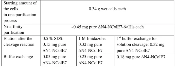
filters with 10 kDa molecular weight cut-off), but this step can be optimized for each target protein. The brief summary of the purification is provided in Table 2.
Fig. 3 – near here Table 2 – near here
Verification of the specificity of the Ni(II)-induced cleavage by MS
The ∆N4-NColE7 is made up of 127 amino acids and its sequence is:
PGKATGKGKPVNNKWLNNAGKDLGSPVPDRIANKLRDKEFKSFDDFRKKFWEEVSK DPELSKQFSRNNNDRMKVGKAPKTRTQDVSGKRTSFELHHEKPISQNGGVYDMDNIS VVTPKRHIDIHRGK. The mass spectrum of the purified N4-NColE7 protein was recorded.
The deconvoluted mass spectrum resulted in 14501.56 Da average molecular mass for the singly charged (MH+) intact protein (Fig. 4). This value is in a good agreement with the theoretically calculated protonated average mass of 14501.37 Da.
Fig. 4 – near here
A collision-induced dissociation (CID) and higher-energy collisional dissociation (HCD) was performed in order to investigate the specificity of Ni-cleavage at the C-terminus.
The most abundant ion of intact protein at 726 m/z (z = 20) was chosen for HCD fragmentation.
A fragment peak of 582 m/z (z = 3) was identified as the y15+3 ion, i.e., C-terminal SVVTPKRHIDIHRGK fragment of the protein. In a second step, this ion was selected in the ion trap for further fragmentation with CID (Fig. 5). The m/z values of third-generation ions detected in the MS3 spectrum correspond to the expected fragments of y15+3, thereby it gives clear evidence that the designed cleavage occurred at the C-terminus, without any modification or remaining residues, but resulting in complete removal of the hexahistidine affinity tag from the protein.
Fig. 5 – near here
Structural investigation of the purified protein in solution
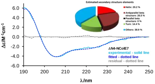
CD spectroscopy was applied to check the solution structure of the N4-NColE7 protein after the nickel(II)-induced cleavage and buffer exchange procedures. The recorded spectrum is characteristic for a folded protein including α-helical segments. The evaluation of the CD
spectra for the secondary structure element fractions by BeStSel program [53] yielded ~ 20 % α-helical content (Fig. 6). This is slightly less than it can be calculated from the crystal structure of a mutated N4-NColE7 in its metal ion bound form (PDB id: 3ZFK [54]) by BeStSel program: 29.2 % of helices; 10.4 % of β strands; 15.9 % turns and 44.4 % others, but the latter is a solid state structure. The obtained result, however, corresponds well to the secondary fractions calculated from the CD spectrum of NColE7 solution with the same program [55], suggesting that the folding of the protein is complete in HEPES buffer after the purification and solvent exchange procedures.
Fig. 6 – near here
The spectral intensity was also applied to determine the precise protein concentration based on the comparison with previous unpublished data. Accordingly, the protein yield was ~ 3.0 mg protein / L of the bacterial culture expressing N4-NColE7 protein.
Conclusion
Affinity-based protein purification methods are indispensable to obtain large amounts of pure proteins in a simplified procedure as compared to the multistep chromatographic purification protocols often leading to a significant loss of target proteins. A number of affinity tags are available nowadays with suitable solid state resins for protein binding. Combination of tags can result in even higher selectivity and thus, higher purity [26,35,39,56,57].
Various methods have been elaborated to cleave and remove the affinity tags after getting rid of impurities. Specific protein based cleavage enzymes, however, display several disadvantages. Apart from their high price they may be sensitive to reaction conditions such as high salt concentration, buffers, temperature. Steric hindrance may also limit protease efficiency. Secondary site cleavages, and the removal of the protease can pose further difficulties. To express N-terminal affinity tags, the start codon should be moved to the 5’-end of the whole gene (instead of having it at the 5’-end of the target protein’s gene), while C- terminal tags will move the stop codon from the 3’-end of the target protein gene to the 3’-end of the tag. Therefore, in almost all cases shorter or longer amino acids strings will remain at the termini of the purified protein after the enzymatic cleavage. These may disturb the folding
and/or the function of the target protein. Therefore, it is a topical issue to develop purification procedures for proteins with native sequences [33,55,58-60].
In this work, we described a new DNA construct providing opportunity for affinity purification strategy of a metalloprotein with a native protein sequence, avoiding any remnant amino acid residues after the removal of the affinity tag, while overcoming several above listed disadvantages of the proteases. The one-step purification method resulted in N4-NColE7 protein samples of high purity with an average yield of ~ 3.0 mg protein from 1 L culture yielding ~ 4.0 g of wet cells.
Ni(II)-induced cleavage of the affinity tag on the resin is recommended for those proteins, which can be deliberated from the solid phase during the cleavage reaction. Thus, only one further buffer exchange step is required for the subsequent applications. Nevertheless, for those proteins, which are difficult to be eluted from the resin, the cleavage in solution can be an alternative strategy. After the hydrolytic reaction the small-sized His tag can be removed by ultrafiltration, while the removal of large-sized affinity tags requires a second affinity chromatographic step. During the cleavage in solution there is a risk of precipitation by slow hydrolysis of the excess metal ions needed for the reaction.
Generally, prior to the design of the purification strategy the amino acid sequence of the target protein has to be checked for the presence of an X-(S/T)-X-H-X motif. Statistically, this segment may occur once in about every 200 amino acids, so that this sequence is more probably present in large proteins. Metalloproteins, which readily use histidine residues for metal ion binding may exhibit increased chance for including the cleavage sequence. Restrictions concerning the quality of the X amino acids (X should not be Cys or Pro) decrease the probability of occurrence of sequences suitable for Ni(II)-cleavage. Nevertheless, even proteins possessing a potentially cleavable sequence can be still purified with the outlined method if the steric requirement of flexibility within the metal ion binding part – so that the Ni(II) complex can not form to initiate the peptide bond cleavage – is not fulfilled. Thus, the described process can easily be generalized for a large number of target proteins.
Acknowledgements
Financial support from the Hungarian National Research, Development and Innovation Office (GINOP-2.3.2-15-2016-00038, GINOP-2.3.2-15-2016-00001 and K_16/120130), from the Hungarian Academy of Sciences and Japanese Society for the Promotion of Science (JSPS),
and from the CALIPSOplus (EU Framework Programme for Research and Innovation HORIZON2020, grant no. 730872) is gratefully acknowledged. H.A.H.A.E. is a Stipendium Hungaricum fellow supported also by Cultural Affairs & Mission Sector in Egypt. E.H. got a fellowship within the frame of UNKP-18-2 New National Excellence Program of the Ministry of Human Capacities.
References
[1] A. Belczyk-Ciesielska, B. Csipak, B. Hajdu, A. Sparavier, M.N. Asaka, K. Nagata, B.
Gyurcsik, W. Bal, Nickel(II)-promoted specific hydrolysis of zinc finger proteins, Metallomics 10 (2018) 1089–1098, DOI: 10.1039/C8MT00098K
[2] W. Bal, J. Lukszo, K. Bialkowski, K.S. Kasprzak, Interactions of Nickel(II) with Histones:
Interactions of nickel(II) with CH3CO-Thr-Glu-Ser-His-His-Lys-NH2, a peptide modeling the potential metal binding site in the "C-tail" region of histone H2A, Chem. Res. Toxicol. 11 (1998) 1014–1023, DOI: 10.1021/tx980051y
[3] W. Bal, R. Liang, J. Lukszo, S.-H. Lee, M. Dizdaroglu and K. S. Kasprzak, Ni(II) specifically cleaves the C-terminal tail of the major variant of histone H2A and forms an oxidative damage-mediating complex with the cleaved-off octapeptide, Chem. Res. Toxicol.
13 (2000) 616–624, DOI: 10.1021/tx000044l
[4] M. Mylonas, A. Krezel, J.C. Plakatouras, N. Hadjiliadis, W. Bal, The binding of Ni(II) ions to terminally blocked hexapeptides derived from the metal binding -ESHH- motif of histone H2A, J. Chem. Soc., Dalton Trans. (2002) 4296–4306, DOI: 10.1039/b206585a
[5] E. Kurowska. J. Sasin-Kurowska, A. Bonna, M. Grynberg, J. Poznanski, L. Knizewski, K.
Ginalski, W. Bal, The C2H2 zinc finger transcription factors are likely targets for Ni(II) toxicity, Metallomics 3 (2011) 1227–1231, DOI: 10.1039/c1mt00081k
[6] N.E. Wezynfeld, K. Bossak, W. Goch, A. Bonna, W. Bal, T. Fraczyk, Human annexins A1, A2, and A8 as potential molecular targets for Ni(II) ions, Chem. Res. Toxicol. 27 (2014) 1996–
2009, DOI: 10.1021/tx500337w
[7] N.E. Wezynfeld, A. Bonna, W. Bal, T. Fraczyk, Ni(II) ions cleave and inactivate human alpha-1 antitrypsin hydrolytically, implicating nickel exposure as a contributing factor in pathologies related to antitrypsin deficiency, Metallomics 7 (2015) 596–604, DOI:
10.1039/c4mt00316k
[8] A. Krezel, E. Kopera, A. M. Protas, J. Poznanski, A. Wysłouch-Cieszynska, W. Bal, Sequence-specific Ni(II)-dependent peptide bond hydrolysis for protein engineering.
Combinatorial library determination of optimal sequences, J. Am. Chem. Soc. 132 (2010) 3355–3366, DOI: 10.1021/ja907567r
[9] E. Kopera, A. Krezel, A. M. Protas, A. Belczyk, A. Bonna, A. Wyslouch-Cieszynska, J.
Poznanski, W. Bal, Sequence-specific Ni(II)-dependent peptide bond hydrolysis for protein engineering: reaction conditions and molecular mechanism, Inorg. Chem. 49 (2010) 6636–
6645, DOI: 10.1021/ic1005709
[10] A.M. Protas, H.H.N. Ariani, A. Bonna, A. Polkowska-Nowakowska, J. Poznanski, W. Bal, Sequence-specific Ni(II)-dependent peptide bond hydrolysis for protein engineering: Active sequence optimization, J. Inorg. Biochem. 127 (2013) 99–106, DOI:
10.1016/j.jinorgbio.2013.07.037
[11] A. Belczyk-Ciesielska, I. A. Zawisza, M. Mital, A. Bonna, W. Bal, Sequence-specific Cu(II)-dependent peptide bond hydrolysis: similarities and differences with the Ni(II)- dependent reaction, Inorg. Chem. 53 (2014) 4639–4646, DOI: 10.1021/ic5003176
[12] T. Fraczyk, N.E. Wezynfeld, K. Bossak, A. Bonna, W. Bal, Human annexins A1, A2, and A8 as potential targets for Ni(II) and Pd(II) ions, J. Biol. Inorg. Chem. 19 (2014) S474, DOI 10.1007/s00775-014-1095-8
[13] C. Harford, B. Sarkar, Amino terminal Cu(II)- and Ni(II)-binding (ATCUN) motif of proteins and peptides: Metal binding, DNA cleavage, and other properties, Acc. Chem. Res. 30 (1997) 123–130, DOI: 10.1021/ar9501535
[14] D.P. Mack, P.B. Dervan, Sequence-specific oxidative cleavage of DNA by a designed metalloprotein, Ni(II)GGH(Hin 139–190), Biochemistry 31 (1992) 9399–9405, DOI:
10.1021/bi00154a011
[15] X. Huang, M.E. Pieczko, E.C. Long, Combinatorial optimization of the DNA cleaving Ni(II)-Xaa-Xaa-His metallotripeptide domain, Biochemistry 38 (1999) 2160–2166, DOI:
10.1021/bi982587o
[16] Z. Yu, M. Han, J.A. Cowan, Toward the design of a catalytic metallodrug: Selective cleavage of G-quadruplex telomeric DNA by an anticancer copper–acridine–ATCUN complex, Angew. Chem. Int. Ed. 53 (2014) 1901–1905, DOI: 10.1002/anie.201410434
[17] M.E. Kimple, A.L. Brill, R.L. Pasker, Overview of affinity tags for protein purification, Curr. Protoc. Protein Sci. 73 (2013) 608–616, DOI: 10.1002/0471140864.ps0909s73
[18] V. Gaberc-Porekar, V. Menart, Potential for using histidine tags in purification of proteins at large scale, Chem. Eng. Technol. 28 (2005) 1306–1314, DOI: 10.1002/ceat.200500167
[19] L.A. Collins-Racie, J.M. McColgan, K.L. Grant, E.A. Diblasio-Smith, J.M. McCoy, E.R.
Lavallie, Production of recombinant bovine enterokinase catalytic subunit in Escherichia coli using the novel secretory fusion partner dsba, Bio/Technology 13 (1995) 982–987, DOI:
10.1038/nbt0995-982
[20] D.S. Waugh, An overview of enzymatic reagents for the removal of affinity tags, Protein Expr. Purif. 80 (2011) 283–293, DOI:10.1016/j.pep.2011.08.005
[21] X. Zhao, G. Li, and S. Liang, Several affinity tags commonly used in chromatographic purification, J. Anal. Methods Chem. (2013) article ID 581093, DOI:10.1155/2013/581093 [22] J.C. Carrington, W.G. Dougherty, A viral cleavage site cassette: identification of amino acid sequences required for tobacco etch virus polyprotein processing, Proc. Natl. Acad. Sci.
USA 85 (1988) 3391–3395, DOI:10.1073/pnas.85.10.3391
[23] R.B. Kapust, J. Tőzsér, T.D. Copeland, D.S. Waugh, The P1′ specificity of tobacco etch virus protease, Biochem. Biophys. Res. Commun. 294 (2002) 949–955, DOI: 10.1016/S0006- 291X(02)00574-0
[24] S. Harper, D.W. Speicher, Purification of proteins fused to glutathione S-transferase in Protein chromatography (Eds. D. Walls, S.T. Loughran) Methods Mol. Biol. 681 (2011) 259–
280, DOI 10.1007/978-1-60761-913-0
[25] T. Schmidt A. Skerra, The Strep-tag system for one-step affinity purification of proteins from mammalian cell culture in Affinity Chromatography (Ed. S. Reichelt) Methods Mol. Biol.
1286 (2015) 83–95, DOI:10.1007/978-1-4939-2447-9
[26] S. Honey, B.L. Schneider, D.M. Schieltz, J.R. Yates, B. Futcher, A novel multiple affinity purification tag and its use in identification of proteins associated with a cyclin-CDK complex, Nucl. Acids Res. 29 (2001) e24, DOI:10.1093/nar/29.4.e24
[27] K. Terpe, Overview of tag protein fusions: From molecular and biochemical fundamentals to commercial systems, Appl. Microbiol. Biotechnol. 60 (2003) 523–533, DOI:
10.1007/s00253-002-1158-6
[28] M. Futatsumori-Sugai, R. Abe, M. Watanabe, M. Kudou, T. Yamamoto, D. Ejima, T.
Arakawa, K. Tsumoto, Utilization of Arg-elution method for FLAG-tag based chromatography, Protein Expr. Purif. 67 (2009) 148–155, DOI: 10.1016/j.pep.2009.03.012
[29] T.R. Butt, S.C. Edavettal, J.P. Hall, M.R. Mattern, SUMO fusion technology for difficult- to-express proteins, Protein Expr. Purif. 43 (2005) 1–9, DOI: 10.1016/j.pep.2005.03.016 [30] T. Asai, L.A. Wims, S.L. Morrison, An interaction between S•tag and S•protein derived from human ribonuclease 1 allows site-specific conjugation of an enzyme to an antibody for targeted drug delivery, J. Immunol. Methods 299 (2005) 63–76, DOI:
10.1016/j.jim.2005.01.020
[31] J.M. Moon, G.Y. Kim, H. Rhim, A new idea for simple and rapid monitoring of gene expression: requirement of nucleotide sequences encoding an N-terminal HA tag in the T7 promoter-driven expression in E. coli, Biotechnol. Lett. 34 (2012) 1841–1846, DOI:
10.1007/s10529-012-0966-8
[32] M.C. Hillman, L.S. Yang, S. Sun, J.L. Duke, K.T. O'Neil, J.E. Kochie, A. Karjoo, P. Nath, L.A. Breth, K. Murphy, O.H. Ross, T.C. Burn, G.F. Hollis, R. Wynn, A comprehensive system for protein purification and biochemical analysis based on antibodies to c-myc peptide, Protein Expr. Purif. 23 (2001) 359–368, DOI: 10.1006/prep.2001.1514
[33] E.A. Peroza, E. Freisinger, Tris is a non-innocent buffer during intein-mediated protein cleavage, Protein Expr. Purif. 57 (2008) 217–225, DOI: 10.1016/j.pep.2007.10.003
[34] P.M. Schmidt, L.G. Sparrow, R.M. Attwood, X. Xiao, T.E. Adams, J.L. McKimm- Breschkin, Taking down the FLAG! How insect cell expression challenges an established tag- system, PLoS ONE 7 (2012) e37779, DOI:10.1371/journal.pone.0037779
[35] D.K. Yadav, N. Yadav, S. Yadav, S. Haque, N. Tuteja, An insight into fusion technology aiding efficient recombinant protein production for functional proteomics, Arch. Biochem.
Biophys. 612 (2016) 57–77, DOI: 10.1016/j.abb.2016.10.012
[36] E.N. Kosobokova, K.A. Skrypnik, V.S. Kosorukov, Overview of fusion tags for recombinant proteins, Biochemistry (Moscow) 81 (2016) 187–200, DOI:
10.1134/S0006297916030019
[37] A.S. Pina, C.R. Lowe, A.C.A. Roque, Challenges and opportunities in the purification of recombinant tagged proteins, Biotechnol. Adv. 32 (2014) 366–381, DOI:
10.1016/j.biotechadv.2013.12.001
[38] C.L. Young, Z.T. Britton, A.S. Robinson, Recombinant protein expression and purification: A comprehensive review of affinity tags and microbial applications, Biotechnol.
J. 7 (2012) 620–634, DOI: 10.1002/biot.201100155
[39] D.W. Wood, New trends and affinity tag designs for recombinant protein purification, Curr.Opin. Plant Biol. 26 (2014) 54–61, DOI: 10.1016/j.sbi.2014.04.006
[40] H. Block, B. Maertens, A. Spriestersbach, J. Kubicek, F. Schäfer, Proteolytic affinity tag cleavage, Methods Enzymol. 559 (2015) 71–97, DOI: 10.1016/bs.mie.2014.11.009
[41] J. Arnau, C. Lauritzen, G.E. Petersen, J.Pedersen, Current strategies for the use of affnity tags and tag removal for the purification of recombinant proteins, Protein Expr. Purif. 48 (2006) 1–13, DOI: 10.1016/j.pep.2005.12.002
[42] E. Kopera, A. Belczyk, W. Bal, Application of Ni(II)-assisted peptide bond hydrolysis to non-enzymatic affinity tag removal, PloS ONE 7 (2012) e36350, DOI:
10.1371/journal.pone.0036350
[43] E. Kopera, S. Krzywda, M. Lenarcic Živkovic, A. Dvornyk, B. Kłudkiewicz, K. Grzelak, I. Zhukov, W. Zagórski-Ostoja, M. Jaskólski, W. Bal, Atomic resolution structure of a protein prepared by non-enzymatic His-tag removal. Crystallographic and NMR study of GmSPI-2 inhibitor, PloS ONE 9 (2014) e106936.
[44] N. E. Wezynfeld, T. Fraczyk, W. Bal, Metal assisted peptide bond hydrolysis: Chemistry, biotechnology and toxicological implications, Coord. Chem. Rev. 327–328 (2016) 166–187, DOI: 10.1016/j.ccr.2016.02.009
[45] S.G. Grant, J. Jessee, F.R. Bloom, D. Hanahan, Differential plasmid rescue from transgenic mouse DNAs into Escherichia coli methylation-restriction mutants, Proc. Natl. Acad. Sci. USA 87 (1990) 4645–4649. DOI: 10.1073/pnas.87.12.4645
[46] F.W. Studier, A.H. Rosenberg, J.J. Dunn, J.W. Dubendorff, Use of T7 RNA polymerase to direct expression of cloned genes, Methods Enzymol. 185 (1990) 60–89. DOI:
10.1016/0022-2836(86)90385-2
[47] J. Sambrook, D.W. Russel, Condensed Protocols from Molecular Cloning: A Laboratory Manual: Cold Spring Harbor Laboratory Press, Cold Spring Harbor, New York (2006).
[48] M.J. Sui, L.C. Tsai, K.C. Hsia, L.G. Doudeva, W.Y. Ku, G.W. Han, H.S. Yuan, Metal ions and phosphate binding in the H-N-H motif: crystal structures of the nuclease domain of ColE7/Im7 in complex with a phosphate ion and different divalent metal ions, Protein Sci. 11 (2002) 2947–2957. DOI: 10.1110/ps.0220602
[49] H. Schägger, Tricine–SDS-PAGE, Nature Protoc. 1 (2006) 16–22, DOI:
10.1038/nprot.2006.4
[50] A.J. Miles, S.V Hoffmann, Y. Tao, R.W. Janes, B.A. Wallace, Synchrotron radiation circular dichroism (SRCD) spectroscopy: New beamlines and new applications in biology, Spectroscopy 21 (2007) 245–255, DOI: 10.1155/2007/282713
[51] A.J. Miles, R.W. Janes, A. Brown, D.T. Clarke, J.C. Sutherland, Y. Tao, B.A. Wallace, S.V. Hoffmann, Light flux density threshold at which protein denaturation is induced by synchrotron radiation circular dichroism beamlines, J. Synchrotron Radiat. 15 (2008) 420–422, DOI: 10.1107/S0909049508009606
[52] M.R. Beccia, T. Biver, B. García, J.M. Leal, F. Secco, R. Ruiz, M. Venturinia, Mechanism of Ni2+ and NiOH+ interaction with hydroxamic acids in SDS: evaluation of the contributions
to the equilibrium and rate parameters in the aqueous and micellar phase, Dalton Trans. 41 (2012) 7372–7381, DOI: 10.1039/c2dt30621b
[53] A. Micsonai, F. Wien, L. Kernya, Y-H. Lee, Y. Goto, M. Réfrégiers, J. Kardos, Accurate secondary structure prediction and fold recognition for circular dichroism spectroscopy, Proc.
Natl. Acad. Sci. USA 112 (2015) E3095–E3103. DOI: 10.1073/pnas.1500851112
[54] A. Czene, E. Tóth, E. Németh, H. Otten, J.-C.N. Poulsen, H.E.M. Christensen, L. Rulíšek, K. Nagata, S. Larsen, B. Gyurcsik, A new insight into the zinc-dependent DNA-cleavage by the colicin E7 nuclease: a crystallographic and computational study, Metallomics 6 (2014) 2090–2099, DOI: 10.1039/c4mt00195h
[55] E. Németh, R.K. Balogh, K. Borsos, A. Czene, P.W. Thulstrup, B. Gyurcsik, Intrinsic protein disorder could be overlooked in cocrystallization conditions - An SRCD case study, Protein Sci. 25 (2016) 1977–1988. DOI: 10.1002/pro.3010
[56] Z. Wang, N. Li, Y. Wang, Y. Wu, T. Mu, Y. Zheng, L. Huang, X. Fang, Ubiquitin-intein and SUMO2-intein fusion systems for enhanced protein production and purification, Protein Expr. Purif. 82 (2012) 174–178, DOI: 10.1016/j.pep.2011.11.017
[57] Y. Han, W. Guo, B. Su, Y. Guo, J. Wang, B. Chu, G. Yang, High-level expression of soluble recombinant proteins in Escherichia coli using an HE-maltotriose-binding protein fusion tag, Protein Expr. Purif. 142 (2018) 25-31, DOI: 10.1016/j.pep.2017.09.013
[58] H.C. Goh, R.M. Sobota, F.J. Ghadessy, S. Nirantar, Going native: Complete removal of protein purification affinity tags by simple modification of existing tags and proteases, Protein Expr. Purif. 129 (2017) 18-24, DOI: 10.1016/j.pep.2016.09.001
[59] S. Frey, D. Görlich, A new set of highly efficient, tag-cleaving proteases for purifying recombinant proteins, J. Chromatography A 1337 (2014) 95–105, DOI:
10.1016/j.chroma.2014.02.029
[60] M. Cheriyan, S.-H. Chan, F. Perler, Traceless splicing enabled by substrate-induced activation of the Nostoc punctiforme Npu DnaE intein after mutation of a catalytic cysteine to serine, J. Mol. Biol. 426 (2014) 4018–4029, DOI: 10.1016/j.jmb.2014.10.025
Table 1: Examples of widely used proteases for removal of affinity tags for protein purification [17-34]. (GST = Glutathione-𝑆-transferase; MBP = Maltose-binding protein; CBP = Calmodulin-binding peptide; CBD = Chitin-binding domain.) The last two lines provide examples for expression systems without specific protease cleavage sites.
Protease Recognition sequence
Affinity Tag Tag position Vector example
Factor Xa IEGR↓ 10×His-tag
GST MBP
N-terminal N-terminal N-terminal
pET16b pGEX-3X pMAL-c2X
Thrombin LVPR↓GS GST N-terminal pGEX-2T
Rhinovirus 3C ("PreScission") protease
LEVLFQ↓GP GST
S-tag
HA tag
c-Myc
N-terminal C-terminal
C-terminal
C-terminal
pGEX-6P-1 pSF-OXB20- COOH-3C- STag pSF-OXB20- COOH-3C- HA
pSF-OXB20- COOH-3C- CMyc TEV protease ENLYFQ↓G(His)7
ENLYFQ↓G/S ENLYFQ↓G/S
MBP Strep-tag Strep-tag
C-terminal N-terminal C-terminal
pDZ2087 pASK-IBA16 pSF-OXB20 - COOH-TEV- STREP Enterokinase (DYK)DDDDK↓ MBP
FLAG tag
N-terminal N- or C- terminal
pMAL-c2E p3xFLAG- CMV Ulpl-specific
protease
SUMO-GG↓ SUMO tag N-terminal pSUMOUlp1
pET SUMO
Genenase I PGAAHY↓ MBP N-terminal pMAL-c2G
- self-cleavable intein tag
CBD C-terminal
N-terminal
pTXB1 pTYB2
- - CBP N-terminal pST50Trc1-
CBPDHFR
- - 6×His-tag C-terminal pET21b
Table 2: Comparison of the N4-NColE7 protein purification strategies.
Starting amount of the cells
in one purification process
0.34 g wet cells each
Ni-affinity purification
~0.45 mg pure N4-NColE7-6×His each Elution after the
cleavage reaction
0.5 % SDS:
0.15 mg pure
N4-NColE7
1 M Imidazole:
0.32 mg pure
N4-NColE7
1st buffer exchange for solution cleavage: 0.32 mg pure N4-NColE7
Buffer exchange 0.05 mg pure
N4-NColE7
0.25 mg pure
N4-NColE7
0.18 mg pure N4-NColE7
Caption of Scheme and Figures
(For interpretation of the references to colour in figures, the reader is referred to the web version of this article.)
Scheme 1: The Ni(II)-initiated hydrolytic cleavage of a peptide or protein at the N-terminus of Ser/Thr amino acids within the X-(Ser/Thr)-X-His-X sequence. The formation of the Ni(II)- ATCUN complex can be observed among the products.
Scheme 2: The redesigned cloning site includes all the restriction enzyme recognitions sites of the pET-21a vector and two additionally inserted BsmBI recognition sites (the arrows pointing downwards show the BsmBI cleavages outside the recognition sites). Cloning the gene of the target protein between the two BsmBI sites allows the expression from the atg start codon (underlined) within the NdeI restriction site. The Ni(II)-initiated hydrolytic cleavage of the expressed protein at the N-terminus of the SRHS sequence will fully remove the C-terminal affinity tag and the linker region encoded by further restriction enzyme recognition site sequences.
Fig. 1: SDS PAGE analysis of the batch Ni(II)-affinity purification procedure. Lane 2: 20 µl aliquot of the total protein fraction obtained after resuspending the pellet in 10 mL 1×binding buffer. Lane 3: 20 µl aliquot of the soluble protein fraction, i.e., from the supernatant after the centrifugation of total protein fraction. Lane 4: 20 µl aliquot of the supernatant after binding to the Ni-NTA resin. Lane 5 (Wash1): 1 µl aliquot of the resin after washing with 4×2 bed volume wash buffer (20 mM Tris pH 7.9; 60 mM Imidazole; 500 mM NaCl; 0.1 % Triton X-100. Lane 6 (Wash2): 1 µl aliquot of the resin after washing with 2×2 bed volume wash buffer with an increased imidazole concentration of 100 mM. Lane 7 (Wash3): 1 µl aliquot of the resin after washing with 2×2 bed volume of 100 mM pH 8.2 HEPES buffer.
Fig. 2: Panel A: The ∆N4-NColE7-6×His batch cleavage with Ni(II) ions. Lanes 2 and 3 show the aliquots from the supernatant and resin, respectively, before starting the cleavage reaction.
Lane 4 shows the absence of ∆N4-NColE7 in the supernatant after 72 hours cleavage reaction, and lane 5 is the aliquot of the ∆N4-NColE7 bound to the resin with the predicted size after 72 hours cleavage reaction. Panel B: SDS PAGE analysis of the elution of the cleaved ∆N4- NColE7 protein from the resin (lane 2) using 0.5 M NaCl containing 10 mM HEPES pH 8.2
buffer solution in the presence of either 1 M imidazole (lanes 3 and 4) or 0.5 % SDS (lanes 5 and 6). Lanes 3 and 5 show the band of the eluted protein in supernatant, while lanes 4 and 6 show the band of the protein remained on the resin after the 15 minutes long elution steps.
Fig. 3: The ∆N4-NColE7-6×His cleavage with Ni(II) ions in solution. Lanes 2 and 3 show the aliquots from the protein solution before starting the cleavage reaction and after 72 hours of incubation with Ni(II) ions, respectively.
Fig. 4: The recorded m/z spectrum of purified N4-NColE7 (A) and the deconvoluted average m/z spectrum (B).
Fig. 5: MS3 spectrum showing the fragmentation pattern of the selected [SVVTPKRHIDIHRGK]+3 precursor ion from the C-terminus of the protein.
Fig. 6: The CD spectrum of ∆N4-NColE7 fitted by BeStSel program between 190-250 nm. The output of the estimation of secondary structure is seen in the inset.
Scheme 1
Scheme 2.
NdeI BsmBI BamHI BsmBI EcoRI SacI 5’-catatggagacgggatcccgtctctccaggcactcggaattcgagctc
M E T G S R L S R H S E F E L
Ni(II) cleavage site
SalI HindIII NotI XhoI
gtcgacaagcttgcggccgcactcgagcaccaccaccaccaccactga-3’
V D K L A A A L E H H H H H H - 6 His-tag Stop
Figure 1.
kDa
20 15
1 -Marker 2 -Total 3 -Soluble 4 -Sup. 5 -Resin/wash1 6 -Resin/wash2 7 -Resin/wash3 8 -Marker
∆N4-NColE7-6 His
Figure 2.
1 -Marker 2 -0 hSup. 3 -0 hResin 4 -72hSup. 5 -72hResin
∆N4-NColE7-6 His
∆N4-NColE7 cleaved kDa
20 15
A
Figure 3.
kDa
20 15
∆N4-NColE7-6 His
∆N4-NColE7 cleaved
1 -Marker 2 -0 h 3 -72 h
Figure 4.
Figure 5.
Figure 6.
-6.0 -4.0 -2.0 0.0 2.0 4.0 6.0
190 200 210 220 230 240 250
e/M-1 cm-1
l/nm
N4-NColE7
experimental - solid line fitted - dotted line residual - dotted line
Estimated secondary structure elements Helix: 20.2 % Antiparallel beta structure: 26.5 % Parallel beta structure: 0 % Turn: 14.4 % Others: 38.9 %
Highlights
- The newly designed cloning site allows for expression of proteins with native sequence - Ni(II) induced peptide bond hydrolysis for affinity tag removal
- Mass spectrometry proved the specificity of the proteolytic cleavage
- CD spectrum of N4-NColE7 is characteristic for proteins with high -helical content
![Table 1: Examples of widely used proteases for removal of affinity tags for protein purification [17-34]](https://thumb-eu.123doks.com/thumbv2/9dokorg/1285681.102774/18.892.130.767.293.931/table-examples-widely-proteases-removal-affinity-protein-purification.webp)