Address for correspondence: Milosz J. Jaguszewski, First Department of Cardiology, Medical University of Gdansk, ul. Dębinki 7, 80–952 Gdańsk, Poland, e-mail: mjaguszewski@escardio.com.pl
Expert consensus for the diagnosis
and treatment of patient with hyperuricemia and high cardiovascular risk
Claudio Borghi1, Andrzej Tykarski2, Krystyna Widecka3, Krzysztof J. Filipiak4, Justyna Domienik-Karłowicz5, 6, Katarzyna Kostka-Jeziorny2, 6, Albert Varga7,
Milosz Jaguszewski6,8, Krzysztof Narkiewicz9, Giuseppe Mancia10
1Department of Medical and Surgical Sciences, University of Bologna, Italy
2Department of Hypertension, Angiology and Internal Diseases, Poznan University of Medical Sciences, Poznan, Poland
3Department of Hypertension and Internal Medicine, Pomeranian Medical University, Szczecin, Poland
4First Department of Cardiology, Medical University of Warsaw, Poland
5Department of Internal Medicine and Cardiology with the Center for Diagnosis and Treatment of Venous Thromboembolism, Medical University of Warsaw, Poland
6Club 30, Polish Cardiac Society, Poland
7Institute of Family Medicine, University of Szeged, Hungary
8First Department of Cardiology, Medical University of Gdansk, Poland
9Department of Hypertension and Diabetology, Medical University of Gdansk, Poland
10Università Milano-Bicocca, Milan, Italy
Definition and epidemiology:
The burden of hyperuricemia is rising The definition of hyperuricemia (HU) varies widely in different studies, which makes the epi- demiological reports somewhat inconsistent. HU remains the culprit of the pathogenesis of gout which occurs in 3–6% of men and 1–2% of women from Western countries [1]. The linear relation- ship between serum uric acid (sUA) levels and the risk of gout have been demonstrated in many studies and registries including the large-scale Framingham study and Normative Aging study in the United States of America (USA) [1]. The United States National Health and Nutrition Examination Survey (NHANES) study estimated the prevalence of gout to be 3.9% (5.9% for men, 2.0% for women) and the HU prevalence to be 21.4% (21.2% for men and 21.6% for women) [2].
The prevalence of HU depends on sex age and ethnicity, reaching a plateau at the age of 70. At variance from USA and other Western countries (see above) in developing countries, it occurs in
less than 1% of the general population, but this may represent an underestimation of the true pre- valence due to insufficient epidemiological data [3]
and a very high prevalence has been found in some developing subsets of the population, such as, for example, the Taiwanese aboriginals (41%).
Reports are also available of ethnic difference such as, in the USA, a higher prevalence among African Americans than among non-African Americans (25.7%, 22.1%, respectively) [4]. Over the 10 years, epidemiological data consistently show an increase in the prevalence of both HU and gout.
This might be caused by rapid economic develop- ment and a change of dietary habits and lifestyle and among patients with a higher socioeconomic status [5, 6]. Interestingly, sUA levels are higher in subsets of subjects at high cardiovascular (CV) risk, including postmenopausal women, non-white patients with hypertension or chronic kidney dis- ease (CKD). The adoption of western lifestyle by natives of other countries and cultures together with a change in the socioeconomic background through immigration to Western countries, as well
Cardiology Journal 2018, Vol. 25, No. 5, 545–564
DOI: 10.5603/CJ.2018.0116 Copyright © 2018 Via Medica
ISSN 1897–5593
POSITION PAPER
as movements from rural to urban communities, has influenced sUA levels [7, 8]. Currently, > 1.2 million patients in the USA and United Kingdom are being prescribed allopurinol, the xanthine oxidase inhibitor (XOI), which successfully lowers their sUA level [9].
The relationship between hyperuricemia and gout
Although the definition of asymptomatic HU is still being discussed, it is after all merely, a biochemical disorder. Gout is a chronic disease with periods of inflammation, also known as flares, which are caused by the deposition of monoso- dium urate (MSU) crystals as a result of HU [10].
The crystallization of MSU in vitro takes place at a concentration of ≥ 6.8 mg/dL (temperature of 37°C) or > 6.0 mg/dL (temperature of 35°C) sUA at pH 7.0. This process may occur at a lower sUA concentration at lower pH levels and at a lower tissue temperature in peripheral joints. The reso- lution of tophaceous deposits occurs when sUA level is < 5 mg/dL [11]. Subsequently, the host’s inflammation response to the deposited crystals shows clinical symptoms of gout [12].
Although the presence of HU is essential for the formation of crystals, only part of the patient population with HU develop gout i.e. 2–36% of patients in studies with a 5–10-year period of follow-up [13, 14]. Thus, the finding of HU is not sufficient to diagnose gout. Conversely, Schles- inger et al. [15], confirmed a “true” normal sUA level (≤ 6 mg/dL) in 14% of patients suffering from an acute attack of gout. Interestingly, gout is unlikely in patients with a low sUA level, even when a confirmed normal sUA level occurs during an acute flare [16]. This indicates a more complex relationship between sUA levels and gout flares.
Thus, the definition based on the solubility of UA may need to be fundamentally reconsidered [4].
High sUA levels are predominantly caused by an insufficient elimination (90%) or overproduction (10%) of sUA. Therefore, the aim of the treatment is dissolution and prevention of further formation of MSU crystals.
Since different mechanisms interact with the pathogenesis of gout, the clinical presentation of the disease is heterogeneous. This includes the genesis of elevated sUA, the process of crystal formation and growth, and subsequent inflamma- tion, activated by MSU crystals. In fact, four stages can be distinguished in the development of gout:
i) HU without symptoms of MSU deposition;
ii) deposition of MSU crystals without sympto- matic gout; iii) deposition of MSU crystals with acute exacerbations of gout; iv) advanced gout characterized by the occurrence of gouty nodules (the effect of an organized, chronic inflamma- tory reaction involving interleukin 1b, TNF-a, and TGF-b1), chronic arthritis and joint damage identified in radiological reports. In the absence of adequate treatment, advanced gout usually appears approximately 10 years after the first “flare-up”.
This mostly occurs in the distal part of the lower limb (i.e., first metatarsophalangeal joint), and un- dergoes spontaneous resolution within 1–2 weeks.
In the absence of adequate treatment, flares occur more frequently affecting more and more joints.
Ultimately the disease changes to stage 4 with the onset of tophi and joint inflammation [17].
A large body of evidence describes the link between HU, CKD and cardiovascular disease (CVD). Undoubtedly, cardiovascular and renal comorbidities play a key role in the management strategies of patients with HU and gout.
The pathophysiological effect of HU on CVD: Purine catabolism, oxidative metabolism, inflammation,
“uric acid paradox”
The primary baseline risk factor for HU in patients is a mutational inactivation of a gene for uricase, an enzyme which degrades urate to the more soluble allantoin [18]. Notwithstanding, to generate HU, additional risk factors are required.
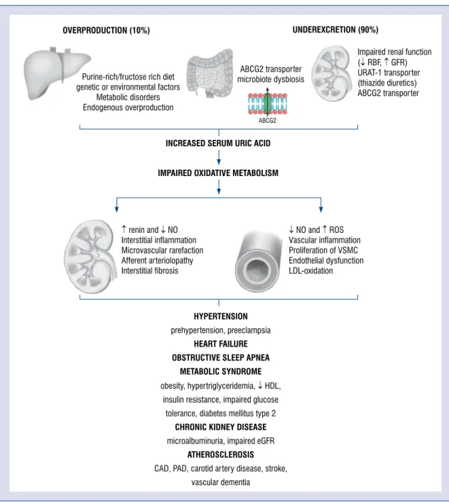
First and foremost, an increased sUA level is the result of a purine/fructose-rich diet, genetic or environmental factors, metabolic disorders, as well as its endogenous overproduction or, in most cases, by insufficient excretion (Fig. 1). The synthesis is mainly affected by phoribosyl-pyrophosphate synthetase or purine salvage pathway influenced by hypoxanthine-xanthine phosphoribosyl trans- ferase. Fructose causes mostly the depletion of ATP increasing the generation and release of UA [19]. An excessive purine turnover may be present in some clinical scenarios, comprised of leukemias, hemolytic anemias, etc. On the other hand, ca. 90%
of UA is reabsorbed in the proximal tubular cells by urate-anion transporter 1 (URAT-1) and organic anion transporters (OATs) clustered in the renal cortex according to their roles as reuptake- or se- cretory transporters (Fig. 2) [20, 21]. The natural mechanism of further breakdown of UA is impos- sible due to the lack of human uricase (hominids lost expression of gene encoding, therefore, they
cannot convert UA to allantoin) [22]. Thus, it is the kidneys that may play a key role in the process of underexcretion.
Impaired oxidative mechanism
The increased activity of xanthine oxidase (XO) influences the XO-derived reactive oxygen species (ROS) formation resulting in vascular endothelial dysfunction. This is the case when an
increased catabolism of purines and subsequent UA production xanthine oxidoreductase converts intensively hypoxanthine to xanthine and xanthine to UA. Xanthine oxidoreductase exists in two iso- forms: xanthine dehydrogenase and XO. XO uses molecular oxygen as an electron acceptor, thus, generating superoxide anion and other reactive oxygen species as by-products which can trigger endothelial dysfunction (e.g. reduces endothe-
Figure 1. Pathophysiological aspects of hyperuricemia and its influence on cardiovascular and kidney disease;
ABCG2 — ATP binding cassette multidrug transporter; CAD — coronary artery disease; eGFR — estimated glomerular filtration rate; HDL — high density lipoprotein; LDL — low density lipoprotein; NO — nitric oxide; PAD — peripheral arterial disease; ROS — reactive oxygen species; RBF — renal blood flow; URAT-1 — uric acid transporter 1; VSMC
— vascular smooth muscle cells.
lial nitric oxide [NO] production) and thereby contribute to the incidence of hypertension and target organ damage. Circulating and endothelial- bound XO is over-expressed in ischemic tissues.
Oxidative stress generated by XO overactivity has a detrimental effect on the vascular endothelium in- cluding coronary arteries [23, 24]. Thus, increased sUA levels impair an oxidative metabolism, stimu- lating the renin–angiotensin system and inhibits the release of endothelial NO. This contributes to, microvascular damage to afferent arterioles, renal vasoconstriction and, permanent sodium- sensitive hypertension [25, 26]. Persistent renal vasoconstriction may contribute to arteriolosclero- sis and the development of primary hypertension.
Indeed, in many experimental studies, UA induces vascular smooth-muscle cell (VSMC) proliferation, inflammation processes, oxidative stress and, con- sequently, local renin–angiotensin system activity [27–29]. Whether hypertension exists or not, the influence of an increased level of sUA on endothe- lial cells and VSMC develops microvascular renal disease [28, 30, 31]. On one hand, preglomerular arteriolar disease impairs renal autoregulatory response and glomerular hypertension. On the other in diabetic patients, elevated sUA levels are a well-known predictor of microalbuminuria and renal dysfunction [32, 33] being associated with an impaired glomerular filtration rate (GFR) [34].
Lastly, preclinical research supports the hy- pothesis that both endothelial dysfunction and the inflammatory and oxidative changes in adipocytes which remain the key factors in causing metabolic syndrome [35]. Some studies reported an associa- tion between sUA levels, obesity, and insulin resist- ance. Subsequently, HU has even been proposed, as a component of metabolic syndrome [36]. Although UA has an inflammatory and prooxidative effect on vascular cells and adipocytes, in some neurologic disorders, i.e., multiple sclerosis or Parkinson’s disease, it may function as an antioxidant thus being protective. Many scientists focus on low-grade sys- temic inflammation, XO activity, or the unfavorable effects of HU [37], however, the entire mechanism that links these diseases is not fully understood.
Genetics and the environment:
Individualized diagnosis and care strategy. Key advances in the understanding of factors associated
with an increased risk of HU
In the development of HU, the influence of genetics and environmental factors, cannot be ig- nored [38]. The inheritance of sUA levels and the fractional excretion of urate have been reported in studies which examine the phenotypic relation- ship between twins and establish it as 43–73% and
Figure 2. Renal proximal tubule urate transportasome; ABCG2 — ATP binding cassette multidrug transporter;
OAT — organic anion transporter; URAT-1 — uric acid transporter 1.
46–96%, respectively [38, 39]. Indeed, genome-wide association studies identified the genetic basis of HU as dominated by loci containing urate transporters and interacting proteins involved in the excretion of urate (SLC2A9, ABCG2, SLC22A11, SLC17A1–
–SLC17A4 and PDZK1) and proteins associated with metabolic pathways (e.g. GCKR, A1CF, IGF1R). For instance, reduced intestinal excretion is associated with polymorphisms of the ABCG2 gene [40]. Con- sequently, some research has shown a relationship between a genetic risk score of a high sUA and CV death or renal function [41, 42]. In other studies, a different SNP in the SLC2A9 gene was associated with an increased sUA level and normal blood pres- sure (BP) or hypertension [43, 44]. Since sUA is a result of the effects of inherited genetic variation especially on kidneys, gastrointestinal tracts and/or the liver and different processes, further specialized genome-wide association studies is required.
Currently, the explanation of the genetic impact on the development of gout from HU is still insuf- ficient. First and foremost, genes encoding proteins are involved in the NLRP3 (NOD-, LRR- and pyrin domain-containing 3) inflammasome pathway feature [45]. The genetic effect depending on glycolysis genes as well as the interactions between genes and environmental factors — i.e., diuretics, alco- hol — have already been researched. Interestingly, genome-wide and targeted sequencing revealed the population-specific and penetrant genetic variants.
Which have provided insight into the pathogenesis of gout, as well as the prediction of likely responsiveness to urine-lowering therapy [45]. These advances lead to individualized patient care. Genetic data can inform about the prognosis in patients suffering from HU, and help clinicians in their selection and dosage of urate-lowering therapy (ULT) and the correct advice on lifestyle changes.
Environmental factors that influence HU in- clude diet and medication: i.e. probenecid, benzbro- marone and sulfinpyrazone and the XOI allopurinol, febuxostat, antihypertensive drugs, cholesterol- lowering therapy, steroid medication, some non- steroidal anti-inflammatory drugs and antibiotics as well as small doses of acetylsalicylic acid and thiazides (Table 1). A high intake of the following:
meat, seafood, fructose, alcohol, and sodium are known to increase sUA levels.
Hyperuricemia and comorbidities:
A high sUA level and cardiovascular risk Uric acid remains an end-product of purine catabolism and is considered, an independent factor
for the development of a wide variety of microvas- cular and macrovascular disorders: hypertension [46], metabolic syndrome [47, 48], coronary artery disease (CAD) [49], diabetes [50], cerebrovascular disease [51, 52] and CKD (Fig. 1) [53] as well as other CVDs [54, 55] and, conversely, these comor- bidities increase the incidence of HU [4].
As mentioned above, the risk of crystallization of MSU increases when the saturation of sUA is >
6.8 mg/dL [18]. This induces the localized inflam- matory reactions in joints and connective tissue [56] leading to gout, in up to 36% of HU-patients within a period of 5–10 years [13, 14]. However, some studies reported a relation between sUA level and CVD not only in patients with clearly diagnosed HU, but also with values considered as normal to high, i.e. > 5.2–5.5 mg/dL [57–59]. Notably, since a high sUA concentration is associated with inflammatory markers, i.e. C-reactive protein or neutrophil count, inflammation seems to be a major link between HU and CVD and kidney diseases [4].
Patients with HU or gout require systematic screening for diseases associated with high sUA concentrations. A large body of evidence confirms the contribution of HU in the worsening of car- diovascular, diabetic, lipid and renal diseases [41, 60, 61]. Thereby supporting the evidence that an increased sUA level indicates an emerging CV Table 1. Drug effect on the serum level of uric acid [4].
Acetylsalicylic acid ≠ (at low doses) Ø (at high doses)
Clopidogrel ´
Ticagrelor ≠
Beta-blockers (propranolol, atenolol, metoprolol, timolol) ≠
ACEIs and ARBs ´
Losartan Ø
Diuretics (loop diuretics, thiazide-type diuretics, amiloride, triamterene, spironolactone, eplerenone)
≠
Alpha-blockers ´
Amlodipine ´
Fenofibrates Ø
Atorvastatin Ø
Simvastatin ´
Hypoglycemic agents Ø
SGLT2 inhibitors
≠ — increase; Ø — decrease; ACEIs — angiotensin-converting enzyme inhibitors; ARBs — angiotensin receptor blocker;
SGLT2 — sodium glucose cotransporter 2
risk [62, 63]. Therefore, the management of such conditions should be an integral part of a patients’
treatment strategy (Tables 2 and 3) [4, 64–66].
In the NHANES, which included 5707 partici- pants aged 20 years and above, hypertension was diagnosed in 74% of patients, CKD in 71%, obesity (body mass index [BMI] ≥ 30 kg/m2) in 53%, type 2 diabetes in 26%, nephrolithiasis in 24%, past myocardial infarction (MI) in 14%, chronic heart failure (HF) in 11%, had suffered a stroke 10%
[67]. The negative results mentioned above were more frequent in patients with HU than in patients without. In the group of patients with a sUA level of > 10 mg/dL, these conditions were even more frequent, i.e.: 86% of patients suffered from chronic disease, 66% hypertension, 65% obesity, 33% HF, 33% type 2 diabetes, 23% past MI, and 12% had suffered a stroke in the past.
Hyperuricemia and hypertension
It has been known for a while, that renal injury can be caused by advanced arteriosclero- sis, glomerulosclerosis, interstitial fibrosis, i.e.
a deposition of urate crystal. This was reflected in a majority of patients with HU/gout who suf- fered from hypertension and were in an older age group [68]. One of the hypothesis attributes HU in hypertension to an impaired urate clearance and tubular secretion [69] as a result of a reduced renal blood flow [70] with an impaired delivery of urate to the sites of tubular secretion in the peritubular space. Nevertheless, recent preclinical and clinical studies consistently support the hypothesis that increased levels of sUA may lead to hypertension [27, 50, 71–82]. Remarkably, HU is more frequent in primary hypertension vs. white-coat or second- ary hypertension [57]. Moreover, it is common among patients with prehypertension or microal- buminuria [32, 83].
An ample body of evidence widely acknowl- edged that the association between an increase in the relative risk of hypertension and high levels of sUA remains independent of traditional risk factors [77, 79, 84–90]. Interestingly, the as- sociation between sUA, hypertension and CVD remain stronger in women, younger patients, and patients with fewer CV risk factors [4]. In a large meta-analysis involving 18 studies (n = 55,607), Grayson et al. [84] confirmed that there is an increase of 13% in the incidence of new-onset hy- pertension for every increase of 1% in sUA level.
Kubawara et al. [91] in their retrospective cohort
Table 3. Prevalence of comorbidities according to hyperuricemia and gout in National Health and Nutrition Examination Survey (NHANES US) 2007–2008 [67].
Comorbidities Hyperuricemia
Gout
Prevalence, % (95% CI) No gout
Prevalence, % (95% CI)
Hypertension 77.7 (66.6–88.8) 47.2 (43.0–51.4)
CKD stage ≥ 2 71.8 (61.3–82.3) 70.4 (62.8–78.0)
Obesity — BMI 30 kg/m2 55.6 (45.5–65.7) 54.2 (49.0–59.4)
Diabetes 26.9 (9.7–44.1) 12.2 (8.7–15.6)
CKD stage ≥ 3 22.6 (14.0–31.2) 17.4 (13.2–21.5)
Nephrolithiasis 20.2 (10.3–30.2) 11.6 (8.9–14.3)
Stroke 11.8 (0.4–23.1) 5.1 (3.6–6.7)
Heart failure 11.7 (6.1–17.4) 4.5 (3.4–5.5)
Myocardial infarction 11.6 (4.5–18.7) 4.5 (3.3–5.6)
Depicted are n prevalence (%) and 95% confidence interval (CI).
BMI — body mass index; CKD — chronic kidney disease; GFR — glomerular filtration rate
CKD stage ≥ 2 = GFR < 60 mL/min/1.73 m2 and ≥ 30 mL/min/1.73 m2; CKD stage ≥ 3 = GFR < 30 mL/min/1.73 m2 and ≥ 15 mL/min/1.73 m2
Table 2. Diseases associated with hyperuricemia, requiring clinical evaluation in patients
diagnosed with hyperuricemia.
Excessive alcohol consumption, lead poisoning Lipid disorders, modifiable risk factors for coronary disease/stroke
Use of drugs increasing serum uric acid Obesity
Metabolic syndrome, type 2 diabetes Kidney stones in past medical history Chronic kidney disease
Hypertension
study (n = 3584) reported sUA level as a strong risk marker for the development of hyperten- sion from prehypertension. The cumulative inci- dence of hypertension from prehypertension over 5 years was 25.3%, and the cumulative incidence of hypertension in subjects with HU (n = 726) was significantly higher than in those without HU (n = 2858; 30.7% vs. 24.0%; p < 0.001). In the Pressioni Arteriose Monitorate e Loro Associazioni (PAMELA) study, 2045 participants underwent a complex assessment of CV risk which included sUA level, metabolic, renal and anthropometric variables, left ventricular (LV) mass index, as well as the measurement of, home, office, and ambula- tory BP. Patients were monitored for an average of 16 years. The new-onset of hypertension was recognized in patients with systolic BP (SBP) or diastolic BP (DBP) exceeding the upper normal- ity levels. On subsequent examination or after introducing antihypertensive therapy an increase of sUA by 1 mg/dL was associated with a signifi- cant increase in the risk of developing new-onset home and ambulatory hypertension (odds ratio [OR] 1.34, 95% confidence interval (CI) 1.06–1.7, p = 0.015; OR 1.29, 95% CI 1.05–1.57, p = 0.014, respectively) [85]. Notably, this was the first study to include a measurement of sUA level among the factors included in the algorithm assessing the total CV risk [61].
Hyperuricemia and metabolic syndrome As mentioned above, several studies demon- strated that sUA level is associated with metabolic syndrome, high BMI, waist circumference, high fasting blood glucose levels, and dyslipidemia [92].
These results, and the fact that targeting high levels of serum low-density lipoprotein (LDL) is beneficial for patients at CV risk prompted other groups to investigate whether HU predicts an increase in LDL cholesterol. Actually, Kuwabara et al. [61] confirmed that elevated sUA increased the risk of developing high LDL cholesterol in both men (OR 1.159 per 1 mg/dL increase, 95%
CI 1.009–1.331, p = 0.037) and women (OR 1.215, 95% CI 1.061–1.390, p = 0.005), as well as hyper- triglyceridemia.
Hyperuricemia and chronic kidney disease:
“What comes first?”
First observations of the possible causative role of sUA on CKD were confirmed in large studies, including, the NHANES and the German Chronic Kidney Disease (GCKD) study [93, 94].
Data from the German registry documented gout in
24.3% of patients with CKD. The prevalence of gout was substantially higher in patients with estimated GFR (eGFR) of < 30 mL/min/1.73 m2 vs. ≥ 60 mL/
/min/1.73 m2 [94]. The meta-analysis, which included 18 prospective studies (n = 431,000), revealed that HU predicts the occurrence of CKD and GFR decline [95]. Indeed, HU plays a key role in the development and progression of CKD. It remains an independent factor in the progression of CKD, even after making adjustment for classic comorbidities such as hyper- tension, proteinuria, and dyslipidemia. This relation- ship was confirmed in IgA nephropathy, diabetes nephropathy, renal transplantation, or autosomal- dominant polycystic kidney disease [96–99]. Notably, in normotensive patients, with normal renal function, the association between sUA and the likelihood of a decrease in eGFR was noted. This effect was ap- parent at a concentration level of sUA of 5.5 mg/dl, 5.0 mg/dL in women [100].
Since the pathogenesis of HU is complex and lots of conflicting factors influencing CKD are well described, the question “what comes first?”
remains open [95]. It is worth noting, that hyper- tension can cause CKD and, subsequently, there is a decline in renal function. Moreover, the diuretic therapy may essentially increase the sUA levels.
However, the studies based on healthy subsets revealed a clear association between sUA and CKD in long-term observation [53, 101].
“Uric acid paradox”
Despite the well-known association between HU and CAD, high sUA levels are strongly associ- ated with peripheral carotid vascular disease and vascular dementia [51, 102].
In a study by Ruggiero et al. [103] (n = 1016), it was found that elderly patients with dementia had higher concentrations of sUA. Patients with the highest sUA concentrations had a higher prob- ability to develop dementia (OR 3.32, 95% CI 1.06–
–10.42) [103]. Contrarily, some studies revealed that HU might reduce the risk of neurological diseases, especially Parkinson’s and Alzheimer’s disease, vascular and non-vascular dementia [104]
or osteoporosis [105]. This so-called “uric acid paradox” has already been widely discussed [4].
Hyperuricemia and adverse events:
A high sUA level and its influence on cardiovascular outcome Hyperuricemia and mortality
The Third NHANES presented an increased risk of all-cause death and cardiac death in
patients with an increased sUA level. The associa- tion remained significant even after adjustment for different factors including demography and comor- bidities [106]. In the PAMELA study, patients were randomly recruited from the general population and underwent a thorough assessment of CV risk, including an echocardiogram and out-of-office BP measurement. The analysis suggested that the cut-off sUA level which showed the best trade-off between sensitivity and specificity of risk prediction were 5.4 mg/dL for CV mortality and 4.9 mg/dL for all-cause mortality [85]. Data from the Preventive Cardiology Information System (PreCIS) database documented that for each 1-mg/dL increase in the sUA level, there was a 39% increase in the risk of death. After adjusting for age, sex, weight, BMI, waist circumference, BP, history of CVD, eGFR rate, levels of cholesterol fractions, and plasma glucose levels, smoking status, and alcohol consumption, the sUA level continued to accurately predict the risk of death (hazard ratio [HR] 1.26, 95% CI 1.15–1.38, p < 0.001). Notably, the association occurred wheth- er the patient was taking diuretics or not.
Interestingly, sUA level significantly improved the predictive accuracy of a model that included Framingham Heart Study score factors, compo- nents of metabolic syndrome, and fibrinogen level [107]. In a group of 51,297 male patients included in the Health Professionals Follow-Up Study, those suffering from gout had a higher risk of all-cause death. Curiously, among men without preexisting CAD, the increased mortality risk was primarily a result of an elevated risk of CVD death. Addition- ally, men with gout had a higher risk of nonfatal MI than men without gout (relative risk [RR] 1.59;
95% CI 1.04–2.41) [108]. In a much larger study that included 354,110 subjects without a history of gout at the Chang Gung Memorial Hospital in Taiwan, patients with both high and low sUA levels were at higher risk of all-cause and cardiovascular mortality [109]. The increased risk of death was re- ported among patients with a high sUA level and HF [110]. A large retrospective analysis of patients with symptomatic HF revealed that HU was significantly associated with increased HF events or death [111].
Hyperuricemia and acute myocardial infarction and risk of strokes
Krishnan et al. [112] confirmed the independ- ent risk relationship between HU and acute MI.
Interestingly, gouty arthritis was associated with an excess risk of acute MI, which was not explained by its well-known links with other comorbidities, i.e., renal function, metabolic syndrome, traditional CV
risk factors as well as diuretic therapy. These results have prompted further research in which sUA levels and untreated gout were documented as independent prognostic markers for poor all-cause and cardiac mortality in patients with recent acute MI [112].
In the Rotterdam Study (n = 4385), among patients without a history of MI or who had suf- fered strokes at baseline, high sUA levels were associated with long-term risk of MI and strokes;
an age and sex-adjusted hazard ratios (95% CI) for highest versus lowest quintile of sUA were 1.68 (1.24–2.27) for CVD, 1.87 (1.12–3.13) for MI, 1.57 (1.11–2.22) for strokes, 1.77 (1.10–2.83) for ischemic strokes, and 1.68 (0.68–4.15) for hemor- rhagic strokes [102]. Tscharre et al. [113] suggest an independent association of HU (defined as UA levels > 6.0 mg/dl in women, and > 7.0 mg/dl in men) with long-term major adverse cardiovascular event (MACE), including CV death, MI and strokes, in acute coronary syndrome patients undergoing percutaneous coronary intervention. Patients with HU had a 1.6-fold increased RR for CV death (p = 0.005) and a 1.5-fold increased risk for MI (p = 0.032) [113]. In a small study (n = 140), the prevalence of moderate-to-severe coronary calci- fication in CAD was more severe in hyperuricemic patients with asymptomatic MSU crystal deposi- tion than normouricemic or hyperuricemic patients without such deposits [114].
In an analysis of the large Swedish registry (n
= 417,734) of a group of patients undergoing health check-ups, moderate levels of sUA appeared to be associated with an increased incidence of acute MI, stroke and congestive HF in middle-aged subjects without prior CVD [115]. Finally, sUA level was posi- tively associated with the presence (p = 0.0001), the number (p = 0.001), the size (p = 0.001), and the location of lacunar infarcts (LI) in the basal ganglia (p = 0.0038), the deep white matter (DWM) (p < 0.0001), and the pons (p = 0.0156). Pathologi- cally, it was characterized by arteriolosclerosis and atherosclerosis. Interestingly, the occurrence of LI increased starting from sUA levels of 5.7 mg/dl [116]. HU may substantially increase the risk of a stroke, even in patients at a low risk of atrial fi- brillation, thus, may constitute an independent risk factor for strokes in a hyperuricemic setting.
Hyperuricemia treatment and cardiovascular outcomes:
Allopurinol and febuxostat
Xantine-oxidase inhibitors, especially allopu- rinol, are currently recommended as a first-line
ULT. However, the optimism related to febuxostat has been dispelled by recently published results from the Cardiovascular Safety of Febuxostat and Allopurinol in Patients with Gout and Cardiovas- cular Morbidities (CARES) study [117].
Allopurinol: The influence of XOI on cardiovascular outcomes Allopurinol and mortality
Knowledge of the effectiveness of XOI makes them a subject of major interest [118–126]. How- ever, the causality between HU/gout and these CV outcomes remains controversial, mainly due to confounding variables and common etiological factors [4, 126-128].
The effect of purine XOI on all-cause mortality and CV mortality is probably due to its potent anti- oxidant effect, which has the ability to inhibit reac- tive oxygen species production [129, 130]. Among potential mechanisms, it should be stressed that endothelial dysfunction and low-grade inflamma- tion can occur as a major negative factor [7, 131]. Of note, at high concentrations, XO promotes oxida- tive stress and emphasizes endothelial dysfunction.
Increasing evidence exists to suggest the crucial role of XO in various forms of ischemic and other types of tissue and vascular injury, inflammatory processes and chronic HF [132].
In a record-linkage database (n = 7135), CV event rates were 74.0 (95% CI 61.9–86.1)/1000-per- son-years in the 100 mg allopurinol group, 69.7 (95% CI 49.6–89.8) in the 200 mg allopurinol group and 47.6 (95% CI 38.4–56.9) in the ≥ 300 mg allopurinol group [133]. Moreover, high-dose al- lopurinol, defined as ≥ 300 mg/dL vs. < 299 mg/dL, was associated with a reduced risk of all-cause mortality (adjusted HR 0.65, 95% CI 0.42–0.99) [7, 134]. In the large-scale cohort study by Dubreuil et al. [135] the initiation of allopurinol therapy was associated with an 11% lower risk of all-cause death in patients with HU and 19% lower risk of death in patients with gout.
Allopurinol and CAD
The key role of six-weeks treatment with al- lopurinol (600 mg daily) vs. placebo in a small ran- domized controlled trial (RCT) of 65 patients (aged 18–85 years) who had angiographically documented CAD, a positive exercise tolerance test, and stable chronic angina pectoris showed that allopurinol vs.
placebo increased the median time to ST depres- sion to 298 s (interquartile range [IQR] 211–408) vs. 249 s (IQR 200–375, p = 0.0002) [136]. Simi-
larly, a small, randomized, double-blind, placebo- -controlled study (n = 65) in patients with CAD and LV hypertrophy [137] proved a significant reduction of LV mass and LV end-systolic volume in patients receiving 600 mg of allopurinol vs. placebo (–5.2 ± 5.8 g vs. –1.3 ± 4.48 g, p = 0.007; –2.81 ±
± 7.8 mls vs. +1.3 ± 7.22 mls, p = 0.047, respec- tively) [137]. Higgins et al. [138] in a systematic review and meta-analysis of 40 studies confirmed that XOI improves endothelial function and reduces markers of oxidative stress. A steep-dose response relationship between allopurinol and endothelial func- tion has been presented and described showing the mechanistic effect of vascular oxidative stress [139].
Allopurinol and congestive HF
In a large observational study of patients with HF and a history of gout (n = 25,090), > 30 day-therapy with XOI was associated with reduced readmissions to hospital, caused by HF or death (adjusted RR 0.69;
95% CI 0.60–0.79, p < 0.001), and all-cause mortal- ity (adjusted RR 0.74; 95% CI 0.61–0.90, p < 0.001) [111]. In contrast, investigators from Oxypurinol Therapy for Congestive Heart Failure (OPT-CHF) study did not find clinical improvements in the unse- lected patient cohort in a group of patients (n = 405) with moderate-to-severe HF (New York Heart Asso- ciation [NYHA] functional class III/IV) due to systolic dysfunction. Data from EXACT-HF study, involving patients (n = 253) with symptomatic HF, left ven- tricular ejection fraction (LVEF) ≤ 40%, receiving allopurinol (600 mg daily) for 24 weeks showed no improvement in LVEF and no significant difference in clinical status between the allopurinol- and placebo- treated patients (worsened: 45% vs. 46%, unchanged:
42% vs. 34%, improved: 13% vs. 19%, respectively;
p = 0.68) [140].
Interestingly, the post-hoc analysis from OPT- -CHF suggested that sUA reduction due to oxypu- rinol correlated with favorable clinical response [141–144] and that sUA may serve as a biomarker to target XO inhibition in congestive HF. These results show the positive potential role of an early intervention of implementing the ULT in patients with congestive HF.
Allopurinol and hypertension
An ample amount of studies investigated the effect of XOI on hypertension. In the United Kingdom Clinical Practice Research Datalink, al- lopurinol use was independently associated with a fall in both SBP and DBP [145]. In the short-term, crossover study presenting preliminary results in adolescents with newly diagnosed hypertension,
treatment with XOI resulted in a lowering of BP.
Treatment with allopurinol (200 mg twice a day for 6 weeks) vs. placebo caused a mean change in SBP (–6.9 mmHg, 95% CI –4.5 to –9.3 mmHg vs.
–2.0 mmHg, 95% CI 0.3 to –4.3 mmHg, p = 0.009) and DBP (–5.1 mmHg, 95% CI –2.5 to –7.8 mmHg vs. –2.4 mmHg, 95% CI 0.2 to –4.1 mmHg, p =
= 0.05) [146]. In meta-analysis of 10 clinical studies with 738 participants treated with allopurinol, SBP decreased by 3.3 mmHg (95% CI 1.4–5.3 mmHg, p = 0.001) and DBP by 1.3 mmHg (95% CI 0.1–2.5 mmHg, p = 0.03) [147]. These findings represent a new potentially positive therapeutic result which requires confirmation in future larger clinical trials.
Whether sUA level is a causative mediator of increased BP and impaired vascular compliance remains unclear. However, in the study of treated hypertensive patients, allopurinol increased aortic compliance irrespective of the antihypertensive drugs used [148]. This is in line with the observa- tion that inhibition of XO with allopurinol signifi- cantly reduces arterial wave reflection measured by augmentation index, a marker of arterial stiffness in stroke survivors [149].
In a randomized, double-blinded, placebo- controlled trial, two mechanisms of sUA reduc- tion were investigated in prehypertensive, obese, adolescents, aged 11–17 years by using XOI or probenecid. Subjects treated with ULT experienced a reduction in clinical SBP 10.2 mmHg and DBP 9.0 mmHg. ULT also resulted in a significant reduc- tion in systemic vascular resistance. This data indi- cates that, at least in young patients with prehyper- tension, their sUA level influences the BP and that it can be significantly reduced by ULT [147, 150].
In a new retrospective analysis of Survival of Myocardial Infarction Long-Term Evaluation (SMILE studies), researchers evaluated the 1-year combined occurrence of MACE, death or hospi- talization for CV causes in patients after acute MI using angiotensin converting enzyme inhibitors (ACEI), such as zofenopril or captopril, plus XOI.
The rate of survival free from MACE was signifi- cantly higher in a group of patients from the XOI- -arm than with other ACEI with no XOI (HR 2.29, 1.06–4.91, p = 0.034). Survival time without any events was longer in patients treated with zofeno- pril and XOI than in those treated with a placebo or other ACEI without XOI (log rank test, p = 0.033) [151]. The survival benefit of XOI therapy has been proven in another RCTs and cohort studies [152]. In a recent study published in “Hypertension” allopu- rinol lowered BP in adolescents and was associated with a significantly lower risk of both stroke (HR
0.50, 95% CI 0.32–0.80) and major cardiac events (HR 0.61, 95% CI 0.43–0.87). High-dose treatment, i.e., ≥ 300 mg daily, was associated with a lower risk of stroke (HR 0.58, 95% CI 0.36–0.94) and major cardiac events (HR 0.65, 95% CI 0.46–0.93).
Summarizing, a decrease in sUA levels, achieved by treating patients with ULT, mainly XOI, has, in general, been associated with an improve- ment in CV effects and amelioration in BP control [62, 118], but further large studies are essential [128]. Nevertheless, allopurinol may also be con- sidered in hypertensives with asymptomatic HU, particularly those at high CV risk. Among the drugs recommended for the treatment of co-morbidities with HU, great attention should be paid to the key drugs that influence sUA levels (Table 1) [153–159].
Allopurinol and renal function
It has been known for a long time, that therapy with XOI might essentially improve renal func- tion [4]. A comprehensive meta-analysis of RCTs revealed statistically significant improvement in eGFR and serum creatinine after ULT, favoring the strategy with allopurinol [160]. Goicoechea et al. [161] documented the slower progression of CKD and reduction of the rate of proteinuria in the group randomly assigned to the arm with XOI vs. placebo. Another meta-analysis confirmed that ULT decreases the risk of kidney failure events and end-stage renal disease (ESRD) by 55% (RR 0.45, 95% CI 0.31 ± 0.64) and 41% (RR 0.59, 95% CI 0.37 ± 0.96, respectively), compared with standard treatment or placebo [162]. Siu et al. [163] reported a decrease in sUA levels and preservation of renal function in patients treated with XOI at 12 months.
The meta-analysis of 12 studies by Sampson et al. [164] (n = 1187) in a wide range of patients revealed improved renal function by ULT at one year assessed by the reduced serum creatinine and raised eGFR. In another population-based cohort study (n = 111,992) investigating the association between HU and renal disease, patients treated with ULT who achieved sUA level of 6 mg/dL showed a 37%-outcome reduction defined as GFR reduction of ≥ 30% or endstage renal disease [165].
Febuxostat: Causes more harm to cardiovascular patients than
clinical benefits
In 2009, a new drug, febuxostat, was approved by the USA Food and Drug Administration (FDA).
Febuxostat is a nonpurine XOI and provides highly selective and potent inhibition of XO and greater
hypouricemic activity vs. commonly used doses of allopurinol. Unexpectedly, in 2017 the FDA alerted the public that preliminary results from a safety clinical trial show an increased risk of cardiac death with febuxostat vs. allopurinol. The first reports suggested a modestly higher rate of CV events with febuxostat [166]. Consequently, a large-scale, intensive RCT was designed (CARES) to investigate whether febuxostat remains non- -inferior to allopurinol with regard to MACE in patients with gout and high CV risk [117]. In the meantime, a meta-analysis of 35 studies did not show a significant difference between febuxostat and allopurinol in CV events (RR 1.69, 95% CI 0.54–5.34, p = 0.37) [126]. Moreover, ULT with febuxostat reduced the renal decline in subjects with gout [167]. In the multicenter FEATHER study, where patients (n = 467) were randomly assigned in a 1:1 ratio to receive febuxostat or placebo for 108 weeks, febuxostat did not mitigate the decline in kidney function in patients with stage 3 CKD and asymptomatic HU [168].
Interestingly, during the European Society of Cardiology scientific sessions in 2018 the results of FREED study were announced, where over 1000 elderly patients with HU, defined as sUA level
> 7.0 mg/dL up to 9.0 mg/dL, with one or more risks for cerebral, cardiovascular, or renal disease were treated with febuxostat (up to 40 mg daily) or non-febuxostat (non-treatment or low 100 mg daily of allopurinol). Febuxostat lowered sUA level to mean 4.5 ± 1.5 mg/dL, while non-febuxostat group achieved only 6.76 ± 1.45 mg/dL. After 3 years, 25% relative risk reduction in the compos- ite of death to any cause, cerebrovascular disease, non-fatal CAD, HF requiring hospitalization, the arteriosclerotic disease requiring treatment, renal impairment, and atrial fibrillation was observed in febuxostat group compared to the non-febuxostat group. Febuxostat also prevented the development and progression of CKD in this study. Notably, renal impairment was the only significantly dif- ferent event, at 16.2% in the febuxostat group vs. 20.5% in the control group (HR 0.745, 95% CI 0.562–0.987). There was no difference in CV clini- cal outcomes examined separately with febuxostat vs. control treatment. Herewith we should depict that only 27% of patients in the non-febuxostat group received allopurinol and more studies are warranted to confirm these results [Kijoma S, ESC Congress 2018]. The question remains whether the mortality results of the CARES trial are due to beneficial effects of allopurinol or deleterious effects of febuxostat.
In the last report, with findings from 32 months follow up (n = 6190), CARES investigators con- firmed that all-cause and CV mortality were higher in the febuxostat group than in the allopurinol group (HR for death from any cause 1.22, 95%
CI 1.01–1.47; HR for CV death 1.34, 95% CI 1.03–1.73) [117].
The first phase 3 RCTs, unfortunately, pro- vided a false hope that febuxostat was safe that led to the FDA-approval. As a result, febuxostat has been prescribed to hundreds of thousands of patients over the past decade who were unaware of the potential danger. Since the last CARES report published in 2018, strong evidence has emerged that the serious CV harms of febuxostat substan- tially outweigh any purported clinical benefit [117].
Thus, based on CARES study, treatment with febuxostat in patients at high CV risk should not be recommended.
Management strategies:
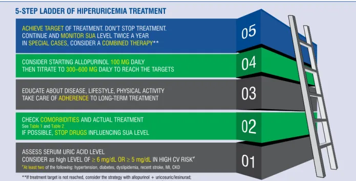
Current recommendation for the treatment of patients with increased sUA levels (Fig. 3) The primary aim of treatment of HU is to lower the sUA level (STEP 1). The measurement of sUA concentration was included in routine tests by ex- perts of both the European Society of Cardiology and European Society of Hypertension.
Our recommendation, based on the available data and a number of available guidelines, points to an optimal target of sUA level at 6 mg/dL (360 μmol/L) which is in line with the 2016 evidence- based recommendation for the management of gout of patients on ULT. Thus, sUA levels should be monitored and maintained at < 6 mg/dL [169]. It is also in accordance with the 2012 American College of Rheumatology (ACR) Guidelines, which indicate ULT for people with symptomatic HU and recom- mends a target sUA level of less than 360 μmol/L (6 mg/dL) for all patients on ULT [170]. Accord- ing to the British Society for Rheumatology, gout management by rheumatologists from the United Kingdom concords well with guidelines for most audit standards. Of note, 2016 guideline points to a lower sUA target (< 5 mg/dL; 300 μmol/L) for patients with severe gout in order to facilitate faster dissolution of UA crystals, especially when tophi, chronic arthropathy, or frequent attacks occur, until a total crystal dissolution and resolu- tion of gout. Furthermore, the British Society of Rheumatology recommends a target of sUA of
< 5 mg/dL for all patients with gout [171]. Notably,
the PAMELA study supported the sUA level of ca.
5 mg/dL in patients at high CV risk. Thus, based on current knowledge, in our opinion, one should consider sUA target of < 5 mg/dL in patients with high CV risk comprising at least two of the follow- ing: hypertension, diabetes, dyslipidemia, recent stroke or MI, CKD and < 6 mg/dL for patients not suffering from the above. Still, more RCTs are needed to support this opinion.
While optimizing the management strategy of HU, great attention should be paid to the key drugs prescribed due to comorbidities that may substan- tially influence sUA levels (Table 1, STEP 2).
Especially since a large body of literature con- firms that HU coexists with a wide variety of microvascular- and macrovascular diseases in- cluding hypertension [4], metabolic syndrome [48], CKD [53] or other CVD [54, 55] and these comorbidities increase the incidence of HU [4].
Consequently, the management of such conditions should be an integral part of a patients’ treatment strategy (Table 2).
As previously reported, also in the United Kingdom, less than half of the patients treated achieved the desired sUA levels during 12 months of treatment. Desired sUA levels ≤ 6 mg/dL and
< 5 mg/dL were achieved by 45% and 25% of patients, respectively [172]. In a cohort study
including 6042 patients, among 1035 allopurinol users, less than half (44.7%) reached target urate concentration [133].
Thus, we conclude that doctors often un- derestimated HU. An important step toward im- proved management of HU/gout in clinical routine is to improve adherence to guidelines, raising awareness of HU and associated comorbidities, prompting specific monitoring and treatment of these, as well as building multidisciplinary teams for optimal diagnostic and management strategy (STEP 3).
Recommended life-style changes (STEP 3) The following dietary factors have shown an adverse effect on sUA levels: low-sodium diet [173], consumption of red meat, seafood, fructose, and sugar-sweetened beverages, alcohol [174]. Di- etary factors known to decrease sUA include coffee, dairy products, cherries [155, 175], and ascorbic acid [176]. Some studies have also confirmed that weight loss and regular physical activity are effec- tive in reducing sUA levels [177, 178]. Therefore, maintenance of healthy weight and an increase in physical activity should be strongly recommended.
Patients with a high concentration of sUA should avoid, fructose-rich and animal-derived high purine meals and alcohol.
Figure 3. Management strategy for patients suffering from hiperuricemia. CV — cardiovascular; CKD — chronic kidney disease; MI — myocardial infarction; SUA — serum uric acid.
Xantine oxidase inhibitors:
First-line therapy (STEP 4)
As mentioned above, XOI, especially allopurinol, are recommended as a first-line ULT. They are rap- idly metabolized to oxypurinol, which is eliminated by the kidneys, therefore this requires dose adjustment according to renal function [169]. According to the Summary of product characteristics of allopurinol in patients with CKD, the maximum allopurinol dos- age should be adjusted to eGFR [169]. The recom- mended initial allopurinol dosage is 100 mg daily, with a gradual titration, e.g. 300–600 mg daily, up to the achievement of the desired sUA target level [169].
In accordance to meta-analysis, which included 15 papers concerning allopurinol, 44.4% of patients reached the target of sUA with this therapy and the average reduction in sUA was 33.8% [179].
If the sUA target cannot be achieved, the patient should be switched to benzbromarone with or without allopurinol (STEP 5), except in patients with eGFR of < 30 mL/min [180]. Although there is growing evidence of a benefit of a high-dose al- lopurinol regimen, i.e. ≥ 600 mg daily, especially in patients with high levels of sUA, we should still be aware of rare complications. Physicians should carefully consider dose escalation to achieve opti- mal treatment goals, especially due to allopurinol hypersensitivity syndrome or severe cutaneous allergic reactions (SCARs) — which usually occur after 8 weeks of therapy [181–183]. Several factors contribute to the development of this syndrome, including initial doses which are too high, CKD, accompanying use of diuretics, the presence of HLA-B*5801 [184, 185].
As mentioned above, XOI improves endothe- lial function [138, 186], oxidative stress markers, as well as cardiac physiology, including LVEF [187], cardiac index [187], end-systolic volume [188], and myocardial efficiency [189]. Thus, allopurinol may be considered in hypertensives with asympto- matic HU, particularly those with CVD. Although high-dose allopurinol defined as ≥ 300 mg/dL is associated with reduced risk of all-cause mortality [7, 134], consideration of optimal dose seems to be a major factor in the design of future research.
Febuxostat
Febuxostat, as a non-purine selective XOI, is metabolized in the liver and excreted by the kidneys. A side effect of febuxostat can be adverse skin effects. Previously, dose-adjusted febuxostat was considered in cases where the sUA target could not be achieved with allopurinol. Based on the meta-analysis of 16 papers, including febuxostat
treatment, 70.3% of patients on febuxostat reached the target of sUA (6 mg/dL) and the reduction in sUA was 45.3% with respect to baseline values [179]. In first studies, febuxostat (dose of ≤ 120 mg/day) resulted superior over allopurinol (dose
≤ 300 mg/day) in terms of probability to achieve the recommended target of sUA (OR 2.64, 95% CI 1.74–4.01) and percentage reduction of uricemia (mean difference: 13.08, 95% CI 7.6–18.55). Fur- thermore, febuxostat showed better results than allopurinol in patients with CKD (OR 0.85, 95% CI 0.75–0.97) [120, 179].
However, the safety concerns are already raised after results in patients with gout and CVD.
Thus, due to substantially higher all-cause and cardiovascular mortality rate in the febuxostat group vs. as compared to in the group receiving al- lopurinol [117] febuxostat cannot be recommended, especially in patients at high CV risk.
Lesinurad
Lesinurad is an oral selective inhibitor of URAT1 and OAT4 renal transporters, which increases renal UA excretion and lowers sUA levels by inhibiting UA reabsorption. A dose of 200 mg daily is recom- mended in combination with XOI in patients who do not achieve treatment targets. Lesinurad can increase efficiency compared to XOI applied alone or prevent maximal doses of XOI [190]. In a CLEAR study, les- inurad of 200 mg or 400 mg together with allopurinol significantly increased the proportion of patients who achieved the sUA target level compared to allopurinol (54.2%, 59.2%, and 27.9%, respectively, p < 0.0001) [191]. Similarly, the combination of lesinurad at 200 mg and 400 mg and maximum dose of febuxostat for 12 months compared to febuxostat monotherapy was more efficient in achieving sUA target levels in the CRYSTAL trial [192, 193].
To summarize, lesinurad in combination with an allopurinol is a novel option for the treatment of HU in adults with gout who have not achieved their target sUA levels with allopurinol alone [194]. Results from three clinical studies investi- gating the effectiveness of lesinurad suggest the addition of lesinurad to allopurinol or febuxostat is superior to XOI monotherapy in reducing sUA concentrations. However, this therapy increased the risk of renal-related adverse events. Thus, the recommendation is to use lesinurad 200 mg orally per day, in combination with an XOI, among patients with adequate renal function (i.e., above 45 mL/min) in cases with inadequately controlled HU associated with gout, should be treated with allopurinol (STEP 5) [193].
Once the sUA target is achieved continuously, the dose of ULT should be maintained indefinitely with ongoing monitoring of sUA levels twice a year (STEP 5).
Many unresolved questions remain:
Areas in need of further study
The prooxidant and antioxidant effect make the role of sUA complex. Furthermore, whether sUA is an independent risk factor of hard endpoints remains unclear and has been further complicated by the interaction of sUA levels and renal function.
Thus, the role of sUA in patients and its influence on their CV system has not been discovered. The superiority of XOI over uricosuric agents is due to potential inhibition of ROS production and its antioxidant effect. The treatment target based on the solubility of UA may need to be reconsidered, especially since data from PAMELA study identified a lower clinically meaningful cut-off level of sUA, i.e. < 5 mg/dL, and even lower for women. Uptill now a patients’ sUA level is not taken into consid- eration in the algorithm for assessing their total CV risk. Last, but by no means least, there is no evidence to support the treatment of asymptomatic HU, although, a large body of evidence does show the beneficial effect of ULT on CV results. In this light, when HU accompanies other CV risk factors, therapy with XOI should at least be considered.
Most relevant recommendations:
The take home message for the clinical practitioners
A remarkable number of epidemiological stud- ies have demonstrated that HU is strongly related to increased CV risk and predicts CV mortality and morbidity in such diseases as hypertension or diabetes. The fact that there is a significant increase in the onset of newly diagnosed hyperten- sion, metabolic syndrome in healthy patients with elevated levels of sUA, its role has been proved to be an independent risk factor in CV patients.
Therefore, all considerations presented, point to the need for greater attention to monitoring sUA levels when diagnosing patients, not only from a rheumatological standpoint but also regarding CV and renal risk, including eGFR measurements [4, 195]. This will play a key role in the prevention of CVD and improve future treatment strategies.
The need for active screening of CV risk factors in patients who had been diagnosed with gout is highly recommended both by the ACR and The
European League Against Rheumatism (EULAR) [169, 196, 197].
In conclusion, we would like to summarize our recommendations which should be helpful to clinicians treating with patients suffering from HU who also have a high CV risk:
— All patients with HU should be informed about pharmacological and epidemiological factors influencing HU, comorbidities, and the CV risk factors.
— All patients should receive precise informa- tion about required lifestyle modification, modification to their diet, a need for weight loss if necessary, and strict adherence to rec- ommended treatment
— Physical activity increase to a moderate level is strongly recommended.
— As we have established that the sUA level in CV patients contributes to the identification of patients at high risk, they should be regularly monitored.
— Both patients and physicians, including pri- mary care physicians, should strive to obtain and maintain lifelong sUA levels lower than 6 mg/dL. In cardiovascular patients the target level should be < 5 mg/dL.
— As soon as a diagnosis of HU is reached, ULT with XOI should be prescribed. Consider start- ing allopurinol 100 mg daily, then titrate to 300–600 mg daily to reach the targets.
— Once the desired sUA target is achieved, the dose of ULT should be maintained indefinitely and the patient’ sUA level should be monitored twice a year.
— A dosage of allopurinol of 300–600 mg/daily is recommended. If it is not tolerated by the patient or desired target of sUA is not reached, the combined therapy of allopurinol + uricos- uric/lesinurad, should be prescribed.
— In patients with renal impairment, the allopu- rinol dosage should be adjusted to eGFR.
— In patients with HU receiving diuretics, treat- ment modification is needed.
— Febuxostat should be avoided, especially in patients with a high CV risk.
Conflict of interest: Claudio Borghi: has received honoraria for participation as speaker in national/
/international meetings from Menarini Interna- tional, Berlin-Chemie, Servier, Takeda, Astellas, Novartis, Grunenthal, Sanofi; he is a mem- ber of Advisory Boards for: Menarini, Servier, Novartis, Grunenthal, BMS, Alfasigma; Andrzej Tykarski: has received honoraria for participation
as speaker in national/international meetings from Servier, Berlin-Chemie Menarini, Egis, Biofarm, Krka, Gedeon Richter, Merck; Krystyna Widecka:
report no conflict of interest; Krzysztof J. Filipiak:
has received honoraria for participation as speaker in national/international meetings from Egis and Berlin-Chemie/Menarini; Justyna Domienik- -Karłowicz: has received consulting honoraria from Egis; Katarzyna Kostka-Jeziorny: has received honoraria for participation as speaker in national/
/international meetings from Egis; Albert Varga:
report no conflict of interest; Milosz Jaguszewski:
has received speaker or consultation fees from Boheringer Ingelheim, Servier, Sandoz, Pfizer, Egis; Krzysztof Narkiewicz: has received hono- raria or consultation fees from Servier, Krka, Berlin-Chemie/Menarini, Egis, Sandoz, Idorsia, Medtronic, Mylan, Polpharma, Adamed, Gedeon Richter; Giuseppe Mancia: has received honoraria for participation as speaker or chairman in national/
/international meetings from: Abbott, Boehringer Ingelheim, Daichi Sankyo, Ferrer, Medtronic, Menarini, Merck, Novartis, Recordati, Sanofi, Servier.
References
1. Kuo CF, Grainge MJ, Zhang W, et al. Global epidemiology of gout: prevalence, incidence and risk factors. Nat Rev Rheumatol.
2015; 11(11): 649–662, doi: 10.1038/nrrheum.2015.91, indexed in Pubmed: 26150127.
2. Zhu Y, Pandya BJ, Choi HK. Comorbidities of gout and hyper- uricemia in the US general population: NHANES 2007-2008.
Am J Med. 2012; 125(7): 679–687.e1, doi: 10.1016/j.am- jmed.2011.09.033, indexed in Pubmed: 22626509.
3. Bardin T, Richette P. Impact of comorbidities on gout and hyper- uricaemia: an update on prevalence and treatment options. BMC Med. 2017; 15(1): 123, doi: 10.1186/s12916-017-0890-9, indexed in Pubmed: 28669352.
4. Borghi C, Rosei EA, Bardin T, et al. Serum uric acid and the risk of cardiovascular and renal disease. J Hypertens. 2015; 33(9):
1729–1741, doi:10.1097/HJH.0000000000000701, indexed in Pubmed: 26136207.
5. Conen D, Wietlisbach V, Bovet P, et al. Prevalence of hyperuri- cemia and relation of serum uric acid with cardiovascular risk factors in a developing country. BMC Public Health. 2004; 4:
9, doi: 10.1186/1471-2458-4-9, indexed in Pubmed: 15043756.
6. Qiu L, Cheng Xq, Wu J, et al. Prevalence of hyperuricemia and its related risk factors in healthy adults from Northern and North- eastern Chinese provinces. BMC Public Health. 2013; 13: 664, doi: 10.1186/1471-2458-13-664, indexed in Pubmed: 23866159.
7. Feig DI, Kang DH, Johnson RJ. Uric acid and cardiovascular risk. N Engl J Med. 2008; 359(17): 1811–1821, doi: 10.1056/
NEJMra0800885, indexed in Pubmed: 18946066.
8. Johnson RJ, Titte S, Cade JR, et al. Uric acid, evolution and primitive cultures. Semin Nephrol. 2005; 25(1): 3–8, indexed in Pubmed: 15660328.
9. Struthers A, Shearer F. Allopurinol: novel indications in cardio- vascular disease. Heart. 2012; 98(21): 1543–1545, doi: 10.1136/
/heartjnl-2012-302249, indexed in Pubmed: 22801998.
10. Ragab G, Elshahaly M, Bardin T. Gout: An old disease in new per- spective: a review. J Adv Res. 2017; 8(5): 495–511, doi: 10.1016/j.
jare.2017.04.008, indexed in Pubmed: 28748116.
11. Perez-Ruiz F, Calabozo M, Pijoan JI, et al. Effect of urate-lower- ing therapy on the velocity of size reduction of tophi in chronic gout. Arthritis Rheum. 2002; 47(4): 356–360, doi: 10.1002/
/art.10511, indexed in Pubmed: 12209479.
12. McCarty DJ. A historical note: Leeuwenhoek’s description of crystals from a gouty tophus. Arthritis Rheum. 1970; 13(4):
414–418, indexed in Pubmed: 4914047.
13. Lin KC, Lin HY, Chou P. The interaction between uric acid level and other risk factors on the development of gout among asymp- tomatic hyperuricemic men in a prospective study. J Rheumatol.
2000; 27(6): 1501–1505.
14. Hall AP, Barry PE, Dawber TR, et al. Epidemiology of gout and hyperuricemia. A long-term population study. Am J Med. 1967;
42(1): 27–37, indexed in Pubmed: 6016478.
15. Schlesinger N, Norquist JM, Watson DJ. Serum urate during acute gout. J Rheumatol. 2009; 36(6): 1287–1289, doi: 10.3899/
jrheum.080938, indexed in Pubmed: 19369457.
16. Logan JA, Morrison E, McGill PE. Serum uric acid in acute gout. Ann Rheum Dis. 1997; 56(11): 696–697, indexed in Pubmed: 9462177.
17. Forbess LJ, Fields TR. The broad spectrum of urate crystal deposition: unusual presentations of gouty tophi. Semin Ar- thritis Rheum. 2012; 42(2): 146–154, doi: 10.1016/j.semar- thrit.2012.03.007, indexed in Pubmed: 22522111.
18. Martillo MA, Nazzal L, Crittenden DB. The crystallization of monosodium urate. Curr Rheumatol Rep. 2014; 16(2): 400, doi: 10.1007/s11926-013-0400-9, indexed in Pubmed: 24357445.
19. Nakagawa T, Hu H, Zharikov S, et al. A causal role for uric acid in fructose-induced metabolic syndrome. Am J Physiol Renal Phys- iol. 2006; 290(3): F625–F631, doi: 10.1152/ajprenal.00140.2005, indexed in Pubmed: 16234313.
20. Enomoto A, Kimura H, Chairoungdua A, et al. Molecular iden- tification of a renal urate anion exchanger that regulates blood urate levels. Nature. 2002; 417(6887): 447–452, doi: 10.1038/
nature742, indexed in Pubmed: 12024214.
21. Borghi C. The management of hyperuricemia: back to the patho- physiology of uric acid. Curr Med Res Opin. 2017; 33(sup3): 1–4, doi:
10.1080/03007995.2017.1378502, indexed in Pubmed: 28952386.
22. de Oliveira EP, Burini RC. High plasma uric acid concentration:
causes and consequences. Diabetol Metab Syndr. 2012; 4: 12, doi: 10.1186/1758-5996-4-12, indexed in Pubmed: 22475652.
23. Prasad M, Matteson EL, Herrmann J, et al. Uric acid is asso- ciated with inflammation, coronary microvascular dysfunction, and adverse outcomes in postmenopausal women. Hyperten- sion. 2017; 69(2): 236–242, doi: 10.1161/HYPERTENSIONA- HA.116.08436, indexed in Pubmed: 27993955.
24. Farquharson CA, et al. Allopurinol improves endothelial dysfunc- tion in chronic heart failure. Circulation. 2002; 106(2): 221–226.
25. Watanabe S, Kang DH, Feng L, et al. Uric acid, hominoid evo- lution, and the pathogenesis of salt-sensitivity. Hypertension.
2002; 40(3): 355–360, indexed in Pubmed: 12215479.
26. Lin C, Zhang Pu, Xue Y, et al. Link of renal microcirculatory dysfunction to increased coronary microcirculatory resist- ance in hypertensive patients. Cardiol J. 2017; 24(6): 623–632, doi: 10.5603/CJ.a2017.0074, indexed in Pubmed: 28653312.
27. Mazzali M, Hughes J, Kim YG, et al. Elevated uric acid in- creases blood pressure in the rat by a novel crystal-independent mechanism. Hypertension. 2001; 38(5): 1101–1106, indexed in Pubmed: 11711505.
28. Kang DH, Park SK, Lee IK, et al. Uric acid-induced C-reactive protein expression: implication on cell proliferation and nitric ox- ide production of human vascular cells. J Am Soc Nephrol. 2005;
16(12): 3553–3562, doi: 10.1681/ASN.2005050572, indexed in Pubmed: 16251237.
29. Corry DB, Eslami P, Yamamoto K, et al. Uric acid stimulates vascular smooth muscle cell proliferation and oxidative stress via the vascular renin-angiotensin system. J Hypertens. 2008;
26(2): 269–275, doi: 10.1097/HJH.0b013e3282f240bf, indexed in Pubmed: 18192841.

![Table 3. Prevalence of comorbidities according to hyperuricemia and gout in National Health and Nutrition Examination Survey (NHANES US) 2007–2008 [67].](https://thumb-eu.123doks.com/thumbv2/9dokorg/1289857.103350/6.871.67.784.808.1070/prevalence-comorbidities-according-hyperuricemia-national-health-nutrition-examination.webp)