STATE-OF-THE-ART REVIEW
Update on Cardiac Catheterization in Patients With Prior Coronary Artery Bypass Graft Surgery
Iosif Xenogiannis, MD,a,bPeter Tajti, MD,a,b,cAllison B. Hall, MD,aKhaldoon Alaswad, MD,dStéphane Rinfret, MD,e William Nicholson, MD,fDimitri Karmpaliotis, MD,gKambis Mashayekhi, MD,hSergey Furkalo, MD,i
João L. Cavalcante, MD,aM. Nicholas Burke, MD,aEmmanouil S. Brilakis, MD, PHDa,b
JACC: CARDIOVASCULAR INTERVENTIONSCME/MOC/ECME
This article has been selected as this issue’s CME/MOC/ECME activity, available online athttp://www.acc.org/jacc-journals-cmeby selecting the JACCJournals CME/MOC/ECME tab.
Accreditation and Designation Statement
The American College of Cardiology Foundation (ACCF) is accredited by the Accreditation Council for Continuing Medical Education to provide continuing medical education for physicians.
The ACCF designates this Journal-based CME activity for a maximum of 1 AMA PRACategory 1 Credit(s)TM. Physicians should claim only the credit commensurate with the extent of their participation in the activity.
Successful completion of this CME activity, which includes participation in the evaluation component, enables the participant to earn up to 1 Medical Knowledge MOC point in the American Board of Internal Medicine’s (ABIM) Maintenance of Certification (MOC) program.
Participants will earn MOC points equivalent to the amount of CME credits claimed for the activity. It is the CME activity provider’s responsibility to submit participant completion information to ACCME for the purpose of granting ABIM MOC credit.
Update on Cardiac Catheterization in Patients With Prior Coronary Artery Bypass Graft Surgerywill be accredited by the European Board for Accreditation in Cardiology (EBAC) for 1 hour of External CME credits. Each participant should claim only those hours of credit that have actually been spent in the educational activity. The Accreditation Council for Continuing Medical Education (ACCME) and the European Board for Accreditation in Cardiology (EBAC) have recognized each other’s accreditation systems as substantially equivalent. Apply for credit through the post-course evaluation. While offering the credits noted above, this program is not intended to provide extensive training or certification in thefield.
Method of Participation and Receipt of CME/MOC/ECME Certificate To obtain credit for this CME/MOC/ECME, you must:
1. Be an ACC member orJACC: Cardiovascular Interventionssubscriber.
2. Carefully read the CME/MOC/ECME-designated article available on- line and in this issue of the journal.
3. Answer the post-test questions. A passing score of at least 70% must be achieved to obtain credit.
4. Complete a brief evaluation.
5. Claim your CME/MOC/ECME credit and receive your certificate electronically by following the instructions given at the conclusion of the activity.
CME/MOC/ECME Objective for This Article:Upon completion, the reader should be able to: 1) select optimal vascular access for patients with prior CABG; 2) evaluate strategies to prevent and treat distal emboli- zation; 3) compare the outcomes of DES versus BMS in SVG PCI;
4) identify the optimal treatment strategy for acute graft failure;
5) select the optimal method of revascularization in patients with prior CABG; and 6) decide between native vessel versus graft PCI in prior CABG patients.
CME/MOC/ECME Editor Disclosure:JACC: Cardiovascular Interventions CME/MOC/ECME Editor Michael C. McDaniel, MD, has reported that he is a Penumbra-Investigator on the EXTACT-PE trial.
Author Disclosures:Dr. Alaswad has received consulting fees from Ter- umo and Boston Scientific; and has been an unpaid consultant for Abbott Laboratories. Dr. Rinfret has received speaker and proctorship honoraria from Boston Scientific, Abbott Vascular Canada, Medtronic Canada, SoundBite, and Terumo US. Dr. Nicholson has been a proctor, consultant, and advisory board member for Abbott Vascular, Boston Scientific, and Medtronic. Dr. Karmpaliotis has received speaker hono- raria from Abbott Vascular and Boston Scientific. Dr. Mashayekhi has received consulting/speaker fees from Ashai Intecc, AstraZeneca, Bio- tronik, Boston Scientific, Cardinal Health, Daiichi-Sankyo, Medtronic, Teleflex, and Terumo. Dr. Cavalcante has received research grants from Medtronic and Abbott; and has been a consultant/speaker for Med- tronic, Circle Cardiovascular Imaging, and Siemens Inc. Dr. Burke has received consulting and speaker honoraria from Abbott Vascular and Boston Scientific. Dr. Brilakis has received consulting/speaker honoraria from Abbott Vascular, American Heart Association (associate editor:
Circulation), Boston Scientific, Cardiovascular Innovations Foundation (board of directors), CSI, Elsevier, GE Healthcare, InfraRedx, and Med- tronic; has received research support from Regeneron and Siemens; is a shareholder in MHI Ventures; and has served on the board of trustees for the Society of Cardiovascular Angiography and Interventions. All other authors have reported that they have no relationships relevant to the contents of this paper to disclose.
Medium of Participation:Print (article only); online (article and quiz).
CME/MOC/ECME Term of Approval Issue Date: September 9, 2019 Expiration Date: September 8, 2020
ISSN 1936-8798/$36.00 https://doi.org/10.1016/j.jcin.2019.04.051
Update on Cardiac Catheterization in Patients With Prior Coronary Artery Bypass Graft Surgery
Iosif Xenogiannis, MD,a,bPeter Tajti, MD,a,b,cAllison B. Hall, MD,aKhaldoon Alaswad, MD,dStéphane Rinfret, MD,e William Nicholson, MD,fDimitri Karmpaliotis, MD,gKambis Mashayekhi, MD,hSergey Furkalo, MD,i
João L. Cavalcante, MD,aM. Nicholas Burke, MD,aEmmanouil S. Brilakis, MD, PHDa,b
ABSTRACT
Patients who undergo coronary bypass graft surgery often require subsequent cardiac catheterization and repeat coro- nary revascularization. Saphenous vein graft lesions have high rates for distal embolization that can be reduced with use of embolic protection devices. They also have high restenosis rates, which are similar with drug-eluting and bare-metal stents. Percutaneous coronary interventions of native coronary arteries is generally preferred over saphenous vein graft interventions, but can often be complex, requiring expertise and specialized equipment. Prolonged dual-antiplatelet therapy and close monitoring can help optimize subsequent clinical outcomes. (J Am Coll Cardiol Intv 2019;12:1635–49)
© 2019 the American College of Cardiology Foundation. Published by Elsevier. All rights reserved.
P
atients who undergo coronary artery bypass graft surgery (CABG) often require additional revascularization because of bypass graft failure or progression of native coronary artery dis- ease (Figure 1) (1,2). Due to the high risk of redo CABG, coronary revascularization is performed by percutaneous coronary intervention (PCI) in nearly all prior CABG patients, but is associated with several challenges, both clinical (high-risk patient characteristics) and technical (such as treatment of failing bypass grafts, chronic total occlusions [CTOs], and severe calcification). We sought toprovide an overview of novel developments in car- diac catheterization and PCI in prior CABG patients, as well as practical recommendations (Central Illustration).
ACCESS SITE SELECTION
Engagement of arterial grafts and saphenous vein grafts (SVGs) for angiography and/or PCI can be per- formed using either femoral or radial approach, however femoral access is associated with lower contrast and radiation dose(3). Although systematic
From theaMinneapolis Heart Institute, Abbott Northwestern Hospital, Center for Complex Coronary Interventions, Minneapolis, Minnesota;bMinneapolis Heart Institute Foundation, Abbott Northwestern Hospital, Complex Coronary Artery Disease Science Center, Minneapolis, Minnesota;cUniveristy of Szeged, Department of Invasive Cardiology, Second Department of Internal Medicine and Cardiology Center, Szeged, Hungary;dHenry Ford Hospital, Department of Interventional Cardiology, Detroit, Michigan;eMcGill University Health Centre, McGill University, Department of Interventional Cardiology, Montreal, Quebec, Canada;fWellSpan Cardiology, Department of Interventional Cardiology, York, Pennsylvania;gColumbia University Medical Center, Center for Interventional Vascular Therapy, New York, New York;hDepartment of Cardiology and Angiology II University Heart Center Freiburg Bad Krozingen, Bad Krozingen, Germany; and theiDepartment of Endovascular Surgery and Angiography, National Institute of Surgery and Transplantology of AMS of Ukraine, Kiev, Ukraine. Dr. Alaswad has received consulting fees from Terumo and Boston Scientific; and has been an unpaid consultant for Abbott Laboratories. Dr. Rinfret has received speaker and proctorship honoraria from Boston Scientific, Abbott Vascular Canada, Medtronic Canada, SoundBite, and Terumo US.
Dr. Nicholson has been a proctor, consultant, and advisory board member for Abbott Vascular, Boston Scientific, and Medtronic.
Dr. Karmpaliotis has received speaker honoraria from Abbott Vascular and Boston Scientific. Dr. Mashayekhi has received consulting/speaker fees from Ashai Intecc, AstraZeneca, Biotronik, Boston Scientific, Cardinal Health, Daiichi-Sankyo, Medtronic, Teleflex, and Terumo. Dr. Cavalcante has received research grants from Medtronic and Abbott; and has been a consultant/speaker for Medtronic, Circle Cardiovascular Imaging, and Siemens Inc. Dr. Burke has received consulting and speaker honoraria from Abbott Vascular and Boston Scientific. Dr. Brilakis has received consulting/speaker honoraria from Abbott Vascular, American Heart Association (associate editor:Circulation), Boston Scientific, Cardiovascular Innovations Foundation (board of directors), CSI, Elsevier, GE Healthcare, InfraRedx, and Medtronic; has received research support from Regeneron and Siemens; is a shareholder in MHI Ventures; and has served on the board of trustees for the Society of Cardiovascular Angiography and In- terventions. All other authors have reported that they have no relationships relevant to the contents of this paper to disclose.
Manuscript received February 17, 2019; revised manuscript received March 26, 2019, accepted April 2, 2019.
reviews of mainly observational studies have sug- gested similar success with radial and femoral access, in the RADIAL CABG (RADIAL Versus Femoral Access for Coronary Artery Bypass Graft Angiography and Intervention) trial, diagnostic coronary angiography via radial access was associated with a higher mean contrast volume (14239 ml vs. 17172 ml; p<0.01), longer procedure time (21.9 6.8 min vs. 34.2 14.7 min; p<0.01), greater patient air kerma radiation exposure (1.080.54 Gray vs. 1.290.67 Gray; p¼ 0.06), and higher operator radiation dose (first oper- ator 1.31.0 mrem vs. 2.61.7 mrem; p<0.01) but higher patient satisfaction as compared with femoral access(4). In observational studies, however, radial access was associated with fewer vascular complica- tions, and reduced hospital stay(4–6). If radial access is selected, the left radial artery should be used in most cases to facilitate engagement of the left inter- nal mammary artery (LIMA) and the other bypass grafts. When graft engagement is challenging using radial access, early conversion to femoral access should be considered(7).
PHYSIOLOGICAL ASSESSMENT OF SVGs
Although fractionalflow reserve (FFR) measurement is the standard of care for assessing intermediate native coronary artery lesions, its use in SVG lesions has been controversial (Table 1)(8–10)and is subject to important limitations. First, FFR of a SVG is the result of SVGflow,flow through the native coronary artery (unless the latter is occluded), and flow via collateral vessels; hence, FFR may be normal, even when a SVG has severe stenosis. Second, the variable
rate of progression of SVG lesions affects the utility of physiological assessment in deciding to defer revascularization, and more data are warranted to determine the utility of physiological assessment(11–14).
INTERMEDIATE SVG LESIONS
As mentioned in the preceding text, in contrast to native coronary artery lesions, intermediate SVG lesions have high rates of progression, which limits the value of physi- ological assessment in this lesion subgroup.
Despite promising early results with prophy- lactic stenting of such lesions in the VELETI (Moderate VEin Graft LEsion Stenting With the Taxus Stent and Intravascular Ultra- sound) trial, the larger VELETI II trial did not show any improvement of clinical outcomes with stenting of intermediate SVG lesions as compared with medical therapy alone during 3-year follow-up (12–14). In addition to the 2 currently proven treatments to prevent SVG failure (aspirin and statins[15–18]), intensive low-density cholesterol lowering with Proprotein convertase subtilisin/kexin type 9 (PCSK9) inhibitors holds promise for preventing progression of SVG atherosclerosis and is currently being investigated for slowing the progression of intermediate SVG lesions (Alirocumab for Stopping Atherosclerosis Progression in Saphenous Vein Grafts [ASAP-SVG];NCT03542110).
PREVENTION AND TREATMENT OF DISTAL EMBOLIZATION DURING SVG PCI
SVG PCIs represent approximately 6% of all PCIs performed in the United States(2,19). The 2 key lim- itations of SVG PCI are: 1) distal embolization and no reflow in the acute phase; and 2) high rates of reste- nosis and/or SVG disease progression during follow-up.
SVG PCI has high risk for no reflow, likely due to embolization of atheromatous material to the distal vasculature and intense vasospasm caused by microembolization of platelet-rich thrombi that release vasoactive agents resulting in microvascular obstruction(1,20). No reflow during SVG PCI has been associated with high risk of subsequent adverse car- diac events. Hong et al. (21) demonstrated that compared with patients who did not develop no reflow, those who did had higher risk for myocardial infarction (MI) (14.36% vs. 55.2%; p ¼ 0.036) and death (13.33% vs. 52.19%; p ¼ 0.039) during 5-year follow-up.
HIGHLIGHTS
Additional revascularization is often needed after coronary artery bypass graft surgery and carries increased risk.
Optimal saphenous vein graft percuta- neous coronary intervention requires embolic protection devices and vasodilators.
If feasible, recanalization of the native coronary artery is preferred over bypass graft recanalization.
Novel technical developments and phar- macotherapy are needed to improve outcomes after coronary bypass graft surgery.
A B B R E V I A T I O N S A N D A C R O N Y M S
BMS= bare-metal stents CABG= coronary artery bypass graft surgery
CI= confidence interval CTO= chronic total occlusion DAPT= dual-antiplatelet therapy
DES= drug-eluting stents EPD= embolic protection device
FFR= fractionalflow reserve IMA= internal mammary artery LIMA= left internal mammary artery
MACE= major adverse cardiac events
MI= myocardial infarction OR= odds ratio
PCI= percutaneous coronary intervention
SVG= saphenous vein grafts
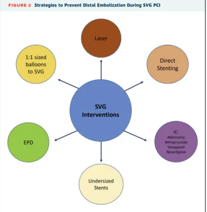
Several strategies can be used to reduce the risk of distal embolization and no reflow (Figure 2). The only strategy that has been tested in randomized controlled trials is use of embolic protection devices. Other strategies include vasodilator administration, direct stenting (22), and use of undersized stents (23).
Nicardipine is often preferred due to prolonged dura- tion of action and less hypotensive effect and is often administered both before and after PCI. Other phar- macological agents such as adenosine, nitroprusside, and verapamil have also shown to prevent or improve no reflow events after intragraft administration (24–31). By contrast, platelet glycoprotein IIb/IIIa re- ceptor inhibitors can cause harm during SVG inter- vention and should not be used routinely in SVG PCI (32). Laser may result in“vaporization”of thrombus and plaque components, potentially reducing the risk for distal embolization; however, it may lead to perforation, especially in highly angulated SVGs(33).
The only currently available embolic protection devices (EPDs) arefilters: the FilterWire (Boston Sci- entific, Natick, Massachusetts) and the Spider (Med- tronic, Santa Rosa, California) (Table 2)(34–40). Both require a distal landing zone for deployment; hence, they cannot be used in distal anastomotic lesions (unless thefilter is deployed in the native coronary artery). The FilterWire is directly advanced through the target SVG lesion, whereas the Spider can be delivered over any guidewire advanced through the
SVG lesion. A proximal occlusion device (Proxis, St.
Jude Medical, Saint Paul, Minnesota) was dis- continued in 2012, and a distal occlusion device (Guardwire, Medtronic) was discontinued in 2017.
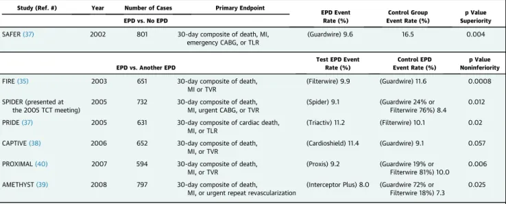
In the first randomized controlled trial of EPD versus no EPD for SVG (SAFER [Saphenous vein graft Angioplasty Free of Emboli Randomized] trial) that randomized 801 patients, use of the Guardwire was associated with lower incidence of MI (8.6% vs.
14.7%; p¼0.008) and“no reflow”(3% vs. 9%; p¼0.02) (36). Given the results of the SAFER trial, subsequent EPD trials in SVG PCI compared one device with another. The FIRE (FilterWire EX Randomized Evaluation) trial compared the FilterWire with the GuardWire in 651 patients undergoing SVG-PCI. Thir- ty-day major adverse cardiac events (MACE) rates were similar between the 2 groups (9.9% of FilterWire EX group vs. 11.6% of GuardWire group; p¼0.0008 for noninferiority)(35). In the SPIDER (Saphenous vein graft Protection In a Distal Embolic protection Ran- domized) trial, the SpideRXfilter was compared with FilterWire and GuardWire in 700 patients and was shown to be noninferior with comparable 30-day MACE rates (9.1% vs. 8.4%; p ¼ 0.01 for non- inferiority)(34). In a pooled analysis of 5 controlled trials and 1 registry evaluating EPDs in SVG-PCI, Coolong et al.(41) showed that the benefit of EPDs for reducing 30-day MACE was consistent across various degrees of SVG degeneration scores.
FIGURE 1 Progression of Coronary Artery Disease in Patients With Prior CABG
(A)Causes of angina in patients with prior CABG.(B)PCI target vessel in prior CABG patients during different time intervals from CABG. Image inBreproduced with permission from Brilakis et al.(2). CABG¼coronary artery bypass graft surgery; NCDR¼National Cardiovascular Data Registry; PCI¼percutaneous coronary intervention; pts¼patients; SVG¼saphenous vein grafts.
CENTRAL ILLUSTRATION Cardiac Catheterization in Patients With Prior CABG: A Systematic Approach
Xenogiannis, I. et al. J Am Coll Cardiol Intv. 2019;12(17):1635–49.
BMS¼bare-metal stents; CABG¼coronary artery bypass graft surgery; DAPT¼dual-antiplatelet therapy; DES¼drug-eluting stents; IMA¼internal mammary artery; POBA¼plain old balloon angioplasty; SVG¼saphenous vein grafts.
Despite the aforementioned trials and the Amer- ican College of Cardiology/American Heart Associa- tion guideline recommendation to use EPDs in SVG PCI when technically feasible (Class I, Level of Evi- dence: B), EPDs remain underused: they were used in only 22% of patients undergoing SVG PCI in the United States(42–44)and in an even lower proportion in other countries, likely due to concerns over cost,
prolongation of the procedure, and lack of expertise in use of those devices. The 2018 European Society of Cardiology/European Association for Cardio-Thoracic Surgery (ESC/EACTS) changed the EPD recommen- dation to Class IIa (Level of Evidence: B) from Class I (Level of Evidence: B)(45), referencing observational studies that did notfind an association between EPD use and improved clinical outcomes(46,47). Howev- er, observational studies comparing EPD use versus no EPD use are subject to significant selection bias (higher risk lesions are more likely to be treated with an EPD), therefore their impact on clinical practice should be limited. Prospective, randomized trials of EPDs in SVG PCI would be optimal, but are unlikely to be performed. EPDs may not be required for in-stent restenotic lesions that have low risk for distal embo- lization(48). Recurrent SVG in-stent restenosis may respond to brachytherapy(49).
STENT SELECTION IN SVG PCI
Saphenous vein graft lesions have high rates of in- stent restenosis, which often presents as an acute coronary syndrome (50–52). Whether drug-eluting stents (DES) improve outcomes compared with bare- metal stents (BMS) in SVG lesions has been examined in 6 prospective randomized trials (Table 3) (50–59). During long-term follow-up, the 2 larger studies showed no difference between DES and BMS (54,56), and another showed worse outcomes with DES (59). In a meta-analysis of the 6 previously mentioned randomized trials by Kheiri et al. (60), there were no significant differences between DES and BMS in the long-term incidence of MACE, target lesion revascularization, target vessel revasculariza- tion, stent thrombosis, and all-cause mortality.
TABLE 1 Published Studies on Bypass Graft Physiological Assessment
First Author (Year) (Ref. #) Number of Patients Objective Major Findings
Aqel et al. (2008)(9) 10 patients with 10 SVG lesions with>50%
stenosis
Access the physiological significance of SVG lesions with FFR
The sensitivity, specificity, PPV, NPV, and accuracy of FFR<0.75 for the detection of ischemia on stress MPI were 50%, 75%, 33%, 85%, and 70%, respectively Di Serafino et al. (2013)(10) 233 patients with CABG and
intermediate graft lesions (venous and arterial)
Compare the outcomes between FFR-guided and angiography-guided PCI
Patients with arterial graft stenosis had lower rates of MACE and TVF in the FFR-guided group. Patients with SVG stenosis had no significant difference for both MACE and TVF between the 2 groups
Almomani et al. (2018)(8) 33 patients with SVG lesions vs. 532 patients with native vessel disease
Compare the prognostic value of deferring intervention in lesions with FFR>0.8 in native coronary artery lesions vs. aortocoronary bypass grafts
MACE and TVF rates were significantly higher in the SVG group vs. the native vessel group (36% vs. 21%; p¼0.01 and 27%
vs. 14%; p¼0.01)
CABG¼coronary artery bypass surgery; FFR¼fractionalflow reserve; MACE¼major adverse cardiac events; MPI¼myocardial perfusion imaging; NPV¼negative predictive value; PCI¼percutaneous intervention; PPV¼positive predictive value; SVG¼saphenous vein graft; TVF¼target vessel failure.
FIGURE 2 Strategies to Prevent Distal Embolization During SVG PCI
EPD¼embolic protection devices; IC¼intracoronary; SVG¼saphenous vein grafts.
Because BMS and DES provide similar outcomes in SVGs, BMS should be preferred in countries with significant difference in the prices of DES and BMS.
There are several potential explanations for the failure of DES to improve outcomes as compared with BMS. First, the pathophysiology and physical history of SVG disease differs from that of native
coronary arteries. Whereas atherosclerosis in coro- nary arteries takes decades to develop, accelerated atherosclerosis is observed in SVGs within months to years, often in a more concentric and diffuse pattern with less well-defined fibrous cap that likely responds differently to DES. Second, neo- atherosclerosis occurs earlier in DES compared with
TABLE 3 Randomized Controlled Trials of DES Versus BMS in SVG Lesions
Study (Ref. #)
Year
Published N Primary Endpoint
Drug-Eluting Stent Event Rate (%)
Bare-Metal Stent
Event Rate (%) p Value
RRISC(52,59) 2006 75 6-month angiographic restenosis 13.6 32.6 0.031
2007 MACE at 32 months 58 41 0.130
SOS(50,55) 2009 80 12-month angiographic restenosis 9 51 <0.001
2010 80 Target vessel failure at 35 months 34 72 0.001
ISAR-CABG(56,57) 2011 610 12-month composite of death, MI, and TLR 15 22 0.02
2018 610 60-month composite of death, MI, and TLR 55.5 53.6 0.89
DIVA(54) 2018 597 12-month composite of cardiac death, target-
vessel MI, and TVR
17 19 0.70
2018 597 2.7-yr median follow-up—composite of cardiac death, target-vessel MI, and TVR
37 34 0.44
ADEPT(53) 2018 57 Late lumen loss at 6 months 0.470.95 mm 0.531.09 mm 0.86
Presented
BASKET-SAVAGE* 2016 173 12-month composite of cardiac death, MI, and TVR
2.3 17.9 <0.001
BASKET-SAVAGE* 2016 173 36-month composite of cardiac death, MI, and TVR
12.4 29.8 0.0012
*Presented at the 2016 European Society of Cardiology meeting (Rome, Italy, August 30, 2016).
ADEPT¼Comparison between the STENTYS self-apposing bare metal and paclitaxel-eluting coronary stents for the treatment of saphenous vein grafts; BASKET-SAVAGE¼Basel Kosten Effektivitäts Trial–
SAphenous Venous Graft Angioplasty Using Glycoprotein 2b/3a Receptor Inhibitors and Drug-Eluting Stents trial; BMS¼bare-metal stents; DES¼drug-eluting stents; DIVA¼Drug-Eluting Stents vs. Bare Metal Stents In Saphenous Vein graft Angioplasty; ISAR-CABG¼Is Drug-Eluting-Stenting Associated with Improved Results in Coronary Artery Bypass Grafts? trial; RRISC¼Reduction of Restenosis In Saphenous vein grafts with Cyphersirolimus-eluting stent trial; SOS¼Stenting Of Saphenous vein grafts trial; other abbreviations as inTables 1 and 2.
TABLE 2 Published Trials of EPDs in SVG Interventions
Study (Ref. #) Year Number of Cases Primary Endpoint
EPD Event Rate (%)
Control Group Event Rate (%)
p Value Superiority EPD vs. No EPD
SAFER(37) 2002 801 30-day composite of death, MI,
emergency CABG, or TLR
(Guardwire) 9.6 16.5 0.004
EPD vs. Another EPD
Test EPD Event Rate (%)
Control EPD Event Rate (%)
p Value Noninferiority
FIRE(35) 2003 651 30-day composite of death,
MI or TVR
(Filterwire) 9.9 (Guardwire) 11.6 0.0008 SPIDER (presented at
the 2005 TCT meeting)
2005 732 30-day composite of death,
MI, urgent CABG, or TVR
(Spider) 9.1 (Guardwire 24% or Filterwire 76%) 8.4
0.012
PRIDE(37) 2005 631 30-day composite of cardiac death,
MI, or TLR
(Triactiv) 11.2 (Filterwire) 10.1 0.02
CAPTIVE(38) 2006 652 30-day composite of death,
MI, or TVR
(Cardioshield) 11.4 (Guardwire) 9.1 0.057
PROXIMAL(40) 2007 594 30-day composite of death,
MI, or TVR
(Proxis) 9.2 (Guardwire 19% or Filterwire 81%) 10.0
0.006
AMETHYST(39) 2008 797 30-day composite of death,
MI, or urgent repeat revascularization
(Interceptor Plus) 8.0 (Guardwire 72% or Filterwire 18%) 7.3
0.025
Triactiv is manufactured by Kensey Nash Corp. (West Whiteland Township, Pennsylvania), Cardioshield by MedNova (Galway, Ireland), and Interceptor Plus by Medtronic; other devices as in the text.
AMETHYST¼Assessment of the Medtronic AVE Interceptor Saphenous Vein Graft Filter System; CAPTIVE¼CardioShield Application Protects during Transluminal Intervention of Vein grafts by reducing Emboli; EPD¼embolic protection device; FIRE¼FilterWire EX Randomized Evaluation; MI¼myocardial infarction; PRIDE¼Protection During Saphenous Vein Graft Intervention to Prevent Distal Embolization; PROXIMAL¼Proximal Protection During Saphenous Vein Graft Intervention; SAFER¼Saphenous vein graft Angioplasty Free of Emboli Randomized; SPIDER¼Saphenous Vein Graft Protection In a Distal Embolic Protection Randomized Trial; TLR¼target lesion revascularization; TVR¼target vessel revascularization; other abbreviations as inTable 1.
BMS, which may lead to a catch-up phenomenon (61,62). Third, thin-strut BMS may have lower risk for restenosis in SVGs than thicker strut stents that were used in most prior studies. Fourth, most DES versus BMS studies had mandatory angiography follow-up and were not blinded(50,52,53,57), which may bias outcomes in favor of DES (oculostenotic reflex). The DIVA (Drug-Eluting Stents vs. Bare Metal Stents In Saphenous Vein Graft Angioplasty) trial, the more recent randomized controlled trial that demonstrated no benefit of DES over BMS used blinding and did not mandate routine angiographic follow-up(54).
EARLY POST-OPERATIVE GRAFT FAILURE, ACUTE AND CHRONIC TOTAL SVG OCCLUSIONS
Graft failure during the early post-operative period occurs in up to 12% of the grafts, with approximately 3% of the patients developing symptoms(63). Graft occlusion rates are higher for vein grafts (3% to 12%
before discharge) compared with radial artery (3% to 4%) and internal mammary artery (IMA) (1% to 2.5%) grafts(64). Potential causes include conduit defects, suboptimal anastomosis technique, poor native vessel runoff, and competitive flow with the native vessel.
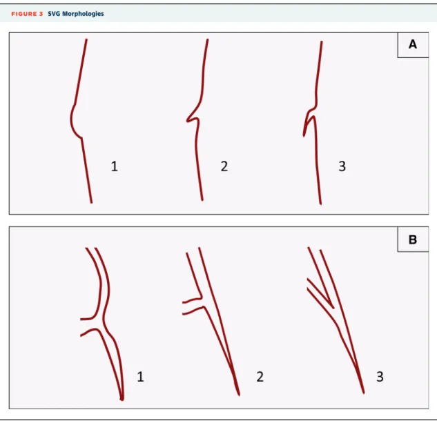
FIGURE 3 SVG Morphologies
(A)Anatomic variants of the proximal cap of occluded saphenous vein grafts (SVGs): retrograde crossing should not be attempted in morphology #1 due to increased risk for perforation.(B)Anatomic variants of bypass graft distal anastomoses. Retrograde guidewire and equipment crossing is more likely to be challenging for morphology #3.
Graft patency is higher when anastomosed to highly stenosed native coronary arteries: in a study of 164 patients who underwent pre-CABG FFR, graft occlusion at coronary angiography after 1 year was 8.9% for bypass grafts on functionally significant le- sions (FFR<0.75) versus 21.4% for bypass grafts on lesions with FFR$0.75(65). Conversely, there is an accelerated rate of disease progression in bypassed native coronary vessels, especially for non-left ante- rior descending artery vessels and when SVGs are used as compared with arterial grafts(66).
Unless the diagnosis of acute graft failure is made in the operating room, PCI is preferred for symp- tomatic graft failure. PCI is best performed in the corresponding native coronary artery instead of the bypass graft, if possible, in part because PCI of the graft anastomosis may lead to suture dehiscence and perforation(45). Redo CABG is recommended when coronary anatomy is not suitable for PCI, a large ter- ritory of myocardium is under jeopardy, multiple significant grafts are occluded, or in case of anasto- motic lesions(46,67).
Acute SVG occlusions carry a high risk for short- and long-term adverse outcomes. Welsh et al. (68) demonstrated that prior CABG patients presenting
with an ST-segment elevation myocardial infarction had similar outcomes compared with patients without prior CABG when the infarct-related artery was a native coronary artery; however, 90-day mor- tality was much higher in prior CABG patients whose culprit vessel was a SVG (19% vs. 5.7%; p ¼ 0.05).
Thrombosed SVGs often have large thrombotic burden which can be approached with thrombectomy and use of embolic protection devices(69). Aspiration thrombectomy is preferred over rheolytic thrombec- tomy to minimize the risk for distal embolization and adverse outcomes(70). Suction should be maintained until removal of the aspiration thrombectomy cath- eter from the guide catheter for optimal thrombus retrieval and reduction of the systemic thromboem- bolism risk. Occasionally, aspiration through a deeply intubated guide catheter or guide catheter extension (balloon-assisted deep intubation—BADI) may be required for retrieval of very large thrombi. Use of laser is another option for such patients, whereas thrombolytic administration has been associated with poor outcomes and is generally avoided(71).
ST-segment elevation myocardial infarction due to SVG obstruction can be very challenging to treat.
Given the suboptimal results of thrombolytic therapy
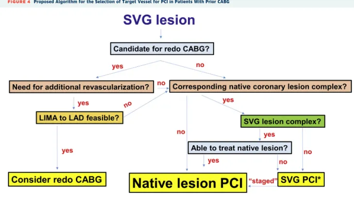
FIGURE 4 Proposed Algorithm for the Selection of Target Vessel for PCI in Patients With Prior CABG
LAD¼left anterior descending coronary artery; LIMA¼left internal mammary artery; other abbreviations as inFigures 1 and 2.
in occluded SVGs, PCI is the preferred reperfusion modality (71). Sometimes, identifying and engaging the grafts can be challenging, often requiring multiple catheters or aortography that may delay reperfusion (72). Use of embolic protection may be useful in such cases, although irreversible injury may have already occurred. SVG lesions are highly friable and rich in thrombus, and carry high risk for no reflow. Throm- bolysis In Myocardial Infarction (TIMI)flow grade 3 post-PCI is achieved less frequently in patients who had SVG as the culprit vessel compared with patients who had a native coronary artery as the culprit vessel, and such patients had higher in-hospital and 30-day mortality and 1-year MACE rates(73,74).
Because of high risk for restenosis, SVG CTOs should generally not be recanalized (Class III indica- tion, Level of Evidence: C) (42), unless no other treatment options exist. Occluded SVGs can be used, however, for retrograde crossing of the corresponding native coronary artery if the occlusion morphology is favorable (Figure 3).
LEFT INTERNAL MAMMARY ARTERY AND ARTERIAL GRAFT PERCUTANEOUS CORONARY INTERVENTION
PCI of arterial grafts and especially of the LIMA is much less common than SVG PCI. The 2 main reasons are the better rates of LIMA patency over SVGs and
possibly performance of redo CABG in cases of LIMA failure (Figure 4). Redo CABG is generally avoided in patients with a patent IMA graft to the left anterior descending coronary artery(75).
There should be high threshold for performing PCI through IMA grafts, given the high risk for ischemia and complications. Straightening of a tortuous LIMA during the advancement of guidewires and micro- catheters may lead to pseudolesions (the so-called
“accordioning effect”), that can lead to flow compromise and ischemia. Pseudolesions must be differentiated from vasospasm and dissection.
Administration of intravenous vasodilators will be ineffective in the presence of the pseudolesions, which should correct with guidewire withdrawal(76).
Deep guide intubation and use of guide catheter ex- tensions may lead to IMA dissection and/or perfora- tion(77,78). Despite the aforementioned limitations, PCI to IMA lesions has been associated with higher rates of restoration of TIMI flow grade 3 and lower rates of periprocedural complications compared with SVG PCI (79). IMA anastomotic lesions may be best treated with balloon angioplasty, whereas proximal and mid-segment lesions are stented in most cases (80). Gruberg et al.(81) analyzed 174 patients who underwent PCI of 128 IMA anastomotic lesions and found a higher need for repeat revascularization after stenting (33%) as compared with balloon angioplasty only (4.3%). Sharma et al. (82) also reported worse
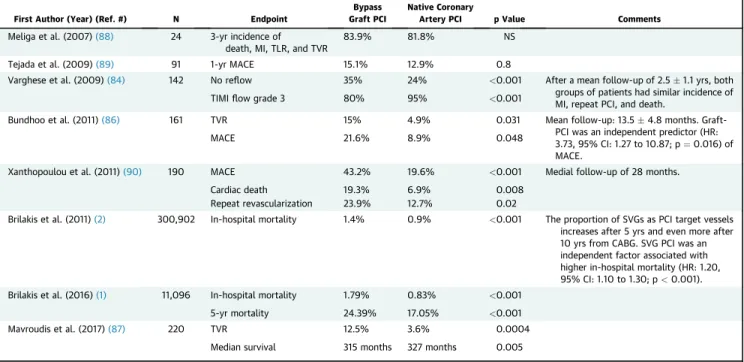
TABLE 4 Major Studies Comparing Bypass Graft Versus Native Coronary Artery PCI
First Author (Year) (Ref. #) N Endpoint
Bypass Graft PCI
Native Coronary
Artery PCI p Value Comments
Meliga et al. (2007)(88) 24 3-yr incidence of death, MI, TLR, and TVR
83.9% 81.8% NS
Tejada et al. (2009)(89) 91 1-yr MACE 15.1% 12.9% 0.8
Varghese et al. (2009)(84) 142 No reflow 35% 24% <0.001 After a mean follow-up of 2.51.1 yrs, both
groups of patients had similar incidence of MI, repeat PCI, and death.
TIMIflow grade 3 80% 95% <0.001
Bundhoo et al. (2011)(86) 161 TVR 15% 4.9% 0.031 Mean follow-up: 13.54.8 months. Graft-
PCI was an independent predictor (HR:
3.73, 95% CI: 1.27 to 10.87; p¼0.016) of MACE.
MACE 21.6% 8.9% 0.048
Xanthopoulou et al. (2011)(90) 190 MACE 43.2% 19.6% <0.001 Medial follow-up of 28 months.
Cardiac death 19.3% 6.9% 0.008
Repeat revascularization 23.9% 12.7% 0.02
Brilakis et al. (2011)(2) 300,902 In-hospital mortality 1.4% 0.9% <0.001 The proportion of SVGs as PCI target vessels increases after 5 yrs and even more after 10 yrs from CABG. SVG PCI was an independent factor associated with higher in-hospital mortality (HR: 1.20, 95% CI: 1.10 to 1.30; p<0.001).
Brilakis et al. (2016)(1) 11,096 In-hospital mortality 1.79% 0.83% <0.001
5-yr mortality 24.39% 17.05% <0.001
Mavroudis et al. (2017)(87) 220 TVR 12.5% 3.6% 0.0004
Median survival 315 months 327 months 0.005
CI¼confidence interval; HR¼hazard ratio; NS¼nonsignificant; other abbreviations as inTables 1, 2, and 3.
outcomes with stenting compared with angioplasty alone at the anastomotic site (25% vs. 4.2%; p ¼ 0.006) in 288 patients with 311 IMA lesions.
In patients with IMA grafts, the proximal subcla- vian artery should be evaluated, because severe le- sions in this location could lead to coronary ischemia and even acute coronary syndromes(83). Subclavian artery stenting can be an effective treatment in such cases.
NATIVE CORONARY ARTERY PCI IN PRIOR CABG PATIENTS
Most PCIs (approximately two-thirds) performed in prior CABG patients are in native coronary artery le- sions(2,84). Native coronary artery lesions in prior CABG patients are often complex, with high rates of calcification, tortuosity, and CTOs. The bypass grafts can often be used for retrograde crossing in such pa- tients, although wiring upstream from the distal anastomosis can be challenging (Figure 3). Shortened guiding catheters are especially recommended for PCI of the distal native vessels through the IMA and also retrograde techniques. Advanced PCI techniques, such as use of atherectomy and CTO PCI are, there- fore, often needed (85). Nevertheless, outcomes after native coronary artery PCI are better than out- comes post-SVG PCI in multiple series (Table 4) (1,2,84,86–90).
In patients presenting with SVG lesions, several operators advocate treating the native coronary ar- tery instead, given the high short- and long-term risks of SVG PCI. The 2018 ESC/EACTS guidelines on myocardial revascularization state that PCI to a native vessel should be preferred over PCI of the bypass graft (Class IIa, Level of Evidence: C) (45). Decision making can be challenging, however, as the corre- sponding native coronary artery lesions are often complex to treat or even totally occluded (often CTOs). One approach is to treat the native coronary artery when it is simple or when both SVG PCI and native coronary PCI are complex, and there is local expertise in treating such lesions (Figure 4). This could also be done in a staged manner: the culprit SVG lesion is initially treated (especially for patients presenting with acute coronary syndromes who have complex native coronary artery lesions), followed by PCI of the native coronary artery weeks or months later(91). If the thrombosed SVG cannot be recanal- ized, PCI of the native coronary artery can sometimes be performed (92). Because SVGs that become occluded due to thrombus have very high rates of reocclusion, staged PCI of the corresponding native
coronary artery should be considered after the initial procedure. In such cases, stenting the distal SVG anastomosis should be avoided, if possible, as it could hinder subsequent treatment of the native coronary vessel. This conceptually appealing approach will need to be validated in clinical studies.
Remainingflow in the SVG after successful native vessel PCI has been a source of concern because competitive flow from the SVG can lead to native stent thrombosis (93,94). Some operators advocate routine SVG coiling after treating the native coronary artery although robust data are missing(95).
COMPLICATIONS
Due to the need to engage and visualize the bypass grafts (and the often high complexity of treated le- sions) angiography and PCI in prior CABG patients requires longer procedural and fluoroscopy time, higher radiation dose, and larger volume of contrast (96–98). As a result (and also because of worse base- line renal function), the risk for contrast nephropathy and possibly hemodialysis is increased in those pa- tients who have had prior CABG compared with those who have not(93,97,99).
Even though coronary perforations were previ- ously considered to be “innocent” complications in prior CABG patients due to pericardial adhesions preventing formation of a pericardial effusion and tamponade, it is now appreciated that they can be lethal events. Coronary perforation in prior CABG patients can lead to loculated hematomas resulting in cardiac chamber compromise and hemodynamic collapse (dry tamponade). In the OPEN-CTO (Out- comes, Patient Health Status, and Efficiency in Chronic Total Occlusion Hybrid Procedures) database, the perforation rate in post-CABG patients was approximately 7%. Four perforations that led to death occurred in the 365 patients with prior CABG (1.1%) (100). Such loculated effusions may require surgery or computed tomography-guided drainage for treatment. Prompt identification and treatment of coronary or graft perforation is, therefore, critical in prior CABG patients(101,102).
POST-PROCEDURAL ANTITHROMBOTIC THERAPY
Long-term dual-antiplatelet therapy (DAPT) is conceptually appealing in prior CABG patients, as they often have extensive, multilevel atherosclerotic disease and high risk for subsequent adverse cardio- vascular events. In a meta-analysis of 22 studies comparing DAPT to aspirin alone following CABG,
DAPT was associated with lower cardiovascular mor- tality (odds ratio [OR]: 0.67; p ¼0.02) and a trend toward lower all-cause mortality (OR: 0.78; p¼0.08), although there was no difference when the analysis was confined to randomized controlled trials. SVG occlusion up to 1 year after CABG was significantly lower with DAPT overall (OR: 0.64; p< 0.01) and in the subset of randomized controlled trials (OR: 0.58;
p < 0.01). Importantly, patients who were treated with DAPT for>6 months had lower stroke rates (OR:
0.47; p¼0.04) but higher incidence of major bleeding (OR: 1.31; p¼0.03)(103).
In another meta-analysis of 9 randomized controlled trials, patients who received ticagrelor or prasugrel in addition to aspirin had lower mortality compared with patients taking clopidogrel and aspirin (relative risk: 0.49; 95% confidence interval [CI]: 0.33 to 0.71; p¼0.0002), whereas there was no significant difference when clopidogrel plus aspirin was compared with aspirin monotherapy(104). In a subanalysis of the PLATO (Platelet Inhibition and Patient Outcomes) trial, the reduction of the primary endpoint of cardiovascular death, MI, and stroke was not statistically significant in post-CABG patients (19.6% vs. 21.4%; adjusted hazard ratio: 0.91 [inter- quartile range: 0.67 to 1.24])(105). Large, randomized trials are needed in order to clarify the usefulness of DAPT in different clinical settings (acute coronary syndromes vs. stable coronary artery disease) and the optimal antiplatelet combination.
In a study of 603 patients who underwent SVG PCI, those taking clopidogrel in addition to aspirin for more than 2 years had lower rates of MI or death during a 5-year follow-up after the cessation of clo- pidogrel, compared with patients who were taking clopidogrel for a shorter time period (106). In the
DAPT (Dual Antiplatelet Therapy) study, patients who underwent SVG PCI had better outcomes with 30- month versus 12-month DAPT(107,108). Administra- tion of DAPT for a longer duration than is usually recommended after native vessel PCI (generally 6 months for stable coronary disease and 1 year for acute coronary events) in patients undergoing SVG PCI, therefore, may be beneficial.
Whether anticoagulation can reduce bypass graft failure and improve clinical outcomes after CABG re- mains controversial. In a substudy of COMPASS (Cardiovascular OutcoMes for People Using Anti- coagulation StrategieS) trial, the combination of 2.5 mg of rivaroxaban twice per day with aspirin did not reduce the incidence of graft failure in patients with prior CABG compared with aspirin administra- tion alone (113 [9.1%] vs. 91 [8.0%]; OR: 1.13; 95% CI:
0.82 to 1.57; p ¼ 0.45). It also did not reduce the composite endpoint of cardiovascular death, MI, and stroke (12 [2.4%] vs. 16 [3.5%], hazard ratio: 0.69;
95% CI: 0.33 to 1.47; p¼0.34)(109).
CONCLUSIONS
Prior CABG patients undergoing cardiac catheteriza- tion have increased risk for complications and often require complex procedures. Several new studies have advanced our understanding of the optimal approach to cardiac catheterization and PCI in these high-risk patients.
ADDRESS FOR CORRESPONDENCE: Dr. Emmanouil S. Brilakis, Minneapolis Heart Institute, 920 East 28th Street #300, Minneapolis, Minnesota 55407. E-mail:
esbrilakis@gmail.com.
R E F E R E N C E S
1.Brilakis ES, O’Donnell CI, Penny W, et al.
Percutaneous coronary intervention in native cor- onary arteries versus bypass grafts in patients with prior coronary artery bypass graft surgery: insights from the veterans affairs clinical assessment, reporting, and tracking program. J Am Coll Cardiol Intv 2016;9:884–93.
2.Brilakis ES, Rao SV, Banerjee S, et al. Percuta- neous coronary intervention in native arteries versus bypass grafts in prior coronary artery bypass grafting patients: a report from the Na- tional Cardiovascular Data Registry. J Am Coll Cardiol Intv 2011;4:844–50.
3.Rigattieri S, Sciahbasi A, Brilakis ES, et al. Meta- analysis of radial versus femoral artery approach for coronary procedures in patients with previous coronary artery bypass grafting. Am J Cardiol 2016;117:1248–55.
4.Michael TT, Alomar M, Papayannis A, et al.
A randomized comparison of the transradial and transfemoral approaches for coronary artery bypass graft angiography and intervention: the RADIAL- CABG trial (RADIAL Versus Femoral Access for Coronary Artery Bypass Graft Angiography and Intervention). J Am Coll Cardiol Intv 2013;6:1138–44.
5.Bundhoo SS, Earp E, Ivanauskiene T, et al.
Saphenous vein graft percutaneous coronary intervention via radial artery access: safe and effective with reduced hospital length of stay. Am Heart J 2012;164:468–72.
6.Rathore S, Roberts E, Hakeem AR, Pauriah M, Beaumont A, Morris JL. The feasibility of percu- taneous transradial coronary intervention for saphenous vein graft lesions and comparison with transfemoral route. J Intervent Cardiol 2009;22:
336–40.
7.Cooper L, Banerjee S, Brilakis ES. Crossover from radial to femoral access during a challenging percutaneous coronary intervention can make the difference between success and failure. Cardiovasc Revasc Med 2010;11:266. e5–8.
8.Almomani A, Pothineni NV, Edupuganti M, et al.
Outcomes of fractional flow reserve-based deferral in saphenous vein graft narrowing. Am J Cardiol 2018;122:723–8.
9.Aqel R, Zoghbi GJ, Hage F, Dell’Italia L, Iskandrian AE. Hemodynamic evaluation of coro- nary artery bypass graft lesions using fractional flow reserve. Catheter Cardiovasc Interv 2008;72:
479–85.
10.Di Serafino L, De Bruyne B, Mangiacapra F, et al. Long-term clinical outcome after fractional flow reserve- versus angio-guided percutaneous coronary intervention in patients with
intermediate stenosis of coronary artery bypass grafts. Am Heart J 2013;166:110–8.
11.Abdel-Karim AR, Da Silva M, Lichtenwalter C, et al. Prevalence and outcomes of intermediate saphenous vein graft lesions:findings from the stenting of saphenous vein grafts randomized- controlled trial. Int J Cardiol 2013;168:2468–73.
12.Rodes-Cabau J, Bertrand OF, Larose E, et al.
Comparison of plaque sealing with paclitaxel- eluting stents versus medical therapy for the treatment of moderate nonsignificant saphenous vein graft lesions: the moderate vein graft lesion stenting with the Taxus stent and intravascular ultrasound (VELETI) pilot trial. Circulation 2009;
120:1978–86.
13.Rodes-Cabau J, Bertrand OF, Larose E, et al.
Five-year follow-up of the plaque sealing with paclitaxel-eluting stents vs medical therapy for the treatment of intermediate nonobstructive saphenous vein graft lesions (VELETI) trial. Can J Cardiol 2014;30:138–45.
14.Rodes-Cabau J, Jolly SS, Cairns J, et al. Sealing intermediate nonobstructive coronary saphenous vein graft lesions with drug-eluting stents as a new approach to reducing cardiac events: a ran- domized controlled trial. Circ Cardiovasc Interv 2016;9:e004336.
15.Collaborative overview of randomised trials of antiplatelet therapy–II: maintenance of vascular graft or arterial patency by antiplatelet therapy.
Antiplatelet Trialists’ Collaboration. BMJ 1994;
308:159–68.
16.Goldman S, Copeland J, Moritz T, et al.
Saphenous vein graft patency 1 year after coro- nary artery bypass surgery and effects of anti- platelet therapy. Results of a Veterans Administration Cooperative Study. Circulation 1989;80:1190–7.
17.Kulik A. Secondary prevention after coronary artery bypass graft surgery: a primer. Curr Opin Cardiol 2016;31:635–43.
18.Okrainec K, Platt R, Pilote L, Eisenberg MJ.
Cardiac medical therapy in patients after under- going coronary artery bypass graft surgery: a re- view of randomized controlled trials. J Am Coll Cardiol 2005;45:177–84.
19.Dehmer GJ, Weaver D, Roe MT, et al.
A contemporary view of diagnostic cardiac cathe- terization and percutaneous coronary intervention in the United States: a report from the CathPCI Registry of the National Cardiovascular Data Registry, 2010 through June 2011. J Am Coll Cardiol 2012;60:2017–31.
20.Lee KW, Norell MS. Management of ’no- reflow’complicating reperfusion therapy. Acute Card Care 2008;10:5–14.
21.Hong YJ, Jeong MH, Ahn Y, et al. Intravascular ultrasound findings that are predictive of no reflow after percutaneous coronary intervention for saphenous vein graft disease. Am J Cardiol 2012;109:1576–81.
22.Leborgne L, Cheneau E, Pichard A, et al. Effect of direct stenting on clinical outcome in patients treated with percutaneous coronary intervention on saphenous vein graft. Am Heart J 2003;146:
501–6.
23.Hong YJ, Pichard AD, Mintz GS, et al. Outcome of undersized drug-eluting stents for percuta- neous coronary intervention of saphenous vein graft lesions. Am J Cardiol 2010;105:179–85.
24.Grygier M, Araszkiewicz A, Lesiak M, Grajek S.
Intracoronary adenosine administered during aor- tocoronary vein graft interventions may reduce the incidence of no-reflow phenomenon. A pilot randomised trial. Kardiol Pol 2014;72:126–33.
25.Hillegass WB, Dean NA, Liao L, Rhinehart RG, Myers PR. Treatment of no-reflow and impaired flow with the nitric oxide donor nitroprusside following percutaneous coronary interventions:
initial human clinical experience. J Am Coll Cardiol 2001;37:1335–43.
26.Kaplan BM, Benzuly KH, Kinn JW, et al.
Treatment of no-reflow in degenerated saphenous vein graft interventions: comparison of intra- coronary verapamil and nitroglycerin. Cathet Car- diovasc Diagn 1996;39:113–8.
27.Kapoor N, Yalamanchili V, Siddiqui T, Raza S, Leesar MA. Cardioprotective effect of high-dose intragraft adenosine infusion on microvascular function and prevention of no-reflow during saphenous vein grafts intervention. Catheter Car- diovasc Interv 2014;83:1045–54.
28.Michaels AD, Appleby M, Otten MH, et al.
Pretreatment with intragraft verapamil prior to percutaneous coronary intervention of saphenous vein graft lesions: results of the randomized, controlled vasodilator prevention on no-reflow (VAPOR) trial. J Invasive Cardiol 2002;14:
299–302.
29.Sdringola S, Assali A, Ghani M, et al. Adenosine use during aortocoronary vein graft interventions reverses but does not prevent the slow-no-reflow phenomenon. Catheter Cardiovasc Interv 2000;
51:394–9.
30.Sharma S, Lardizabal JA, Singh S, Sandhu R, Bhambi BK. Intra-graft abciximab and verapamil combined with direct stenting is a safe and effective strategy to prevent slow-flow and no- reflow phenomenon in saphenous vein graft le- sions not associated with thrombus. Recent Pat Cardiovasc Drug Discov 2012;7:152–9.
31.Zoghbi GJ, Goyal M, Hage F, et al. Pretreat- ment with nitroprusside for microcirculatory pro- tection in saphenous vein graft interventions.
J Invasive Cardiol 2009;21:34–9.
32.RoffiM, Mukherjee D, Chew DP, et al. Lack of benefit from intravenous platelet glycoprotein IIb/
IIIa receptor inhibition as adjunctive treatment for percutaneous interventions of aortocoronary bypass grafts: a pooled analysis of 5 randomized clinical trials. Circulation 2002;106:3063–7.
33.Niccoli G, Belloni F, Cosentino N, et al. Case- control registry of excimer laser coronary angio- plasty versus distal protection devices in patients with acute coronary syndromes due to saphenous vein graft disease. Am J Cardiol 2013;112:1586–91.
34.Dixon S. Saphenous vein graft protection in a distal embolic protection randomized trial. Paper presented at: Transcatheter Cardiovascular Ther- apeutics; October 17-21, 2005; Washington, DC.
35.Stone GW, Rogers C, Hermiller J, et al. Ran- domized comparison of distal protection with a filter-based catheter and a balloon occlusion and
aspiration system during percutaneous interven- tion of diseased saphenous vein aorto-coronary bypass grafts. Circulation 2003;108:548–53.
36.Baim DS, Wahr D, George B, et al. Randomized trial of a distal embolic protection device during percutaneous intervention of saphenous vein aorto-coronary bypass grafts. Circulation 2002;
105:1285–90.
37.Carrozza JP Jr, Mumma M, Breall JA, Fernandez A, Heyman E, Metzger C. Randomized evaluation of the TriActiv balloon-protectionflush and extraction system for the treatment of saphenous vein graft disease. J Am Coll Cardiol 2005;46:1677–83.
38.Holmes DR, Coolong A, O’Shaughnessy C, et al. Comparison of the CardioShieldfilter with the guardwire balloon in the prevention of em- bolisation during vein graft intervention: results from the CAPTIVE randomised trial. Euro- Intervention 2006;2:161–8.
39.Kereiakes DJ, Turco MA, Breall J, et al. A novel filter-based distal embolic protection device for percutaneous intervention of saphenous vein graft lesions: results of the AMEthyst randomized controlled trial. J Am Coll Cardiol Intv 2008;1:
248–57.
40.Mauri L, Cox D, Hermiller J, et al. The PROX- IMAL trial: proximal protection during saphenous vein graft intervention using the Proxis Embolic Protection System: a randomized, prospective, multicenter clinical trial. J Am Coll Cardiol 2007;
50:1442–9.
41.Coolong A, Baim DS, Kuntz RE, et al. Saphe- nous vein graft stenting and major adverse cardiac events: a predictive model derived from a pooled analysis of 3958 patients. Circulation 2008;117:
790–7.
42.Levine GN, Bates ER, Blankenship JC, et al.
2011 ACCF/AHA/SCAI guideline for percutaneous coronary intervention: a report of the American College of Cardiology Foundation/American Heart Association Task Force on Practice Guidelines and the Society for Cardiovascular Angiography and Interventions. J Am Coll Cardiol 2011;58:e44–122.
43.Lee M, Kong J. Current state of the art in ap- proaches to saphenous vein graft interventions.
Interv Cardiol 2017;12:85–91.
44.Mehta SK, Frutkin AD, Milford-Beland S, et al.
Utilization of distal embolic protection in saphe- nous vein graft interventions (an analysis of 19, 546 patients in the American College of Cardiology-National Cardiovascular Data Regis- try). Am J Cardiol 2007;100:1114–8.
45.Neumann FJ, Sousa-Uva M, Ahlsson A, et al.
2018 ESC/EACTS guidelines on myocardial revas- cularization. Eur Heart J 2019;40:87–165.
46.Brennan JM, Al-Hejily W, Dai D, et al. Three- year outcomes associated with embolic protection in saphenous vein graft intervention: results in 49 325 senior patients in the Medicare-linked Na- tional Cardiovascular Data Registry CathPCI Reg- istry. Circ Cardiovasc Interv 2015;8:e001403.
47.Paul TK, Bhatheja S, Panchal HB, et al. Out- comes of saphenous vein graft intervention with and without embolic protection device: a comprehensive review and meta-analysis. Circ Cardiovasc Interv 2017;10:e005538.