Synthesis and Cell Growth Inhibitory Activity of Six Non- glycosaminoglycan-Type Heparin-Analogue Trisaccharides
Erika Lisztes,
[a]Erika Mező,
[b]Fruzsina Demeter,
[b, c, d]Lilla Horváth,
[e]Szilvia Bősze,
[e]Balázs István Tóth,
[a]Anikó Borbás,*
[b]and Mihály Herczeg*
[b, f]The design and synthesis of heparin mimetics with high anticancer activity but no anticoagulant activity is an important task in medicinal chemistry. Herein, we present the efficient synthesis of five Glc-GlcA-Glc-sequenced and one Glc-IdoA-Glc- sequenced non-glycosaminoglycan, heparin-related trisacchar- ides with various sulfation/sulfonylation and methylation patterns. The cell growth inhibitory effects of the compounds were tested against four cancerous human cell lines and two
non-cancerous cell lines. Twod-glucuronate-containing tetra-O- sulfated, partially methylated trisaccharides displayed remark- able and selective inhibitory effects on the growth of ovary carcinoma (A2780) and melanoma (WM35) cells. Methyl sub- stituents on the glucuronide unit proved to be detrimental, whereas acetyl substituents were beneficial to the cytostatic activity of the sulfated derivatives.
Introduction
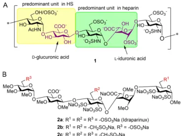
Heparin and heparan sulfate (HS) are linear anionic polysacchar- ides belonging to the family of glycosaminoglycans. Although both are composed of alternatingα-d-glucosamine and hexur- onic acid (β-d-glucuronic acid,β-d-GlcA, orα-l-iduronic acid,α- l-IdoA) units, there are some structural differences between their saccharide sequences and sulfation degree (Figure 1A).
Whereas heparin predominantly consists of the trisulfated l- IdoA-containing disaccharide (highlighted in green in structure 1), the major repeating unit of HS is a d-GlcA-containing disaccharide with a lower sulfation degree (highlighted in
yellow in structure1).[1]The most well-known activity of heparin is the anticoagulant effect which is based on the specific interaction of a unique pentasaccharide domain of heparin with the endogenous coagulation inhibitor antithrombin.[2]
Beyond antithrombin, a large number of proteins, such as growth factors, cytokines, enzymes, membrane receptors as well as viral proteins can interact with both heparin and HS.[3]
Consequently, heparin and HS have many biological effects[4–10]
such as anti-inflammatory,[5] cardiovascular and tissue protection,[5]kidney and nerve protection,[6]angiogenic,[7]meta- stasis and growth factor inhibitory[8]as well as anti-protozoan[9]
and antiviral[10] activity which can be translated to therapeutic application.
The HS/heparin-protein interactions are dominated by charge-charge interactions between the anionic carboxylate [a] Dr. E. Lisztes, Dr. B. István Tóth
Department of Physiology, University of Debrecen PO Box 22, 4012 Debrecen, (Hungary)
[b] Dr. E. Mező, F. Demeter, Prof. A. Borbás, Dr. M. Herczeg Department of Pharmaceutical Chemistry, University of Debrecen Egyetem tér 1, 4032 Debrecen (Hungary)
E-mail: borbas.aniko@pharm.unideb.hu herczeg.mihaly@science.unideb.hu [c] F. Demeter
Doctoral School of Chemistry, University of Debrecen Egyetem tér 1, 4032 Debrecen (Hungary)
[d] F. Demeter
MTA-DE Molecular Recognition and Interaction Research Group, ELKH University of Debrecen
Egyetem tér 1, 4032 Debrecen, Hungary [e] L. Horváth, Dr. Sz. Bősze
MTA-ELTE Research Group of Peptide Chemistry Eötvös Loránd University
Pázmány Péter sétány 1/a, 1117 Budapest (Hungary) [f] Dr. M. Herczeg
MTA-DE Research Group for Oligosaccharide Chemistry, ELKH Egyetem tér 1, 4032 Debrecen (Hungary)
E-mail: herczeg.mihaly@science.unideb.hu
Supporting information for this article is available on the WWW under https://doi.org/10.1002/cmdc.202000917
© 2021 The Authors. ChemMedChem published by Wiley-VCH GmbH. This is an open access article under the terms of the Creative Commons Attribution Non-Commercial NoDerivs License, which permits use and distribution in any medium, provided the original work is properly cited, the use is non- commercial and no modifications or adaptations are made.
Figure 1.A) Representative pentasaccharide unit of heparin/heparan sulfate (HS;1), highlighting the predominant disaccharide repeating units. B) Synthetic non-glycosaminoglycan-type pentasaccharides (2 a-c) with anti- coagulant activity
and sulfate group of the polysaccharide and basic amino acids of the proteins, and, importantly, heparin, due to its higher sulfation degree, can outcompete HS for binding to protein ligands. Indeed, heparin and its derivatives are being inves- tigated for the treatment of a number of disorders, including cancer.[11,12]
Polysulfated oligosaccharides (malto- and isomaltooligom- ers, oligomannuronates) have also been known to be effective in inhibiting the growth, angiogenesis and migration of cancer cells.[13–16]
Based on the above, non-glycosaminoglycan analogues of heparin might be important structures in the development of anticancer agents. Our research group has long been working on the synthesis of heparin-analogue oligosaccharides. Several pathways have been developed to prepare the non-glycosami- noglycan, fully sulfated and fully methylated heparinoid anti- coagulant pentasaccharide idraparinux2 a,[17–20]and its sulfonic acid derivatives 2 b and2 c (Figure 1) in which several sulfate esters were substituted by sulfonatomethyl moieties to improve the binding affinities.[20,21,22] We envisaged that trisaccharides fragments of these highly sulfated/sulfonylated pentasacchar- ides (3–8, Figure 2) might bind to the protein ligands, for example, heparanase, with charge–charge interactions and might display cell growth inhibitory activity. Moreover, advanta- geously, such smaller oligosaccharides lack anticoagulant activity because pentasaccharide is the minimal unit of heparin- oids required for the anticoagulant effect as it is well known from the literature.[23–25]
In this paper, we describe the synthesis of six heparin- analogue trisaccharides (five Glc-GlcA-Glc/one Glc-IdoA-Glc3–8, Figure 2), including three sulfonic acid derivatives (6–8), and present their cell growth inhibitory activity on some healthy and cancerous cell lines. We focused primarily on the synthesis of d-glucuronic acid-containing oligosaccharides because such HS-like structures might have heparanase inhibitory activity.[26,27]
Moreover, the synthesis of the GlcA moiety is much simpler and faster than the preparation of the l-iduronic acid unit. We assumed that this small set of trisaccharides allows us to compare the effect of iduronic acid versus glucuronic acid and sulfate group vs sulfonate group on the biological activity studied.
Results and Discussion
Chemistry
Synthesis of compounds 3 and 6 have been described earlier.[28,29]The preparation of thed-glucuronic acid-containing trisaccharides 4, 5, 7 and the l-iduronic acid-containing trisaccharide 8 was planned by coupling the precursor, non- uronic disaccharide donors 12, 13 and 14 to the properly protected glucoside acceptor (9,10or11) and formation of the uronic acid at the trisaccharide level. The mono- (9, 10 and 11)[30,31,33] and disaccharide (12, 13 and 14)[20,29,31,32]
building blocks used in the synthesis have already been described in our previous works (Figure 3). The sulfonatomethyl group was always introduced at the monosaccharide level and protected in ester form to facilitate the synthesis. Sulfonic acid methyl ester 11[30] was formed by free-radical addition of bisulfite to the corresponding 6,7-unsaturated heptoside followed by methyl esterification of the obtained sulfonic acid by diazo- methane. Noteworthy, this method, requiring a peroxybenzoate catalysis, was incompatible with thioglycosides bearing an oxidisable thio aglycone.
The sulfonic acid ethyl ester moiety (10, 13, 14) was introduced by Horner-Wadsworth-Emmons olefination[20,29,31,32]
of the corresponding 6-aldehyde derivatives followed by catalytic hydrogenation or by nucleophilic displacement of the corresponding primary carbohydrate triflates with the lithiated ethyl methanesulfonate.[31]These two methods worked equally well on O- and S-glycosides, and the disaccharide units used (13,14) were constructed from the sulfonatomethyl-containing thioglycosyl monosaccharide donors.
The synthesis was started with the assembly of the protected glucuronic acid containing trisaccharides 15–17 (Scheme 1). Condensation of the disaccharide donor12and the
Figure 2.Non-glycosaminoglycan heparin-analogue trisaccharides (3-8) in-
volved in this study. Figure 3.The structure of the used mono- and disaccharide building blocks.
monosaccharide acceptor 9[33] using the NIS-TfOH promoter system in dry CH2Cl2provided the needed protected trisacchar- ide 15 with exclusive β-selectivity and good yield. The glycosylation reaction was also performed with the sulfonic acid containing building blocks 13 and 10. In this reaction, the expected sulfonic acid containing protected trisaccharide 16 was formed with excellent yield and complete stereoselectivity.
For the synthesis of thel-iduronic acid containing trisaccharide, the monosaccharide acceptor11bearing a C-6-sulfonatomethyl moiety was glycosylated with the disaccharide imidate14using TMSOTf activation. Under the acidic conditions of the glyco- sylation reaction, theO-PMB group was cleaved from position 4 of the non-reducing end, and the sulfonic acid methyl ester was converted to the corresponding sodium sulfonate by the alkaline work-up procedure. This one-pot three-step trans- formation afforded the l-iduronate-containing partially pro- tected trisaccharide17with 63 % yield.
Deprotection and formation of the uronic acid, methyl ether and sulfate ester functional groups on trisaccharides 15–17 were then performed. Starting from compound 15, two trisaccharides (4and5) were prepared (Scheme 2). First, the 6- O-NAP group was selectively removed from the glucuronic acid precursor unit under oxidative conditions using DDQ, then the liberated hydroxyl group of18was oxidized into carboxylic acid using TEMPO/BAIB reagent combination to result in the d- glucuronic acid containing trisaccharide19.
Removal of the benzyl groups by catalytic hydrogenation afforded20, the liberated hydroxyl groups of which were then sulfated at 50°C using SO3·Et3N complex in DMF to obtain the
first trisaccharide product 4which contains two acetyl groups on the glucuronic acid moiety. Careful alkaline hydrolysis of the two acetyl groups in the presence of sulfate esters provided the partially methylated tetra-O-sulfated trisaccharide 5 in 92 % yield.[34]
For the preparation of the permethylated trisaccharide disulfonic acid 7, the 6-O-NAP ether of the middle unit was selectively removed from compound16. The liberated hydroxyl group of 21 was oxidized into carboxylic acid to produce22.
Next, the acetyl groups were cleaved under Zemplén con- ditions, and the ethyl ester protecting groups were removed from the methylene sulfonates by nucleophilic displacement reaction using NaI reagent (24). Subsequently, the free 2- and 3- OH groups of the glucuronic acid unit were methylated using NaH and MeI in dry DMF. Beside the hydroxyls, the carboxylic acid moiety was also methylated in the reaction. The methyl ester was hydrolyzed under alkaline conditions (25) and the benzyl groups were removed by catalytic hydrogenation (26).
Finally, the liberated hydroxyl groups were sulfated using SO3·Et3N complex to produce trisaccharide 7 bearing two methylene sulfonic acid moieties.
Towards synthesis of the l-iduronic acid containing trisac- charide8, compound17was deacetylated and the obtained27 was treated with methyl iodide in the presence of NaH (Scheme 3). Parallel to the O-methylation conversion of the ethyl sulfonate ester into sodium sulfonate was also observed giving rise to the disodium salt28. The uronate ester was then Scheme 1.Synthesis of the protected trisaccharides (15-17). a) dry CH2Cl2,
NIS,15: TfOH,16: AgOTf, 50 to 15°C, 4 h (15: 78 %;16: 86 %); b) dry CH2Cl2, TMSOTf, 20 to 0°C, 2 h (63 %).
Scheme 2.Transformations of thed-glucuronic acid containing protected trisaccharides15and16. a) CH2Cl2, H2O, DDQ, RT, 30 min (18: 84 %,21: 82 %);
b) CH2Cl2, H2O, TEMPO, BAIB, RT, 24 h (19: 61 %,22: 79 %); c) 96 % EtOH, AcOH, Pd(C), H2, RT, 24 h (20: 98 %,26: 92 %); d) dry DMF, SO3·Et3N, 50°C,4:
24 h;7: 48 h (4: 74 %,7: 68 %); e) MeOH, 3 M NaOH, 0°C to RT, 24 h (92 %); f) i. MeOH, NaOMe, RT, 24 h, ii. acetone, NaI, RT, 24 h (90 % over two steps); g) dry DMF, NaH, MeI, 0°C to RT, 24 h (72 %); h) THF, MeOH, 0.5 M NaOH, RT, 24 h (82 %).
converted to the uronate salt29using aqueous NaOH solution in methanol.
Finally, the benzyl groups were removed by catalytic hydrogenation andO-sulfation of the liberated hydroxy groups was carried out under the previously described conditions. As a result, thel-iduronate-containing trisaccharide disulfonic acid8 has successfully been prepared. After completion of the syn- thesis, the cell growth inhibitory activity and cytotoxicity of these six trisaccharide derivatives were investigated.
Biological evaluation
The biological effect of the above six trisaccharide derivatives 3–8to the cellular viability of A2780 human ovarian carcinoma, WM35 human melanoma and HaCaT spontaneously immortal- ized human keratinocyte cell lines were investigated by MTT assay. Doxorubicin, a generally used chemotherapeutic agent[35]
was used as positive control. The dose-response relationship of doxorubicin was investigated on each cell line prior to testing the trisaccharides and 1μM, as a maximal effective concen- tration (Figure S1 in the Supporting information), was used as positive control in the subsequent experiments.
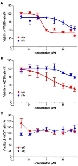
Among the investigated trisaccharides, compound 4and5 reduced the viability of the WM35 melanoma cell line and compound 4 also inhibited the growth of the A2780 ovarian carcinoma cells in a concentration dependent manner (Fig- ure 4A, B). Experimentally determined IC50values of compound 4 were 0.55�0.46 and 1.63�0.61μM on WM35 and A2780 cells, respectively. Compound5was less effective when tested on A2780 cells and its potency is reduced compared to
compound4: IC50values of compound5were 13.28�8.57 and 10.27�5.62μM on WM35 and A2780 cells, respectively. Efficacy of these compounds was lower than that of doxorubicin: they induced only a partial decrease in viability even at the highest concentration (50μM) applied (Figure 4A, B). Importantly, compound 4 and 5 were discriminative between the tumor- driven and nontumorigenic cell lines: In contrast to doxorubicin (Figure S1C), the viability of the non-tumorigenic HaCaT cells did not decline applying these compounds up to 50μM (Figure 4C).
The other four trisaccharides (3,6,7and8) only moderately affected the growth of the cancerous cell lines and did not exhibit dose dependent effect on viability. Considering that compounds3,4and5differ only in the substitution pattern of the glucuronic acid unit, the inactivity of compound 3 demonstrates that the methyl substitution on the uronic acid is detrimental to the inhibitory activity against the cancerous cell Scheme 3.Transformation of thel-iduronic acid containing trisaccharide17.
a) MeOH, NaOMe, RT, 4 h (99 %); b) dry DMF, NaH, MeI, 0°C, 2 h (52 %); c) MeOH, 0.2 M NaOH, RT, 24 h (76 %); d) i. 96 % EtOH, Pd(C), AcOH, H2, RT, 24 h, ii. dry DMF, SO3·Et3N, 50°C, 24 h (59 % over two steps).
Figure 4.Effect of compounds4and5on the viability of cancerous and non-cancerous cell lines. Concentration-dependent effect of compound4 and5on the viability of A) WM35 melanoma, B) A2780 ovarian carcinoma and C) HaCaT keratinocyte cell lines. Viability was determined by MTT assay after 72 h of treatment with the compounds applied in the indicated concentrations and, when it was possible logistic dose-response curves were fitted and IC50values were determined as described in the Experimental Section. Data are presented as mean�SEM,n=6 at each data point.
lines studied. Moreover, the higher activity of compound 4 relative to5shows that acetyl substituents on the uronic acid are advantageous to the biological effect. The growth of HaCaT cells was not influenced by compounds 3, 6, 7 and 8 (Figures S2–S5). The compounds selectivity was also studied on human cancerous cell lines (Ebc-1 and MonoMac6) from differ- ent origin and on non-cancerous non-human primate Vero E6 cells (Figure S6A–F). In this study, compound4and5showed a modest, concentration-dependent cytostatic effect on the cancerous MonoMac6 cell line. Except for that, the compounds were ineffective on all three cell lines.
Conclusions
Using our previous synthetic experience, the targeted four new heparin-analogue trisaccharides were successfully synthesized with excellent yields. The cell growth inhibitory study showed clearly that the six tested trisaccharides have no effect on the growth of healthy keratinocyte derived cells (HaCaT). They do not adversely affect the growth of these cells as opposed to the chemotherapeutic agent Doxorubicin which is used in medi- cine. Moreover, our results have also shown that two of our compounds of glucuronic acid content (4,5) displayed remark- able cell growth inhibitory effects on ovary carcinoma (A2780) and melanoma (WM35) cells.
Methyl substituents on the glucuronide unit proved to be detrimental to the cytostatic activity of the sulfated derivatives.
As it is well demonstrated that the methyl substitution of the uronic acids does not adversely affect the anticoagulant activity of heparinoids,[20–22,36,37]
this effect was unexpected and worthy of further study. The sulfonic acid derivatives, including the iduronate-containing disulfonic acid 8 showed very low or no activity, that, however, can probably be attributed to the methyl ether content of their uronate residue.
In summary, in this short study we identified two glucoro- nate-containing heparinoid trisaccharides of simplified structure that display promising and selective cell growth inhibitory activity.
Experimental Section
General information: Optical rotations were measured at room temperature on a Perkin-Elmer 241 automatic polarimeter. TLC analysis was performed on Kieselgel 60 F254(Merck) silica-gel plates with visualization by immersing in a sulfuric-acid solution (5 % in EtOH) followed by heating. Column chromatography was per- formed on silica gel 60 (Merck 0.063–0.200 mm) and Sephadex LH- 20 (Sigma-Aldrich, bead size: 25–100 mm). Organic solutions were dried over MgSO4and concentrated under vacuum.1H and13C NMR spectroscopy (1H: 360, 400 and 500 MHz; 13C: 90.54, 100.28 and 125.76 MHz) were performed on Bruker DRX-360, Bruker DRX-400 and Bruker Avance II 500 spectrometers at 25°C. Chemical shifts are referenced to SiMe4or sodium 3-(trimethylsilyl)-1-propanesulfonate (DSS, δ=0.00 ppm for 1H nuclei) and to residual solvent signals (CDCl3: δ=77.00 ppm, CD3OD: δ=49.15 ppm for 13C nuclei).
MALDI-TOF MS analyses of the compounds were carried out in the positive reflektron mode using a BIFLEX III mass spectrometer (Bruker, Germany) equipped with delayed-ion extraction. 2,5-
Dihydroxybenzoic acid (DHB) was used as matrix and F3CCOONa as cationising agent in DMF. ESI-TOF MS spectra were recorded by a microTOF Q type QqTOFMS mass spectrometer (Bruker) in the positive ion mode using MeOH as the solvent.
Penta-sodium [methyl (2,3,4-tri-O-methyl-6-O-sulfonato-α-d-glu- copyranosyl)-(1!4)-(2,3-di-O-acetyl-β-d-glucopyranosyl-uronate)- (1!4)-2,3,6-tri-O-sulfonato-α-d-glucopyranoside] (4): Compound 20 (140 mg, 0.206 mmol) was treated with SO3·Et3N complex (747 mg, 4.12 mmol) in dry DMF (5.0 mL). After 24 h of stirring at 50°C, the reaction mixture was neutralized with saturated aqueous solution of NaHCO3(1.73 g, 20.59 mmol), and the resulting mixture was concentrated in vacuo. The crude product was purified by Sephadex gel G-25 in H2O and then treated with Dowex ion exchange resin (Na+form) to give4(165 mg, 74 %) as a white solid.
[α]D= +50.0 (c=0.12, H2O); Rf 0.21 (6 : 7 : 1 EtOAc/MeOH/H2O); 1H NMR (400 MHz, D2O):δ=5.30 (t,J=8.8 Hz, 1H, H-3-E), 5.20 (d, J=
3.4 Hz, 1H, H-1-D), 5.14 (d,J=3.2 Hz, 1H, H-1-F), 4.96-4.92 (m, 2H, H- 1-E, H-2-E), 4.58 (t,J=8.7 Hz, 1H, H-3-F), 4.38-4.34 (m, 3H, H-2-F, H- 6a,b-F), 4.27 (d,J=10.5 Hz, 1H, H-6a-D), 4.16-4.12 (m, 2H, H-4-E, H- 6b-D), 4.03-3.98 (m, 2H, H-4-F, H-5-F), 3.90 (d,J=9.7 Hz, 2H, H-5-E, H-5-D), 3.62, 3.58, 3.48, 3.45 (4 x s, 12H, 4 x OCH3), 3.55-3.50 (m, 1H, H-3-D), 3.33-3.28 (m, 2H, H-2-D, H-4-D), 2.15, 2.13 (2 x s, 6H, 2 x Ac- CH3) ppm;13C NMR (100 MHz, D2O):δ=174.0, 173.0 (3 C, 2 x Ac-CO, COONa), 98.8 (1 C, C-1-E), 97.4 (1 C, C-1-D), 97.2 (1 C, C-1-F), 82.5 (1 C, C-3-D), 80.0 (1 C, C-2-D), 78.1 (1 C, C-4-D), 76.9 (1 C, C-5-E), 75.7 (2 C, C-3-E, C-3-F), 75.1 (2 C, C-2-F, C-4-E), 73.8 (1 C, C-4-F), 72.6 (1 C, C-2-E), 68.9, 68.8 (2 C, C-5-D, C-5-F), 65.9, 65.8 (2 C, C-6-D, C-6-F), 60.2, 60.0, 59.6 (3 C, 3 x OCH3), 55.4 (1 C, C-1-OCH3), 20.5, 20.3 (2 C, 2 x Ac-CH3) ppm; ESI-TOF-MS: m/z calcd for C26H37Na3O31S4: [M- 2Na]2 520.995; found: 520.996.
Penta-sodium [methyl (2,3,4-tri-O-methyl-6-O-sulfonato-α-d-glu- copyranosyl)-(1!4)-(β-d-glucopyranosyl-uronate)-(1!4)-2,3,6-tri- O-sulfonato-α-d-glucopyranoside] (5): Compound 4 (40 mg, 0.036 mmol) dissolved in MeOH (1.5 mL) and 3 M solution of NaOH (0.8 mL) was added at 0°C. After 24 h stirring at room temperature, the reaction mixture was neutralized with AcOH and the resulting mixture was concentrated in vacuo. The crude product was purified by Sephadex gel G-25 in H2O and then treated with Dowex ion exchange resin (Na+form) to give5(34 mg, 92 %) as a white solid.
[α]D= +68.7 (c=0.08, H2O); Rf 0.01 (8 : 2 MeCN/H2O); 1H NMR (400 MHz, D2O):δ=5.64 (d,J=3.7 Hz, 1H, H-1-D), 5.14 (d,J=3.6 Hz, 1H, H-1-F), 4.64 (d,J=7.9 Hz, 1H, H-1-E), 4.62 (t,J=9.4 Hz, 1H, H-3- F), 4.36 (d,J=3.1 Hz, 2H, H-6a,b-F), 4.34 (dd,J=3.4 Hz, J=9.8 Hz, 1H, H-2-F), 4.27 (d,J=10.2 Hz, 1H, H-6a-D), 4.13 (d,J=10.1 Hz, 1H, H-6b-D), 4.07-3.99 (m, 2H, H-4-F, H-5-F), 3.90-3.82 (m, 3H, H-4-E, H- 5-E, H-5-D), 3.74-3.70 (m, 1H, H-3-E), 3.60, 3.57, 3.52, 3.45 (4 x s, 12H, 4 x OCH3), 3.55-3.50 (m, 1H, H-3-D), 3.40 (dd,J=8.1 Hz, J=9.1 Hz, 1H, H-2-E), 3.36-3.30 (m, 2H, H-2-D, H-4-D) ppm;13C NMR (100 MHz, D2O):δ=101.7 (1 C, C-1-E), 98.1 (1 C, C-1-F), 96.8 (1 C, C-1-D), 82.4 (1 C, C-3-D), 81.2 (1 C, C-2-D), 78.9 (1 C, C-4-D), 77.4, 77.2, 77.1 (4 C, C-3-F, C-3-E, C-4-E, C-5-E), 76.1 (1 C, C-2-F), 74.4 (1 C, C-2-E), 73.9 (1 C, C-4-F), 69.8, 69.7 (2 C, C-5-D, C-5-F), 67.2 (1 C, C-6-F), 66.9 (1 C, C-6-D), 61.0, 60.8, 59.0 (3 C, 3 x OCH3), 56.2 (1 C, C-1-OCH3) ppm;
ESI-TOF-MS:m/zcalcd for C22H33Na5O29S4: [M-2Na]2 478.985; found:
478.985.
Penta-sodium [methyl (2,3,4-tri-O-methyl-6-deoxy-6-C-sulfonato- metyl-α-d-glucopyranosyl)-(1!4)-(2,3-di-O-methyl-β-d-glucopyr- anosyl-uronate)-(1!4)-6-deoxy-6-C-sulfonatomethyl-2,3-di-O-sul- fonato-α-d-glucopyranoside] (7): Compound 26 (98 mg, 0.119 mmol) was treated with SO3·Et3N complex (215 mg, 1.19 mmol) in dry DMF (2.9 mL). After 48 h stirring at 50°C, the reaction mixture was neutralized with saturated aqueous solution of NaHCO3 (499 mg, 5.940 mmol) and the resulting mixture was concentrated in vacuo. The crude product was purified by Sephadex gel G-25 in H2O and then treated with Dowex ion
exchange resin (Na+form) to give7(83 mg, 68 %) as a white solid.
[α]D= +66.9 (c=0.13, H2O); Rf 0.19 (6 : 7 : 1 EtOAc/MeOH/H2O); 1H NMR (500 MHz, D2O):δ=5.49 (d,J=3.7 Hz, 1H, H-1”), 5.11 (d,J=
3.7 Hz, 1H, H-1), 4.69 (d,J=7.8 Hz, 1H, H-1’), 4.59 (t,J=9.1 Hz, 1H, H-3), 4.36 (dd,J=3.7 Hz,J=9.6 Hz, 1H, H-2), 3.96-3.89 (m, 3H, H-4’, H-5, H-5’), 3.76 (t,J=9.1 Hz, 1H, H-4), 3.62-3.52 (m, 17H, H-3’, H-5”, 5 x OCH3), 3.50 (t,J=9.7 Hz, 1H, H-3”), 3.44 (s, 3H, C-1O-CH3), 3.33 (dd, J=3.9 Hz, J=10.2 Hz, 1H, H-2”), 3.30 (t, J=9.0 Hz, 1H, H-2’), 3.18- 2.96 (m, 5H, H-4”, H-7a,b, H-7”a,b), 2.46-2.44 (m, 1H, H-6a), 2.21-2.16 (m, 1H, H-6”a), 1.98-1.88 (m, 2H, H-6b, H-6”b) ppm, 13C NMR (500 MHz, D2O):δ=173.3 (1 C,CO), 102.0 (1 C, C-1’), 96.9 (1 C, C-1), 95.3 (1 C, C-1”), 85.5 (1 C, C-3’), 82.6 (1 C, C-2’), 82.2 (1 C, C-4”), 81.4 (1 C, C-3”), 80.4 (1 C, C-2”), 78.4 (1 C, C-4), 76.3 (1 C, C-3), 75.2 (1 C, C-2), 75.1 (1 C, C-5’), 72.8 (1 C, C-4’), 69.3 (1 C, C-5), 68.4 (1 C, C-5”), 60.3, 60.2, 59.6, 59.1, 58.9 (5 C, 5 x OCH3), 55.3 (1 C, C-1-OCH3), 47.2, 47.0 (2 C, 2 x C-7), 26.3 (1 C, C-6’), 26.0 (1 C, C-6) ppm; ESI-TOF-MS:
m/zcalcd for C26H41Na6O27S4: [M+H]+1051.0099; found: 1051.0091.
Hepta-sodium [methyl (6-deoxy-4-O-methyl-2,3-di-O-sulfonato-6- C-sulfonatomethyl-α-d-glucopyranosyl)]-(1!4)-(2,3-di-O-methyl- α-l-idopyranosyl-uronate)-(1!4)-(6-deoxy-2,3-di-O-sulfonato-6- C-sulfonatomethyl-α-d-glucopyranoside)] (8): A mixture of 29 (34 mg, 0.030 mmol) and Pd/C (10 %, 30 mg) was dissolved in 96 % EtOH-AcOH (30 : 1, 3.1 mL) and stirred in an autoclave under H2 atmosphere (at 10 bar) for 24 h. The catalyst was filtered through a pad of Celite, and the filtrate was concentrated. The crude product (23 mg, 99 %, Rf 0.17 (7 : 6 : 1 CH2Cl2/MeOH/H2O)) was used for further reaction without purification. A solution of the crude product (23 mg, 0.029 mmol) in dry DMF (1.5 mL) was treated with SO3·Et3N complex (105 mg, 0.580 mmol, 5.0 equiv./OH). After 24 h stirring at 50°C, the reaction mixture was neutralized with aqueous solution of NaHCO3(244 mg, 25.0 equiv./OH). The resulting mixture was concentrated. The crude product was purified by Sephadex gel G-25 in H2O and then treated with Dowex ion exchange resin (Na+ form), to give8(20 mg, 59 % for two steps) as a white solid. [α]D= +41.3 (c=0.08, H2O); Rf 0.12 (7 : 6 : 1 CH2Cl2/MeOH/H2O); 1H NMR (D2O, 360 MHz):δ=5.39 (d,J1,2=3.1 Hz, 1H, H-1-F), 5.24 (s, 1H, H-1- G), 5.13 (d,J1,2=3.4 Hz, 1H, H-1-H), 4.86 (s, 1H, H-5-G), 4.63-4.57 (m, 2H, H-3-H, H-3F), 4.37 (dd,J1,2=3.3 Hz,J2,3=9.6 Hz, 1H, H-2-H), 4.28 (dd, J1,2=3.0 Hz, J2,3=9.4 Hz, 1H, H-2-F), 4.13 (s, 1H, H-4-G), 3.92- 3.77 (m, 4H), 3.59, 3.57, 3.53, 3.45 (4 x s, 12H, 4 x OCH3), 3.30 (t,J= 8.9 Hz, 1H), 3.20-3.00 (m, 5H), 2.39-1.92 (m, 4H, H-6a,b-F, H-6a,b-H) ppm;13C NMR (D2O, 90 MHz):δ=97.9 (2 C, C x C-1), 94.9 (1 C, C-1), 82.4, 80.0, 78.2, 77.6, 76.6, 76.5, 75.9, 72.7, 71.3, 70.2, 70.1 (12 C, skeleton carbons), 61.3, 60.1, 58.9, 56.3 (4 C, 4 x OCH3), 48.2, 47.9 (2 C, 2 x C-7), 27.3, 27.2 (2 C, 2 x C-6) ppm; ESI-TOF-MS:m/zcalcd for C24H35Na8O33S6: [M+Na]+1226.8561; found: 1226.8538.
Methyl (6-O-benzyl-2,3,4-tri-O-methyl-α-d-glucopyranosyl)-(1! 4)-[2,3-di-O-acetyl-6-O-(2’-naphthyl)methyl-β-d-glucopyranosyl]- (1!4)-2,3,6-tri-O-benzyl-α-d-glucopyranoside (15): To a solution of compound12:[29] (440 mg, 0.948 mmol, 1.5 equiv.) and compound9[33] (500 mg, 0.632 mmol) in dry CH2Cl2 (15 mL) was added 4 Å molecular sieves (0.5 g). After stirring for 30 min at room temperature, the mixture was cooled to 50°C and the solutions of NIS (213 mg, 0.948 mmol, 1.5 equiv. for donor) in dry THF (400μL) and TfOH (25μL, 0.294 mmol, 0.3 equiv. for donor) were added.
Allowed to warm up the solution to 15°C and the mixture were stirred for 4 h at that temperature. When the TLC analysis (1 : 1n- hexane/EtOAc) showed complete consumption of the donor, the reaction mixture was neutralized with Et3N (500μL), diluted with CH2Cl2 (150 mL), and filtered. The filtrate was washed with an aqueous solution of Na2S2O3(10 %, 2 × 50 mL), a saturated aqueous solution of NaHCO3 (2 × 50 mL), and water (2 × 50 mL), dried, and concentrated. The crude product was purified by column chroma- tography on silica gel (1 : 1n-hexane/EtOAc) to give compound15 (562 mg, 78 %) as a colourless syrup. [α]D= +62.2 (c=0.09, CHCl3);
Rf 0.47 (1 : 1 n-hexane/EtOAc);1H NMR (400 MHz, CDCl3):δ=7.81- 7.13 (m, 27H, arom), 5.14 (t,J=9.2 Hz, 1H, H-3-E), 5.08 (d,J=3.5 Hz, 1H, H-1-D), 5.02 (d,J=11.6 Hz, 1H, Bn-CH2a), 4.86 (dd,J=9.3 Hz,J=
8.2 Hz, 1H, H-2-E), 4.76 (d, J=11.7 Hz, 1H, Bn-CH2b), 4.71 (dd, J= 12.1 Hz,J=5.5 Hz, 2H, Bn-CH2), 4.59-4.53 (m, 4H, H-1-E, H-1-F, NAP- CH2), 4.48-4.41 (m, 3H, Bn-CH2), 4.28 (d, J=12.2 Hz, 1H, Bn-CH2b), 3.95-3.84 (m, 3H, H-3-F, H-4-F, H-4-E), 3.76 (dd, J=10.5 Hz, J= 3.0 Hz, 1H, H-6a-F), 3.68-3.60 (m, 5H, H-5-D, H-5-F, H-6a,b-E, H-6b-F), 3.58, 3.42, 3.40, 3.34 (4 x s, 12H, 4 x OCH3), 3.46-3.35 (m, 4H, H-2-F, H-3-D, H-6a,b-D), 3.25-3.21 (m, 1H, H-5-E), 3.19-3.15 (m, 1H, H-4-D), 3.05 (dd,J=9.8 Hz,J=3.5 Hz, 1H, H-2-D), 2.01, 1.94 (2 x s, 6H, 2 x Ac-CH3) ppm;13C NMR (100 MHz, CDCl3):δ=170.0, 169.7 (2 C, 2 x Ac-CO), 139.5, 138.3, 138.1, 137.7, 136.1, 133.3, 132.9 (7 C, 7 x Cq arom), 128.7-125.7 (27 C, arom), 99.9 (1 C, C-1-F), 98.4 (1 C, C-1-E), 97.8 (1 C, C-1-D), 83.2 (1 C, C-3-D), 81.8 (1 C, C-2-D), 80.2 (1 C, C-3-F), 79.3, 79.2 (2 C, C-2-F, C-4-D), 77.0 (1 C, C-4-F), 75.1, 74.9 (3 C, C-3-E, C-4-E, C-5-E), 72.9 (1 C, C-2-E), 71.2 (1 C, C-5-D), 69.9 (1 C, C-5-F), 73.7, 73.6, 73.3 (5 C, 4 x Bn-CH2, NAP-CH2), 68.8, 68.4 (2 C, C-6-D, C- 6-E), 67.9 (1 C, C-6-F), 60.7, 60.4, 59.3 (3 C, 3 x OCH3), 55.3 (1 C, C-1- OCH3), 21.1, 20.8 (2 C, 2 x Ac-CH3) ppm; MALDI-TOF-MS:m/zcalcd for C65H76NaO18: [M+Na]+1167.492; found: 1167.657.
Methyl [ethyl (2,3,4-tri-O-methyl-6-deoxy-6-C-sulfonatomethyl-α- d-glucopyranosyl)]-(1!4)-[2,3-di-O-acetyl-6-O-(2’-naphthyl)meth- yl-β-d-glucopyranosyl]-(1!4)-[ethyl (2,3-di-O-benzyl-6-deoxy-6- C-sulfonatomethyl-α-d-glucopyranoside)] (16): To a solution of compound 13[31] (1.343 g, 1.667 mmol, 1.6 equiv.) and compound10[31] (500 mg, 1.041 mmol) in dry CH2Cl2 (30 mL) was added 4 Å molecular sieves (1.0 g). After stirring for 30 min at room temperature, the mixture was cooled to 50°C and the mixture of the solutions of NIS (412 mg, 1.833 mmol, 1.5 equiv. for donor) in dry THF (1.0 mL) and AgOTf (103 mg, 0.400 mmol, 0.38 equiv. for donor) in dry toluene (1.0 mL) were added. Allowed to warm up the solution to 15°C and the mixture were stirred for 4 h at that temperature. When the TLC analysis (1 : 1n-hexane/EtOAc) showed complete consumption of the donor, the reaction mixture was diluted with CH2Cl2(200 mL), and filtered through a pad of Celite®. The filtrate was washed with an aqueous solution of Na2S2O3(10 %, 2 × 75 mL), a saturated aqueous solution of NaHCO3(2 × 75 mL), and water (2 × 75 mL), dried, and concentrated. The crude product was purified by column chromatography on silica gel (1 : 1 n-hexane/
EtOAc) to give compound16(1.058 g, 86 %) as a colourless syrup.
[α]D= +36.5 (c=0.20, CHCl3);Rf0.28 (1 : 1n-hexane/EtOAc);1H NMR (400 MHz, CDCl3): δ=7.87-7.76 (m, 3H, arom), 7.69 (s, 1H, arom), 7.48-7.33 (m, 3H, arom), 7.31-7.08 (m, 17H, arom), 5.17 (t,J=9.2 Hz, 1H, H-3-E), 5.07-4.73 (m, 5H, H-1-D, H-1-E, H-2-E, BnCH2), 4.70-4.43 (m, 5H, H-1-F, NAPCH2, BnCH2), 4.27 (q,J=7.1 Hz, 2H, SO3CH2CH3), 4.11 (q, 2H, SO3CH2CH3), 3.97-3.80 (m, 2H, H-3-F, H-4-E), 3.69-3.59 (m, 1H, H-5-F), 3.53-3.22 (m, 7H, H-3-D, H-5-D, H-2-F, H-4-F, H-7a-F, H-6a-E, H-6b-E), 3.53, 3.50, 3.38, 3.31 (4 s, 12H, 4 x CH3), 3.21-2.98 (m, 3H, H-5-E, H-7b-F, H-7a-D), 2.95-2.78 (m, 2H, H-2-D, H-7b-D), 2.67 (t, J=9.3 Hz, 1H, H-4-D), 2.39-2.27 (m, 1H, H-6a-F), 2.26-2.11 (m, 1H, H- 6a-D), 2.07, 2.00 (2 s, 6H, 2 x Ac-CH3), 1.88-1.72 (m, 2H, H-6b-F, H-6b- D), 1.39 (t,J=6.9 Hz, 3H, SO3CH2CH3), 1.28 (t, 3H, SO3CH2CH3) ppm;
13C NMR (100 MHz, CDCl3):δ=169.8, 169.7 (2 C, 2 x Ac-CO), 139.1, 137.7, 135.2, 133.1, 132.9 (5 C,Cq arom), 128.3, 128.1, 128.2, 129.0, 127.9, 127.8, 127.5, 126.8, 126.5, 126.0, 125.9, 125.8, 125.6 (17 C, arom), 100.9 (1 C, C-1-D), 97.6 (1 C, C-1-F), 96.8 (1 C, C-1-E), 83.2 (1 C, C-4-D), 82.3 (1 C, C-3-D), 81.9 (1 C, C-2-D), 81.8 (1 C, C-4-F), 79.7 (1 C, C-2-F), 79.7 (1 C, C-3-F), 75.0 (1 C, C-5-E), 74.5 (1 C, C-3-E), 74.1 (1 C, C-4-E), 74.2, 73.3, 73.1 (3 C, 2 x BnCH2, NAPCH2), 72.7 (1 C, C-2- E), 69.2 (1 C, C-5-D), 67.5 (1 C, C-5-F), 67.1 (1 C, C-6-E), 66.1, 65.8 (2 C, SO3CH2CH3), 60.5, 60.2, 58.9, 55.3 (4 C, OCH3), 46.7, 46.6 (2 C, C- 7-D, C-7-F), 25.9 (C-6-D), 25.9 (C-6-F), 20.8, 20.6 (2 C, COCH3), 15.0, 14.1 (2 C, SO3CH2CH3) ppm; MALDI-TOF-MS: m/z calcd for C57H76NaO22: [M+Na]+1199.42; found: 1200.73.
Methyl [ethyl (2,3-di-O-benzyl-6-deoxy-6-C-sulfonatomethyl-α-d- glucopyranosyl)]-(1!4)-[methyl (2-O-acetyl-3-O-methyl-α-l-ido- pyranosyl)-uronate]-(1!4)-[sodium (2,3-di-O-benzyl-6-deoxy-6-C- sulfonatomethyl-α-d-glucopyranoside)] (17): To a solution of compound 14[20] (147 mg, 0.315 mmol, 1.5 equiv.) and compound11[30] (200 mg, 0.210 mmol) in dry CH2Cl2 (6 mL) was added 4 Å molecular sieves (1.0 g). After 30 min, the mixture was cooled to 20°C and a solution of TMSOTf (5μL, 0.021 mmol) in dry CH2Cl2(190μL) was added. After stirring for 2 h, TLC analysis showed the complete consumption of the donor. The reaction mixture was neutralized with Et3N (150μL), diluted with CH2Cl2
(100 mL) and filtered. The filtrate was washed with a saturated aqueous solution of NaHCO3 (2 × 25 mL) and water (2 × 25 mL), dried and concentrated. The crude product was purified by column chromatography on silica gel (1 : 1 n-hexane/EtOAc) to give compound17(151 mg, 63 %) as a colourless syrup.Rf0.12 (1 : 1n- hexane/EtOAc); 1H NMR (360 MHz, CD3OD):δ=7.37-7.26 (m, 20H;
arom), 5.20 (d, 1H,J=2.6 Hz), 4.91-4.57 (m, 12H; 4 x Bn-CH2, 2 x H-1, H-2’, H-5’), 4.26 (q, J=7.1 Hz, 2H, SO3CH2CH3), 3.92-3.18 (m, 10H), 3.49, 3.34 (3 x s, 9H, 3 x OCH3), 3.07-2.84 (m, 4H; 2 x H-7a,b), 2.34- 2.24 (m, 3H, 2 x H-6a), 2.06 (s, 3H, Ac-CH3), 1.94-1.90 (m, 2H, 2 x H- 6b), 1.35 (t, J=7.1 Hz, 3H, SO3CH2CH3) ppm; 13C NMR (90 MHz, CD3OD): δ=172.0, 171.3 (2 C, 2 x CO), 140.3, 140.2, 139.7, 139.5 (4 C, 4 x Cqarom), 129.5-128.3 (20 C, arom), 100.8, 99.4, 98.7 (3 C, 3 x C-1), 82.4, 81.7, 81.1, 80.6, 79.5, 78.1, 76.6, 74.8, 71.8, 70.5, 70.3, 69.8 (12 C, skeleton carbons), 76.2, 75.9, 74.5, 74.0 (4 C, 4 x Bn-CH2), 68.0 (1 C, SO3CH2CH3), 58.8, 55.6, 52.6 (3 C, 4 x OCH3), 47.0 (2 C, 2 x C-7), 28.3, 26.7 (2 C, 2 x C-6), 21.4 (1 C, Ac-CH3), 15.5 (1 C, SO3CH2CH3) ppm.
Methyl (6-O-benzyl-2,3,4-tri-O-methyl-α-d-glucopyranosyl)-(1! 4)-(2,3-di-O-acetyl-β-d-glucopyranosyl)-(1!4)-2,3,6-tri-O-benzyl- α-d-glucopyranoside (18): To a vigorously stirred solution of 15 (550 mg, 0.480 mmol) in CH2Cl2 (8.0 mL) and H2O (0.8 mL) DDQ (163 mg, 0.720 mmol) was added. After 30 min the mixture was diluted with CH2Cl2(200 mL) and extracted with saturated aqueous solution of NaHCO3 (2 × 30 mL), and H2O (2 × 30 mL), dried and concentrated. The crude product was purified by silica gel chromatography (9 : 1 CH2Cl2/acetone) to give compound 18 (405 mg, 84 %) as a colourless syrup. [α]D= +25.0 (c=0.10, CHCl3);
Rf0.40 (9 : 1 CH2Cl2/acetone);1H NMR (400 MHz, CDCl3):δ=7.40-7.24 (m, 20H, arom), 5.10 (t,J=9.2 Hz, 1H), 5.02 (d,J=3.6 Hz, 1H), 4.90- 4.43 (m, 11H), 3.83-3.81 (m, 2H), 3.75-3.71 (m, 2H), 3.63-3.61 (m, 7H), 3.59, 3.45, 3.44, 3.36 (4 x s, 12H, 4 x OCH3), 3.50-3.38 (m, 3H), 3.17 (d, J=9.1 Hz, 1H), 3.09 (dd,J=9.7 Hz,J=3.6 Hz, 2H), 2.01, 1.93 (2 x s, 6H, 2 x Ac-CH3) ppm; 13C NMR (100 MHz, CDCl3): δ=169.8, 169.5 (2 C, 2 x Ac-CO), 139.2, 138.2, 137.9, 137.5 (4 C, 4 x Cqarom), 128.7- 127.2 (20 C, arom), 99.8, 98.4, 98.1 (3 C, 3 x C-1), 83.3, 81.5, 79.8, 79.4, 79.0, 77.0, 75.2, 74.9, 74.7, 72.7, 71.3, 69.8 (12 C, skeleton carbons), 75.3, 73.7, 73.5, 73.4 (4 C, 4 x Bn-CH2), 68.5, 67.5, 61.3 (3 C, 3 x C-6), 60.6, 60.4, 59.4 (3 C, 3 x OCH3), 55.3 (1 C, C-1-OCH3), 20.9, 20.7 (2 C, 2 x Ac-CH3) ppm; MALDI-TOF-MS: m/z calcd for C54H68NaO18: [M+Na]+1027.430; found: 1027.886.
Methyl (6-O-benzyl-2,3,4-tri-O-methyl-α-d-glucopyranosyl)-(1!
4)-[sodium (2,3-di-O-acetyl-β-d-glucopyranosyl)-uronate]-(1!4)- 2,3,6-tri-O-benzyl-α-d-glucopyranoside (19): To a vigorously stirred solution of18(390 mg, 0.388 mmol) in CH2Cl2(8.0 mL) and H2O (4.0 mL) TEMPO (12 mg, 0.077 mmol) and BAIB (375 mg, 1.164 mmol) were added. After 24 h stirring at room temperature, the reaction mixture was quenched by addition of 10 % aqueous solution of Na2S2O3 (35 mL). The phases were separated, and the aqueous layer was extracted with CH2Cl2(3 x 50 mL). The combined organic layers were dried, and concentrated. The crude product was purified by silica gel chromatography (95 : 5 CH2Cl2/MeOH) to give19(245 mg, 61 %) as a colourless syrup. [α]D= +46.1 (c=0.18, CHCl3);Rf0.33 (95 : 5 CH2Cl2/MeOH);1H NMR (400 MHz, CD3OD):δ=
7.33-7.08 (m, 20H, arom), 5.01 (t,J=9.2 Hz, 1H), 4.98 (d,J=2.1 Hz, 1H), 4.89-4.34 (m, 11H), 3.97 (t, J=9.2 Hz, 1H), 3.74-3.47 (m, 9H), 3.45, 3.34, 3.32, 3.21 (4 x s, 12H, 4 x OCH3), 3.30-3.25 (m, 2H), 3.15 (d, J=9.4 Hz, 1H), 2.93 (dd,J=9.7 Hz,J=3.4 Hz, 1H), 1.91, 1.82 (2 x s, 6H, 2 x Ac-CH3) ppm;13C NMR (100 MHz, CD3OD):δ=172.7, 171.5, 171.0 (3 C, 2 x Ac-CO,COONa), 140.4, 139.5, 139.1 (4 C, 4 x Cqarom), 129.8-128.3 (20 C, arom), 101.3, 99.1, 98.8 (3 C, 3 x C-1), 84.4, 82.9, 81.0, 80.6, 80.2, 78.3, 77.8, 77.7, 75.7, 73.5, 72.3, 71.2 (12 C, skeleton carbons), 76.4, 74.6, 74.5, 74.2 (4 C, 4 x Bn-CH2), 69.3, 69.0 (2 C, 2 x C-6), 61.0, 60.8, 59.6 (3 C, 3 x OCH3), 55.6 (1 C, C-1-OCH3), 21.1, 20.7 (2 C, 2 x Ac-CH3) ppm; MALDI-TOF-MS:m/zcalcd for C54H65Na2O19: [M+Na]+1063.391, found 1063.853.
Methyl (2,3,4-tri-O-methyl-α-d-glucopyranosyl)-(1!4)-[sodium (2,3-di-O-acetyl-β-d-glucopyranosyl)-uronate]-(1!4)-α-d-gluco- pyranoside (20): A mixture of 19 (235 mg, 0.226 mmol) in 96 % EtOH/AcOH (19 : 1, 15 mL), and Pd(C) (10 %, 180 mg) was stirred in an autoclave under H2atmosphere (at 10 bar) for 24 h. The catalyst was filtered off through a pad of Celite®, washed with MeOH, and the filtrate was concentrated under reduced pressure. The crude product was purified by Sephadex LH-20 gel chromatography (MeOH) to give20(150 mg, 98 %) as a white powder. [α]D= +106.7 (c=0.03, H2O); Rf 0.38 (1 : 1 CH2Cl2/MeOH); 1H NMR (400 MHz, CD3OD):δ=5.31 (t,J=9.9 Hz, 1H, H-3-E), 5.18 (d, J=3.3 Hz, 1H), 4.90-4.83 (m, 2H), 4.75 (d,J=3.4 Hz, 1H), 4.09 (t,J=9.2 Hz, 1H), 3.94 (d, J=9.4 Hz, 1H), 3.82-3.67 (m, 6H), 3.63-3.47 (m, 4H), 3.59, 3.52, 3.45, 3.38 (4 x s, 12H, 4 x OCH3), 3.26-3.21 (m, 2H), 2.12, 2.10 (2 x s, 6H, 2 x Ac-CH3) ppm;13C NMR (100 MHz, CD3OD):δ=177.6, 173.5, 173.3 (3 C, 2 x Ac-CO,COONa), 100.7, 100.0, 98.2 (3 C, 3 x C-1), 83.4, 81.3, 80.0, 79.4, 76.2, 75.7, 73.4, 72.5, 72.0, 71.9, 71.2 (12 C, skeleton carbons), 60.8, 60.6 (2 C, 2 x C-6), 61.0, 60.5, 60.2 (3 C, 3 x OCH3), 55.9 (1 C, C-1-OCH3), 21.3, 20.9 (2 C, 2 x Ac-CH3) ppm; MALDI-TOF- MS:m/zcalcd for C26H41Na2O19: [M+Na]+703.203, found 703.520.
Methyl (2,3,4-tri-O-methyl-6-deoxy-6-C-ethylsulfonatomethyl-α- d-glucopyranosyl)-(1!4)-(2,3-di-O-acetyl-β-d-glucopyranosyl)- (1!4)-2,3-di-O-benzyl-6-deoxy-6-C-ethylsulfonatomethyl-α-d- glucopyranoside (21): To a vigorously stirred solution of16(1.00 g, 0.850 mmol) in CH2Cl2(12.5 mL) and H2O (1.25 mL) DDQ (289 mg, 1.28 mmol) was added. After 30 min the mixture was diluted with CH2Cl2(250 mL) and extracted with saturated aqueous solution of NaHCO3 (2 × 50 mL), and H2O (2 × 50 mL), dried and concentrated.
The crude product was purified by silica gel chromatography (6 : 4 n-hexane/acetone) to give compound 21 (725 mg, 82 %) as a colourless syrup. [α]D= +48.7 (c=0.15, CHCl3); Rf 0.28 (6 : 4 n- hexane/acetone);1H NMR (400 MHz, CDCl3): δ=7.43-7.22 (m, 10H, arom), 5.24 (t,J=9.3 Hz, 1H, H-3-E), 5.03 (d,J=3.7 Hz, 1H, H-1-D), 4.98 (d,J=11.6 Hz, 1H, BnCH2), 4.90-4.80 (m, 2H, H-2-E, BnCH2), 4.74 (d,J=12.1 Hz, 1H, BnCH2), 4.68 (d,J=8.0 Hz, 1H, H-1-E), 4.62 (d,J= 12.1 Hz, 1H, BnCH2), 4.51 (d,J=3.6 Hz, 1H, H-1-F), 4.29 (q,J=7.1 Hz, 4H, SO3CH2CH3), 3.92-3.82 (m, 2H, H-3-F, H-4-E), 3.73 (dt,J=10.3 Hz, J=2.6 Hz, 1H, H-5-F), 3.62-3.15 (m, 10H, H-5-E, H-6a-E, H-6b-E, H-2-F, H-4-F, H-7a-F, H-3-D, H-5-D, H-7a-D, H-7b-D), 3.56, 3.53, 3.43, 3.35 (4 s, 12H, 4 x CH3), 3.14-3.05 (m, 1H, H-7b-F), 3.02 (dd,J=9.8 Hz,J=
3.6 Hz, 1H, H-2-D), 2.74 (t,J=9.2 Hz, 1H, H-4-D), 2.44-2.34 (m, 1H, H- 6a-F), 2.34-2.24 (m, 1H, H-6a-D), 2.05, 2.03 (2 s, 6H, 2 x AcCH3), 1.93- 1.74 (m, 3H, H-6b-F, H-6b-D), 1.42 (t,J=7.1 Hz, 6H, SO3CH2CH3) ppm;
13C NMR (100 MHz, CDCl3): δ=170.0, 169.7 (2 x CO), 138.9, 137.8 (2 C, Cqarom), 128.5, 128.5, 128.1, 128.0, 127.6, 126.4 (10 C, arom), 100.2 (1 C, C-1-D), 97.9 (1 C, C-1-F), 96.8 (1 C, C-1-E), 83.8 (1 C, C-4- D), 82.7 (1 C, C-3-D), 81.8 (1 C, C-4-F), 81.6 (1 C, C-2-D), 79.5 (1 C, C- 2-F), 78.8 (1 C, C-3-F), 75.3 (1 C, C-4-E), 74.9 (1 C, C-5-E), 74.6, 73.4 (1 C, 2 C, BnCH2), 72.6 (1 C, C-2-E), 72.1 (1 C, C-4-E), 69.5 (1 C, C-5-D), 67.7 (1 C, C-5-F), 66.2, 66.2 (2 C, 2 x SO3CH2CH3), 60.3 (1 C, C-6-E), 60.8, 60.6, 59.5, 55.5 (4 C, 4 x OCH3), 46.7, 46.7 (2 C, C-7-D, C-7-F), 26.3 (1 C, C-6-D), 25.7 (1 C, C-6-F), 20.9, 20.6 (2 C, 2 x AcCH3), 15.1,
15.1 (2 C, 2 x SO3CH2CH3) ppm; MALDI-TOF-MS: m/z calcd for C46H68NaO22S2: [M+Na]+1059.35; found: 1060.06.
Methyl (2,3,4-tri-O-methyl-6-deoxy-6-C-ethylsulfonatomethyl-α- d-glucopyranosyl)-(1!4)-[sodium (2,3-O-acetyl-β-d-glucopyrano- syl)-uronate]-(1!4)-2,3-di-O-benzyl-6-deoxy-6-C-
ethylsulfonatomethyl-α-d-glucopyranoside (22): To a vigorously stirred solution of compound 21(700 mg, 0.680 mmol) in CH2Cl2
(15 mL) and H2O (7.5 mL) TEMPO (21 mg, 0.2 equiv., 0.14 mmol) and BAIB (870 mg, 4.0 equiv., 2.70 mmol) were added and stirred for 24 h at room temperature. The reaction mixture was quenched by the addition of 10 % aqueous solution of Na2S2O3 (10.0 mL). The mixture was then extracted twice with CH2Cl2 (20 mL), and the combined organic layers were dried and concentrated. The crude product was purified by column chromatography to give 22 (625 mg; 79 %) as a white foam. [α]D= +47.3 (c=0.11, CHCl3); Rf
0.40 (98 : 2 CH2Cl2/MeOH); 1H NMR (400 MHz, CDCl3): δ=7.40-7.21 (m, 10H, arom), 5.20 (m, 1H, H-3-E), 5.01 (d,J=3.6 Hz, 1H, H-1-D), 4.92-4.84 (m, 4H, H-2-E, H-1-E, 2 x BnCH2), 4.68, (d,J=12.0 Hz, 1H, BnCH2), 4.53 (d,J=12.0 Hz, 1H, BnCH2), 4.47 (d,J=3.6 Hz, 1H, H-1- F), 4.31-4.29 (m, 4H, SO3CH2CH3), 4.03 (t,J=8.6 Hz, 1H, H-5-E), 3.96- 3.83 (m, 2H, H-3-F, H-4-E, 3.63 (dt,J=9.9, 2.5 Hz, 1H, H-5-F), 3.59- 3.58 (m, 7H, H–3-D, H -5-D, H-2-F, H-4-F, H-7a-D, H-7b-D, H-7a-F), 3.56, 3.53, 3.42, 3.31 (4 s, 12H, 4 x CH3), 3.08-3.00 (m, 2H, H-7b-F, H- 2-D), 2.73 (t,J=9.3 Hz, 1H, H-4-D), 2.34-2.21 (m, 2H, H-6a-D, H-6a-F), 2.06, 2.02 (2 x s, 6H, 2 x AcCH3), 1.90-1.77 (m, 2H, H-6b-D, H-6b-F), 1.41, 1.40 (2 x t, J=6.9 Hz, 6H, 2 x SO3CH2CH3) ppm; 13C NMR (100 MHz, CDCl3):δ=170.0, 169.6 (2 C, 2 xCO), 138.8, 137.9 (2 C, Cq
arom), 128.5, 128.4, 128.2, 128.1, 127.6 (10 C, arom), 100.5 (1 C, C-1- D), 97.9 (1 C, C-1-F), 97.6, 83.8, 82.7, 81.8, 81.4, 79.9, 79.9, 79.5, 75.3, 74.0, 72.5, 69.5, 67.8 (12 C, skeleton carbons), 75.1, 73.5 (2 C, 2 x BnCH2), 66.7, 66.3 (2 C, 2 x SO3CH2CH3), 60.7, 60.7, 59.6, 55.6 (4 C, 4 x OCH3), 46.9, 46.6 (2 C, C-7-D, C-7-F), 26.0, 25.9 (2 C, C-6-D, C-6-F), 20.9, 20.7 (2 C, 2 x AcCH3), 15.2, 15.2 (2 C, 2 x SO3CH2CH3) ppm, MALDI-TOF-MS: m/z calcd for C46H65Na2O23S2: [M+Na]+ 1095.3;
found: 1095.0.
Methyl [sodium (2,3,4-tri-O-methyl-6-deoxy-6-C-sulfonatometh- yl)-α-d-glucopyranosyl]-(1!4)-[sodium (β-d-glucopyranosyl)-uro- nate]-(1!4)-[sodium (2,3-di-O-benzyl-6-deoxy-6-C- sulfonatomethyl)-α-d-glucopyranoside] (23): To the solution of compound 22 (530 mg, 0.490 mmol) in MeOH (20 mL) NaOMe (5 mg, 0.075 mmol, 0.15 equiv.) was added and the mixture was stirred at room temperature for 24 h. The reaction mixture was neutralized with acetic acid and all volatiles were evaporated. The crude product was dissolved in acetone (20 ml) and NaI (222 mg, 1.48 mmol, 3.0 equiv.) was added to the solution and stirred at room temperature for 24 h. The reaction mixture was concentrated under reduced pressure and the crude product was purified by column chromatography (7 : 6 : 1 CH2Cl2/MeOH/H2O) and gel chro- matography (Sephadex LH-20, MeOH) to give23(434 mg, 90 % for two steps) as a colourless syrup. [α]D= +71.0 (c=0.10, MeOH);Rf
0.41 (95 : 5 CH2Cl2/MeOH);1H NMR (400 MHz, CD3OD):δ=7.43-7.36 (m, 2H, arom), 7.35-7.19 (m, 8H, arom), 5.52 (d,J=3.7 Hz, 1H, H-1- D), 4.97 (d,J=11.2 Hz, 1H, BnCH2), 4.76 (d,J=11.2 Hz, 1H, BnCH2), 4.69-4.62 (m, 2H, H-1-E, H-1-F), 4.61-4.53 (m, 2H, BnCH2), 3.91-3.69 (m, 5H), 3.68-3.44 (m, 5H), 3.57, 3.53, 3.53, 3.34 (4 s, 12H, 4 x OCH3), 3.32-3.29 (m, 1H), 3.15 (dd,J=3.7 Hz, 1H, H-2-D), 3.10-2.99 (m, 3H, 3 x H-7), 2.89-2.75 (m, 2H, H-7, H-4-D), 2.60-2.47 (m, 1H, H-6), 2.32- 2.18 (m, 1H, H-6), 1.98-1.75 (m, 3H, 2 x H-6, OH) ppm; 13C NMR (100 MHz, CD3OD): δ=140.2, 139.5 (2 C, Cq arom), 129.4, 129.3, 129.2, 129.1, 128.8, 128.4 (10 C, arom), 103.6, 98.6, 98.6 (3 C, 3 x C- 1), 85.2, 83.9, 83.7, 81.6, 81.4, 80.8, 79.2, 78.6, 78.2, 75.5, 70.8, 70.0 (12 C, skeleton carbons),76.0, 73.9 (2 C, 2 x BnCH2), 60.8, 59.3, 55.5 (4 C, CH3), 28.1, 28.1 (2 C, C-6-D, C-6-F) ppm; MALDI-TOF-MS:m/z calcd for C38H51Na4O21S2: [M+Na]+999.19; found: 999.44.
Methyl [sodium (2,3,4-tri-O-methyl-6-deoxy-6-C-sulfonatometh- yl)-α-d-glucopyranosyl]-(1!4)-[methyl (2,3-O-methyl-β-d-gluco- pyranosyl)-uronate]-(1!4)-[sodium (2,3-di-O-benzyl-6-deoxy-6-C- sulfonatomethyl)-α-d-glucopyranoside] (24): To the solution of compound 23 (482 mg, 0.490 mmol) in dry DMF (15 mL) NaH (36 mg, 1.48 mmol, 60 m/m%, 3.0 equiv.) was added at 0°C. After 30 min stirring at that temperature 315μl MeI (2.22 mmol, 4.5 equiv.) was added to the mixture and stirred for 24 h at room temperature. The reaction mixture was quenched by the addition of MeOH (1.5 mL) and acetic acid (1-2 drops). The solution was concentrated and the crude product was purified by gel chroma- tography (Sephadex LH-20, MeOH) to give24(354 mg, 72 %) as a white foam. [α]D= +33.3 (c=0.09, MeOH); Rf 0.36 (7 : 3 CH2Cl2/ MeOH); 1H NMR (400 MHz, CD3OD):δ=7.27-7.13 (m, 10H, arom), 5.34 (d,J=3.5 Hz, 1H, H-1-D), 4.88 (d,J=11.1 Hz, 1H, BnCH2), 4.58- 4.46 (m, 5H, H-1-E, H-1-F, BnCH2), 3.81 (d,J=9.6 Hz, 1H), 3.74 (t,J=
9.1 Hz, 1H), 3.68 (t,J=9.2 Hz, 1H), 3.63-3.55 (m, 1H), 3.49, 3.49, 3.49, 3.47, 3.44, 3.41, 3.26 (7 s, 21H, 7 x OCH3), 3.54-3.31 (m, 4H), 3.30-3.06 (m, 4H), 3.03-2.66 (m, 4H), 2.54-2.41 (m, 1H, 1 x H-6a), 2.20-2.08 (m, 1H, 1 x H-6a), 1.89-1.85 (m, 2H, 2 x H-6b) ppm;13C NMR (100 MHz, CD3OD):δ=170.5 (1 C,CO), 140.3, 139.4 (2 C, Cqarom), 129.2, 129.1, 128.9, 128.7, 128.2 (10 C, arom), 104.7, 98.6, 96.6 (3 C, 3 x C-1), 86.9, 85.3, 84.5, 84.0, 83.5, 82.8, 81.0, 80.9, 75.3, 74.9, 70.6, 70.2 (12 C, skeleton carbons), 76.2, 74.1 (2 C, 2 x BnCH2), 61.1, 61.1, 60.9, 60.5, 59.4, 55.6, 53.1 (7 C, 7 x OCH3), 48.9, 48.5 (2 C, C-7-D, C-7-F), 37.0, 28.2, 27.7 (2 C, C-6-D, C-6-F) ppm; MALDI-TOF-MS: m/z calcd for C41H58Na3O21S2: [M+Na]+1019.26; found: 1019.47.
Methyl [sodium (2,3,4-tri-O-methyl-6-deoxy-6-C-sulfonatometh- yl)-α-d-glucopyranosyl]-(1!4)-[sodium (2,3-O-methyl-β-d-gluco- pyranosyl)-uronate]-(1!4)-[sodium (2,3-di-O-benzyl-6-deoxy-6-C- sulfonatomethyl)-α-d-glucopyranoside] (25): To the solution of compound24(480 mg, 0.480 mmol) in the mixture of THF (3.0 mL) and MeOH (3.0 mL) 0.5 M NaOH solution (2.9 mL) was added and stirred at room temperature for 24 h. The reaction was neutralized by the addition of 60 v/v % acetic acid solution and the mixture was concentrated. The crude product was purified by gel chromatography (Sephadex, LH-20, MeOH) and converted to sodium salt by ion exchange resin (Dowex, Na+ form, MeOH) to give 25 (398 mg, 82 %) as colorless syrup. [α]D= +42.8 (c=0.10, CHCl3);Rf0.94 (6 : 4 CH2Cl2/MeOH);1H NMR (400 MHz, CD3OD):δ= 7.31 (d,J=7.2 Hz, 2H, arom), 7.26-7.08 (m, 8H, arom), 5.36 (d,J= 3.7 Hz, 1H, H-1-D), 4.88 (d,J=11.4 Hz, 1H, BnCH2), 4.66-4.52 (m, 3H, H-1-E, H-1-F, BnCH2), 4.47 (s, 2H, BnCH2), 3.81-3.69 (m, 2H), 3.68-3.65 (m, 3H), 3.48, 3.47, 3.45, 3.42, 3.40, 3.08 (6 x s, 18H, 6 x OCH3), 3.52- 3.16 (m, 4H), 3.04-2.87 (m, 5H), 2.81-2.63 (m, 2H), 2.54-2.40 (m, 1H, H-6a), 2.21-2.08 (m, 1H, H-6a), 1.88-1.68 (m, 2H, 2 x H-6b) ppm;13C NMR (100 MHz, CD3OD): δ=139.2, 138.2 (2 C, Cq arom), 127.9, 127.5, 126.9 (10 C, arom), 97.3, 97.2, 95.5 (3 C, C-1-D, C-1-E, C-1-F), 86.2, 84.4, 83.9, 82.6, 81.9, 81.2, 79.8, 79.4, 77.0, 74.2, 69.2, 68.8 (12 C, skeleton carbons), 72.5, 72.5 (2 C, 2 x BnCH2), 59.6, 59.5, 59.0, 58.3, 54.6, 54.3 (6 C, 6 x OCH3), 29.4, 27.0 (2 C, C-6-D, C-6-F) ppm;
MALDI-TOF-MS: m/z calcd for C40H55Na4O21S2: [M+Na]+ 1027.23;
found: 1027.90.
Methyl [sodium (2,3,4-tri-O-methyl-6-deoxy-6-C-sulfonatometh- yl)-α-d-glucopyranosyl]-(1!4)-[sodium (2,3-O-methyl-β-d-gluco- pyranosyl)-uronate]-(1!4)-[sodium (6-deoxy-6-C-sulfonatometh- yl)-α-d-glucopyranoside] (26): To the solution of compound 25 (398 mg, 0.400 mmol) in EtOH (15 mL, 96 %) 10 %-os Pd/C (300 mg) and acetic acid (350μL) were added. The mixture was stirred at room temperature for 24 h under 10 bar H2 atmosphere. The mixture was diluted with MeOH and the catalyst was filtrated through a pad of Celite®and then the solution was concentrated.
The crude product was purified by gel chromatography (Sephadex, G-25, H2O) to give26(121 mg, 92 %) as white powder. [α]D= +77.7 (c=0.13, MeOH); Rf 0.53 (7 : 6 : 1 CH2Cl2/MeOH/H2O); 1H NMR
(400 MHz, D2O):δ=5.35 (d,J=3.7 Hz, 1H, H-1”), 4.46 (d,J=7.7 Hz, 1H, H-1’), 3.77-3.51 (m, 5H), 3.51-3.31 (m, 19H), 3.31-3.07 (m, 6H), 3.07-2.77 (m, 5H), 2.32-2.19 (m, 1H, H-6a), 2.09-1.97 (m, 1H, H-6a), 1.82-1.63 (m, 2H, 2 x H-6b) ppm,13C NMR (100 MHz, D2O):δ=174.4 (1 C,CO), 102.3, 98.7, 95.1 (3 C, 3 x C-1), 85.8, 83.4, 82.9, 82.3, 81.2, 80.6, 75.8, 72.9, 71.5, 70.9, 68.9, 68.4 (12 C, skeleton carbons), 60.5, 60.1, 59.5, 59.4, 58.9, 55.1 (6 C, 6 x OCH3), 47.3, 47.1 (2 C, 2 x C-7), 26.2, 25.8 (2 C, 2 x C-6) ppm; MALDI-TOF-MS: m/z calcd for C26H43Na4O21S2: [M+H]+847.13; found: 847.64.
Methyl [ethyl (2,3-di-O-benzyl-6-deoxy-6-C-sulfonatomethyl-α-d- glucopyranosyl)]-(1!4)-[methyl (3-O-methyl-α-l-idopyranosyl)- uronate]-(1!4)-[sodium (2,3-di-O-benzyl-6-deoxy-6-C- sulfonatomethyl-α-d-glucopyranoside)] (27): To a solution of compound17(103 mg, 0.088 mmol) in MeOH (2.5 mL) was added NaOCH3(25 mg, 0.462 mmol). The reaction mixture was stirred for 4 h and monitored by TLC. After the complete disappearance of the starting material, the mixture was neutralized with AcOH and concentrated. The crude product was purified by column chroma- tography on Sephadex LH-20 (MeOH) to give compound 27 (99 mg, 99 %) as a colourless syrup. [α]D= +167.5 (c=0.08, CHCl3);
Rf0.38 (9 : 1 CH2Cl2/MeOH);1H NMR (CD3OD, 360 MHz):δ=7.36-7.24 (m, 20H, arom), 5.16 (s, 1H), 5.10 (d, J=3.4 Hz, 1H), 5.04 (d, J=
2.1 Hz, 1H), 4.90-4.61 (m, 8H), 4.29 (q, J=7.1 Hz, 2H, SO3CH2CH3), 3.95 (s, 1H), 3.83 (s, 1H), 3.76-3.73 (m, 3H), 3.60-3.21 (m, 9H), 3.49, 3.41, 3.35 (3 x s, 9H, 3 x OCH3), 3.19-2.83 (m, 4H, 2 x H-7a,b), 2.34- 1.92 (m, 4H, 2 x H-6a,b), 1.37 (t,J=7.1 Hz, 3H, SO3CH2CH3) ppm;13C NMR (CD3OD, 90 MHz):δ=171.7 (1 C, CO), 140.2, 140.0, 139.4, 138.9 (4 C, 4 x Cqarom), 129.6-128.6 (20 C, arom), 102.4, 98.6, 96.1 (3 C, 3 x C-1), 82.3, 81.9, 80.6, 80.0, 79.2, 75.6, 74.5, 72.4, 71.4, 70.5, 69.1, 67.0 (12 C, skeleton carbons), 76.3, 76.0, 74.8, 73.8 (4 C, 4 x BnCH2), 67.9 (1 C, SO3CH2CH3), 58.5 (1 C, C-3’-OCH3), 55.6 (C-1-OCH3), 52.9 (1 C, COOCH3), 48.6, 47.1 (2 C, 2 x C-7), 28.4, 26.8 (2 C, 2 x C-6), 15.5 (1 C, SO3CH2CH3); ESI-TOF-MS:m/zcalcd for C53H68KO21S2: [M+H]+ 1143.333; found: 1143.394.
Methyl [sodium (2,3-di-O-benzyl-6-deoxy-4-O-methyl-6-C- sulfonatomethyl-α-d-glucopyranosyl)]-(1!4)-[methyl (2,3-di-O- methyl-α-l-idopyranosyl)-uronate]-(1!4)-[sodium (2,3-di-O- benzyl-6-deoxy-6-C-sulfonatomethyl-α-d-glucopyranoside)] (28):
To a solution of 27 (95 mg, 0.084 mmol) in dry DMF (3 mL) was slowly added NaH (8 mg, 0.201 mmol) at 0°C. After stirring for 30 min at 0°C, MeI (11μL, 0.201 mmol) was added. When complete conversion of the starting material into a main spot had been observed by TLC analysis (2 h at 0°C), CH3OH (1.0 mL) was added.
The reaction mixture was stirred for 5 min and the solvents were evaporated. The crude product was purified by column chromatog- raphy on Sephadex LH-20 (MeOH) to give compound28 (50 mg, 52 %) as a colourless syrup. Rf 0.44 (9 : 1 CH2Cl2/MeOH); 1H NMR (CD3OD, 360 MHz):δ=7.39-7.25 (m, 20H, arom), 5.19 (d,J=2.1 Hz, 1H), 5.16 (d,J=3.3 Hz, 1H), 4.86 (d,J=2.7 Hz, 1H), 4.86-4.61 (m, 8H), 3.85-3.84 (m, 1H), 3.76-3.72 (m, 4H), 3.61-3.30 (m, 4H), 3.51, 3.47, 3.46, 3.39, 3.37 (5 x s, 15H, 5 x OCH3), 3.08-2.98 (m, 3H) 2.91-2.77 (m, 4H, 2 x H-7a,b), 2.36-1.89 (m, 4H, 2 x H-6a,b) ppm;13C NMR (90 MHz, CD3OD):δ=171.6 (1 C, CO), 140.1, 140.0, 139.5, 139.4 (4 C, 4 x Cq arom), 129.5-128.6 (20 C, arom), 99.8, 98.7, 95.5 (3 C, 3 x C-1), 84.9, 82.2, 81.9, 80.6, 80.5, 79.4, 78.2, 75.7, 71.9, 71.1, 70.7, 69.8 (12 C, skeleton carbons), 76.2, 76.1, 73.9 (4 C, 4 x BnCH2), 61.4, 59.9, 58.9, 55.6 (4 x OCH3), 52.7 (1 C, COOCH3), 28.5, 28.4 (2 C, 2 x C-6).
Methyl [sodium (2,3-di-O-benzyl-6-deoxy-4-O-methyl-6-C- sulfonatomethyl-α-d-glucopyranosyl)]-(1!4)-[sodium (2,3-di-O- methyl-α-l-idopyranosyl)-uronate]-(1!4)-[sodium (2,3-di-O- benzyl-6-deoxy-6-C-sulfonatomethyl-α-d-glucopyranoside)] (29):
A solution of the trisaccharide 28 (45 mg, 0.040 mmol) in MeOH (2 mL) was treated with 0.2 M aqueous solution of NaOH (1.0 mL).
After 24 h stirring at room temperature the TLC showed complete conversion of the carboxylic esters into sodium salts. The mixture
was neutralized with acetic acid, and concentrated. The crude product was purified by Sephadex gel LH-20 in MeOH to give29 (34 mg, 76 %).Rf0.12 (8 : 2 CH2Cl2/MeOH);1H NMR (360 MHz, D2O):
δ=7.40-7.31 (m, 20H, arom), 5.13 (d, J=2.6 Hz, 1H), 5.06 (d, J= 3.4 Hz, 1H), 4.86-4.55 (m, 10H), 4.05 (s, 1H), 3.90-3.74 (m, 6H), 3.61- 3.37 (m, 4H), 3.57, 3.45, 3.38, 3.37 (4 x s, 12H, 4 x OCH3), 3.17-2.99 (m, 4H, 2 x H-7a,b), 2.40-1.92 (m, 4H, 2 x H-6a,b) ppm; 13C NMR (D2O, 90 MHz): δ=181.5 (1 C, CO), 138.7, 138.5, 138.3 (4 C, 4 x Cq
arom), 129.8-129.2 (20 C, arom), 98.0, 94.6 (3 C, 3 x C-1), 83.6, 81.1, 80.4, 80.2, 79.9, 78.8, 73.3, 71.6, 70.1, 70.0 (12 C, skeleton carbons), 76.6, 76.1, 74.2, 73.8 (4 C, 4 x BnCH2), 61.0, 60.1, 58.8, 56.0 (4 x OCH3), 48.3, 48.1 (2 C, 2 x C-7), 27.2 (2 C, 2 x C-6) ppm.
Biological evaluation: Biological activity of compounds 3-8 was tested by investigating their effect on the cellular viability of A2780 human ovarian carcinoma, WM35 human melanoma and HaCaT spontaneously immortalized human keratinocyte cell lines. WM35 and A2780 cells were cultured in RPMI1640 Medium (ThermoFisher) supplemented with 10 % fetal bovine serum (FBS. ThermoFisher) and antibiotics penicillin and streptomycin (Pen-Strep, Thermo- Fisher). HaCaT cell were cultured in DMEM (ThermoFisher) supplemented with FBS and Pen-Strep. During culturing, cells were incubated in a humified thermostat at constant 37°C and in the presence of 5 % CO2. Media were refreshed every 2-3 days and cells were subcultured when reached ca. 80 % confluence. Cellular viability was determined by MTT assay as described earlier.[38,39,40]
Briefly, the number of viable cells was indirectly determined by measuring the conversion of the tetrazolium salt MTT (3-{4,5- dimethilthiasol-2-il}-2,5-diphenyltetrasolium bromide, Sigma-Al- drich) to formazan by mitochondrial dehydrogenases. Cells were plated in 96-well microplates (10.000 cells per well density) and were cultured for 3 days and treated by the compounds daily.
Negative control group was treated with equal amount of vehicle solvent (DMSO, Sigma-Aldrich) and positive control group was treated with 1μg/mL doxorubicin. Cells were then incubated with 1 mg/ml MTT for 3 h, precipitated formazan crystals were dissolved in acidic isopropanol (10 % 1 M HCl dissolved in isopropanol and supplemented with 10 % Triton X 100, all from VWR) and concentration of formazan was assessed colorimetrical way measur- ing absorbance at 565 nm. Viability was calculated based on the measured absorbance and given as percentage where 100 % viability is determined as the mean absorbance of the negative control (i. e., vehicle treated) samples and 0 % viability is determined as the mean absorbance of the positive control samples measured in parallel on the same microplate. Dose-response relationship of the above compounds was assessed in a concentration range from 0.03-50μM by fitting logistic dose-response curves and calculating the IC50 values using the equationy=A2+(A1 A2)/(1+(x/x0)p) where the parameters are: A1: initial value (ymin), A2: final value (ymax), x0: center (EC/IC50) andp is the calculated power. Fittings were carried out and parameters were calculated using Origin 8.6 software (OriginLab Corporation, Northampton, MA, USA).
Acknowledgements
The authors gratefully acknowledge financial support for this research from the Premium Postdoctoral Program of HAS (PPD 461038, M.H.) and from the EU and co-financed by the European Regional Development Fund under the project GINOP-2.3.2-15- 2016-00008 and from the National Research, Development and Innovation Office of Hungary (K128368, PD134791, E.L.). B.I.T. was supported by the János Bolyai Research Scholarship of the Hungarian Academy of Sciences and the New National Excellence Program of the Ministry for Innovation and Technology (ÚNKP-20-