THE ROLE OF MICRORNAS
IN RENAL ISCHEMIA REPERFUSION INJURY THERAPEUTIC APPLICATION
OF RNA INTERFERENCE
PhD thesis
Tamás Kaucsár, MD
Basic Medicine Doctoral School Semmelweis University
Supervisor: Dr. Péter Hamar, MD, Ph.D, D.Sc Official reviewers:
Dr. Zoltán Giricz, Ph.D Dr. Tamás Csont, MD, Ph.D
Head of the Final Examination Committee:
Dr. Barna Vásárhelyi, MD, Ph.D, D.Sc
Members of the Final Examination Committee:
Dr. Andrea Fekete, MD, Ph.D Dr. Anita Rácz, MD, Ph.D
Budapest, 2014
2
Table of Contents
Table of Contents ... 2
List of Abbreviations ... 5
Introduction ... 8
MicroRNAs (miRNAs): generation and mechanism of action (Figure 1) ... 8
MiRNA nomenclature ... 10
MiRNA function ... 11
Influencing miRNA expression in vivo ... 12
Nucleic acid therapy – problems and solutions of delivery ... 12
Inhibition of miRNA function ... 14
Enhancement of miRNA function ... 16
Kidney specific miRNAome, renal disease specific alterations, and functional investigations of miRNAs in the kidney ... 17
Ischemic acute kidney injury ... 20
Pathophysiology ... 20
Cellular injuries ... 21
miRNAs in ischemic AKI... 22
Objectives ... 25
Methods ... 26
Patients ... 26
Animals ... 26
LNA-modified miRNA oligonucleotides ... 27
Kidney ischemia-reperfusion injury ... 27
Sacrifice, blood and organ collection ... 28
Ex vivo cell purification/sorting ... 28
Plasma Urea and NGAL ELISA ... 29
3
Histology and immunohistochemistry ... 30
Cell Culture experiments ... 32
Transfection Assays ... 32
Scratch wound healing assay of HK-2 cells ... 32
Protein Analysis ... 32
Luciferase Reporter Assays ... 33
RNA preparation ... 33
Multiplex analysis of the microRNA profile ... 34
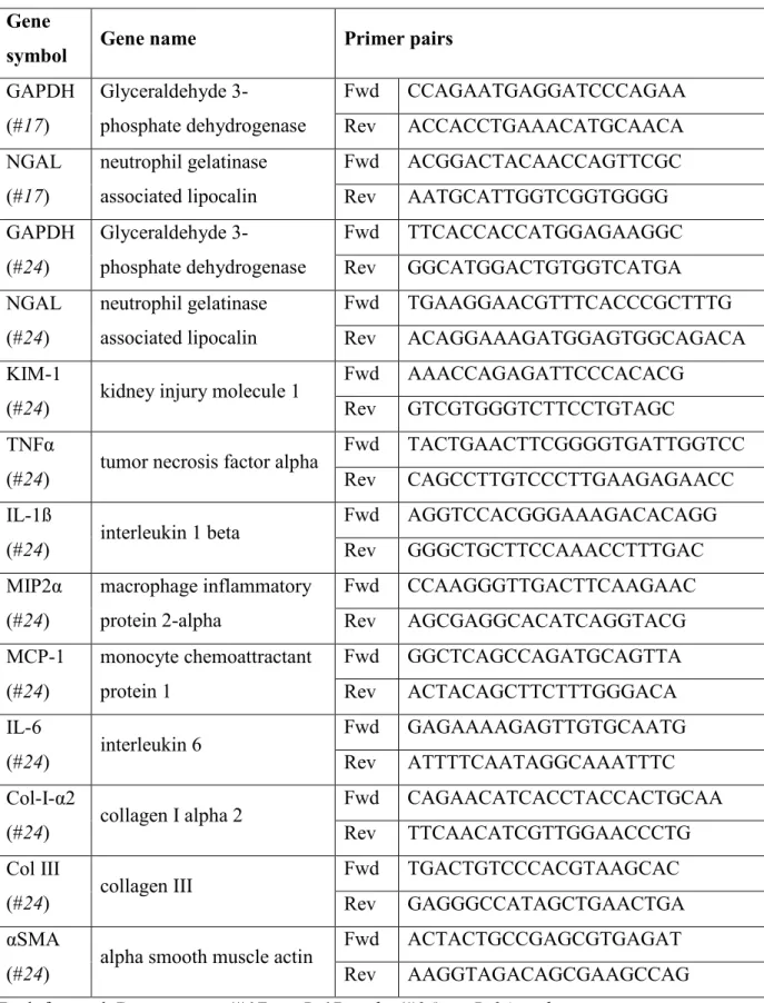
Quantitative real-time PCR analysis of miRNAs and gene expression in renal tissue ... 35
MicroRNA target prediction ... 36
Statistical analysis ... 36
Results ... 39
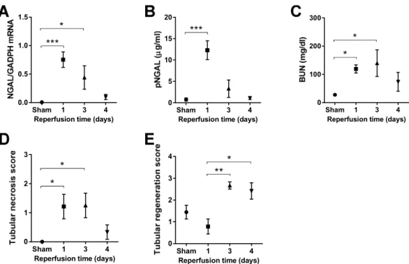
Lethal renal ischemia-reperfusion injury markers ... 39
Kinetics of sublethal renal ischemia-reperfusion injury markers ... 41
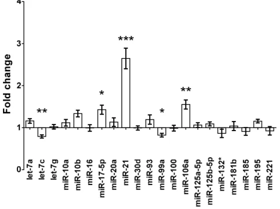
Micro RNA expression changes and time-course of renal miR-17-5p, miR-106a and miR-21 expressions after renal ischemia-reperfusion injury ... 42
Correlation between the two renal miRNA expressions and other markers of renal I/R injury ... 45
miR-24 in renal I/R-injury ... 46
Functional role of miR-24 in tubular epithelial cells ... 48
Sphingosine-1-phosphate receptor 1 (S1PR1), H2A histone family, member X (H2A.X) and Heme Oxygenase-1 (HO-1) are direct targets of miR-24 in vitro ... 48
Markers of kidney damage and endothelial activation in I/R-injury after miR-24 silencing ... 51
Kidney morphology, infiltration of immune cells, level of apoptosis after miR-24 silencing ... 53
4
Regulation of miR-24, survival, kidney function as well as markers of kidney damage
and inflammation in bilateral I/R-injury in vivo ... 56
MiR-24 in the progression from acute kidney injury to chronic kidney disease ... 59
Discussion ... 61
Conclusions ... 66
Summary ... 67
Bibliography ... 69
Bibliography of the candidate’s publications ... 88
Acknowledgements ... 89
5
List of Abbreviations
2’-OM 2’-O-methylated (CH3 is added to the 2’ OH) 3’ UTR 3’ untranslated region
A adenine
ADP adenosine diphosphate AGO Argonaut protein AKI Acute kidney injury Akt serine/threonine-protein
kinase (Ak (mouse strain) transforming protein) AMI Acute myocardial infarction
AMO Anti-miRNA
Oligonucleotides
AMP adenosine monophosphate ANOVA analysis of variance APC allophycocyanin
ASO antisense oligonucleotides ATF3 activating transcription
factor 3
ATP adenosine triphosphate AU arbitrary units
αSMA alpha smooth muscle actin BCL-2 B cell leukemia/lymphoma 2
bp base pair
BSA bovine serum albumin BUN blood urea nitrogen
C cytosine
CD(no.) cluster of differentiation (number)
cDNA complementary (copy) DNA CIT cold ischemia time
CKD chronic kidney disease CL contralateral
Col III collagen, type III Col1α2 collagen type I, alpha 2
chain
CTR control
DAB 3,3' diaminobenzidine DAMP damage(danger)-associated
molecular patterns DAPI 4’,6-diamidino-2-
phenylindole
DGCR-8 DiGeorge syndrome critical region
DN Diabetic nephropathy DNA Deoxyribonucleic acid DNase deoxyribonuclease dsRBD dsRNA-binding domain dsRNA double-stranded RNA dUTP deoxyuridine triphosphate EDTA ethylenediaminetetraacetic
acid
eIF eukaryotic translation initiation factor ELISA enzyme-linked
immunosorbent assay eNOS endothelial nitric oxide
synthase (NOS3) ESRD end stage renal disease Exp5/Xpo5 Exportin-5
FACS fluprescence-associated cell sorting
FC fold change
FFPE formalin fixed paraffin embedded
fwd forward primer
6
G guanine
GAPDH glyceraldehyde-3-phosphate dehydrogenase
GFR glomerular filtration rate H2A.X histone 2A family, member
X (H2AFX)
HAVCR1 hepatitis A virus cellular receptor 1 (KIM1, TIM1) HE hematoxylin eosin
HIF1α hypoxia inducible factor 1, alpha subunit
HK-2 Human kidney 2 (proximal tubular cell line)
HO-1 heme oxygenase 1 hpf high power field HRP horseradish peroxidase
hsa homo sapiens
HSF-1 heat shock transcription factor 1
HSP Heat shock protein i.p. intraperitoneal I/R ischemia reperfusion ICAM-1 Intercellular Adhesion
Molecule 1
IgG immunoglobulin G IHC immunohistochemistry
IL interleukin
IPC ischemic preconditioning KIM1 kidney injury molecule 1
(HAVCR1)
LCN2 lipocalin-2 (NGAL) LNA locked nucleic acid LTA Lotus tetragonolobus
agglutinin
Ly-6G lymphocyte antigen 6 complex, locus G MACS magnetic-activated cell
sorting
MCL-2 myeloid cell leukemia sequence 2
MCP-1 monocyte chemoattractant protein 1
MEM Minimal Essential Medium MFI median fluorescence
intensity
mghv mouse gammaherpesvirus MIP2α macrophage inflammatory
protein 2-alpha miRNA micro RNA
MMP-2 matrix metallopeptidase
mmu mus musculus
mRNA messenger RNA
mTAL medullary thick ascending limb
NCBI National Center for
Biotechnology Information NGAL neutrophil gelatinase
associated lipocalin (LCN2) NO nitric oxide
ODN Oligodeoxyribonucleotides P bodies processing bodies
PAS periodic acid-Schiff PBS phosphate buffered saline PCA principal component analysis PCR polymerase chain reaction PDCD4 programmed cell death
protein 4
PDGFRβ platelet-derived growth factor receptor beta
7 pDNA plasmid DNA
PE Phycoerythrin
PEG poly-ethylene-glycol PEI poly-ethilene-imine PIC polyion complex pNGAL plasma NGAL Pol II RNA polymerase II PON peroxynitrite pre-miRNA precursor miRNA pri-miRNA primary miRNA PTEN phosphatase and tensin
homolog
qPCR quantitative (real-time) PCR RanGTP GTP-binding RAs-related
Nuclear protein RCC Renal cell carcinoma rev reverse primer
RISC RNA-Induced Silencing Complex
RNA Ribonucleic acid RNAi RNA interference RNase Ribonuclease
ROS reactive oxygen species RT reverse transcription S1PR1 sphingosine-1-phosphate
receptor 1
SEM standard error of the mean
shRNA short hairpin RNA siRNA short interfering RNA Sirt1 sirtuin 1
snoRNA Small nucleolar RNA snRNA small nuclear RNA SPF specific pathogen-free STAT signal transducer and
activator of transcription STZ Streptozotocin
T thymine
TAR trans-activation response TBS tris-buffered saline TIM1 T cell-immunoglobulin-
mucin (HAVCR1) TLR Toll-like receptor TMA tissue microarray TMB 3,3',5,5'-
tetramethylbenzidine TNFα tumor necrosis factor alpha TRBP2 TAR RNA binding protein 2 TUNEL transferase dUTP nick end
labeling
VEGF vascular endothelial growth factor
VEGFR2 VEGF Receptor 2
VHL von Hippel-Lindau tumor suppressor
8
Introduction
In the postgenomic era, investigation of the human transcriptome has revealed that the genome encodes many thousands of functional RNAs not transcribed into proteins (non-coding RNAs) (1). Micro RNAs (miRNAs)compose a large family: about 1% of the genes encoded in the genome belong to the miRNA family (2). First described in 1993 (3) miRNAs are short, being composed of only 18-25 nucleotides (nt). Presently, it is hypothesized, that miRNA sequences play an important role in gene-expression regulation, through RNA interference (RNAi), controlling protein synthesis from most human genes at the posttranscriptional level (post-transcriptional regulation of gene- expression) (4). Emergingknowledge surrounding the role of miRNAs in theregulation of post-transcriptional protein expression has dramaticallyaltered the view of how target genes are regulated.
The regulatory functions of our body comprise networks. The hypothesis, that miRNAs exert their regulatory function in networks is supported by the high number of non-coding RNAs which are functionally active, e.g. miRNAs that have been shown to target signaling molecules (5). Furthermore, some genes encoding miRNAs are closely located (clustered) in the genome and in some cases different miRNAscontrol a single messenger RNA (mRNA) target or vice versa a single miRNA may influence expression of multiple different target proteins. MiRNA expression profiles during different disease states can be determined by microarray studies. Such systemic approaches together with individual analysis of different miRNAs provide insight into an exciting new regulatory network of the miRNAome.
Micr Micr
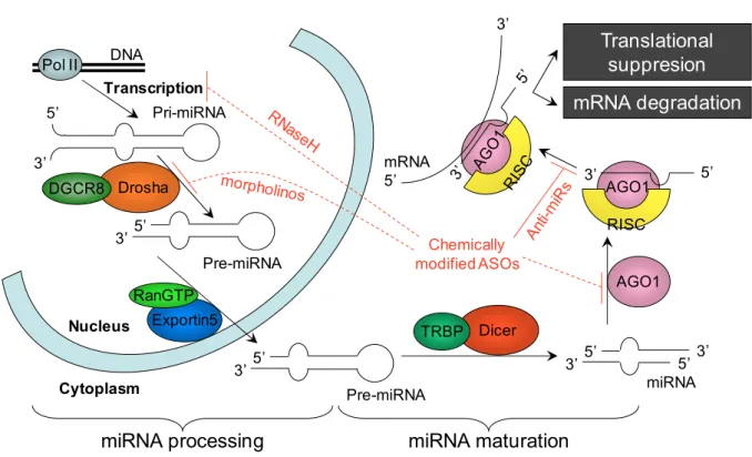
Micr Micro o oRNAs (miRNAs): generation and mechanism of action ( o RNAs (miRNAs): generation and mechanism of action ( RNAs (miRNAs): generation and mechanism of action ( RNAs (miRNAs): generation and mechanism of action (Figure Figure Figure Figure 1 1 1)))) 1
MiRNAs are generated from endogenous hairpinstructured transcripts throughout the genome (2). MiRNA encoding genes are transcribed by RNApolymerase II (pol II) providing long precursor transcripts, known as primary miRNAs (pri-miRNAs) (6). After transcription, still inside the nucleus, Drosha Ribonuclease (RNase): a type III nuclear RNase cleaves nucleotides from the pri-miRNA, processing it into shorter pre-miRNAs and defining their 3’ end. Efficient cleavage requires a double-stranded RNA-binding domain (dsRBD) containing cofactor: DiGeorge syndrome critical region (DGCR)-8. The
9
stem-loop (hairpin) structured pre-miRNA has a characteristic 5’ phosphate and 3’
hydroxy termini with a two nucleotide 3’ single-stranded overhanging end (7). This end structure is recognized by the nuclear export factor Exportin-5 (Exp5/Xpo5), which uses Ran-GTPas a co-factor (8) andtransports the pre-miRNA to the cytoplasm (9).Further cytoplasmic processing by Dicer (another type III ribonuclease in the cytoplasm) performs a second cleavage at the hairpin structure, and defines the 5’ end of the mature miRNA. The Dicer also uses a double stranded (ds)RNA-binding domain (dsRBD) containing cofactor: trans activation response (TAR) RNA binding protein 2 (TRBP2).
As a result of the cleavage by Dicer a double-stranded18- to 25-nucleotide-long miRNA is generated (10). The mature miRNA is one of the strands of the dsRNA (miRNA/miRNA* duplex). One of the two strands is loaded on an Argonaut family protein (AGO1): the catalytic site of the RNA induced silencing complex (RISC), thus assembling the RISC-ribonucleoprotein complex. Unlike short interfering (si)RNAs which bind to AGO2, miRNAs bind to AGO1.
The guide strand of the miRNA is incorporated into the RNA-induced silencing complex (RISC) (11), andremains stably associated with RISC, becoming themature miRNA. The opposite (passenger) strandis disposed. The miRNA guidesRISC to the target messenger (m)RNA with complementary sequence.
Translation of the target mRNA is silenced in case of incompletely complementary sequence, and the mRNA is spliced up (cleaved) by the RISC in case of fully complementary sequence. As endogenous miRNAs often contain mismatches, the more common (primary) mechanism is translational repression: AGO1 does not cleave the mRNA, but binds to it and allosterically inhibits translation. Unlike RNAi induced by siRNA, cleavage (degradation) of the mRNA occurs more seldom, only by complete match between the miRNA and the mRNA (12).
It is interesting, that many distinct ways exist to obtain post transcriptional gene silencing by miRNA interference. Protein translation can be inhibited at translation initiation by inhibiting different eukaryotic translation initiation factors (eIF)s or at translation elongation. Furthermore, instead of translation inhibition, co-translational degradation of the nascent polypeptide chain or without interfering with the translation machinery by sequestering and processing mRNAs in discrete cytoplasmic foci: P bodies are possible ways of post-transcriptional gene silencing by miRNAs (12), (13).
10
Figure 1. The miRNA machinery and sites of intervention. Micro RNA biogenesis and function (based on (14), (15), (16), (17), (18)).
Pol II: RNApolymerase II. DGCR-8: DiGeorge syndrome critical region (cofactor of Drosha RNase III). RanGTP: cofactor of Exportin-5. TRBP2: TAR RNA binding protein 2 (cofactor of Dicer (a cytoplasmic RNase III, which cuts the hairpin of the pre-miRNA.
RISC: RNA-Induced Silencing Complex. AGO1: Argonaut protein (the catalytic site of the RISC). mRNA cleavage or translational suppression depends on the level of complementarity. Possible interventions (red): chemically modified AntiSense Oligonucleotides (ASOs) can block the RISC active site or inhibit mature miRNA binding to the RISC, but can interfere with miRNA processing early steps as well.
MiRNA nomenclature MiRNA nomenclature MiRNA nomenclature MiRNA nomenclature
The continuous discovery of new miRNAs necessitates a consistent gene naming scheme. Therefore, every mature miRNA has a “miR” prefix (precursor miRNAs are denoted with “mir”) and a unique identifying number, which are assigned sequentially, in order of discovery. Identical miRNAs have the same identifying number, even between different organisms. The host organism can be designated by an abbreviated 3 or 4 letter prefix (e.g., hsa-miR for Homo sapiens, mmu-miR for Mus musculus, mghv-mir-M1-2
Pol II DNA
Pri-miRNA
Drosha
Pre-miRNA
Nucleus Exportin5
Cytoplasm
Dicer
miRNA
miRNA processing miRNA maturation
Pre-miRNA
mRNA degradation Translational
suppresion
Transcription 5’
3’
3’ 5’
3’ 5’ 3’ 5’
5’3’
mRNA 5’
3’
AGO1 RISC
3’ 5’
AGO1 RanGTP
DGCR8
TRBP Chemically modified ASOs
11
for mouse gammaherpesvirus etc.). Furthermore, identical miRNAs encoded in different chromosomal locations (in case of multiple copies) have numbered suffixes (ascending in order of discovery, (not chromosome number), e.g.: hsa-miR-194-1 and hsa-miR-194- 2 are located on chromosome 1 and 11, respectively). Paralogous miRNA sequences, which differ only by one or two nucleotides have lettered suffixes (e.g., hsa-miR-200a, hsa-miR-200b, hsa-miR-200c). Where two different mature miRNAs are processed from the same hairpin precursor, the ending (3’ or 5’) of the arm of provenance has to be specified (e.g., miR-17-5p, miR-17-3p) or an asterisk can be applied to the less predominantly expressed transcript (strand) (e.g., miR-199*). The miRNA encoding genes are named using the same three-letter prefix, which can be modified according to the conventions of the host organism (capitalization, hyphenation or italics).
Nevertheless, online databases also exist (e.g., http://mirbase.org/, http://rfam.janelia.org/) to prevent accidental overlap when naming newly discovered miRNAs. The new identifying number will be assigned just after the paper describing the miRNA has been accepted for publication (19), (20).
MiRNA function MiRNA function MiRNA function MiRNA function
A single microRNA may alter the expression of a large number of target genes, thus influencing a specific pathology by regulating whole disease-specific pathways and signaling cascades rather than a single gene (21). This unique function underlines the immense importance of these small molecules.
MiRNAs are involved in gene regulation in different processes such as embryonic (22) or hematopoetic (23) development, apoptosis (24), or tumor initiation and progression (miR-17–92 cluster, miR-21, miR-372) (25). MiRNAs are also involved in many physiological (2) and pathophysiological processes (26). The most investigated role of miRNAs is in oncogenesis. In nephrology, the involvement of miRNAs in many renal diseases is also under intense investigation, including diabetic nephropathy, immunologic renal diseases such as allograft rejection and autoimmune renal diseases, and genetically determined renal diseases such as polycystic kidney disease (27).
12
Influencing miRNA expression in vivo Influencing miRNA expression in vivo Influencing miRNA expression in vivo Influencing miRNA expression in vivo
Members of the miRNAome are explored in different disease states by genome- wide search tools such as microarrays, and substantial data has been accumulated already in several disease states and organ systems, including the kidney. Data obtained with microarray analysis has to be validated by quantitative qPCR (28). A new tool: next generation sequencing can be also applied to detect multiple miRNAs from an experimental sample (29). In many pathological processes, miRNA levels have been found to be up- or down-regulated. A functional investigation of selected miRNAs is ongoing. Presently, experimental strategies aimed at interfering with miRNAome are based on transfection of small, pre-determined nucleic acid sequences into target cells.
Nucleic acid therapy Nucleic acid therapy Nucleic acid therapy
Nucleic acid therapy – – – problems and solutions of delivery – problems and solutions of delivery problems and solutions of delivery problems and solutions of delivery
The major problem of in vivo therapies with nucleic acids such as short interfering RNA (siRNA), micro RNA (miRNA), antisense oligonucleotide (ASO) or plasmid DNA (pDNA) (nucleic acid therapy or nucleic acid-based next generation biopharmaceuticals (30)) is delivery itself into target organs and target cells (31). Instability of nucleotides in the extra cellular surrounding and high sensitivity to degradation by nucleases, can lead to inactivation of the applied nucleic acid (32). Small oligonucleotide molecules are rapidly cleared from the bloodstream by nucleases (33) and by the kidney (34). Thus, injected small nucleic acids disappear by enzymatic digestion and renal clearance.
Finally, nucleic acids are negatively charged, hence they do not likely penetrate cell membranes and enter cells (35). Several approaches have been developed to enhance in vivo half-life of therapeutic nucleic acids and to promote their delivery to target organs and cellular uptake.
1. Physical forces: Naked, unmodified as well as chemically modified nucleic acid delivery can be amplified by enhanced pressure injections (local or systemic:
hydrodynamic tail vein injection) – first applied for RNA interference in the kidney by Hamar et al. (36): the solvent bolus protects from nuclease degradation, and the hydrodynamic pressure forces the nucleic acid into the interstitium of parenchymal organs, and induces pore openings on parenchymal cell membranes. Such pore openings can be enhanced by local application of ultrasound (sonoporation (37)) or electric field, similarly to in vitro electroporation (38), (39).
13
2. Chemical modifications of the therapeutically applied nucleic acids include modifications of the ribose-phosphate backbone (40), or terminal modifications: addition of functional groups such as methyl, alkyl (41) or cholesteryl groups (34). Furthermore, more fundamental chemical modifications have been investigated such as morpholinos (nonionic DNA analogs) (42) or locked nucleic acid analogues (LNA) (34), (43).
Chemical modifications aim to enhance resistance to nuclease enzymatic breakdown, but preserve function. In some cases chemical modifications may lead to loss or reduction of nucleic acid function (unpublished observations).
3. Delivery can be enhanced by packaging the therapeutic nucleic acids into vectors or instead of delivery of the therapeutic nucleic acids themselves, plasmid DNA (pDNA) encoding the therapeutic nucleic acid is applied widely. Nucleic acids or encoding plasmids can be delivered by viral (adenovirus, adeno associated virus, lentivirus) or non- viral: chemical complex delivery systems. Chemical complexes are formed between positively charged polyion complexes (PIC) and negatively charged nucleic acids. Such carriers (transfection reagents) include cationic liposomes (for eg.: LipofectamineTM RNAiMax® /Invitrogen/) (44) in which nucleic acids are encapsulated in lipid vesicles, lipoplexes (self-assembling multi-lamellar lipid complexes) (45), (46), or cationic polymers: polyplexes (47) (for eg.: ciclodextrin (48), poly-ethilene-imine (PEI) (34), poly-ethylene-glycol (PEG), polyamines: such as poly-L-lysine or siPORT® /Ambion/).
More recently, “nanocarriers” such as carbon nanotubes (49), iron nanoparticles combined with a magnetic field (50) or gold nanorods (51) have also been developed.
These vectors or nanocarriers protect the nucleic acids from renal filtration, enzymatic degradation and enhance cellular uptake. Electrostatic surface coating of delivery particles can enhance or target their delivery (52).
4. Conjugation of the nucleic acid or the nucleic acid-delivery particle with cell surface receptor ligands can enhance cell specificity (targeting ligands) and cellular uptake (32), (53).
5. Therapeutic nucleic acid delivery can be enhanced by depo-products (carriers) with prolonged deliberation of the therapeutic nucleic acid or complex particle such as gelatin (54), hydrogels (55), athelocollagen (56), chitosan (34) or cyclodextrin (53).
14
Functional investigations of miRNAs include miRNA expression blockade with AntiSense Oligonucleotides (ASOs) or enhancement with different nucleic acid structures designed to target any miRNA of interest (17).
Inhibition of miRNA function Inhibition of miRNA function Inhibition of miRNA function Inhibition of miRNA function
MiRNAs can be blocked at multiple levels (Figure 1). A non-sequence specific, direct method to reduce miRNA activity is to interrupt its synthesis by targeting components of the miRNA biogenesis machinery. However, this method might lead to global reduction of all miRNAs and related side-effects.
More specifically, targeted degradation of the pri-miRNA transcript in the nucleus can be achieved with antisense OligoDeoxyriboNucleotides (ODN). RNaseH recognizes RNA–DNA duplexes, cleaving the RNA strand: the pri-miRNA with ODN complementary sequence (57), (58). However, whether this is an effective approach to target miRNAs requires further study (17). Targeting the hairpin structure by siRNA, or RNaseH-ODN in the pri-miRNA/pre-miRNA state is not likely to be effective due to difficulties in accessing the loop structure with a short nucleotide sequence and which may be protected by pre-miRNA binding factors (59).
Morpholino modified antisense oligonucleotides (morpholinos) were also used to target miRNA precursors in zebra fish embryos to inhibit miRNA maturation at Drosha or Dicer processing (60).
The most effective miRNA inhibitors act on the mature miRNA (Figure 1) (17).
Anti-miRNA Oligonucleotides (AMOs) are actually AntiSense Oligonucleotides, a class of ASOs that are chemically engineered short RNAs, which effectively and specifically silence miRNAs. Unlike RNaseH-ODNs, AMOs target mature miRNA in the cytosol, more specifically in the RISC.
Chemical modification of AMOs is usually applied to stabilize the AMOs against nuclease degradation, improve affinity for target miRNA and to promote tissue uptake for in vivo delivery. Prolongation of in vivo half – life of small RNAs is a crucial problem.
Furthermore, improving hybridization affinity for the target RNA is necessary, as RISC- bound miRNA has a strong binding capacity for the target mRNA. Possible chemical modifications include 2′sugar modifications, locked nucleic acid (LNA) as well as phosphorothioate backbone modifications of the AMO (17). All of the 2′ modifications
15
improve affinity to target RNA. The phosphorothioate backbone, reduces target affinity, however provides resistance to nuclease degradation. Krüztfeldt and colleagues used 2’- O-methylated (2’-OM) sugar, phosphorothioate backbone and a cholesterol moiety containing a single – stranded RNA (also called antagomir) (61). Three low volume (end volume = 0.2 ml) tail vein injections significantly reduced miR-16, miR-122, miR-192 and miR-194 expression in vivo in many target organs: lung, liver, heart, intestine, bone marrow, ovaries and adrenals including the kidney. Furthermore, they also characterized the properties and function of AMOs in mice. They demonstrated that AMOs require a length >19-nt for highest efficiency to discriminate between a single nucleotide mismatch of the target miR (61).
Locked nucleic acids (LNAs) are a class of nucleic acid analogues, with high binding affinity to complementary mRNA targets leading to mRNA inhibition. Strong RNA binding ability (62) of LNA enable their utilization to inhibit also miRNAs (63).
Similar to the 2’-OM AMO approach, LNA AMOs prevent miR – RISC interaction (64).
LNA AMOs enable specific miRNA detection by northern blot analysis (65) and in situ hybridization (66). LNA AMOs have already been successfully used for inhibition of miRNA function in vitro (67) and might be utilized in cancer diagnostics and therapeutics (68). LNA AMOs, injected intravenously, effectively antagonized miR-122 in mouse liver (69) and non-human primates (70). Depletion of miR-122 by tail vein injection of unconjugated and phosphorothioated AMOs into mice reduced plasma cholesterol without toxicity. Furthermore, intraperitoneal injection of phosphorothioate backbone LNA AMO was also efficient (69). Inhibition of miR-21 was similarly effective with 2'- OM, LNA AMO or cationic liposomes (71). The 2’sugar, phosphorothioate backbone and LNA AMOs are commercially available.
AMOs function: According to one hypothesis, AMOs bind to the single stranded sense miRNA loaded into the RISC, hence preventing miRNA-RISC binding to the complementary mRNA (72). Another hypothesis is that they interfere with miRNAs (complementary pairing) before loading into the RISC (57). Recently, miRNA-AMO duplexes were demonstrated to degrade in a distinct cytosolic compartment from P- bodies, thus antagomir induced miRNA degradation is probably independent of previously described RNAi pathways (61). However, further research is necessary to elucidate the acting mechanisms of these molecules and to discover further methods of
16
miRNA regulation (15). The formation of stable heteroduplexes between LNA AMO and miRNA can be detected by northern analysis (69).
Enhancement of miRNA function Enhancement of miRNA function Enhancement of miRNA function Enhancement of miRNA function
Besides inhibition, enhancement of miRNA function is also possible by enhancing endogenous miRNA function or by inserting short, double stranded RNA sequences (mimics) into cells with an identical nt sequence to the target miRNA.
Restoring miRNA function is important if pathologic processes are coupled with miRNA loss of function or reduced expression. Based on structural-functional homologies, exogenous siRNAs introduced into target cells may function as regulatory miRNAs.
To experimentally induce a miRNA function, cells or organs are transfected with miRNA encoding short hairpin RNAs (shRNAs: pre-miRNA hairpin sequences) that mimic natural miRNA molecules. Following intracellular delivery, pre-miRNA hairpin sequences are processed into mature miRNAs by Dicer. Short hairpin RNA coding vectors provide a powerful method for miRNA expression (73). Thus, transfection with pre-miRNA hairpin sequences mimic or increase the desired miRNA effects.
Besides delivery, another road-block to nucleic acid therapy is the incompletely mapped side-effect spectrum. Possible side-effects can be off-target effects including the induction of the antiviral interferon response, or sequence mismatched silencing of other miRNAs or mRNA-protein expression. Furthermore, it has been reported, that overloading the endogenous miRNA machinery may be harmful, even lethal (74).
However, optimal dosing may circumvent these problems (75). Regarding clinical applications, presently, lethal diseases such as cancer or diseases of compartmentalized organs such as the eye or lung are the primary targets of nucleic acid therapy. These compartmentalized organs have the advantage, that they can be accessed directly (i.e.
nose, eye) and not only through the systemic circulation, thus systemic side effects such as the interferon response or off-target silencing in non-targeted organs can be avoided.
Direct access may also enable more efficient delivery, and protection from RNase degradation in the blood.
17
Kidney specific miRNAome, renal disease specific alterations, and functional Kidney specific miRNAome, renal disease specific alterations, and functional Kidney specific miRNAome, renal disease specific alterations, and functional Kidney specific miRNAome, renal disease specific alterations, and functional investigations of miRNAs in the
investigations of miRNAs in the investigations of miRNAs in the
investigations of miRNAs in the kidney kidney kidney kidney
The role of miRNAs is currently under intense investigation in many disease areas. After detecting expression profiles, research is now trying to influence expression of miRNA in different disease states. The kidney seems to have its own miRNA network, and disease-specific alterations may provide future diagnostic tools and therapeutic targets.
Human and murine kidney – specific miRNA expression profiles have been already reported. The initial studies on miRNA expression in the kidney involved the isolation, detection, and validation of miRNAs from the whole kidney. Sun et al.
compared miRNA expression in six different human organs, including the kidney, and found a highly kidney specific miRNA cluster (76). Another miRNA cluster related to the kidney was found by Sawera et al. They demonstrated that all the precursors and most of the mature miRNAs of the porcine miR-17-92 cluster were expressed in the kidney.
The mir-17-92 micro RNA cluster (represented by miR-17, 18, 19a/b, 20, 25, 92, 93 and 106a/b) is of particular interest, because of its evolutionary conservation (77). In a study where a homology search was conducted using human miRNAs to query the pig genome, two of the miRNAs previously associated with kidney (miR-92 and miR-194) and two other (miR-31 and miR-210) were expressed in porcine kidney (78). Another study, this time on mice, demonstrated that miR-10b and miR-200b were expressed exclusively in kidney, and miR-192, together with miR-194, was expressed both in kidney and liver (79). It is important to mention, that though several studies suggested that most miRNAs are conserved among related species, other studies provided evidence that many miRNAs are species specific (80).
Further research has been conducted in order to identify local miRNA expression profiles. This has shown that some miRNA are only present or are predominant in the cortex while others preferentially localize to the medulla, suggesting functional differences (Table 1) (28). Based on simultaneous proteomic expression profile changes cortical and medullar miRNA-target protein pairs were established by computational algorithms suggesting a role of cortical miRNAs in oxidative stress related processes (28).
18 Table 1. Localization of some renal miRNAs
miR Kidney expression pattern References
Let-7a/b/c whole kidney (28)
miR-10a/b whole kidney (79)
miR-23 whole kidney (80)
miR-26a whole kidney (28)
miR-30 whole kidney (80)
miR-31 whole kidney (78)
miR-99 whole kidney (80)
miR-204 whole kidney (76)
miR-210 whole kidney (78)
miR-215 whole kidney (76)
miR-216 whole kidney (76)
miR-18 Cortex (80), (81)
miR-19 Cortex > medulla (77), (81) miR-20 Cortex > medulla (77), (81) miR-92 Cortex > medulla (77), (78), (81) miR-192 Cortex > medulla (28), (76) miR-194 Cortex > medulla (76), (78), (28)
miR-203 Cortex (28)
miR-27a/b Medulla (28)
miR-125a/b Medulla (28)
miR-17 medulla > cortex (77), (81) miR-200 medulla > cortex (28), (79) , (80)
Mapping the renal miRNAome with expression array studies was the first step.
Next, miRNA expression patterns typical of kidney diseases were explored, to provide information about which miRNAs could be deregulated in the injured kidney. The summary of these miRNAs can be found in our review published in 2010 (82). The microarray based studies generally identify large numbers of deregulated miRNAs in different pathologies. Therefore just those miRNAs will be mentioned which were further
19
studied by the authors of the respective studies, or those which expression level had the greatest fold change value.
Probably, the most investigated renal miRNA expression profile changes are those, characteristic of diabetic nephropathy (DN) (14). Many miRNAs have been described in diabetic nephropathy. From these we have identified miR-21 and miR-17 during our experiments to be major regulators of ischemia-reperfusion injury of the kidney. MiR-21 was downregulated in early DN, and upregulation of miR-21 inhibited mesangial cell proliferation (83). Furthermore, transgenic over expression of miR-17 repressed fibronectin expression in mice, suggesting a possible therapeutic approach (84).
Renal fibrosis is the final common pathway of end stage renal disease leading to renal failure in many different renal diseases such as diabetic, hypertensive or chronic allograft nephropathy. Renal fibrosis is usually initiated with glomerular damage, with podocyte detachment and focal sclerosis marked by albuminuria. Albuminuria may induce subsequent tubular damage. Several podocyte associated miRNAs are involved in the regulation of glomerular ultrafiltration. Podocytes are highly differentiated cells which are implicated in many progressive renal diseases and are responsible for maintaining the glomerular architecture and synthesis and composition of the slit diaphragm and the glomerular basement membrane, a major part of the glomerular filtration barrier. Podocyte specific deletion of Dicer resulted in podocyte apoptosis, with consequent glomerular damage and proteinuria (85), (86). MiR-23b, 24, 26a, and 30 seem to be responsible for podocyte homeostasis (87), (88).
One of the earliest associations between miRNAs and disease was made in the field of oncology. However, many aspects of this research can be applied to renal diseases due to similar pathomechanisms such as hypoxia and fibrotic processes. Many miRNAs have been identified in renal tumors. From these, relevance to our studies include miR- 17 and miR-106a. Kort and colleagues found that miR-17-92 cluster (oncomiR-1) were upregulated in Wilm’s tumor (89) and miR-17 and miR-221 were upregulated in renal cell carcinoma (RCC) (90). Some of the most commonly deregulated miRNAs (miR-20a, 21 and 106a) can modulate von Hippel-Lindau tumor suppressor (VHL) gene. Moreover, some RCC associated miRNAs (miR-21, 26a, 27a, 106a and 210) can be induced also by hypoxia. These data highlight the importance of miRNA regulation in cancer angiogenesis (91) and renal fibrosis.
20
Another important process which involves both immunological response and ischemia is kidney transplantation. Microarray analysis in allograft biopsy specimens sustained the argument that miRNA expression patterns could be valuable biomarkers in clinical transplantation by reflecting the allograft status (92). Sui et al. identified 20 miRNAs differently expressed in acute rejection after renal transplantation (93). These data may also help to better understand the pathophysiologic background of kidney graft rejection.
Ischemic acute kidney injury Ischemic acute kidney injury Ischemic acute kidney injury Ischemic acute kidney injury
Acute kidney injury (AKI) is a frequent complaint in clinical nephrology and it may develop in about 30% of patients at the intensive care unit (94) resulting in a mortality rate of up to 50% (95), thus represents a major socioeconomic health problem (96). The most frequent cause of AKI has prerenal etiology (e.g. hypoperfusion in circulatory shock or during cardiac surgery) often leading to intrinsic parenchymal lesions through ischemia-reperfusion (I/R) injury of the kidney (97). Moreover, it is commonly associated with the transplantation procedure and thus an unavoidable phenomenon in transplanted kidneys (98). AKI is also increasingly recognized as a cause of chronic kidney disease (99). Currently, a targeted and specific therapy for this important clinical disorder is not available (100).
Pathophysiology
In case of decreased blood flow to the kidney, renal autoregulation tries to preserve the glomerular filtration rate (GFR). Therefore blood volume is diverted to the urinary space and less blood supply will reach the medulla (101). However the renal microvascular system due to the countercurrent exchange of oxygen leads to an a priori gradient of decreasing oxygen tension from cortex to medulla. The tubular epithelial cells of the medulla have a high energy demand established by their high reabsorptive capacity.
Therefore, when oxygen supply further decreases tubular cells, mainly in the S3 segment of the proximal tubules, located in the outer medulla will be injured and acute tubular necrosis develops (102). Since the tubular epithelial cells of the distal nephron convert much easier from aerobic (oxidative) to anaerobic (glycolytic) metabolism, are more resistant to hypoxia (102). Moreover, the tubular cells from the medullary thick ascending
21
limb (mTAL) portion of the distal tubules are spatially close to proximal tubules.
Therefore, several studies indicate a paracrine interaction between these cells: distal tubular cells might reduce injury and/or improve regeneration of the proximal tubular cells (103). Endothelial cell injury also occurs. On one hand this leads to endothelial dysfunction which impairs the autoregulatory mechanisms and enhances vasoconstriction (104). On the other hand a variety of adhesion molecules are up-regulated (ICAM-1, P- selectin, E-selectin) inducing endothelial-leukocyte interactions (105), which together with endothelial cell swelling and coagulation disorders contribute to the development of no reflow phenomena in the reperfusion phase (106). In this way renal blood flow might remain compromised even after the ischemic event.
Reperfusion itself, though vital to the restoration of kidney function, is associated with significant additional cellular injury (107). Inflammation is activated partly by the injured epithelial cells where first leukocytes adhere. Microvascular permeability is also increased due to alterations in contacts between endothelial cells and breakdown of perivascular matrix, allowing immune cells to infiltrate the renal parenchyma (104).
Injured tubular cells also contribute to inflammations by activation of Toll-like receptors (TLR4, TLR2) by damage/danger-associated molecular patterns (DAMPs) (108) and by the generation of several pro-inflammatory cytokines such as TNFα, IL-6, IL-8, IL-1β, MCP-1, TGFβ (104). First, neutrophil-infiltration occurs, however macrophages and later also lymphocytes play an important role in the cellular immune response and oxidative stress during reperfusion (105).
Cellular injuries
Oxygen distribution impairment during ischemia interferes with oxidative phosphorylation. The lack of oxygen, which serves as an electron acceptor in the electron transport chain leads to decreased ATP production (109). In addition, the accumulated ADP and AMP is degraded to hypoxanthine which is further processed by xanthine dehydrogenase. However, due to low ATP, Ca2+ will not only leak out from the endoplasmic reticulum into the cytoplasm, but the externalization of cytosolic Ca2+ is also diminished (105). The high intracellular Ca2+ activates numerous enzymes (110), thus xanthine dehydrogenase will be converted by proteolysis or by sulfhydryl oxidation to xanthine oxidase which enables the generation of reactive oxygen species (111).
Furthermore, cytoskeletal structures are also degraded by the activated proteases leading
22
to loss of epithelial and endothelial cell polarity and function (102). Activated phospholipases result in instable membrane structures, hence necrosis frequently develops during the ischemic phase (104). Ion permeability of the membranes are also disturbed and further increase in intracellular Ca2+, Na+ and water content causes cellular swelling/edema (112). The mitochondrial membranes are also affected. However, the anaerobic glycolysis results in intracellular acidosis which keeps the mitochondrial permeability transition pores closed (113). During reperfusion, intracellular acidosis is ameliorated to the cost of mitochondrial damage whereupon reactive oxygen species (ROS) and cytochrome C is released into the cytoplasm (114). This triggers apoptosis through caspases (105), (104) but necrosis or even necroptosis in case of insufficient ATP provisions might also occur (115). Reactive oxygen radicals may interact with the increased NO to form peroxynitrite (PON), which further aggravates oxidative damage by protein nitrosylation (116). Sublethal ischemic injury also induces hypoxia inducible factors (HIFs) together with signal transducer and activator of transcription (STAT) 3 which mediate among other anti-oxidant (e.g. heme-oxygenase (HO)-1) and angiogenic (VEGF) genes (116). Stress response (heat shock) proteins (HSPs) are also activated and could have a protective effect in renal ischemia-reperfusion injury (117).
miRNAs in ischemic AKI
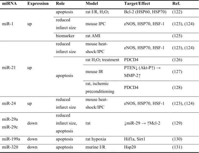
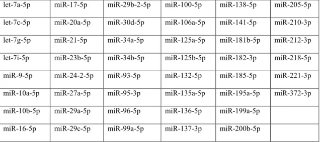
The first studies regarding the role of miRNAs in ischemia-reperfrusion (I/R) injury were carried out mainly on acute myocardial infarction (AMI) models and initially there were no information about the kidneys. These AMI associated miRNAs are presented in Table 2, and they served as a guide for our preliminary experiments.
Meanwhile, evidence to the importance of miR-24 in I/R injury also grew (118). It has been shown that miR-24 was critically involved in endothelial apoptosis during cardiac I/R-injury as well as apoptosis of cancer and T cells (119), (120), (121).
23
Table 2. List of the first identified miRNAs in relation to ischemia-reperfusion (of the heart, in acute myocardial infarction).
miRNA Expression Role Model Target/Effect Ref.
miR-1 up
apoptosis rat I/R, H2O2 Bcl-2 (HSP60, HSP70) (122) reduced
infarct size mouse IPC eNOS, HSP70, HSF-1 (123), (124)
biomarker rat AMI (125)
miR-21 up
reduced infarct size
mouse heat-
shock/IPC eNOS, HSP70, HSF-1 (123), (124)
apoptosis
rat H2O2 treatment PDCD4 (126) mouse IR PTEN↓ (Akt-P↑) →
MMP-2↑ (127)
rat, ischemic
preconditioning PDCD4 (128)
miR-24 up reduced
infarct size
mouse heat-
shock/IPC eNOS, HSP70, HSF-1 (123), (124) miR-29a
miR-29c down
reduced infarct size, apoptosis
rat ↓miR-29 → ↑Mcl-2 (129)
miR-199a down apoptosis rat hypoxia Hif1a, Sirt1 (130)
miR-320 down apoptosis murine I/R Hsp20 (131)
The role of miRNAs in the I/R-induced AKI was first reported in a proximal tubular cell targeted Dicer knockout mouse model, in which microRNA depletion attenuated renal ischemic damage (132). Unfortunately in the first two microarray studies performed on post-ischemic kidney no overlap in miRNA expression pattern could be found (132), (133). Later, two other microarray studies were published, which confirmed miR-21, miR-362, miR-685 and miR-1894-3p upregulation, and miR-805 downregulation (132), (133), (134), (135). Godwin et al. reported the course of changes in the expression of miRNAs differentially expressed after I/R injury (133), however only the histological damage and immune infiltration were assessed without measuring kidney function in this study. This same group also emphasized a prominent role for miR-21 in the prevention of tubular cell death after hypoxia in vitro (133), and further investigated the data with principal component analysis (PCA) combined with spherical geometry and found that after I/R injury there is a distinct miRNA expression pattern compared to sham
24
controls (136). MiR-21 also contributed to the beneficial effects of the delayed renal ischemic- preconditioning (137), (138). Others found, that miR-494 promoted inflammation and apoptosis and thus aggravated kidney injury in AKI by targeting ATF3, a stress response molecule (139). The HIF1α regulated miR-127 was also shown to contribute to renal I/R-injury in rats through modulation of cell trafficking, evidenced on proximal tubule cells in vitro (140). Another hypoxia inducible miRNA: miR-210 (141) was shown to be involved in the angiogenic processes during renal I/R injury, by enhancing VEGF and VEGFR2 expression (134). Vascular regeneration after ischemic AKI was also achieved with hematopoietic over-expression of miR-126, which reduced kidney injury and improved kidney function as well (142). An interesting study by Cantaluppi et al. found, that an injection of microvesicles, derived from cultured endothelial cell progenitors ameliorated kidney injury (143). However, if endothelial cells were previously transfected with miR-126 and miR-296 substantial reduction in the described protective effect could been observed (143). The beneficial paracrine effect of vesicle-transported miRNAs in AKI is further supported by the discovery of non-platelet RNA-containing particles which were found to mediate kidney regeneration (144).
Besides their involvement in the pathological processes, miRNAs were also evaluated as biomarkers in renal I/R induced AKI. MiR-10a and miR-30d not only increased in the urine, but also correlated with the injury severity in ischemia and STZ induced AKI mouse model (145). In AKI patients urine miR-21 and miR-155 just changed slightly (146), but miR-494 showed a more prominent increase compared to healthy controls (139). Furthermore, miR-21 might also serve as a prognostic marker after cardiac surgery-induced AKI (147).
25
Objectives
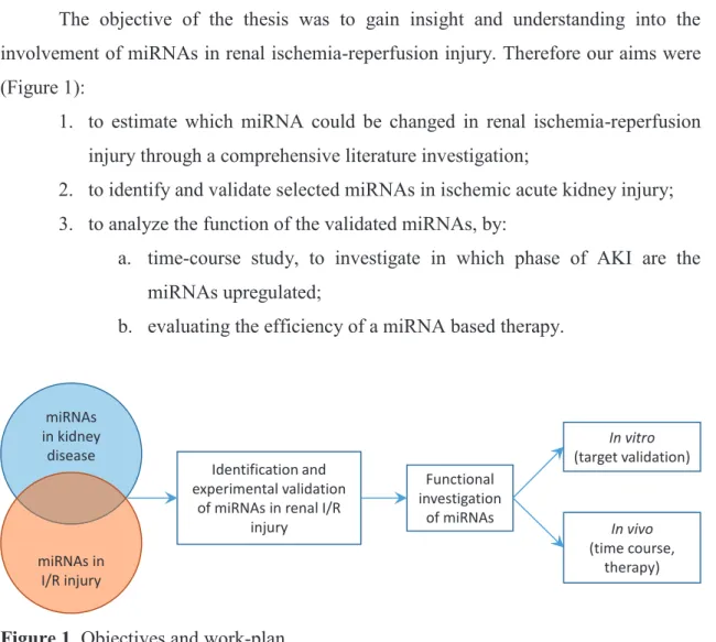
The objective of the thesis was to gain insight and understanding into the involvement of miRNAs in renal ischemia-reperfusion injury. Therefore our aims were (Figure 1):
1. to estimate which miRNA could be changed in renal ischemia-reperfusion injury through a comprehensive literature investigation;
2. to identify and validate selected miRNAs in ischemic acute kidney injury;
3. to analyze the function of the validated miRNAs, by:
a. time-course study, to investigate in which phase of AKI are the miRNAs upregulated;
b. evaluating the efficiency of a miRNA based therapy.
Figure 1. Objectives and work-plan.
The thesis has three major parts based on the published articles. First, the comprehensive literature investigation, partly presented in the introduction was published as a review article about the involvement of miRNA networks in kidney diseases. Second, the identification of the miR-17 family in a murine renal I/R injury model and the time- course study of several miRNAs, carried out in the laboratory of Dr. Péter Hamar, Semmelweis University resulted in an original research article (further referred to as:
miR-17 study). The investigation of miR-24 in ischemic AKI, containing target validation
and in vivo evaluation of a miR-24 based therapeutic strategy, was carried out in the laboratory of Prof. Dr. Thum Thomas, Hannover Medical School and lead to another research article (further referred to as: miR-24 study).
miRNAs in kidney
disease
miRNAs in I/R injury
Identification and experimental validation
of miRNAs in renal I/R injury
Functional investigation
of miRNAs
In vivo (time course,
therapy) In vitro (target validation)
26
Methods Patients Patients Patients Patients
Renal biopsies from kidney transplant patients were obtained from the Interdisciplinary Transplant Center for Clinical Research, Hannover Medical School.
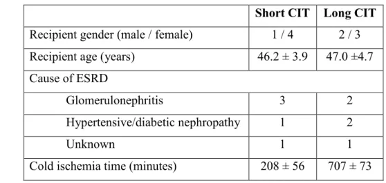
From the available biopsies with prolonged (12 ± 1 hours, n=5) and short (3.5 ± 1 hours, n=5) cold ischemia time (CIT), clinical and demographic data were collected. The patient study was confirmed by the Ethics Committee of the Hannover Medical School and all patients gave their written informed consent (approval number: 2765). Patient characteristics are shown in Table 3.
Table 3: Demographics of transplant patients with long and short cold ischemia time (CIT)
Short CIT Long CIT Recipient gender (male / female) 1 / 4 2 / 3 Recipient age (years) 46.2 ± 3.9 47.0 ±4.7 Cause of ESRD
Glomerulonephritis 3 2
Hypertensive/diabetic nephropathy 1 2
Unknown 1 1
Cold ischemia time (minutes) 208 ± 56 707 ± 73
Animals Animals Animals Animals
Male C57BL/6 mice (Charles River, Germany) were maintained under specific pathogen-free (SPF) conditions with access to standard rodent chow (Altromin standard diet, Germany) and tap water ad libitum. Mice weighing 29.4±2.9 g were used in the miR- 17 study, and approval from the Semmelweis University Animal Research Ethical Committee has been obtained under the registration no.: XIV-I-001/2103-4/2012. Mice that were 10 to 12 weeks old weighing 20 to 30 g were used for experiments carried out in the miR-24 study, and the animal experimental procedures were in agreement with institutional and legislational regulations and were approved by the local authorities (approval number: 08/1434). In vivo studies conformed to the Guide for the Care and Use
27
of Laboratory Animals published by the US National Institutes of Health (NIH publication No. 85-23, revised 1996).
LNA LNA LNA
LNA----modified miRNA oligonucleotides modified miRNA oligonucleotides modified miRNA oligonucleotides modified miRNA oligonucleotides
The oligonucleotides were provided by miRagen Therapeutics (Boulder, CO 80301, U.S.A.). The antimiR-24 is a 16-mer oligonucleotide chemistry composed of LNA and DNA directed against base 2-17 of mature miR-24. In brackets the mature miR-24 sequence is given, underlined are the binding sites of the LNA-modified antimiR directed against miR-24 (GACAAGGACGACUUGACUCGGU). The control antimiR has a comparable chemical composition but is directed against a microRNA expressed in Caenorhabditis elegans.
Kidney ischemia Kidney ischemia Kidney ischemia
Kidney ischemia----reperfusion injury reperfusion injury reperfusion injury reperfusion injury
Renal ischemia-reperfusion (I/R) injury was performed as established in our lab and described previously (36), (148). Briefly, the experiments were carried out at room temperature 24±0.5 °C using standard operating procedures. The intra-abdominal temperature was maintained using a heating pad (HK-3, DOPS, Czech Republic). For surgeries in the miR-17 study, the animals were narcotized with an intraperitoneal (i.p.) injection of 80 mg/kg ketamine and 4 mg/kg xylazin cocktail (CP-Pharma Handelsgesellschaft mbH, Germany). After median laparotomy, the left renal pedicle was prepared and clamped, to obtain sublethal (20 min) and lethal (30 min) ischemia as determined in previous studies (36), (149). After removing the clamps, we observed if the kidneys are properly reperfused. The right kidney was then removed. Sham operated control mice were also prepared in the same manner as those subjected to I/R except that the renal pedicle was not clamped. Sterile physiological salt solution was used for moistening the exposed viscera. The abdominal muscles and the skin were sutured separately with 3-0 vicryl suture (Ethicon Inc., Mexico).
In the miR-24 study the surgical interventions were carried out following isoflurane anesthesia. As in the miR-17 study, following median laparotomy, the left renal pedicle was prepared and the vascular clamp was applied for 27 minutes. Mice were dosed with intraperitoneal injections (i.p.) of a locked nucleic acid (LNA) targeting miR-24
28
(LNA-24) as well as control (mismatch) LNA (LNA-CTR) at a concentration of 10 mg/kg 24 hours before the operation (n=20/group). Survival analyses and a part of the animal operation were performed at Phenos GmbH, Hanover, Germany. For the survival analysis, bilateral renal I/R or sham (n = 4) operations were performed. During the bilateral renal I/R first the left, then the right renal pedicle were prepared. The vascular clamps were placed within 15 seconds on the left and the right renal pedicle. After 27 minutes ischemia the clamps were removed, in the same order as positioned. Proper reperfusion of the kidneys was then observed. In the sorting experiment the renal pedicle was only clamped on the left side (unilateral I/R-injury). In this setting the contralateral kidney served as control to the injured kidney (I/R-kidney). The animals were sacrificed on day 1, day 3 and day 7 after renal I/R injury (n=7/group/timepoint).
Sacrifice, blood and organ collection Sacrifice, blood and organ collection Sacrifice, blood and organ collection Sacrifice, blood and organ collection
In the miR-17 study, animals were sacrificed at the specified time-points under ether anesthesia. Right before sacrifice, 500 U/mouse heparin (Merckle GmbH., Germany) was injected i.p., and blood was collected from the thoracic cavity after cross- sectioning the vena cava. After blood urea measurements blood was centrifuged at 1500 g for 8 min at 4 °C to obtain plasma for later analysis. The kidney was removed after a slow transcardiac perfusion with 20 ml physiological salt solution, pre-cooled to 4 °C.
Plasma and renal tissue samples were snap frozen in liquid nitrogen and kept at -80 °C until use. In the miR-24 study kidney tissue samples and blood were harvested on days 0, 1, 3 and 7.
Ex vivo cell purification/sorting Ex vivo cell purification/sorting Ex vivo cell purification/sorting Ex vivo cell purification/sorting
The cellular origin of miR-24 following induction of I/R-injury was investigated by fluorescence-associated cell sorting (FACS) analysis using specific antibodies following a protocol by Chau et al. with modifications (150). Following clamping of the right renal pedicle for 27 minutes and a reperfusion period of 1, 3 and 7 days both kidneys were extracted, de-capsulated, homogenized, then incubated at 37ºC for 45 min with CollagenaseII (81 U/ml) in Hank’s Balanced Salt Solution (HBSS, Gibco). After filtration
29
(70µm), cells were centrifuged and then re-suspended in 1 ml FACS buffer (Millipore) containing 1% BSA.
Cells were separated using the following specific antibodies or lectins: rat antimouse-CD31-PE (1:400 BD Pharmingen) for endothelial cells, Lotus tetragonolobus agglutinin (LTA) (1:200, DAKO) for proximal epithelium and anti-mouse Tim1-biotin (1:200, E Bioscience) followed by streptavidin-APC (1:1000, BD Pharmingen) for injured proximal epithelium. PDGF-Receptor beta+ pericytes were separated from kidneys following a protocol by Schrimpf et al (151). Cells were incubated with rabbit anti-PDGF Receptor beta antibodies (Abcam) for 15 minutes on ice. After washing, cells were incubated with goat anti-rabbit IgG microbeads (Miltenyi Biotech) (15 minutes at 4°C) and resuspended and isolated by MACS magnetic bead separation.
Plasma Urea and NGAL ELISA Plasma Urea and NGAL ELISA Plasma Urea and NGAL ELISA Plasma Urea and NGAL ELISA
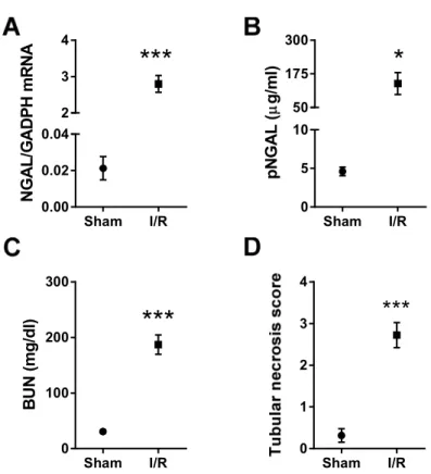
Renal function was evaluated by determination of blood urea nitrogen (BUN) retention. Blood urea levels were measured from 32 µl whole blood obtained during organ collection with Reflotron® Urea test strips (Roche Diagnostics GmbH, Mannheim, Germany) on Reflotron® Plus device (Roche Diagnostics GmbH, Mannheim, Germany) as described in the manufacturer’s protocol. Urea values were divided by 2.14 to obtain BUN levels. In the miR-24 study, renal function parameters (serum-urea and –creatinine) were analyzed on a Beckman Analyzer (Beckman Instruments GmbH, Munich, Germany).
Neutrophil gelatinase associated lipocalin (NGAL, Lipocalin-2) has been demonstrated to be a sensitive marker of tubular epithelial damage (152). The plasma NGAL level was determined with a mouse Lipocalin-2/NGAL DuoSet ELISA Development kit (R&D Systems, USA) as described by the manufacturer. Shortly, the 96 well plates (Nunc™ GmbH & Co. KG, Langenselbold, Germany) were coated with the capture antibody, and the non-specific binding sites were blocked with reagent diluent (1% BSA in PBS, pH 7.2 - 7.4). Adequately diluted (103 fold for sham- and 105 fold for ischemic-) samples were incubated on the plate in duplicates for 2 hours, and then the detection antibody was added. Next, Streptavidin-HRP was linked to the biotinylated detection antibody, followed by a short incubation with TMB Substrate (Sigma-Aldrich Chemie GmbH, Germany). A washing session (5 times with 300 µl of washing buffer)
30
was performed after each step until the addition of the substrate solution. The enzymatic reaction was terminated by stop solution containing H2SO4. The optical density was measured with Victor3™ 1420 Multilabel Counter (PerkinElmer, WALLAC Oy, Finland) at 450 nm with wavelength correction set to 544 nm. The NGAL concentrations were calculated with WorkOut software (Dazdaq Ltd., England), using a four parameter logistic curve-fit.
Histology and immu Histology and immu Histology and immu
Histology and immunohistochemistry nohistochemistry nohistochemistry nohistochemistry
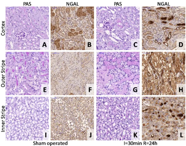
Renal tissue samples fixed in 4% buffered formaldehyde were dehydrated and embedded in paraffin wax (FFPE) for histology and immunohistochemistry. Kidney samples from the miR-17 study were evaluated morphologically by tissue microarray (TMA) as described previously (153). Briefly, blocks of 70-sample TMAs contained duplicates of 2 mm diameter cylinders cut by the computer-controlled puncher of the TMA Master Device (3DHISTECH Kft, Budapest, Hungary) from the I/R sensible cortico-medullary junction of each FFPE kidney. For morphology and immunohistochemistry, 4 µm thick sections were cut from the TMA blocks. Renal tubular necrosis and regeneration were evaluated in Periodic acid-Schiff (PAS) stained TMA sections. A histological score of 0 to 4 was given by a pathologist blinded to the origin of the tissue as follows: 0= no lesion; 1= minimal or focal changes affecting less than 20%
of the field; 2= mild changes or the extension of the lesion/regeneration to approx. 25%
of the field; 3= moderate changes or the extension of the lesion/regeneration to less than 50% of the field; 4=severe changes or the extension of the lesion/regeneration to more than 50% of the field).
Renal tubular cell damage was evaluated by neutrophil gelatinase-associated lipocalin (NGAL, Lipocalin-2) immunostaining. Dewaxed and rehydrated TMA sections were cooked at 100 °C for 25 min in a 0.01M Tris-HCl and 0.1 EDTA buffer (TBS; pH 9.0) for antigen retrieval. The immunostaining involved consecutive incubations of TMA sections in 1% bovine serum albumin (BSA) in TBS (pH 7.4) for 15 min, rabbit anti- human NGAL IgG (1:100; R&D Systems, Minneapolis, USA) for 16 h and in goat anti- rabbit IgG EnVision-peroxidase polymer kit (Dako, Glostrup, Denmark) for 40 min, all at room temperature. Tissue-bond peroxidase activity was developed with a DAB/H2O2
chromogen/substrate kit (Dako). Immunostained TMA slides were digitalized using a
31
Pannoramic Scan instrument, and the results were analyzed with the Pannoramic Viewer software (3DHISTECH).
In the miR-24 study the severity of morphologic renal damage was assessed in a blinded manner using an arbitrary score based on HE-stained kidney sections following a modification of a protocol developed by Broekema et al (154). Briefly, the extent of four typical I/R injury–associated damage markers (i.e., dilatation, denudation, intraluminal casts, loss of brush border membrane and cell flattening) was expressed in arbitrary units (AU) in a range of 0 to 4 according to the percentage of damaged tubules within a high power field of view: 0: no damage; 1: less than 25% damage; 2: 25%–50%
damage; 3: 50%–75% damage; and 4: more than 75% damage.
In the miR-24 study immunostainings were performed in cryosections. For inflammatory cell influx the following primary antibodies were used: monoclonal rat anti- mouse F4/80 for macrophages (Serotec, Oxford, United Kingdom), monoclonal rat anti- mouse CD45 for leucocytes (BD Pharmingen, BD Biosciences, Santa Cruz, CA), affinity- purified rat anti-mouse Ly-6G/Gr-1 for neutrophils (eBioscience, San Diego, CA), purified Rat Anti-Mouse CD4 for T helper lymphocytes (BD Pharmingen, BD Biosciences, Santa Cruz, CA). Capillary rarefaction in the outer medulla was evaluated after fluorescent immunohistochemical staining for polyclonal rabbit anti-mouse CD31 for endothelial cells (Abcam, Cambridge, UK). Deparaffinized kidney sections were boiled in citrate buffer for antigen retrieval, blocked with 5% milk, and incubated overnight at 4°C with primary antibodies. This was followed by antibody visualization using Alexa 488/Alexa 547 secondary antibodies (Molecular Probes/Invitrogen, Carlsbad, CA). Quantification of CD45-, F4/80-, CD31-, CD4-, and Ly-6G-expressing cells was done by counting of positive cells in ten randomly chosen, non-overlapping fields in the outer medulla. A fluorescein in situ cell death detection kit was used according to the manufacturer’s instructions for Terminal deoxynucleotidyl transferase dUTP nick end labeling (TUNEL) assay (Roche Applied Science, Mannheim, Germany).
TUNEL-positive tubular cells and total DAPI (4’,6-diamidino-2-phenylindole)-positive tubular cells were counted in ten non-overlapping fields of outer medulla in each sample.
Data are presented as a percent ratio of TUNEL-positive epithelial cells versus total DAPI-positive epithelial cells.
32
Cell Cultur Cell Cultur Cell Cultur
Cell Culture experiments e experiments e experiments e experiments
For in vitro analyses immortalized human kidney proximal tubular epithelial cells (HK-2) were used. HK-2 cells were maintained in Keratinocyte Growth Medium 2 with supplements.
Cells were grown to 60% to 70% confluence and used for further analyses.
Apoptosis was determined by TUNEL staining (Roche Applied Science, Mannheim, Germany). All assays were done according to the manufacturer’s instructions.
Transfection Assays Transfection Assays Transfection Assays Transfection Assays
Transient liposomal transfection of miRNAs was performed according to the
manufacturers’ instructions. Briefly, cells were split 1 day before transfection to reach 60% to 70% confluence on the day of transfection. Specific pre-miRNAs and control miRNA (pre-neg) and Lipofectamine 2000 (Invitrogen) were mixed separately and incubated for 5 minutes with Opti-MEM I media (Invitrogen). Complexes were added together and incubated for 20 minutes. Media were changed to antibiotic-free media before the addition of liposomal miRNA complexes (final miRNA concentration: 100 nmol/L ). Cells were incubated for 4 hours before the media were changed to fresh media. Silencing miRNA targets was monitored for 72 hours after transfection by Western blot analysis.
Scratch wound healing assay of HK Scratch wound healing assay of HK Scratch wound healing assay of HK
Scratch wound healing assay of HK----2 cells 2 cells 2 cells 2 cells
Transfected HK-2 cells were cultivated in human keratinocyte medium at 37°C, 5% CO2. The scratches in the cell monolayer were generated with a 100-µl pipett-tip, and the cells were photographed at 0, 8, and 24 hours with a Nikon Ti 90 microscope (Germany). Subsequently, the cell free area was calculated.
Protein Analysis Protein Analysis Protein Analysis Protein Analysis
Downstream mechanisms were investigated by Western blot analysis using 10 to 40 µg of total protein. Tissue was homogenized, cells were pelleted. Cell lysis was performed (Cell lysis buffer, Cell Signaling, Technology, Danvers, MA, U.S.A.) and protein electrophoresis initiated. Proteins were transferred to polyvinylidene difluoride (PVDF) membranes, blocked with 5% milk in TBS-Tween, and probed overnight at 4°C