ORIGINAL ARTICLE
Dual-Stained Cervical Cytology and Histology with Claudin-1 and Ki67
Tímea Szekerczés1&Ádám Galamb2&Adrienn Kocsis3,4&Márta Benczik3,4,5&Tibor Takács3,4,5&Attila Martonos2&
Balázs Járay1&András Kiss1&Csaba Jeney3,6&Miklós Nyíri3,4&Zsuzsa Schaff1 &Gábor Sobel2
Received: 18 October 2017 / Accepted: 30 January 2018 / Published online: 13 February 2018
#Arányi Lajos Foundation 2018
Abstract
Several biomarkers are in use to improve the sensitivity and specificity of cervical cancer screening. Previously, increased expres- sion of tight junction protein claudin-1 (CLDN1) was detected in premalignant and malignant cervical lesions and applied for cytology screening. To improve the specificity, a double immunoreaction with CLDN1/Ki67 was developed in the recent study.
Parallel p16/Ki67 (CINtec®PLUS) and CLDN1/Ki67 dual-stained cytology and histology were performed and compared. p16/
Ki67 immunoreaction showed positivity in 317 out of 1596 smears with negativity in 1072 and unacceptable reactions in 207 samples. CLDN1/Ki67 dual staining was positive in 200 of 1358 samples, negative in 962, whereas 196 smears could not be evaluated due to technical reasons. Considering the high-grade squamous intraepithelial lesion cytology as gold standard, sensitivity of CLDN1/Ki67 reaction was 76%, specificity was 85.67%, while for p16/Ki67 sensitivity was 74% and specificity was 81.38%.
Comparison of CLDN1/Ki67 and p16/Ki67 dual stainings showed the results of the two tests not to be significantly different.
Analysing histological slides from 63 cases, the results of the two tests agreed perfectly. As conclusion the sensitivity and specificity proved to be similar using p16/Ki67 and CLDN1/Ki67 double immunoreactions both on LBC samples and on histological slides.
Keywords Cervical cancer . Claudin-1/Ki67 immunochemistry . p16/Ki67 reaction . Claudins
Introduction
Papanicolaou (Pap) smear-screening significantly reduced the incidence and mortality of cervical cancer, however, because of the low sensitivity and poor reproducibility in contrast to the high specificity, moreBsophisticated^methods have been introduced in the last decades and new screening guidelines have been proposed [1–6].
High-risk human papillomaviruses (hrHPV) play crucial role in the development of premalignant and malignant cervi- cal lesions and HPV DNA-based testing has significantly in- creased the sensitivity of primary cervical screening [3,5,7].
HPV testing however, is less specific than cytology, mainly because the majority of infections are transient and spontane- ously eliminated in the majority of cases [3,6,8].
New biomarkers aiming to improve the specificity of screening have been developed, which might be used to dif- ferentiate the productive and transforming HPV infection and/
or predict disease severity [6,9,10]. Several new tests were introduced, such as p16INK4A, dual p16INK4a/Ki67 staining, ProexC (combined MCM2/TOP2A), etc. [6,10,11].
Previously, our group as well as others demonstrated sig- nificantly increased expression of the tight junction (TJ) pro- tein claudin-1 in premalignant and malignant cervical lesions [12–14]. Claudin-1 (CLDN1) as a biomarker has been sug- gested to be used for both cytology and histology with similar diagnostic potential as p16INK4a [9]. Claudins are the main functional and structural components of TJs, playing role in paracellular permeability, maintaining cellular polarity and participating in signal transduction [15]. The limitation of our previous study was the relatively low specificity of CLDN1 staining similarly to p16INK4a testing [9]. For this Tímea Szekerczés and Ádám Galamb equally contributed
* Zsuzsa Schaff
schaff.zsuzsa@med.semmelweis-univ.hu
1 2nd Department of Pathology, Semmelweis University, Üllői út 93, Budapest H-1091, Hungary
2 2nd Department of Obstetrics and Gynecology, Semmelweis University, Budapest, Hungary
3 Cellcall Ltd., Budapest, Hungary
4 NEUMANN Diagnostics Ltd., Budapest, Hungary
5 SYNLAB Hungary Ltd., GenoID Molecular Diagnostic Laboratory, Budapest, Hungary
6 Department of Medical Microbiology, Semmelweis University, Budapest, Hungary
https://doi.org/10.1007/s12253-018-0384-x
reason, the aim of the present study was to test a double im- munoreaction using antibodies against CLDN1 and the pro- liferation marker Ki67 on cytological and histological sam- ples, in order to improve specificity. The results of the p16/
K i 6 7 ( C I N t e c® P L U S ) a n d C L D N 1 / K i 6 7 d u a l immunostainings were compared with each other, as well as with the cervical cytological readings and also with the histo- logical results, and then analysed statistically.
Materials and Methods
Patient Population and Tissues Samples
An outpatient population of 2907 women between 18 and 65 years of age who attended routine gynaecological screen- ing between 2013 and 2015 were enrolled in the TRACE clinical study conducted in Hungary [11] (Fig.1). All clinical samples were obtained with the approval of the National Ethics Committee (asset number: V-R-021/04346–4/2013), all patients gave informed consent and the trial was performed in accordance with the Declaration of Helsinki. Cervical cy- tology samples were collected after colposcopy assisted visual inspection of the cervix by gynecologists and were kept in preservation solution for subsequent liquid-based cytology (LBC) analysis. When required by the cytological diagnosis, confirmatory histological diagnosis was made (cone or punch biopsy).
Cytological Diagnosis
All samples obtained for cytology were collected in PreservCyt® Solution (Hologic™Inc. Marlborough, MA,
USA) and processed using ThinPrep® 2000 Processor (Hologic™Inc.). The first slide from each vial was taken for cytology smears, Pap-stained and reported using the 2001 Bethesda Reporting System [16,17]. Further ThinPrep slides were prepared from the same container for immunocytochem- ical reactions and for subsequent HPV testing (see below).
ThinPrep smears were evaluated if at least 500 cervical epi- thelial cells were present.
HPV Testing
HPV viral DNA detection was performed on LBC samples using CONFIDENCE HPV™test, which detects HPV16 and HPV18 separately and other high-risk types in groups based on multiplex real-time PCR technology [11].
Immunocytochemistry and Immunohistochemistry Parallel ThinPrep slides from the residual material from each vial were used for the immunoreactions (CINtec® PLUS, CLDN1/Ki67 sequence). For immunohistochemistry the 3–4 μm thick, formalin fixed paraffin embedded (FFPE) sections were cut and prepared further.
CINtec®PLUS Immunoreaction (p16
INK4a/Ki67 Testing)
ThinPrep slides were prepared for CINtec®PLUS (Roche mtm Laboratories AG, Mannheim, Germany) reaction accord- ing to the manufacturer’s instructions. When accessing the reactions, the cases were called positive if one or more cell(s) out of at least 500 normal or altered squamous epithelial cells
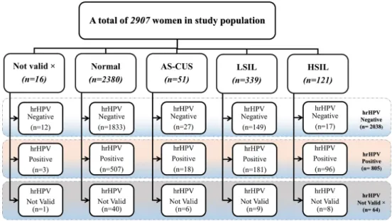
Fig. 1 Cytology (liquid-based cytology) diagnosis and hrHPV testing of the study population.
hrHPV = high-risk human papilloma virus, AS-CUS = atypical squamous cells of undetermined significance, LSIL = low-grade squamous intraepithelial lesion, HSIL = high-grade squamous intraepithelial lesion
on the slides stained both with a brown cytoplasmic/nuclear (p16) and a red nuclear (Ki67) reaction irrespective of the interpretation of morphologic abnormalities.
CLDN1/Ki67 Immunoreaction
Parallel LBC (3rd slide from the collection vials) and FFPE slides were prepared for CLDN1 and Ki67 reactions.
Similarly as in the case of CINtec® PLUS reaction, a case was considered positive if one or more cervical epithelial cell(s) had both a brown membrane stain (CLDN1) and a red nuclear reaction (Ki67).
CLDN1 reaction was carried out in a Ventana ES automatic immunostainer (Ventana Medical Systems Inc.; Tucson, AZ, USA). The slides were incubated for 30 min at 42 °C with the primary polyclonal rabbit antibody against CLDN1 in 1:100 dilution (Cell Marque, Roclin, CA, USA), followed by a HRP multimer-based, biotin-free detection method. Secondary an- tibody and reagents were obtained from Ventana (iView DAB Detection Kit; Ventana Medical Systems Inc.). Bluing reagent was not used because CLDN1 immunoreactions were follow- ed manually by Ki67 reaction. In the first step, antigen retriev- al was used again for 10 min at 95 °C with Antigen Unmasking Solutions (Vector Laboratories, Burlingame, CA, USA, in 1:100 dilution). Thereafter the slides were incu- bated for 30 min with the mouse monoclonal primary anti- body against Ki67 (Dako, Glostrup, Denmark) diluted 1:100, at room temperature. The secondary antibody reagent (ImmPRESS AP Reagent; Vector Laboratories) included a polymer reagent conjugated to alkaline phosphatase (AP) and goat anti-mouse Fab′ antibody fragments for detection of Ki67, applied for 30 min at room temperature (1:100 dilu- tion). Vector Red Alkaline Phosphatase Substrate Kit (Vector Laboratories) was applied for 25 min using Fast Red as chro- mogen. Finally, the slides were counterstained with alcohol- free hematoxylin and coverslipped with xylene-based Cytoseal XYL Mounting Medium (Richard-Allan Scientific, Thermo Scientific, Kalamazoo, MI, USA).
Statistical Analysis
Statistical analysis was carried out using STATISTICA soft- ware v12.0 (StatSoft; Tulsa, OK, USA) and MedCalc (v12.4.0.0; MedCalc Software, Mariakerke, Belgium).
Differences were considered statistically significant when p< 0.05. McNemar’s test was used to evaluate differences in positivity for p16/Ki67 and CLDN1/Ki67 in cytological sam- ples. Kappa test was used to measure the agreement of the two immunochemical tests. Sensitivity, specificity, positive pre- dictive value (PPV), negative predictive value (NPV), with 95%-confidence intervals were calculated for endpoint which was CIN2/CIN2+ (cervical intraepithelial neoplasia,
high-grade squamous intraepithelial lesion - HSIL). Receiver Operating Characteristic(ROC) curves and Area under the curve (AUC) were computed to further compare CINtec® PLUS with CLDN1/Ki67.
Results
1. Patient Population, Cytology and Histology
Out of a total of 2907 women who underwent LBC for routine cervical cancer screening, 2891 samples provided acceptable results, from which 2380 proved to be normal as shown together with the results of hrHPV testing (Fig.1).
High-risk HPVs (hrHPV) were detected in 805 samples, negative results were found in 2038 samples and no accept- able results were received in 64 cases (Fig.1). The diagno- sis of the 63 histological samples is demonstrated on Table1.
2. Comparing Cytology and Histological Diagnosis to p16/
Ki67 (CINtec®PLUS) and CLDN1/ Ki67 Dual-Stainings a) Comparing Cytology Diagnosis and p16/Ki67 (CINtec®
PLUS) Reaction
CINtec® PLUS reactions were performed in 1596 cases (Table 2). The positive reaction presented a brownish cytoplasmic/nuclear reaction for p16 and a red immunostain- ing for Ki67 simultaneously in one or more cervical epithelial cells (Fig. 2a). Out of the 1596 samples, 1386 presented evaluable results with both Pap test and CINtec®PLUS reac- tions, in 207 cases one of the tests could not be evaluated because of technical reasons (Table2).
b) Comparing Cytology Diagnosis with CLDN1/Ki67 CLDN1-positive cells gave an intense brown linear circular/semi-circular or spotted reaction along the cellular membranes, sometimes in the cytoplasm, together with a bright red nuclear Ki67 reaction in the same cells (Fig.2b, c, d; Fig. 3a, b). CLDN1/Ki67 dual-immunostaining was per- formed in 1358 cases and 1159 provided acceptable immuno- staining as shown on Table3.
c) Comparing Histology Diagnosis with CLDN1/Ki67 and p16/Ki67 (CINtec®PLUS)
Parallel slides from the FFPE blocks were used for immu- nohistochemistry. Ki67 positivity was seen in the cells of the basal layer in normal cervical squamous epithelium both by CINtec® PLUS and by CLDN1/Ki67 reaction (Fig.4a, b).
Brownish reaction for p16 (Fig.4c) and CLDN1 (Fig.3c, d;
Fig.4d) and numerous Ki67 positive red nuclei could be dem- onstrated by both tests in the CIN2+ lesions. By comparison of the two tests (CINtec®PLUS and CLDN1/Ki67), the results agreed perfectly (Table1).
3. Comparing Test Performance for CLDN1/Ki67 and p16/
Ki67 (CINtec® PLUS) Considering the Cytological Evaluation
CINtec®PLUS and CLDN1/Ki67 could be performed par- allel in 1352 cases. Both reactions were accepted in 1097 cases (Table4). Assessment of the two immune tests equalled in 1003 samples (840 negative and 163 positive, 91.4%) (Table4). The value of agreement between the two tests by Kappa tests and the result of the two tests agreed accordingly (κ= 0.724; 95% CI from 0.672 to 0.776).
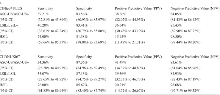
Furthermore, comparison of the test performances for CINtec® PLUS and CLDN1/Ki67 considering cytological evaluation and efficiency values [sensitivities, specificities, positive (PPVs), and negative predictive values (NPVs)] for all screening methods to detect ASC-US, LSIL or HSIL is summed in Table5a and b. It is worthy of note that among women with LSIL cytology CINtec®PLUS showed slightly higher sensitivity than CLDN1/Ki67 (40.28 vs. 35.07%) but the specificity values were lower (83.41 vs 87.13%). For pa- tients with HSIL, the sensitivity and specificity values reached a good, evaluable range, over 70–80%. CLDN1/Ki67 showed slightly better sensitivity [76.00% (95% CI from 61.83% to 86.94%)] and specificity [85.67% (95% CI from 83.40% to 87.74%)] (Table5b) than the CINtec®PLUS values [sen- sitivity 74.00% (95% CI from (59.66% to 85.37%) and specificity 81.38% (95% CI from 78.88% to 83.69%)].
Table 1 P16/Ki67 (CINtec® PLUS) and CLDN1/Ki67 on histological section (n= 63)
Histological diagnosis
Number of cases
Average ages (yrs)
CINtec®PLUS CLDN1/Ki67
Positive Negative Positive Negative
Carcinoma in situ 7 40.57 7 0 7 0
HSIL (CIN 2 or 3) 30 33.50 30 0 30 0
LSIL (CIN 1) 3 42.33 0 3 0 3
No dysplasia, chronic cervicitis
23 35.61 0 23 0 23
CLDN1claudin-1,HSILhigh grade squamous intraepithelial lesion,LSILlow grade squamous intraepithelial lesion,CINcervical intraepithelial neoplasia
Table 2 Results of p16/Ki67 (CINtec®PLUS) on LBC specimens.
AS-CUSatypical squamous cells of undetermined significance,LSILlow grade squamous intraepithelial lesions,HSILhigh grade squamous intraepithelial lesion
Regarding the efficiency of the CINtec®PLUS and CLDN1/
Ki67 test analysis, Receiver Operating Characteristic (ROC) curves were demonstrated to assess differences between the two immunoreaction performances. The result of the analysis are given in Fig.5. Data were similar to the previous compar- ison above, CLDN1/Ki67 had slightly higher curve [AUC = 0.806 (95% CI from (0.781 to 0.829)] than CINtec®PLUS [AUC = 0.774 (95% CI from 0.748 to 0.798)], but no signifi- cant difference (p= 0.177) was found between the two tests since the difference area was only 0.0317.
Discussion
Several biomarkers have been introduced for cervical cancer screening so as to improve sensitivity and specificity as com- pared with Pap cytology readings and HPV testing [10,18,19].
Among these, immunochemical methods are already commer- cially available and several studies have proved their usefulness in diagnostic practice [20].
Detection of p16INK4ais one of the extensively evaluated methods for recognition of altered dysplastic cervical cells,
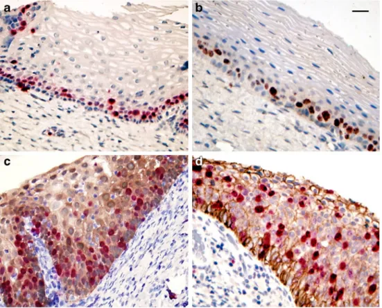
Fig. 3 CLDN1/Ki67 immunreactions on LBC (a,b) and histological slides (c,d). Red nuclei and brown membranous staining show positive cells.
CLDN1 = claudin-1, LBC = liquid- based cytology. Scalebar:
35μm (a,b) and 50μm (c,d) Fig. 2 Dual immunoreactions on LBC (ThinPrep) cytology slides (a). p16/Ki67 (CINtec®PLUS) reaction shows positive cells with red nuclei and brown cytoplasmic staining (a). CLDN1/Ki67 positive cells have red nuclei and brown membranous linear or spotted staining (b,c,d). LBC = liquid-based cytology, CLDN1 = claudin-1. Scalebar: 35μm
and the specificity of the reaction increased significantly after being combined in a dual immunostaining with Ki67 [5,6, 20–22]. The results have been confirmed in a multicentric, prospective, pan-European study (PALMS study), which
found that positive predictive values for CIN2+ were higher for p16/Ki67 immunocytochemistry versus HC2 HPV testing [10]. More recently it has been shown that p16/Ki67 reaction is associated with hrHPV persistence and CIN2+ lesions [21].
Fig. 4 CINtec®PLUS (a,c) and CLDN1/Ki67 (b,d)
immunoreactions on histological slides. Ki67 positive red nuclei are detected in basal cells of the normal cervical epithelium(a,b) by both stainings. Nuclear reactions are more extended in the HSIL samples together with brown p16 cytoplasmic reaction with CINtec® PLUS (c) and membranous reaction with CLDN1 (D). CLDN1 = claudin- 1, HSIL = high-grade squamous epithelial lesion. Scalebar: 50μm
Table 3 Results of CLDN1/Ki67 on LBC specimens.
CLDN1claudin-1,AS-CUSatypical squamous cells of undetermined significance,LSILlow grade squamous intraepithelial lesions,HSILhigh grade squamous intraepithelial lesion
The objective of our current study was to compare side-by- side the results of the commercially available test, p16INK4a/ Ki67 (CINtec®PLUS) dual staining with our newly devel- oped CLDN1/Ki67 double immunoreaction. In our previous study the detection of tight junction protein CLDN1 proved to have similar diagnostic potential as p16I N K 4 a [9].
Concordance of CLDN1 immunocytochemistry of HSIL pos- itives was 84.0%, being 69.0% for negatives, suggesting CLDN1 to be a competing marker for detection of premalig- nant and malignant cervical lesions [9].
Claudins (CLDNs) belong to the family of integral mem- brane proteins [23] and are the main components of the belt- like networks of tight junctions (TJs). TJs act as important
paracellular seals, semi-barriers for trafficking different mole- cules, ions and water and participate in the maintenance of cellular polarity. TJs are dynamic structures undergoing con- tinuousBmolecular remodelling^and respond to several stim- uli as to cytokines and growth factors or epigenetic events [24]. CLDNs interact with other proteins and participate in signal transduction through a PDZ domain, influencing cell proliferation and transformation [24]. Deregulation of TJ pro- teins - increase, decrease or loss - in association with altered cellular morphology during carcinogenesis and with the pro- gression of the lesions is well documented [13,25,26]. A significant upregulation of CLDN1 was demonstrated in pre- malignant and malignant cervical squamous [12, 14] and
Table 5 Performance of p16/Ki67 (a) and CLDN1/Ki67 (b) in abnormal cervical lessions (n=1097) a.
CINtec®PLUS Sensitivity Specificity Positive Predictive Value (PPV) Negative Predictive Value (NPV)
ASC-US/ASC-US+ 39.21% 83.56% 38.36% 84.05%
(95% CI) (32.81% to 45.89%) (80.93% to 85.97%) (32.07% to 44.95%) (81.43% to 86.42%)
LSIL/LSIL+ 40.28% 83.41% 36.64% 85.43%
(95% CI) (33.61% to 47.24%) (80.79% to 85.80%) (30.43% to 43.19%) (82.90% to 87.72%)
HSIL 74.00% 81.38% 15.95% 98.50%
(95% CI) (59.66% to 85.37%) (78.88% to 83.69%) (11.48% to 21.31%) (97.44% to 99.20%) b.
CLDN1/Ki67 Sensitivity Specificity Positive Predictive Value (PPV) Negative Predictive Value (NPV)
ASC-US/ASC-US+ 34.36% 87.36% 41.49% 83.61%
(95% CI) (28.20% to 40.93%) (84.96% to 89.49%) (34.37% to 48.89%) (81.04% to 85.96%)
LSIL/LSIL+ 35.07% 87.13% 39.36% 84.93%
(95% CI) (28.65% to 41.92%) (84.75% to 89.27%) (32.33% to 46.73%) (82.43% to 87.19%)
HSIL 76.00% 85.67% 20.21% 98.68%
(95% CI) (61.83% to 86.94%) (83.40% to 87.74%) (14.72% to 26.67%) (97.71% to 99.32%)
CLDN1claudin-1,AS-CUSatypical squamous cell of undetermined significance,LSILlow grade squamous intraepithelial lesions,HSILhigh grade squamous intraepithelial lesion
Table 4 Comparison of CLDN1/Ki67 and p16/Ki67 (CINtec®PLUS) results on LBC samples.
CLDN1claudin-1
glandular lesions [13,27], in other squamous carcinomas as in esophageal [28] cancer, in different forms of breast cancer [29] etc. Other claudins such as claudin-3 and -4 were highly expressed in cholangiocarcinomas [30], in prostate [31] and ovarian cancers [32] etc. Increased expression of certain claudins does not necessary mean theBincreased tightness^
orBbetter^function of TJs, it is rather a deregulation of the interaction between TJ proteins in the multiprotein complexes and expresses disturbed proteinBpartnering^with other pro- teins in these dynamic structures.
Based on our own previous observations as well as of others, increased expression of CLDN1 characterizes the neoplastic cervical epithelial cells, which can be demonstrated both by histology, cytology and by cDNA array technique [9,12,14, 33]. It was proved that the expression of CLDN1 increases with the severity of the cervical lesions from CIN1 to CIN2+ and invasive cancer [12]. Previously CLDN1 has been suggested to be a biomarker in cervical cytology however, in that particular study the proliferative marker was not considered [9].
In the recent study, expressions of CLDN1 and prolifera- tion marker Ki67 were combined in dual immunostaining and compared with the results of p16/Ki67 (CINtec®PLUS) reac- tion. The latter test showed a sensitivity of 74.00% and a specificity of 81.38%, while for CLDN1/Ki67 they were 76.00% and 85.67%, respectively using Pap cytology read- ings as reference.
Taking ethical questions into consideration, the first slide from each LBC vial was used for Pap cytology diagnosis evaluated by the Bethesda system, the second slide for CINtec®PLUS and the third for CLDN1/Ki67 immunoreac- tion. Those samples where the cell number was not sufficient for any of the three tests were excluded from further evalua- tion, decreasing the power of the statistical analysis.
One of the main goals of the current study was the com- parison of the recently available and widely used dual immu- nostaining method for p16/Ki67 (CINtec® PLUS) with our
newly developed reaction (CLDN1/Ki67). The relatively large number of samples available for comparing the two tests seemed to be sufficient for statistical analysis. In addition, the fact that the 3rd sample from the LBC collecting vials was used for the CLDN1/Ki67 staining, probably with a less num- ber of cells, further strengthens our results, which demonstrat- ed that no significant differences were found between the CLDN1/Ki67 and CINtec®PLUS dual stainings.
Previously, significantly increased CLDN1 expression was demonstrated with the severity and progression of histological grade during cervical carcinogenesis detected by immunohis- tochemistry [12]. In the recent study, the CLDN1/Ki67 reac- tion gave a result similar to the reaction with CINtec®PLUS.
This suggests that the CLDN1/Ki67 dual staining might be used in the future for the detection of CIN2/CIN2+ lesions on histological slides too.
A new cervical biomarker test needs to be compared with existing ones, in this case CLDN1/Ki67 to p16/Ki67 (CINtec®PLUS) for cervical immunochemistry and immuno- histochemistry, as discussed above. Wentzensen et al. [6] eval- uated the clinical relevance of the dual immunoassay with p16/Ki67 compared with Pap cytology diagnosis. In their study, the double staining demonstrated lower positivity than cytology at ASC-US threshold. For CIN2+, the double stain- ing had similar sensitivity and higher specificity as compared with cytology, and the pattern for CIN2+ was similar [6].
Bergeron et al. [10] in the PALMS study involving 5 European countries, using colposcopy-guided biopsy diagno- sis of CIN2/CIN2+ as clinical endpoints, found that the p16/
Ki67 dual staining cytology gave comparable results for ASC- US as HC2 (Qiagen, Hilden, Germany) testing and lower for LSIL, but higher specificity compared to HC2 HPV testing both for ASC-US and LSIL.
In our recent study the p16/Ki67 (CINtec® PLUS) and CLDN1/Ki67 dual reactions were compared in a case-control manner. The sensitivity and specificity of the two tests were performing equally in a large number of sample sets, which suggests that the CLDN1/Ki67 double immunoreactions might be used in the future for the detection of premalignant and malignant cervical lesions.
Author Contributions TSz performed and evaluated the immunreactions and statistics, ÁG and AM collected the clinical samples and data, BJ and AKi evaluated the cytology, ZsS and GS designed and coordinated the study and wrote the manuscript. AKo and MB are clinical experts of the TRACE clinical study (Cellcall). TT is the inventor of the HPV detection technology applied in the CONFIDENCE HPV™ (NEUMANN Diagnostics) testing. CsJ was former head of the TRACE clinical study (Cellcall). ZsS accepts full responsibility for the work. All authors have read and approved the final manuscript.
Funding This work was supported by grant #NKFP_07_2-SPE-SAFE, Jedlik Ányos 2. subprogram KMR_12–1–2012-0032 by the Hungarian National Research and Development Fund and grant #OTKA PD105019 by the Hungarian National Research Foundation.
Fig. 5 ROC analysis curves of the two dual stainings. CLDN1/Ki67 (red) has slightly higher curve than CINtec® PLUS (green), but no significant difference (p= 0.177) was found. ROC = Receiver Operating Characteristic, CLDN1 = claudin-1
Compliance with Ethical Standards
Conflict of Interest The authors declare that they have no conflict of interest with respect to the research, authorship, and/or publication of this article.
AKo is an employee of Cellcall and NEUMANN. MB receives con- sultancy fees from Cellcall and NEUMANN, she is an employee of SYNLAB GenoID Laboratory, which participated in the TRACE clinical study (all related expenses were charged to Cellcall and NEUMANN).
TT is a part time employee of Cellcall, he receives consultancy fees from NEUMANN, he is minority owner of NEUMANN, the inventor of the HPV detection technology described in the manuscript covering which Cellcall submitted a patent application (license now owned by NEUMANN Diagnostics Ltd.); he is a part time employee of SYNLAB GenoID Laboratory. CsJ was a former employee of Cellcall. MNy is an employee of Cellcall and NEUMANN.
References
1. Hillemans P, Soergel P, Hertel H, Jentschke M (2016) Epidemiology and early detection of cervical cancer. Oncol Res Treat 39:1–6
2. Sawaya GF, Smith-McCune K (2016) Cervical cancer screening.
Obstet Gynecol 127:459–467
3. Lees BF, Erickson BK, Huh WK (2016) Cervical cancer screening:
evidence behind the guidelines. Am J Obstet Gynecol 214:438–443 4. Ocque R, Austin RM (2016) Follow-up of women with negative pap test results and abnormal clinical signs or symptoms. Am J Clin Pathol 145:560–567
5. Ikenberg H, Bergeron C, Schmidt D, Griesser H, Alameda F, Angeloni C, Bogers J, Dachez R, Denton K, Hariri J, Keller T, von Knebel Doeberitz M, Neumann HH, Puig-Tintore LM, Sideri M, Rehm S, Ridder R, PALMS Study Group (2013) Screening for cervical cancer precursors with p16/Ki-67 dual-stained cytology:
results of the PALMS study. J Natl Cancer Inst 105:1550–1557 6. Wentzensen N, Fetterman B, Castle PE, Schiffman M, Wood SN,
Stiemerling E, Tokugawa D, Bodelon C, Roitras N, Lorey T, Kinney W (2015) p16/Ki-67 dual stain cytology for detection of cervical precancer in HPV-positive women. J Natl Cancer Inst 107:
1–8
7. Cuzick J, Clavel C, Petry KU, Meijer CJ, Hoyer H, Ratnam S, Szarewski A, Birembaut P, Kulasingam S, Sasieni P, Iftner T (2006) Overview of the European and north American studies on HPV testing in primary cervical cancer screening. Int J Cancer 119:
1095–1101
8. Plummer M, Schiffman M, Castle PE, Maucort-Boulch D, Wheeler CM, ALTS Group (2007) A 2-year prospective study of human papillomavirus persistence among women with a cytological diag- nosis of atypical squamous cells of undetermined significance or low-grade squamous intraepithelial lesion. J Infect Dis 195:1582– 1589
9. Benczik M, Galamb Á, Koiss R, Kovács A, Járay B, Székely T, Szekerczés T, Schaff Z, Sobel G, Jeney C (2016) Claudin-1 as a biomarker of cervical cytology and histology. Pathol Oncol Res 22:
179–188
10. Bergeron C, Ikenberg H, Sideri M, Denton K, Bogers J, Schmidt D, Alameda F, Keller T, Rehm S, Ridder R, PALMS Study Group (2015) Prospective evaluation of p16/Ki-67 dual-stained cytology for managing women with abnormal Papanicolaou cytology:
PALMS study results. Cancer Cytopathol 123:373–381
11. Kocsis A, Takács T, Jeney C, Schaff Z, Koiss R, Járay B, Sobel G, Pap K, Székely I, Ferenci T, Lai HC, Nyíri M, Benczik M (2017) Performance of a new HPV and biomarker assay in management of
hrHPV positive women: Subanalysis of the ongoing multicenter TRACE Clinical Trial (n>6,000) to evaluate POU4F3 as a potential biomarker of cervical precancer and cancer. Int J Cancer 140:1119– 1133
12. Sobel G, Páska C, Szabó I, Kiss A, Kádár A, Schaff Z (2005) Increased expression of claudins in cervical squamous intraepithelial neoplasia and invasive carcinoma. Hum Pathol 36:
162–169
13. Szabo I, Kiss A, Schaff Z, Sobel G (2009) Claudins as diagnostic and prognostic markers in gynecological cancer. Histol Histopathol 24:1607–1615
14. Lee JW, Lee SJ, Seo J, Song SY, Ahn G, Park CS, Lee JH, Kim BG, Bae DS (2005) Increased expressions of claudin-1 and claudin-7 during the progression of cervical neoplasia. Gynecol Oncol 97:53– 59
15. Lal-Nag M, Morin PJ (2009) The claudins. Genome Biol 10:235 16. Nayar R, Wilbur DC (2015) The pap test and Bethesda 2014.
Cancer Cytopathol 123:271–281
17. Solomon D (2015) Foreword. In: Nayar F, Wilbur DC (ed) The Bethesda system for reporting cervical cytology. Definitions, criteria, and explanatory notes, 3rd ed. Springer International Publishing Switzerland, Cham, pp v-vii
18. Wentzensen N, von Knebel Doeberitz M (2007) Biomarkers in cervical cancer screening. Dis Markers 23:315–330
19. Lee S, Rose MS, Sahasrabuddhe VV, Zhao R, Duggan MA (2017) Tissue-based immunohistochemical biomarker accuracy in the di- agnosis of malignant glandular lesions of the uterine cervix: a sys- tematic review of the literature and meta-analysis. Int J Gynecol Pathol 36:310–322
20. Polman NJ, Uijterwaal MH, Witte BI, Berkhof J, van Kemenade FJ, Spruijt JW, van Baal WM, Graziosi PG, van Dijken DK, Verheijen RH, Helmerhorst TJ, Steenbergen RD, Heideman DA, Ridder R, Snijders PJ, Meijer CJ (2017) Good performance of p16/ki-67 dual- stained cytology for surveillance of women treated for high-grade CIN. Int J Cancer 140:423–430
21. Yu LL, Guo HQ, Lei XQ, Qin Y, Wu ZN, Kang LN, Zhang X, Qiao YL, Chen W (2016) p16/Ki-67 co-expression associates high risk human papillomavirus persistence and cervical histo- pathology: a 3-year cohort study in China. Oncotarget 7:64810– 64819
22. Possati-Resende JC, Fregnani JHTG, Kerr LM, Mauad EC, Longatto-Filho A, Scapulatempo-Neto C (2015) The accuracy of p16/Ki67 and HPV test in the detection of CIN2/3 in women diag- nosed with ASC-US or LSIL. PLoS One 10:e0134445
23. Furuse M, Fujita K, Hiiragi T, Fujimoto K, Tsukita S (1998) Claudin-1 and -2: novel integral membrane proteins localizing at tight junctions with no sequence similarity to occludin. J Cell Biol 141:1539–1550
24. Morin PJ (2005) Claudin proteins in human cancer: promising new targets for diagnosis and therapy. Cancer Res 65:9603–9606 25. Soini Y (2005) Expression of claudins 1, 2, 3, 4, 5 and 7 in various
types of tumours. Histopathology 46:551–560
26. Cunniffe C, Brankin B, Lambkin H, Ryan F (2014) The role of claudin-1 and claudin-7 in cervical tumorigenesis. Anticancer Res 34:2851–2857
27. Akimoto T, Takasawa A, Murata M, Kojima Y, Takasawa K, Nojima M, Aoyama T, Hiratsuka Y, Ono Y, Tanaka S, Osanai M, Hasegawa T, Saito T, Sawada N (2016) Analysis of the expression and localization of tight junction transmembrane proteins, claudin- 1,−4,−7, occludin and JAM-A, in human cervical adenocarcino- ma. Histol Histopathol 31:921–931
28. Győrffy H, Holczbauer Á, Nagy P, Szabó Z, Kupcsulik P, Páska C, Papp J, Schaff Z, Kiss A (2005) Claudin expression in Barrett’s esophagus and adenocarcinoma. Virchows Arch 447:961–968 29. Tőkés AM, Kulka J, Paku S, Szik Á, Páska C, Novák PK, Szilák L,
Kiss A, Bögi K, Schaff Z (2005) Claudin-1,−3 and−4 proteins and
mRNA expression in benign and malignant breast lesions: a re- search study. Breast Cancer Res 7:R296–R305
30. Lódi C, Szabó E, Holczbauer Á, Batmunkh E, Szíjártó A, Kupcsulik P, Kovalszky I, Paku S, Illyés G, Kiss A, Schaff Z (2006) Claudin-4 differentiates biliary tract cancers from hepato- cellular carcinomas. Mod Pathol 19:460–469
31. Szász AM, Nyírády P, Majoros A, Szendrői A, Szűcs M, Székely E, Tőkés AM, Romics I, Kulka J (2010) Beta-catenin expression and claudin expression pattern as prognostic factors of prostatic cancer progression. BJU Int 105:716–722
32. English DP, Santin AD (2013) Claudins overexpression in ovar- ian cancer: potential targets for Clostridium Perfringens entero- toxin (CPE) based diagnosis and therapy. Int J Mol Sci 14:
10412–10437
33. Vázquez-Ortíz G, Ciudad CJ, Pina P, Vazquez K, Hidalgo A, Latorre B, Garcia JA, Salamanca F, Peralta-Rodriguez R, Rangel A, Salcedo M (2005) Gene identification by cDNA arrays in HPV- positive cervical cancer. Arch Med Res 36:448–458