The clinicopathological potential of Ki67 labeling index in breast cancer
PhD thesis
Balázs Ács M.D.
Doctoral School of Pathological Sciences Semmelweis University
Supervisor: Attila Marcell Szász M.D., Ph.D., consultant pathologist
Official reviewers:
Lilla Reiniger M.D., Ph.D., assistant professor
Ilona Kovács M.D., Ph.D., consultant pathologist, head of division
Head of the Final Examination Committee:
Péter Nagy M.D., D.Sc., professor
Members of the Final Examination Committee:
Katalin Dezső M.D., Ph.D., assistant professor
László Landherr M.D., Ph.D., consultant oncologist, head of division Budapest
2017
1 TABLE OF CONTENTS
LIST OF ABBREVIATIONS ... 3
1. INTRODUCTION ... 5
1.1. Epidemiology of breast cancer ... 5
1.2. Diagnosis of breast cancer ... 6
1.3. Subtypes of breast cancer ... 6
1.4. Prognostic and predictive factors of breast cancer in daily practice ... 8
1.5. Therapy of breast cancer ... 11
1.5.1. Surgery ... 11
1.5.2. Radiation therapy ... 11
1.5.3. Adjuvant therapy ... 11
1.5.4. Neoadjuvant therapy ... 12
1.6. Ki67: Gene, function, detection, role in breast cancer ... 13
1.6.1. Ki67 gene, structure, function ... 13
1.6.2. Detection of Ki67 ... 14
1.6.3. Ki67 in breast tissues ... 15
1.6.4. Ki67 and its relationship with other markers of breast cancer ... 16
1.6.5. Ki67 in breast cancer: Prognostic and predictive potential ... 16
1.6.6. Standardization efforts of the application of Ki67 in daily practice... 18
2. OBJECTIVES ... 21
3. METHODS ... 22
3.1. Patients ... 22
3.2. Tissue preparation ... 22
3.3. Immunohistochemistry ... 23
3.4. Semi-quantitative evaluation of Ki67 reactions ... 24
3.5. Digital image analysis of Ki67 reactions ... 25
3.6. Statistical analysis ... 27
2
4. RESULTS ... 28
4.1. The validity of five Ki67 antibodies ... 31
4.1.1. Comparison of Ki67 LI score of the different antibodies ... 31
4.1.2. Concordance of Ki67 LI score of the different antibodies ... 33
4.1.3. Capacity of the different Ki67 antibodies to predict disease-free survival 36 4.2. The reproducibility between different Ki67 evaluations ... 42
4.2.1. Comparison of semi-quantitative (SQ) evaluations ... 42
4.2.2. Comparison of digital image analyses (DIA) evaluations ... 48
4.2.3. Comparison of semi-quantitative (SQ) and digital image analyses (DIA) evaluations in prognosis prediction ... 48
4.3. The role of Ki67 in neoadjuvant setting ... 53
4.3.1. Defining cut-off points for Ki67 LI in the pCR and pNR groups ... 53
4.3.2. Defining cut-off points for Ki67 LI based on survival (DMFS and OS) ... 53
4.3.3. Association between Ki67 LI, subtype and pathological response ... 55
4.3.4. Prognostic potential of Ki67 LI, subtype and pathological response ... 59
4.3.5. Ki67 LI in the partial responder group (pPR)... 61
5. DISCUSSION ... 64
6. CONCLUSIONS ... 80
7. SUMMARY ... 81
8. ÖSSZEFOGLALÁS ... 82
9. BIBLIOGRAPHY ... 83
10. BIBLIOGRAPHY OF THE CANDIDATE’S PUBLICATIONS ... 103
10.1. Publications related to the PhD thesis ... 103
10.2. Publications not related to the PhD thesis ... 104
ACKNOWLEDGEMENTS ... 107
3 LIST OF ABBREVIATIONS
ASCO American Society of Clinical Oncology CCC Concordance correlation coefficient
CK Cytokeratin
DCIS Ductal carcinoma in situ DFS Disease-free survival DIA Digital image analysis
DMFS Distant metastases-free survival ER Estrogen receptor
ESMO European Society for Medical Oncology FDA US Food and Drug Administration FFPE Formalin-fixed paraffin embedded FISH Fluorescent in situ hybridization FNAB Fine-needle aspiration biopsy HER2 Human epidermal growth factor 2 HR Hormone receptor
ICC Intra-class correlation coefficient IHC Immunohistochemistry
Ki67 LI Ki67 labeling index
NAC Neoadjuvant chemotherapy NPI Nottingham Prognostic Index
NQ NuclearQuant
OS Overall survival
pCR Pathologic complete response (to neoadjuvant therapy) PgR Progesterone receptor
pNR No response to neoadjuvant therapy pPR Partial response to neoadjuvant therapy PQ PatternQuant
ROC Receiver operating characteristic
RT-qPCR Real-time quantitative reverse-transcription analysis SQ Semi-quantitative evaluation
TMA Tissue microarray
4 TNBC Triple-negative breast cancer WHO World Health Organization
5 1. INTRODUCTION
1.1. Epidemiology of breast cancer
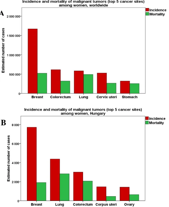
Breast cancer represents a major public health issue globally ranking as the 1st of the most frequently occurring cancers among women with over 1,600,000 new cases annually estimated from GLOBOCAN statistics in 2012 [1]. Each year, more than 500,000 women die of breast cancer, making it the first-leading cause of cancer related deaths among women worldwide (Figure 1A) [1].
A
B
Figure 1: Incidence and mortality of the most common cancers among women worldwide (A) and in Hungary (B). Source of the data: GLOBOCAN [1] and National Cancer Registry Database [2] 2012.
6
In Hungary with over 7000 new cases and 1900 deaths per year, breast cancer is the most commonly diagnosed cancer and the third-leading cause of death after lung and colorectal malignancies among women (Figure 1B) [1,2]. The mortality of breast cancer is decreasing from the 1990’s because of spreading of breast cancer screening and emerging new therapies. However, the morbidity of breast cancer shows an increasing trend as mean age rising as well as obesity, early menarche and childbearing in later age are getting more common in societies.
1.2. Diagnosis of breast cancer
Finding breast cancer early and getting appropriate cancer treatment are the most important strategies to decrease the mortality of breast cancer. Since many women with breast cancer have no symptoms, the diagnosis of breast cancer often begins with lesion found on x-ray mammography screening test. Breast cancer is sometimes found on a routine clinical – or self-examination when a new lump appears. This is followed by a breast ultrasound test, or when the clinical picture is ambiguous, a magnetic resonance imaging [3]. The only way to confirm a potentially malignant breast lesion detected on imaging test is performing biopsy. The most widely used biopsy techniques are core biopsy and fine-needle aspiration biopsy (FNAB). While only cells are obtained with FNAB, core biopsy allows to examine the histological structures as well. Furthermore, molecular tests are more commonly applied on core biopsy samples and thus used for clinical decision making. Based on the results of imaging and biopsy tests, the multidisciplinary team reviews the medical condition and sets up treatment options for each patient individually. The management of breast cancer requires close cooperation of different disciplines. Therefore, multidisciplinary oncoteam includes oncologist, surgeon, radiologist, radiation oncologist and pathologist.
1.3. Subtypes of breast cancer
It has long been known that breast cancer cannot be considered as a homogenous disease but rather a group of heterogeneous tumors in the same anatomical localization [4]. Breast tumors originate from the epithelial - or the mesenchymal tissue of the breast. Histologically, breast cancer classification is performed according to the regularly updated atlas of the World Health Organization (WHO) considering the
7
tumor’s morphological characteristics [5]. The clear majority of malignant breast tumors arise from the epithelium. The most common is invasive breast carcinoma no special type (NST), then invasive lobular carcinoma, followed by many ‘special types’
such as tubular, medullary, mucinous, micropapillary, metaplastic carcinoma, etc. [6].
This heterogeneity has brought to the fore the concept, that gene defects and failure of gene regulatory networks driving to carcinogenesis and metastases might show stronger association with the phenotype and clinical behavior of tumors than the morphological classification. In the last decade, various molecular techniques have been used increasingly to help refine breast cancer classification. The pioneer studies have proposed a new description of the heterogeneous group of breast cancers at the molecular level by cDNA microarray analyses revealing several molecular/intrinsic subtypes beyond the traditional hormone receptor (HR)+ and HR – types [7,8]. The most reproducibly defined intrinsic subtypes among the HR+ cancers are the luminal A and luminal B subgroups [9]. The human epidermal growth factor 2 (HER2)-enriched and basal-like subgroups are the major intrinsic subtypes identified among HR- breast cancers [9]. The rationale underlying this classification is that these breast cancer molecular subtypes differ in their gene expression patterns, clinical features, prognosis and response to treatment (Table 1) [9,10].
Despite the discovery of these intrinsic subtypes, the immunohistochemical (IHC) expression of estrogen receptor (ER), progesterone receptor (PgR), Her2 and the HER2 gene amplification detected by fluorescent in situ hybridization (FISH) are used to assess breast cancers in clinical practice [9,11]. This approach is most widely used to approximate the intrinsic subtypes of breast cancer, as defined by gene expression profiling because of financial reason and because it clearly defines whether an actual breast cancer can be treated with targeted therapy [11]. In the surrogate definitions of intrinsic subtypes, the ER+ tumors can be divided into luminal A-like and luminal B- like groups based on the IHC expression of ER, PgR, Her2, Ki67 and/or HER2 gene amplification [9,11,12]. In ER- tumors, HER2 and Triple-negative (TNBC) subtypes are distinguished based on the IHC expression of Her2 and/or HER2 gene amplification.
Both subgroups are characterized by negative IHC reactions of ER and PgR. Ki67 expression may vary in the ER- subgroups therefore has no relevance in therapy decision making (Table 1) [9,11,12].
8 Table 1:Subtypes of breast cancer.
Intrinsic subtypes [9]
Clinicopathologic surrogate definition
(based on IHC and FISH tests) [12] Notes
Luminal A Luminal A-like
ER + a, HER2 –, PgR high b, low Ki67 c, low-risk molecular
signature d
Luminal B
Luminal B-like HER2 –
ER + a, HER2 –, PgR low b or high Ki67 c, high-risk molecular
signature d
Luminal B-like HER2 + ER + a, HER2 +, any PgR, any Ki67
HER2-
enriched HR – and HER2 + ER/PgR – and HER2 +
Basal-like TNBC ER/PgR – and HER2 –
aER + ≥1%
bSuggested threshold: 20%
cKi67 threshold varies between laboratories
dif available
1.4. Prognostic and predictive factors of breast cancer in daily practice
Breast cancer represents great diversity not only in its morphological and molecular features but also in its prognosis [13]. Several studies have shown that the histological subtypes have different prognosis [14-16]. Mucinous, tubular, medullary, invasive cribriform, infiltrating lobular, tubulo-lobular, adenoid cystic carcinoma, and low grade adenosquamous carcinoma have all been reported to have a more favorable prognosis than invasive carcinoma NST [14-22]. Primary soft tissue sarcoma of the breast is very rare and usually associated with poor outcome [23].
Age is one of the most important risk factor for breast cancer. As the age is rising, the probability of developing cancer is increasing [24]. However, many studies indicated the adverse risk related to younger age as breast cancer is rarer in young women, but if it occurs, it is often presenting with advanced stage resulting in poor prognosis [25].
9
It has long been known that the size of the tumor and the regional lymph node involvement are strongly linked to the prognosis of breast cancer [26,27]. Smaller tumor holds the promise of higher chance for complete recovery. However, smaller tumors with early lymph node metastases have more unfavorable prognosis compared to larger tumors without lymph node involvement [28].
The clinical and pathological stage (TNM) comprising the size of the tumor (T), lymph node status (N) and presence of metastasis (M) largely determines the course of breast cancer as well as the treatment options [29].
During routine microscopic examination of breast cancer, pathologists evaluate histologic grade (tubular differentiation + nuclear pleomorphism + mitotic count), lymphatic – and blood vessel invasion, Nottingham Prognostic Index (NPI = /0.2 x tumor size in cm/ + lymph node stage + tumor grade) that are strong and easily assessed prognostic factors [30-32].
IHC examinations may help breast cancer classification, as E-cadherin expression is absent in most lobular cancers distinguishing lobular and NST cancers in equivocal cases [33-35]. Cytokeratin (CK) 5/6, CK14 and p63 are used to reveal basal/
myoepithelial origin of breast cancer [36-38]. The IHC and/or FISH detection of ER, PgR and Her2 forms the backbone of the clinical management of breast cancer as they not just assign the surrogate intrinsic subtypes referring to prognosis but also basically define treatment options [12]. The IHC detection of p53 has prognostic potential as p53+ tumors have unfavorable prognosis, but they also show better response to chemotherapy [39,40]. However, p53 has not been included in the compulsory routine IHC tests. High Ki67 expression in tumor cells means higher proliferation capacity that has been linked to poor prognosis [41,42]. Various gene mutations have been linked to the genesis and prognosis of breast cancer, such as TP53, BRCA1 and 2, CDH1 etc.
[43,44].
Although evaluation of individual prognostic and predictive factors has allowed the identification of clinically distinct subgroups of patients with breast cancer, there is urgent need to evolve a comprehensive profile of the biological and molecular characteristics of breast cancer that may improve assessment of prognosis and prediction of response to therapies in individual patients [9]. During the last few decades, several platforms of multigene classifiers have been developed to identify
10
patients with favorable prognosis, who can omit chemotherapy, and those with poor prognosis and higher risk of metastasis [9]. Several tests are now commercially available as follows: MammaPrint (Agendia, Amsterdam, Netherlands) is a microarray test approved by the US Food and Drug Administration (FDA) applying a set of 70 genes to accurately predict poor prognosis disease for patients with TNM stage 1 or 2, node-negative, invasive breast cancer of tumor size ≤5 cm [45,46]. The 76-gene signature test of Veridex investigating 60 genes for patients with ER-positive disease and 16 genes for ER-negative disease has a strong prognostic factor for 5 years’ distant metastasis free survival [47]. MapQuant Dx (Ipsogen SA, Marseille, France) can stratify grade II breast carcinomas into grade I-like and grade III-like cancers in ER+ disease [48,49]. All three assays are based on DNA microarrays and require fresh or frozen samples implying a challenge in the daily practice and in prospective validation studies [10]. The technique of real-time quantitative reverse-transcription analysis (RT-qPCR) has also been used for developing prognostic assays extracting RNA from formalin- fixed paraffin-embedded (FFPE) tissue samples, thus relieving the sample procurement [10]. The simplified version of MapQuant Dx, Oncotype DX, and Theros platforms are using this method to predict prognosis [50-53]. The prognostic use of Oncotype DX is supported by level I evidence, and this test has been included in the National Comprehensive Cancer Network - and in the American Society of Clinical Oncology guidelines as a predictor of recurrence and a guide when making therapeutic decisions among early ER positive node negative breast cancers [54]. The largest criticism raised against these assays is that despite similar prognostic and/or predictive value, there is very little overlap in the genes included in these tests [55]. Furthermore, several studies demonstrate that a part of clinically diagnosed and validated breast cancers with Her2 + phenotype are not stratified into the HER2-enriched subtype by gene expression assays.
Vice versa, a part of breast cancers characterized as HER2-enriched subtype do not occur as a Her2 + breast cancer in the daily practice [56-61]. Increasing evidence confirms that the expression of proliferation genes in luminal cancers does not form distinct subgroups (luminal A and luminal B) but a continuum. Thus, stratifying breast cancers into two luminal subgroups based on gene expression assays is artificial [56,62]. The prognostic information of these assays are almost exclusively based on the expression of the proliferation-related genes [54]. Considering the latter and that these
11
assays are too expensive to be widely applied, the question can be raised whether the use of cost-effective and widely available proliferation markers (e.g.: Ki67) should receive greater emphasis in daily practice.
1.5. Therapy of breast cancer 1.5.1. Surgery
The surgical treatment of primary breast cancer has been shifted to breast-conservation treatment. In some patients, mastectomy is implemented due to: Tumor size, multicentricity of the tumor, positive surgical margins after multiple resections, contraindications to radiation therapy, or patient choice. The biopsy of the sentinel lymph nodes is now the standard of care instead of full axillary nodal clearance, unless axillary node involvement is confirmed. However, axillary dissection can be omitted for patients with one or two metastatic sentinel lymph nodes [12,63].
1.5.2. Radiation therapy
Postoperative radiation therapy is indicated after breast-conservation surgery. Shorter fractionation schemes (15–16 fractions with 2.5–2.67 Gy single dose) have been confirmed in large studies and are generally indicated. Boost (tumor bed) irradiation further reduces the risk by 50% and it is recommended for patients with factors indicating high risk of local recurrence. Post-mastectomy radiation therapy is indicated for patients with involved axillary nodes and/or with T3–T4 tumors. Axillary irradiation is recommended for patients with involved lymph nodes [12,63].
1.5.3. Adjuvant therapy
The decision on systemic adjuvant therapies is based on the surrogate intrinsic phenotype determined by ER/PgR, HER2 and Ki67 assessment or on the genomic- based intrinsic subtype.
Endocrine therapy (anti-estrogens, selective estrogen receptor modulators, aromatase inhibitors) should be offered for all breast cancer patients with detectable ER expression (≥1% of invasive cancer cells).
Chemotherapy of breast cancer usually includes four to eight cycles of anthracycline- and/or taxane-based regimen. Sequential use of anthracyclines and taxanes is preferred
12
over concomitant use. Chemotherapy is indicated in most of triple-negative, HER2- positive breast cancers. For luminal B-like Her2− tumors, the indications for chemotherapy are based on the individual risk of relapse (e.g.: High Ki67). Luminal B- like Her2+ tumors should be treated with chemotherapy, endocrine therapy and Her2 targeted therapies. Most luminal A-like tumors, except those with T3/T4 size and extensive nodal involvement, require no chemotherapy. Her2 subtype (HR−) benefit from chemotherapy and Her2 targeted therapies. Triple-negative tumors should be treated with chemotherapy, with possible exclusion of low-risk subtypes such as adenoid cystic carcinomas [12,63].
1.5.4. Neoadjuvant therapy
In breast cancer cases with large tumor size and/or with locally advanced stage, neoadjuvant therapy may allow for achieving operability or decreasing the extent of surgery. All modalities (chemotherapy, endocrine therapy and targeted therapy) applied in adjuvant treatment may also be used in neoadjuvant setting. If chemotherapy is used, it is recommended to deliver all planned treatment without unnecessary breaks, irrespective of the magnitude of tumor response. In this case, the probability of achieving a pathologic complete remission is increased. Although neoadjuvant therapy has not been shown to improve survival superior to those of postoperative adjuvant therapy alone, there is increasing support for neoadjuvant cytotoxic therapy in Stage II triple-negative subtype. In HER2+ breast cancer especially with larger size, Her2 targeted therapy should be started in the neoadjuvant setting, combined with taxane- based chemotherapy. In patients with luminal breast cancer, neoadjuvant cytotoxic chemotherapy is not supported unless to achieve breast conservation surgery, since ER+
and Her2− tumors are usually less responsive to primary chemotherapy compared to Her2+ subtype. However, neoadjuvant endocrine therapy is generally given to post- menopausal women with breast cancer for 4 months preoperatively and continued adjuvant after surgery for 5 to 10 years [12,63].
13
1.6. Ki67: Gene, function, detection, role in breast cancer 1.6.1. Ki67 gene, structure, function
The Ki67 antigen was originally described by Gerdes et al. in the 1980s, by use of a mouse monoclonal antibody against a nuclear antigen from a Hodgkin's lymphoma- derived cell line L428 [64]. It was finally identified by the same group in 1991 and this non-histone protein was named after the researchers' location, Ki as Kiel University, Germany and the 67 label referring to the clone number on the 96-well plate [64]. The complete gene locus of the Ki67 protein, encompassing a 74 basepairs (bp) 5′ region and a 264 bp 3′ region, has been sequenced and aligned to a continuous sequence of 29 965 bp length located on the long arm of human chromosome 10 (10q25-ter). The gene comprises 15 exons with sizes from 67 to 6845 bp and 14 introns with sizes from 87 to 3569 bp. Three introns consist homologue copies of “Alu-repeats”. Exon 13 at the
“center” of this gene is composed of 16 homologous segments with 366 bp (called Ki67 repeats), each including a highly-conserved motif of 66 bp (Ki67 motif). This is highly conserved between species and nine of the Ki67 motif regions contain a highly immunogenic five amino acid sequence that forms the epitope which is the target of several Ki67 antibodies like MIB1 and SP6. Two Ki67 protein isoforms with molecular weights of 345 and 395 kDa have been described [65-68]. The cellular location of Ki67 protein is strongly cell cycle-dependent: The protein is found primarily in the nucleolar cortex and in the dense fibrillar components of the nucleolus during interphase; during mitosis, it becomes linked with the periphery of the condensed chromosomes [69-71].
The half-life of the Ki67 protein has been found to be approximately 60-90 minutes, regardless of the cell position in the cell cycle making it a feasible marker of proliferating cells [72,73]. During the different cell-cycle phases, the expression of Ki67 varies: Its levels are low during the G1 and early S phase and rise to their peak level in mitosis. Later during mitotic phase (anaphase and telophase), a sharp decrease in Ki67 expression levels occurs. These differences seem to reflect variable de novo synthesis and not due to the accumulation of non-degraded proteins. It is not expressed during the resting phase G0 [74,75]. The exact function of Ki67 is not known. Studies have described the involvement of Ki67 in the early steps of polymerase I dependent rRNA synthesis and its important function in cell division [76,77]. Ki67 protein is
14
phosphorylated via serine and threonine and its inhibition results in the arrest of cell proliferation [70,72,78].
1.6.2. Detection of Ki67
There are two approaches for measuring Ki67 expression in breast cancer: (i) a quantitative analysis of the Ki67 (MKI67) mRNA content form frozen or FFPE samples; and (ii) determining the percentage of Ki67 positive cancer cells detected by IHC. Only few data are available about the comparison of RNA and IHC based Ki67 measurement in the same samples. A study reported weak correlation between Ki67 measurements of RT-qPCR and ICH detection in breast cancer [79]. Tan et al. has found significant correlation between Ki67 gene expression levels and IHC results of cases with Ki67 score of >10% [80]. Strong linear association was found between the recurrence score and Ki67 IHC results in a study investigating 53 breast cancer cases [81]. However, the IHC staining for Ki67 detection has the following advantages compared to RNA based method: Only cancer cells are considered when the pathologist assesses the IHC Ki67 score and inflammatory- or stromal cells showing positivity can be excluded. Besides this, the IHC method is widely available and relatively inexpensive [82]. Thus, in daily practice, Ki67 is most often measured on FFPE sections by IHC method.
In general, scoring systems of Ki67 IHC detection are based on the percentage of tumor cells stained by the antibody. In one method, the pathologist examines three to ten high- power fields (×40), counts at least 1000 tumor cells with a standard light microscope and the Ki67 labeling index (Ki67 LI) is defined as the percentage of total number of tumor cells with nuclear staining [83]. However, counting 1000 tumor cells is time- consuming and monotonous. Therefore, in the daily pathological practice this approach has limitations [84]. Thus, some pathologists estimate the percentage of positive tumor cells in different areas of the tumor giving an overall Ki67 LI. However, estimating the percentage of tumor cells has high chance of failure, which might lead to results with low reproducibility [84].
It is also important to note that throughout the cell cycle, the localization and the pattern of IHC Ki67 positivity varies. In early G1 phase the IHC reaction is granular/focal in the nucleus, while in late G1 phase the positivity is seen in the nucleolus. In G2 phase
15
the reaction is granular/focal or diffuse in the nucleus and in the S phase both the nucleolus and nucleus are positive. In the M prophase there is a fine net-like positive reaction in the nucleus, while in the M metaphase the positivity occurs on the surface of the chromosomes and following breakdown of the nuclear membrane some cytoplasmic positivity may also be detected [85]. Consequently, Ki67 LI should include all types of nuclear staining irrespective of the intranuclear localization (nuclear or nucleolar or nuclear membrane) and the distribution (granular or diffuse) and regardless of intensity [82].
One of the greatest challenges in Ki67 scoring is the selection of fields for evaluating because of the variations in cellular proliferation caused by intra-tumoral heterogeneity.
In addition to spatial heterogeneity, a temporal heterogeneity may also occur because of neoadjuvant therapy [86-88]. Tumor heterogeneity is one of the causes for high inter- observer variability. Because of this, at least 500-1000 tumor cells should be evaluated when giving Ki67 LI to achieve an acceptable error rate [89].
1.6.3. Ki67 in breast tissues
Ki67 is expressed in normal breast tissue at low level (<3%) [90]. Numerous studies have reported that ER expression and Ki67 antigen are detected in separate cell populations in normal human breast epithelium. It is an important observation that Ki67 is expressed exclusively in ER-negative breast epithelial cells, which means that ER- positive luminal cells do not proliferate in normal human breast tissue. This separation between ER expression and proliferation does not exist in malignant breast tissue [91].
The association has been described between expression of Ki67 and breast density as well as with precancerous lesions [92,93]. Moreover, the continuous increase of Ki67 expression has been found from benign breast disease to ductal carcinoma in situ to invasive breast cancer [94-96].
It has also been shown that Ki67 expression decreased when aromatase inhibitor was given concomitantly in a study investigating high-risk women [97].
High levels of Ki67 expression were found in about 40% of ductal carcinoma in situ (DCIS). Increased Ki67 levels are associated with comedo necrosis, higher grade lesions, presence of microinvasion as well as the recurrence of DCIS [98,99].
16
Ki67 expression is one of the parameters that can help to distinguish several rare subtypes of breast cancers: The lipid-rich and sebaceous breast carcinomas typically express high Ki67 levels [100,101]. Invasive lobular cancer usually shows a low Ki67 index, and some researchers found low Ki67 level associated with the prognosis of lobular carcinoma [102].
1.6.4. Ki67 and its relationship with other markers of breast cancer
Many studies have demonstrated a strong correlation between Ki67 and histological grade [103-105]. However, this relation is not so surprising, since the mitotic index is one of the three components of histological grade [30]. Both Ki67 and mitotic index are widely applied in the daily practice to measure proliferation. However, some authors propone the use of Ki67 as prognostic marker superior to mitotic index because mitotic index is more subject to individual evaluation as distinguishing apoptotic- and mitotic figures is not always certain [84]. The association between lymph node status and Ki67 has also been intensively investigated and several studies involving large number of patients have revealed a positive correlation [106-108]. The relation between tumor size and Ki67 has also been demonstrated [109]. The HR status has been found to have an inverse relation with Ki67, so that ER and PgR positivity is mostly found in the least proliferating tumors [107,110,111]. The association between Ki67 and Her2 expression is controversial [112-114]. Mutation of p53 oncogene is mostly found in breast cancers expressing higher levels of Ki67 [115].
1.6.5. Ki67 in breast cancer: Prognostic and predictive potential
Several studies were published about the importance of proliferation measured by Ki67 expression in breast cancer, including the Oncotype DX that measures gene expression level of KI67 as one of the 16 genes [51,116,117]. Furthermore, the proliferation group is the mostly weighted in the algorithm of the Recurrence Score [51]. It has been long acknowledged - and more recently several studies have demonstrated - that the immunohistochemical detection of the Ki67 positive cells provides important prognostic information in breast cancer [118-120]. In one of the largest studies, Petrelli et al. [121]
performed a systematic review of the literature which was followed by a meta-analysis of the involved studies. In total, 41 studies enrolling more than 64,000 patients were
17
investigated. Although different cut-off points in a range between 10% to >25% were applied, the study has shown that elevated levels of Ki67 are independently associated with adverse outcome in patients with breast cancer [121]. Moreover, breast pathologists had been undertaking retrospective studies for showing that Ki67 LI was almost as good as the Oncotype DX for the prediction of prognosis in ER + breast cancer cohorts [116,120,122]. However the optimal threshold of Ki67 LI is still uncertain: The St. Gallen Consensus Conference in 2013 recommended a 20% cut-off to distinguish between HER2 negative luminal B-like and luminal A-like breast carcinomas [123], while the majority of the panel in 2015 intended to accept a Ki67 LI threshold of 20-29% [63]. However, the panel also acknowledged that the threshold between Ki67 high and low breast cancers varies between laboratories [63]. Cserni G. et al suggested that different thresholds may be generated for different clinical purposes [124]. According to Denkert et.al. [89], an optimal threshold of Ki67 does not exist, because many cut-off points have a similar prognostic performance. Thus, they recommend to use Ki67 LI as a continuous marker, which reflects the biology of tumor proliferation [89]. In contrast to the guidelines of The St. Gallen Consensus Conference, the International Ki67 in Breast Cancer Working Group is more cautious about the recommendation of Ki67 in daily practice [116]. The European Society for Medical Oncology (ESMO) Clinical Practice Guidelines suggests that Ki67 may provide useful information, if the assay can be standardized [125]. The American Society of Clinical Oncology (ASCO) did not recommend the use of Ki67 for prognosis in newly diagnosed breast cancer patients because of lack of reproducibility across laboratories [126,127].
In addition to ongoing debate on its prognostic utility, Ki67 has also been investigated as a potential predictive marker in neoadjuvant and adjuvant settings. For neoadjuvant chemotherapy of breast cancer, Ki67 was significantly associated with clinical or pathological response in several studies [84,128]. However, in a recent research involving 506 breast cancer patients, Ki67 did not represent an independent predictive potential for neoadjuvant therapy [129]. In contrast to this, the systematic review by Luporsi et al. has determined a level of evidence of II-B for Ki67 regarding neoadjuvant treatment response [120]. The other setting for prediction of response to therapy is the evaluation of survival in adjuvant studies. In adjuvant setting, the predictive role of
18
Ki67 is even more uncertain. In the IBCSG 8/9 trial, no predictive potential of Ki67 for response to chemotherapy vs. no chemotherapy was found [130]. The elevated Ki67 was associated with a higher efficacy of docetaxel in PACS01 [131], but the evaluation of BCIRG001 did not confirm this [132]. The controversial predictive potential of Ki67 between neoadjuvant and adjuvant settings was addressed in the review by Denkert et al. expounding that Ki67 affects in opposite directions for assessment of prognosis and for assessment of response to neoadjuvant therapy [89]. ]. An elevated level of Ki67 is associated to unfavorable prognosis, as well as to better response to neoadjuvant therapy. It has also been shown that in some, but not all breast cancer subtypes, response to neoadjuvant therapy is linked to improved prognosis [89]. These two contrary effects cause an overlap in adjuvant studies. Thus, it is not possible to separate the negative prognostic effect of high Ki67 in non-responding cases from the positive prognostic effect of high Ki67 tumors that respond to therapy [89]. The authors also suggest distinguishing three groups of tumors in relation with Ki67 expression and responsiveness to therapy as follows: i) low Ki67 tumors that do not respond to chemotherapy but also have a good prognosis i.e.: Luminal A-like subtype (Low Ki67 associated with good outcome). ii) High Ki67 tumors with response to chemotherapy has better outcome (high Ki67 associated to favorable outcome) compared to iii) high Ki67 tumors that are chemotherapy-resistant (high Ki67 associated to poor outcome) [89].
1.6.6. Standardization efforts of the application of Ki67 in daily practice
Ki67 is currently one of the most promising yet controversial biomarker in breast cancer [133]. Despite the promise of Ki67 as a prognostic and/or predictive tool, controversy exists regarding its applied methodology in clinical practice. Therefore, there is an urgent need for reproducible methodology and consistent scoring methods of Ki67 LI.
To overcome this struggle, the International Ki67 in Breast Cancer Working Group has introduced a recommendation for the application of Ki67 IHC in daily practice [116].
According to this, parameters that predominantly influence the IHC results of Ki67 include pre-analytical, analytical, interpretation and scoring, and data analysis steps [116].
19
Several pre-analytical issues might negatively affect Ki67 measurement as follows:
Type of biopsy, time to fixation, type of fixative, time in fixative, and how the specimen is stored for long term [116]. Two studies have found that in general, Ki67 IHC has better tolerance in preanalytical variability than other IHC assays [134,135]. However, alterations in the appearance of stained nuclei were observed: The well-fixed core biopsies showed well-circumscribed, uniformly stained nuclei, while highly variable staining was found in nuclei of poorly-fixed specimens [116]. Tissue handling guidelines that are already established for ER (8–72 hours of neutral buffered formalin fixation) are adequate for Ki67 IHC [116].
The analytical issues of Ki67 IHC encompass the type of the used Ki67 antibody and IHC protocol. Ki67 IHC is most often performed using the MIB1 antibody and the International Ki67 in Breast Cancer Working Group has endorsed its use in daily practice [84,116]. However, little emphasis had been put so far on a very evident technical question, namely, are all commercially available Ki67 antibodies detecting the same amount of proliferating tumor cells in each case? Can we use the different antibodies interchangeably? Most published studies concluded that there are indeed differences between the protein expression levels of different Ki67 antibodies; however, the different results were not linked to the prognosis [136-139]. Regarding IHC protocol, positive and negative controls should be used in each group of Ki67 IHC;
positive nuclei of non-malignant cells and mitotic figures provide the quality of a section. The best evidence supports the use of heat-induced antigen retrieval by microwave processing [116]. Chromogen development and counterstaining for Ki67 IHC do not differ from other antigen - antibody systems. The chromogenic staining needs optimization as negative nuclei represent usually the clear majority of overall cell population [116]. Thus, weak counterstaining can lead to overestimation of the Ki67 LI.
Difficulties in evaluating immunoreactions can also be responsible for discrepancies of Ki67 scoring reproducibility. Ki67 LI values are usually defined as the percentage of positive tumor cell nuclei, counted in 3-10 high-power fields by testing at least 500- 1000 tumor cells [116]. Another method is to estimate the mean Ki67 LI in the entire lesion. Both methods are monotonous, time-consuming and exhausting with a chance of leading to controversial results and inaccurate reproducibility [84]. Although the counting method has been recommended by the International Ki67 in Breast Cancer
20
Working Group, other studies have demonstrated the counting method is not superior to visual estimation [124,140,141]. Biological heterogeneity of Ki67 staining can occur across the specimen and it has large impact on the Ki67 scoring. One approach is to evaluate Ki67 IHC in “hot-spot” fields that contain the most proliferating tumor cells.
The other way is to give a representative score by averaging fields across the section [82]. This issue is currently being investigated to assess which method is more robust [116]. Although, recommendations published in 2011 provide a suitable landmark to improve pre-analytical and analytical validity, related protocols still show high variety and poor reproducibility linked with the context of different sampling, fixation, antigen retrieval, staining and scoring methods [116,120,142].
Rapid development of digital microscopy by now allows fast digitalization of histological slides at high-resolution, which can firmly support education, research and diagnostics in pathology [143,144]. The emergence of digital image analysis (DIA) platforms improved the capacity, precision and reproducibility of in situ biomarker evaluation [145]. However, these features alone may not be enough for diagnostic accuracy, which must be based on histological pattern recognition as the most relevant requirement of precise sample selection and assessment of immunoreactions [146]. DIA platforms are able to assess Ki67 LI, however it has not been clarified yet, if their results can meet the requirements of the daily diagnostic practice and reduce variability of Ki67 scoring [147].
21 2. OBJECTIVES
In my PhD thesis, three aspects of clinical validity of Ki67 LI are investigated as follows: i) The comparison of different Ki67 antibodies used in daily practice. ii) The reproducibility between pathologists evaluating Ki67 LI and the potential of DIA in Ki67 scoring. iii) The role of Ki67 in neoadjuvant setting.
Therefore, in the breast cancer working group of 2nd Department of Pathology, Semmelweis University we aimed to:
1, Compare the semi-quantitatively defined Ki67 LI of five commercially available Ki67 IHC antibodies in a consecutive breast cancer patient population.
2, Correlate the prognosis prediction potential of each Ki67 antibodies with that of conventional clinicopathological factors in univariate and multivariate analyses.
3, Investigate the reproducibility of Ki67 LI among three pathologists, based on their conventional visual estimation.
4, Test the agreement of semi-quantitative and DIA Ki67 scoring.
5, Determine and compare the outcome prediction potential of each semi-quantitative and DIA assessments with that of conventional clinicopathological factors.
6, Find optimal cut-off values for Ki67 expression in neoadjuvant patient cohort that best correlates with response rates to neoadjuvant therapy and with distant metastasis- free survival as well as with overall survival.
7, Investigate the association between Ki67, subtype and pathological response.
8, Investigate the prognostic potential of Ki67 in neoadjuvant setting with multivariate analysis.
22 3. METHODS
3.1. Patients
Two distinct breast cancer patient cohorts were enrolled in the investigations encompassing 498 patients totally without any overlap: 1) 378 consecutive breast cancer cases from the Buda MÁV Hospital Pathology Unit, Budapest, Hungary diagnosed between 1999 and 2002 with 99.80 months median follow up (disease-free survival, DFS). All patients’ breast cancers had been surgically removed. Pathological features were retrieved from the pathology reports or the original H&E stained slides were reviewed. Treatment data were retrieved from patients’ medical records.
2) 120 patients diagnosed with invasive breast cancer and treated with neoadjuvant chemotherapy (NAC) at Semmelweis University, Budapest, Hungary between 2002 and 2013 were retrospectively recruited. Patients were enrolled only if they had completed NAC, thereafter underwent surgery. The median follow up time for overall survival (OS) and distant metastases-free survival (DMFS) was 60.5 and 59 months, respectively. Degree of response to NAC was categorized according to Pinder et al.
(2007) [18] in the histological sections of the post-treatment surgical specimens as follows: Pathologic complete response (pCR) was defined as no residual invasive tumor and the absence of any residual invasive tumor in the lymph nodes. Partial response to therapy (pPR), either <10% of tumor remaining (pPRi), or 10-50% tumor remaining (pPRii), or >50% of tumor remaining but some evidence of response to therapy is present (pPRiii). Non-responders (pNR) were defined as no evidence of response to therapy.
The study was approved by the Institutional Review Board of Semmelweis University (TUKEB, #7-1/2008 and TUKEB 120/2013). Regarding the definition of surrogate molecular subtypes of breast cancer, we referred to the St. Gallen recommendations from 2013 that include five categories (luminal A, luminal B/HER2-, luminal B/HER2+, HER2+ and triple negative [123].
3.2. Tissue preparation
Tissue microarrays (TMA) were built from 10% neutrally buffered FFPE representative tissue blocks of the 378 consecutive cases. Tumor areas were selected by pathologists
23
based on hematoxylin & eosin stained slides. Duplicate cores (each 2 mm in diameter) were punched (TMA Master, 3DHISTECH Ltd., Budapest, Hungary) from each case, resulting 10 TMA blocks.
Regarding the neoadjuvant cohort involving 120 cases, the pre-treatment core biopsy specimens and in case of non pCR, the surgical specimens were investigated.
3.3. Immunohistochemistry
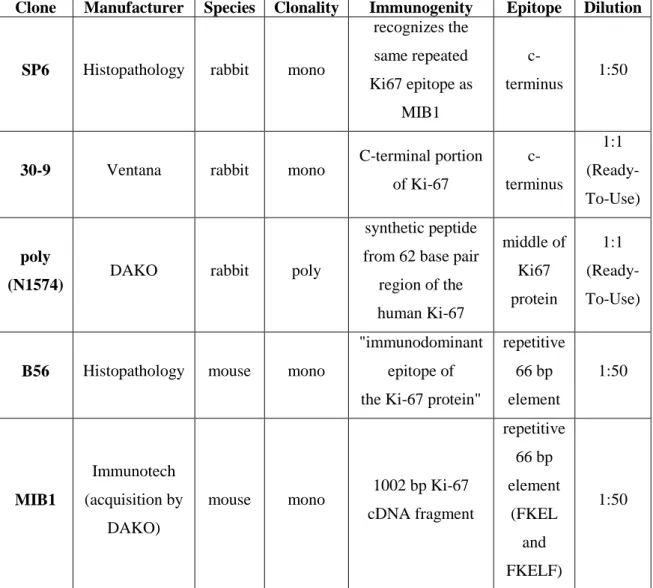
Paraffin sections of 3 μm thickness were cut from the TMA blocks for IHC. The following five antibodies (Table 2) were used for IHC detection of Ki67 on TMA blocks: SP6 (Histopathology), 30-9 (Ventana), N1574-poly (DAKO), B56 (Histopathology), MIB1 (Immunotech).
Table 2:Characteristics of the used Ki67 antibodies.
Clone Manufacturer Species Clonality Immunogenity Epitope Dilution
SP6 Histopathology rabbit mono
recognizes the same repeated Ki67 epitope as
MIB1
c-
terminus 1:50
30-9 Ventana rabbit mono C-terminal portion of Ki-67
c- terminus
1:1 (Ready- To-Use)
poly
(N1574) DAKO rabbit poly
synthetic peptide from 62 base pair
region of the human Ki-67
middle of Ki67 protein
1:1 (Ready- To-Use)
B56 Histopathology mouse mono
"immunodominant epitope of the Ki-67 protein"
repetitive 66 bp element
1:50
MIB1
Immunotech (acquisition by
DAKO)
mouse mono 1002 bp Ki-67 cDNA fragment
repetitive 66 bp element
(FKEL and FKELF)
1:50
24
Furthermore, Ki67-MIB1 was investigated with immunofluorescent labeled (MIB1-IF) antibody (IR 626 DAKO) as well. The IHC reactions were performed in an automated immunostainer (Ventana Benchmark XT, Roche, Basel, Switzerland) according to the manufacturer’s protocol (at 42 °C for 32 minutes) after antigen retrieval using the pH 9.0 CC1 buffer at 42 °C for 30 minutes. For antibody visualization, UltraView DAB Detection kit (Ventana, Tucson, USA) was applied. Immunofluorescent staining was performed manually.
To detect Ki67 in core biopsy and surgical specimens of the neoadjuvant breast cancer cohort MIB1 antibody was used with the same protocol.
Furthermore, ER, PgR and Her2 IHC were also performed using the following antibodies: 1:200 anti-ER (clone 6F11), 1:200 anti-PgR (clone 312) and 1:150 anti- HER2 (clone CB11) antibodies purchased from Novocastra Laboratories Ltd (Newcastle upon Tyne, UK) with the same protocol. The cut-off value for ER and PgR positivity was 1% positive tumor cells with nuclear staining. Hormone receptor (HR) negativity was defined as being negative for both ER and PgR. HER2 IHC positivity was defined as score 3+ complete, strong membrane staining in >10 % of tumor cells.
For IHC 2+ samples, FISH was performed to confirm gene amplification by using Ventana Benchmark automatic staining system with INFORM® Her-2/neu FISH test until 2008 and Zytovision® ERBB2/CEN17 dual FISH probe after 2008. HER2 status was defined according to the ASCO/CAP guideline valid at the time of diagnosis (ASCO/CAP guideline 2007 and ASCO/CAP guideline 2013) [148,149].
3.4. Semi-quantitative evaluation of Ki67 reactions
Semi-quantitative (SQ) evaluation of Ki67 IHC of 378 consecutive cases was performed on digital slides using the TMA Module software on the PannoramicViewer (v1.11.49.0) platform (all 3DHISTECH, Budapest, Hungary) as follows: Ki67 LI was defined as the percentage of positive tumor cell nuclei, estimated on average in 3-10 high-power fields, in each core. Any nuclear positivity was considered, including nuclear, nucleolar or nuclear membrane localization irrespective of the pattern (granular or diffuse) in a range of 100–500 cells, depending on the cellularity of the TMA cores.
Duplicate cores were evaluated separately and their mean Ki67 LI was finally analyzed.
25
During the comparison of five Ki67 antibodies, the IHC reactions were evaluated by two pathologists independently and if any discrepancy occurred, the inconsistent cases were reassessed and a consensus Ki67 LI score was given.
When the reproducibility was investigated between observers, the IHC reactions of MIB1 antibody were evaluated by three pathologists (SQ-1, SQ-2, SQ-3) independently.
The three pathologists have considerable but different level of experience in Ki67 scoring of breast cancer. SQ1 is the youngest with a pathology specialist status for a year only. SQ-2 and SQ-3 are consultant pathologists with substantial experience in diagnostic practice and special focus on breast pathology. Dichotomization of Ki67 LI values either at 14% or 20% and 30% thresholds was also performed [123,63].
Regarding the neoadjuvant cohort, the Ki67 IHC reactions were evaluated by two pathologists independently and if any discrepancy occurred, the inconsistent cases were reassessed and a consensus Ki67 LI score was given.
3.5. Digital image analysis of Ki67 reactions
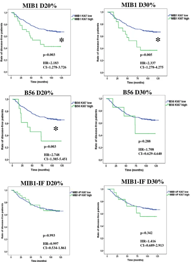
TMA slides were digitized with Pannoramic Flash II slide scanner using x20 objective (NA=0.83), collecting sharp signals from 7 focal planes in “Extended-focus” mode through the 3 µm section thickness at 80 jpeg image quality factor. DIA was performed on the IHC reactions of MIB1 antibody using the PatternQuant (PQ) software of the QuantCenter package module enabling automated tissue pattern recognition by separating epithelial elements from stroma. All digital hardware and software tools were from 3DHISTECH Ltd. (Budapest, Hungary). Designation of training tissue patterns to be recognized and the calibration were done in co-operation by a pathologist and an IT expert to achieve the best recognition pattern (achieved at a PQ training magnification of 1.5x; a gamma level of 1; dilution of 3; a contour of 0). So, as the detection and quantification of tumor cell nuclei using NuclearQuant (NQ) at the following settings:
Blur: 15; Radius minimum: 1.5; Radius maximum: 8; Area min: 15; Intensity minimum:
30; Contrast minimum 30 (Figure 2). The brown DAB and the hematoxylin counterstain were separated with digital color deconvolution [150]. Based on these settings of PQ and NQ, automated Ki67 evaluation was performed on each core (DIA-1 analysis). In the other DIA test, automated annotations were assessed by pathologists on each core, and when it was necessary, DIA settings were adjusted independently (from the Ki67 LI
26
results of DIA-1, SQ-1, SQ-2, SQ-3) to exclude artifacts, underestimation or overestimation of positive/negative cells and false detections (DIA-2 analysis).
A B
D C
E
Figure 2:Workflow of 3DHistech DIA assessment. Examples of desired tissue patterns were given, demarcated with the red and green lines (red = epithel pattern, green = stroma pattern) [A,B], that we wanted to be recognized and distinguished by the software named PatternQuant [C]. Then the software named NuclearQuant counts the recognized negative (blue) and positive (red) cells only in the annotations designated by PatternQuant (red areas on picture C) [D,E].
27 3.6. Statistical analysis
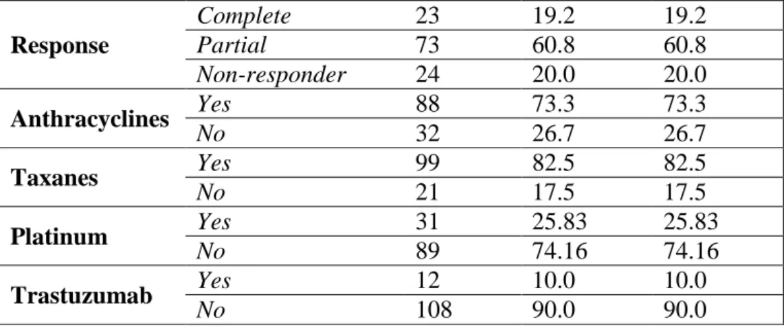
For statistical analysis SPSS 22 software (IBM, Armonk, USA) and MedCalc 13.3.3.0 (MedCalc Software, Ostend, Belgium) software were used. Degree of agreement among different antibodies detecting Ki67 was evaluated by using intra-class correlation coefficient (ICC), concordance correlation coefficient (CCC), Cohen's kappa and Bland- Altman plot. To assess statistical differences between each antibody, Wilcoxon signed- rank and McNemar tests were applied, since our data were not normally-distributed, even after log-transformation (Shapiro-Wilk and Kolmogorov-Smirnov tests).
The reproducibility between pathologists was estimated with ICC and CCC. Altman’s guideline was followed for the interpretation of ICC [151]. CCC was interpreted according to McBride [152]. Degree of agreement among different observers (SQ-1, SQ-2, SQ-3, DIA-1, and DIA-2) was evaluated by using Cohen's kappa and Bland- Altman Plot. To assess statistical differences between observers the Wilcoxon signed- rank and McNemar tests were applied, since our data were not normally-distributed, even after log-transformation (Shapiro-Wilk and Kolmogorov-Smirnov tests).
Differences in the distribution of characteristics between the parameters of patients with pCR or pPR and patients with pNR were evaluated using two-sided Fisher’s Exact Test.
Two-sided Mann-Whitney-Wilcoxon test was used to define age distributions in pCR vs. pNR and vs. pPR. The optimal cut-off value for Ki67 percentage to discriminate response to treatment was assessed by receiver operating characteristic (ROC) curve analysis. To identify the optimal Ki67 threshold for NAC, only pCR and pNR cases were involved in ROC analyses, because pPR status is considered as a soft endpoint.
Kaplan-Meier analysis supported with log-rank test was executed to assess prognostic potential. To compare prognosis prediction potential, multivariate Cox-regression analysis was applied. OS was defined as the elapsed time from the date of diagnosis of the tumor by core biopsy to the date of death, or when patients were last censored if still alive. DMFS was defined as time from the date of primary diagnosis to the occurrence of first distant metastases. DFS was defined as time from the date of primary diagnosis to the occurrence of first relapse. In all statistical analysis, the level of significance was set at p< 0.05.
28 4. RESULTS
Clinicopathological characteristics of the 378 breast carcinomas are shown in Table 3.
Mean patient age was 59 years (range: 27-94 years). Most of the cases were pT1 and pT2, the majority with low mitotic index and histological grade of 1 or 2 and of luminal A - like subtype. Most patients had an axillary stage of pN0-1 (55.8%). In 92 cases (24.3%) axillary surgery was not performed due to clinical or patient related reason (see Table 3). More than half of the patients (57.7%) underwent postoperative breast irradiation, and slightly fewer patients (42.1%) received adjuvant chemotherapy in this cohort. All patients with ER positive breast cancer received endocrine treatment.
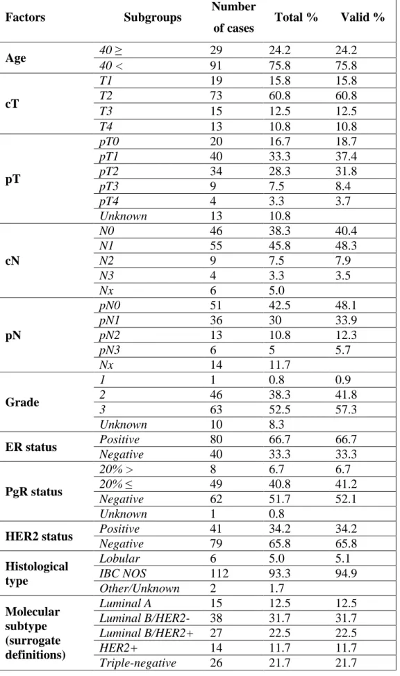
Aggregate clinicopathological features of the 120 cases in the neoadjuvant cohort are displayed in Table 4. Mean patient age was 50.6 years (range: 29-74 years). Most patients (59.6 %) had node-positive disease and cT2 tumors (60.8 %). Tumors were ER- positive in 66.7 % of cases and presented PgR positivity >20.0 % in 41.2 % of the analyzed samples. In 34.2 % of cases HER2 positivity was detected. Of the 120 tumors, 12.5 % were of luminal A, 31.7 % of luminal B/HER2 negative, 22.5 % of luminal B/HER2 positive, 11.7 % of HER2+ and 21.7 % of TNBC subtype. Twenty three out of 120 patients (19.2 %) achieved pathologic complete remission (pCR), 73 (60.8 %) showed partial remission (pPR), whereas no response to NAC (pNR) was detected in 24 cases (20.0 %). In the group of patients who obtained pPR, residual tumor was detected in lymph nodes only in 7 patients (9.6 %), major response (>90 % tumor regression) to NAC was observed in 8 cases (11.0 %), a response rate between 50-90% was detected in 26 cases (35.6 %), whereas a response rate <50% was observed in 32 cases (43.8 %).
29
Table 3: Clinicopathological data of the 378 breast carcinomas.
Patients (n, %) 378 100%
Age (mean ± SD, range) 58.90 ± 12.98 27-94 Tumor size (mm) (mean ± SD) 23.58 ± 15.56
Mitotic index (n/10HPF)
(mean ± SD) 9.07 ± 10.89
Grade 1 (n, %) 146 38.6%
2 (n, %) 143 37.9%
3 (n, %) 89 23.5%
Subtype LUMA (n, %) 184 48.7%
LUMB (n, %) 124 32.8%
HER2 (n, %) 20 5.2%
TNBC (n, %) 49 13.0%
no data (n, %) 1 0.3%
Lymph node status (TNM 7)
0 (n, %) 133 35.2%
1 (n, %) 85 22.5%
2 (n, %) 39 10.3%
3 (n, %) 29 7.7%
no data# (n, %) 92 24.3%
Vascular Invasion none (n, %) 117 31.0%
present (n, %) 251 66.4%
no data (n, %) 10 2.6%
Necrosis none (n, %) 277 73.3%
present (n, %) 95 25.1%
no data (n, %) 6 1.6%
Chemotherapy no (n, %) 213 56.3%
yes (n, %) 159 42.1%
no data (n, %) 6 1.6%
Radiation therapy no (n, %) 154 40.7%
yes (n, %) 218 57.7%
no data (n, %) 6 1.6%
Follow-up time (n, median, IQT*)
334, 99.80 57.93
*interquartile range. #29 cases were small, screen detected lesions before the nationwide screening was introduced. No sentinel lymph node technique was available at that time. Six patients developed second primary carcinoma in the same breast previously undergoing breast conserving surgery with axillary block dissection. In 2 cases, no lymph nodes were found in the removed axillary fat tissue. In 35 cases, due to co- morbidities or advanced age of patients axillary staging was omitted. In the remaining 20 cases, recurrent breast carcinoma was diagnosed (in these cases the primary tumors were not available).
30
Table 4: Clinicopathological data of the 120 breast carcinomas.
Factors Subgroups Number
of cases Total % Valid %
Age 40 ≥ 29 24.2 24.2
40 < 91 75.8 75.8
cT
T1 19 15.8 15.8
T2 73 60.8 60.8
T3 15 12.5 12.5
T4 13 10.8 10.8
pT
pT0 20 16.7 18.7
pT1 40 33.3 37.4
pT2 34 28.3 31.8
pT3 9 7.5 8.4
pT4 4 3.3 3.7
Unknown 13 10.8
cN
N0 46 38.3 40.4
N1 55 45.8 48.3
N2 9 7.5 7.9
N3 4 3.3 3.5
Nx 6 5.0
pN
pN0 51 42.5 48.1
pN1 36 30 33.9
pN2 13 10.8 12.3
pN3 6 5 5.7
Nx 14 11.7
Grade
1 1 0.8 0.9
2 46 38.3 41.8
3 63 52.5 57.3
Unknown 10 8.3
ER status Positive 80 66.7 66.7
Negative 40 33.3 33.3
PgR status
20% > 8 6.7 6.7
20% ≤ 49 40.8 41.2
Negative 62 51.7 52.1
Unknown 1 0.8
HER2 status Positive 41 34.2 34.2
Negative 79 65.8 65.8
Histological type
Lobular 6 5.0 5.1
IBC NOS 112 93.3 94.9
Other/Unknown 2 1.7
Molecular subtype (surrogate definitions)
Luminal A 15 12.5 12.5
Luminal B/HER2- 38 31.7 31.7 Luminal B/HER2+ 27 22.5 22.5
HER2+ 14 11.7 11.7
Triple-negative 26 21.7 21.7
31 Response
Complete 23 19.2 19.2
Partial 73 60.8 60.8
Non-responder 24 20.0 20.0
Anthracyclines Yes 88 73.3 73.3
No 32 26.7 26.7
Taxanes Yes 99 82.5 82.5
No 21 17.5 17.5
Platinum Yes 31 25.83 25.83
No 89 74.16 74.16
Trastuzumab Yes 12 10.0 10.0
No 108 90.0 90.0
4.1. The validity of five Ki67 antibodies
4.1.1. Comparison of Ki67 LI score of the different antibodies
We investigated the Ki67 LI score of the 5 antibodies, and the following median values were observed: SP6 antibody: 8.00%, 30-9 antibody: 8.00%, poly antibody: 5.75%, MIB1 antibody: 3.50%, B56 antibody: 3.50%, MIB1-IF antibody: 3.50% (Figure 3).
Figure 3: Boxplot of Ki67 LI of the five antibodies.
32
Significant difference occurred between all Ki67 LI assessments of the 5 antibodies (p values for all comparisons ≤ 0.005). Dichotomizing Ki67 LI scores at 20% threshold, we found no significant difference between MIB1, poly and MIB1-IF (MIB1 vs. poly p=0.052; MIB1vs. MIB1-IF p=0.230; poly vs. MIB1-IF p=0.405) (Table 5). At 30%
cut-off score, no significant difference occurred between MIB1, poly and MIB1-IF (MIB1 vs. poly p=0.115; MIB1vs. MIB1-IF p=0.988; poly vs. MIB1-IF p=0.230), similarly to the results at 20% threshold. Furthermore, 30-9 and poly did not differ significantly at 30% cut-off score (p=0.096) (Table 5).
Table 5: Statistical comparisons of the five Ki67 antibodies.
Wilcoxon signed-rank
test p
SP6 30-9 poly MIB1 B56 MIB1-
IF SP6 - ≤ 0.001 ≤ 0.001 ≤ 0.001 ≤ 0.001 ≤ 0.001 30-9 ≤ 0.001 - ≤ 0.001 ≤ 0.001 ≤ 0.001 ≤ 0.001 poly ≤ 0.001 ≤ 0.001 - ≤ 0.001 ≤ 0.001 0.005 MIB1 ≤ 0.001 ≤ 0.001 ≤ 0.001 - 0.002 0.002 B56 ≤ 0.001 ≤ 0.001 ≤ 0.001 0.002 - ≤ 0.001 MIB1-IF ≤ 0.001 ≤ 0.001 0.005 0.002 ≤ 0.001 - McNemar
test p
SP6 D20%
30-9 D20%
poly D20%
MIB1 D20%
B56 D20%
MIB1- IF D20%
SP6 D20% - ≤ 0.001 ≤ 0.001 ≤ 0.001 ≤ 0.001 ≤ 0.001 30-9 D20% ≤ 0.001 - 0.006 ≤ 0.001 ≤ 0.001 0.002 poly D20% ≤ 0.001 0.006 - 0.052 ≤ 0.001 0.405
MIB1
D20% ≤ 0.001 ≤ 0.001 0.052 - ≤ 0.001 0.230 B56 D20% ≤ 0.001 ≤ 0.001 ≤ 0.001 ≤ 0.001 - ≤ 0.001
MIB1-IF
D20% ≤ 0.001 0.002 0.405 0.230 ≤ 0.001 - McNemar
test p
SP6 D30%
30-9 D30%
poly D30%
MIB1 D30%
B56 D30%
MIB1- IF D30%
SP6 D30% - 0.001 ≤ 0.001 ≤ 0.001 ≤ 0.001 ≤ 0.001 30-9 D30% ≤ 0.001 - 0.096 0.001 ≤ 0.001 0.001 poly D30% ≤ 0.001 0.096 - 0.115 ≤ 0.001 0.230
MIB1
D30% ≤ 0.001 0.001 0.115 - ≤ 0.001 0.988 B56 D30% ≤ 0.001 ≤ 0.001 ≤ 0.001 ≤ 0.001 - 0.003
MIB1-IF
D30% ≤ 0.001 0.001 0.230 0.988 0.003 - D20% = dichotomized at 20% threshold
D30% = dichotomized at 30% threshold
![Figure 2: Workflow of 3DHistech DIA assessment. Examples of desired tissue patterns were given, demarcated with the red and green lines (red = epithel pattern, green = stroma pattern) [A,B], that we wanted to be recognized and distinguished by the softw](https://thumb-eu.123doks.com/thumbv2/9dokorg/1382207.114066/27.892.134.746.163.960/workflow-dhistech-assessment-examples-patterns-demarcated-recognized-distinguished.webp)