Received: January 8, 2016 Accepted: March 24, 2016 Correspondence to:
Magdolna Dank
Semmelweis Univ. Dept. of Clinical Oncology
Tomo str. 25-29
Budapest: H-1083, Hungary dank.oncology@gmail.com Aim To identify breast cancer subtypes likely to respond
to primary systemic therapy (PST or neoadjuvant therapy) and to assess the accuracy of physical examination (PE) and breast ultrasonography (US) in evaluating and predicting re- sidual size of breast carcinoma following PST.
Methods 116 patients who received at least two cycles of PST between 1998 and 2009 were selected from a prospec- tively collected clinical database. Radiological assessment was done by mammography and US. Prior to PST, tumors were subclassified according to core biopsy (NCB) and/or fine-needle aspiration-based immunohistochemical profiles of NCB. Pathological response rates were assessed following the surgeries by using Chevallier classification. Tumor mea- surements by PE and US were obtained before and after PST.
Different clinical measurements were compared with histo- logical findings. Disease-free survival (DFS) was assessed.
Results Pathological complete remission (pCR = Cheval- lier I/II) was observed in 25 patients (21.5%), 44% of whom had triple negative histology, 28% Her2 positive and 76%
had high-grade tumor. Of 116 patients, 24 received taxane- based PST, 48 combined taxane + anthracycline treatment, 8 trastuzumab combinations, 21 anthracycline-based treat- ments, and 15 other treatments. In the taxane treated group, the pCR rate was 30%, in the taxane + anthracycline group 25%, in the anthracycline group 9.5%, and in trastuzumab group 37.5%. After PST, PE and US were both significantly associated with pathology (P < 0.001 and P = 0.004, respec- tively). Concerning OS, significant difference was observed between the Chevallier III and IV group (P = 0.031) in favor of Chevallier III group. In the pCR group, fewer events were observed during the follow-up period.
Conclusions Our results show that even limited, routinely used immunohistochemical profiling of tumors can predict the likelihood of pCR to PST: patients with triple negative and Her2-positive cancers are more likely to achieve pCR to PST. Also, PE is better correlated with pathological findings than US.
Morphological and pathological response in primary systemic therapy of patients with breast cancer and the prediction of disease free survival: a single center observational study
Gyöngyvér Szentmártoni1, Anna-Mária Tőkés2, Timea Tőkés1, Krisztián Somlai3, Attila Marcell Szász2, László Torgyík1, Janina Kulka2, Magdolna Dank1
1Semmelweis University, Department of Clinical Oncology, Budapest, Hungary
22nd Department of Pathology, Budapest, Hungary
3Surgical Division of St. Margit Hospital, Budapest, Hungary
There are many controversial data on the benefits and risks of primary systemic therapy (PST) of breast cancer. It is ge- nerally accepted that PST results in various clinical respons- es in 60%-90% of patients, while pathologic complete re- mission (pCR), the predictor of overall survival, occurs only in 3%-16% of patients (1-3).
Although the response rate of breast cancers to PST is a short-term marker, it has a long-term outcome and impor- tant influence on patients’ life. Therefore, it is important to identify new and reliable factors that may predict response to PST. Several studies have been conducted with the aim to identify predictive factors for pCR after administration of PST. Early identification of features that can predict pCR may allow a better selection of patients. However, there is no global consensus on the predictive factors. The role of hormone receptor status, tumor grade, and tumor cell proliferation has already been established (1-8). A large number of studies showed that women with luminal A type cancer (ER-positive/Her2-negative) were unlikely to achieve a pCR after optimal neoadjuvant chemotherapy (9,10). Based on this observation, some experts consider that patients with luminal A tumors are not eligible for pre- operative chemotherapy (11).
Controversies exist in the assessment of the accuracy of physical examination, sonography, and mammography in predicting the residual size of breast tumors following PST (12,13). Physical examination is one of the accepted clinical standards in the evaluation of tumor size before, during, and after neoadjuvant chemotherapy, while pathological evaluation is the gold standard and the ultimate assess- ment modality of the residual tumor size after neoadjuvant chemotherapy (14,15). Ultrasound (US) is used primarily for diagnostic purposes – size and biopsy – and for wire local- ization. It is considered complementary to mammography and PE. The sensitivity and specificity of PE and US vary in different studies (1,12,16,17). Sperber et al correlated the findings of PE, US and mammography performed by the same oncologic and radiologic team in patients with lo- cally advanced breast cancer or a tumor/breast tissue ratio that precludes breast-conserving surgery. They found that none of these methods adequately delineated the real ex- tent of the disease in the breast and axillary lymph nodes (18). Peintinger et al calculated the agreement between the predicted and the pathologic responses and the predicted and pathologic tumor sizes by using PE, mammography, and US at diagnosis and before surgery in 162 breast can- cer patients who received PST. They found that the overall agreement between predicted and pathologic responses
was 53% for PE, 67% for mammography plus US, and 63%
for PE plus mammography and US. The sensitivity of mam- mography and US in predicting pCR was 78.6%, the speci- ficity was 92.5%, and the accuracy was 88.9. Agreement of residual tumor size in mammography and US with patho- logic residual tumor size was moderate (18,19). In a recent study, the US estimated pathological tumor size correctly in 63%, overestimated it in 20%, and underestimated it in 17% of 182 patients who underwent PST. However, US was as least as good as breast MRI (20).
Given the important role of the assessment of residual tu- mor size in determining the surgical procedure after neo- adjuvant chemotherapy the aims of this study were:
1) to prospectively evaluate the accuracy of PE and US for clinical staging of primary breast cancer in women receiv- ing neoadjuvant chemotherapy. Until rebiopsy after first cycle of therapy or novel molecular imaging methods will be available in the everyday practice, we need to estab- lish which of the conventional evaluating methods has the highest predictive value for pCR;
2) to compare the results with pathologic measurement performed on surgical specimens;
3) to determine the breast cancer subgroups likely to re- spond to neoadjuvant chemotherapy;
4) to correlate the results of pathological response evalua- tion (ie, Chevallier classification) with the disease free sur- vival (DFS).
MeThoDS Patients
116 patients who received at least two cycles of PST be- tween 1998 and 2009 were selected from a prospective- ly collected clinical database. Patients were not included in the study if there was evidence of inflammatory breast cancer, metastatic disease, previous hormonal therapy for breast cancer, and surgery and radiotherapy. Radiological assessment was done by mammography and US (PET/CT and MRI were only available in the second part of the ana- lyzed period therefore not considered in this study). Tumor measurements by PE, and US were performed before and after PST. Prior to PST, tumors were subclassified accord- ing to core biopsy (NCB) and/or fine-needle aspiration- based immunohistochemical profiles of NCB. Pathologi-
cal response rates were assessed following the surgeries by using Chevallier classification. Different clinical mea- surements were compared with histological findings. Dis- ease-free survival was assessed. Distant metastases were screened by chest x-ray, abdominal sonography, or by CT scan/PET CT.
Clinical assessment
Clinical measurements (physical examination and/or breast sonography) were performed before treatment, at every two or three cycles during therapy, and at the end of neoadjuvant treatment. The average number of treat- ment cycles was 5.6; the majority of patients underwent 6 cycles.
Data on the PE of the tumors were available in 108/116 patients and on US in 58/116 patients. Clinical palpation, ultrasonography, and treatment were performed by the same well trained team consisting of oncologists and radi- ologists at Semmelweis University. Regarding PE, palpation and caliper measurements were performed by the treat- ing physician. Breast US was routinely performed before and after PST by the same experienced radiologist in our institute. Those US results that did not meet these criteria were excluded from the analysis. Patients’ demographics, tumor characteristics, and the largest diameter of the mul- tidimensional tumor measurement obtained by physical examination and/or sonography were recorded. The find- ings were compared with pathological staging.
The clinical response to neoadjuvant chemotherapy was classified according to the Union for International Cancer Control (UICC) criteria (cCR – complete response; cPR – partial response; cSD – stable disease; and cPD – progres- sive disease) (21,22).
Pathological assessment
Histopathological diagnosis, hormone receptor status, and Her2/neu status were determined based on the core biop- sy or fine-needle aspiration biopsy (FNAB) before neoadju- vant therapy. Estrogen (ER) and progesterone receptor (PR) status were determined by using 6F11 and the PR clone 312 (both from Novocastra Laboratories Ltd, Burlingame, CA, USA), respectively, and standard immunohistochemi- cal methods. Tumors with >10% stained cells were con- sidered to have positive receptor status. HER2/neu status was assessed by immunohistochemistry (HER2/neu CB11, Novocastra Laboratories Ltd). CB11 was scored by experi-
enced pathologists according to approved guidelines (23).
In 23 cases, the fluorescence in situ hybridization (FISH) data were available. FISH was performed again in this cases by using a fluorescein-labeled HER2 probe (ERBB2, Her2/
Neu, Kreatech Diagnostics, Amsterdam, The Netherlands) and automated technique (Ventana Medical Systems, Inc., Tucson, AZ, USA) (24-26).
Sections analyzed by FISH were adjacent to the section used for immunohistochemistry and the same areas of the tumors were evaluated.
Biological subtypes of tumors were defined according to the recommendations of the 13th St. Gallen Internation- al Breast Cancer Conference as follows (26): tumors with positive ER status, positive or negative PR status, no Her2 overexpression, and low Ki 67 were grouped into the “lu- minal A” group; those with ER positivity, PR positivity, and high Ki 67 or ER positivity and Her2 overexpression into the “luminal B” group; those with ER/PR- and Her2 positive phenotype into the “Her2” group; and tumors with neither hormone receptors nor Her2 amplification into the “triple negative” group.
Pathological response rates were assessed following surgi- cal removal of tumors on hematoxylin and eosin stained slides. The pathological response to neoadjuvant chemo- therapy was defined by using the Chevallier classification (I-IV) (class I – no residual carcinomas in breast or axillary nodes; class II – only in situ carcinoma remaining, nodes are negative; class III – invasive carcinoma with stromal fi- brosis; and class IV – no or few modifications in the tumor), and class I and II is considered as pCR (27).
Statistics
Statistical analyses were performed using Statistica 64 v12 (Statsoft Inc., Tulsa, OK, USA) and SPSS 15.0 Fam- ily Pack (SPSS, Inc., Chicago, IL, USA). For categorical vari- ables, numbers were allocated for every investigated cat- egory. For continuous variables, the results are shown as means ± standard deviations, and median with interquartile range (IQR). Categorical variables were compared using χ2 test or Fisher exact method, depending on the number of the variables in the contingency tables. Disease-free and overall survival was estimated from the date of pathologi- cal diagnosis (core-biopsy sampling) to the date of last follow-up or death using the Kaplan-Meier survival prob- ability estimator. Log-rank test was used to evaluate the ef- fect of different variables on DFS and OS. All statistical tests
were two-sided. Differences were considered to be statisti- cally significant at P < 0.05.
ReSuLTS
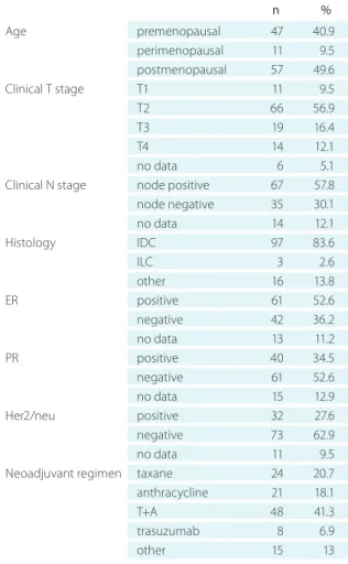
We assessed the clinicopathological characteristics of the included 116 patients (Table 1). The patients’ median age at the time of diagnosis was 49.94 years (IQR 38-59). The me- dian pretreatment tumor size assessed by PE was 40 mm (IQR 30-50) and if assessed by breast US it was median 27 mm (IQR 22-36 mm). According to the preoperative data the vast majority of patients had invasive ductal carcinoma (83.62%), while the others had invasive carcinoma not oth- erwise characterized, and lobular, mixed, and other types of carcinoma, each amounting to <10%. Most patients had
T2 tumors (56.9%), 16.4% had T3, 12% had T4 tumors, and only 9.5% had T1 tumors.
Among 116 patients there were 67 node-positive cases (57.8%). With regard to hormone receptor status, 52.6% of the tumors were ER positive and 34.5% were PR positive.
Of 116 patients, 24 received taxane-based PST, 48 combined taxane + anthracycline treatment, 8 trastuzumab combina- tions, 21 anthracycline-based treatments, and 15 other treat- ments. In the taxane treated group, the pCR rate was 30%, in the taxane + anthracycline group 25%, in the anthracycline group 9.5%. and in trastuzumab group 37.5%.
Upon pathological review of tumor and nodal sta- tus, pathological complete or near-complete remission (pCR = Chevallier I and II) was observed in 25 of 116 cases (21.5%), 44% of whom had triple negative histology and 76% had high-grade tumor. According to the preoperative characteristics of the 25 tumors achieving pCR, 11 of the cases were triple negative, 7 were luminal B, and 7 were Her2 positive. Only 10 luminal-A patients were enrolled in this study, and all of these patients failed to achieve pCR.
The same was true for the majority of luminal B tumors (35/42, 83.4%).
Univariate regression analysis was used to estimate the effects of clinical and pathological characteristics on re- sponse to neoadjuvant chemotherapy. Negative ER and PR status and Her2 positivity were the factors associated with an increased percentage of pCR (Table 2).
The menopausal status was not associated with the likeli- hood of achieving pCR. We did not find any significant cor- relation (Chi square: 4.76, df = 2, P = 0.093). But in the pCR group patients’ mean age was significantly lower than in the non-pCR group (44.4 ± 12.3 vs 50.8 ± 11.8, P = 0.017).
PE and US measurements were also compared with the residual pathologic tumor size. According to the PE TAbLe 1. Pretreatment patient and tumor characteristics of 116
patients*
n %
Age premenopausal 47 40.9
perimenopausal 11 9.5
postmenopausal 57 49.6
Clinical T stage T1 11 9.5
T2 66 56.9
T3 19 16.4
T4 14 12.1
no data 6 5.1
Clinical N stage node positive 67 57.8
node negative 35 30.1
no data 14 12.1
Histology IDC 97 83.6
ILC 3 2.6
other 16 13.8
ER positive 61 52.6
negative 42 36.2
no data 13 11.2
PR positive 40 34.5
negative 61 52.6
no data 15 12.9
Her2/neu positive 32 27.6
negative 73 62.9
no data 11 9.5
Neoadjuvant regimen taxane 24 20.7
anthracycline 21 18.1
T+A 48 41.3
trasuzumab 8 6.9
other 15 13
*T – tumor, N– node, IDC– invasive ductal carcinoma, ILC – invasive lobular carcinoma, eR – estrogen receptor, PR – progesterone recep- tor, her2 – human epidermal growth factor receptor 2, T+A– tax- ane + anthracycline.
TAbLe 2. univariate predictors of pCR to neoadjuvant chemo- therapy for breast cancer*
Characteristics P
PR negativity 0.004
HER2 positivity 0.027
ER negativity 0.002
Therapy NS
*pCR – pathological complete response, eR –estrogen receptor, PR – progesterone receptor, her2 – human epidermal growth factor receptor 2, NS – not significant.
data and UICC evaluation criteria, 27.6% of the patients achieved a clinical CR. However, the pathological com- plete response rate was lower: 21.5%. According to the results obtained by US, the clinical CR rate was 15.5% but we had the US measurement data for only 58 patients (Tables 3 and 4).
Of the 25 patients who achieved a complete pathological response, 9 were clinically described as partial clinical re- sponders; the remaining were described as complete re- sponders using PE. Based on the results of US for clinical evaluation of the 14 patients with available data from this group 5 achieved a complete pathological response and 9 achieved partial response (Tables 3 and 4). After neoad- juvant chemotherapy, both PE- and US-measured clinical remission associated significantly with pathological remis- sion, (P < 0.001 and P = 0.004, respectively).
We further analyzed whether in pCR cases US added an additional value to PE evaluation. We found that in cases when PE correctly identified pCR, only 50% of US examina- tions showed complete remission – the false positivity rate was high. In those pCR cases when PE was false positive, only one US examination contradicted the result of PE by showing clinical complete remission. Thus, US did not add any additional diagnostic value to PE.
The median follow-up was 56.1 months (IQR 36.3-77.1 months). Concerning DFS, pCR was not associated with
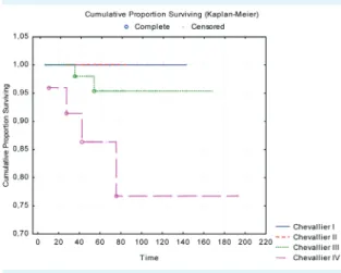
better outcome (P = 0.804), however the number of pa- tients with early disease progression in the pCR group was lower than in the non-pCR group (3 vs 15), but the differ- ence was not significant (Figure 1). We also did not find significantly better OS in the pCR group (P = 0.237), but it should be noted that in the pCR group there were fewer events (CH I-II) during the follow-up period. Nonetheless, when we compared the four Chevallier subgroups regard- ing OS, we still not find differences (P = 0.079) however with subgroup analysis between the Chevallier III and IV groups we detected significant differences in the OS time (P = 0.031) (Figure 2).
TAbLe 3. The results of physical examination compared to pathological response after primary systemic therapy (n = 105 patients, unknown Pe data in 11 cases)*
Pe - CR Pe - PR Pe - SD
Chevallier I+II 16 (55.2%) 9 (15.3%) 0 (0%) Chevallier III 12 (41.4%) 38 (64.4%) 8 (47.1%) Chevallier IV 1 (3.4%) 12 (20.3%) 9 (52.9%)
Total 29 (100%) 59(100%) 17 (100%)
*Pe – physical examination, CR – complete response, PR – partial response, SD – stable disease.
TAbLe 4. The results of breast ultrasonography compared to pathological response after primary systemic therapy (n = 58 patients, uS restaging was incomplete in 58 patients)*
uS - CR uS - PR uS - SD uS - PD Chevallier I+II 5 (55.6%) 9 (24.3%) 0 (0%) 0 Chevallier III 4 (44.4%) 25 (67.6%) 8 (72.7%) 0 Chevallier IV 0 (%) 3 (8.1%) 3 (27.3%) 1 (100%) Total 9 (100%) 37 (100%) 11 (100%) 1 (100%)
*uS – ultrasound, CR – complete response, PR – partial response, SD – stable disease, PD –progressive disease.
FIGuRe 1. Disease free survival of pathological complete response (pCR) group compared to non pCR group, not signifi- cant P = 0.804.
FIGuRe 2. overall survival in the four different Chevallier groups, not significant (P = 0.07). Significant differences we found between the Chevallier III and IV groups (P = 0.031).
DISCuSSIoN
In this study, we evaluated the accuracy of PE and US for clinical staging of primary breast cancer in women receiv- ing PST, by correlating the results with pathologic measure- ment performed on surgical specimens and to determine the breast carcinoma subgroups likely to respond to PST.
The assessment of residual tumor size is important in plan- ning the initial treatment course and also in monitoring disease response to the treatment. There are controversies regarding the reliability of the methods used to evaluate the size of residual breast carcinomas. Physical examina- tion, US, mammography, and MRI have all been used to assess tumor size before, during, and after neoadjuvant chemotherapy in the everyday practice. A recent study by our team assessed the tumor response by our novel, breast cancer specific FDG-PET/CT criteria, which accurately dif- ferentiated pCR from non-pCR patients (28). However, the availability and costs of novel methods indicate that we should evaluate and investigate more classical techniques of tumor measurement. PE, US, and mammography are frequently used techniques for tumor measurements, but high false positivity rates (20%, 65%, and 46%, respectively) and notable false negativity rates (57%, 10%, and 20%, re- spectively) were published. Earlier studies suggested that PE was the best noninvasive predictor of the real size of breast cancer, but MRI can give the best correlation with pathology (12,29-31). When comparing the methods used for clinical assessment with final pathological findings the published results are heterogeneous, but still showing high correlation for PE and for US (32). We found that both PE and US were associated significantly with the final his- tology, however, PE showed slightly better results than US.
The limitation of PE is that tumors smaller than 2 cm some- times are not detectable. In contrast, if a large tumor shows considerable decrease in size by clinical examination, there could also be remaining small tumor foci with minimal re- sidual disease. These small foci, scattered in a relative large area, could be defined as residual tumor or stable disease by the final pathological assessment. This result implies that the clinical diagnosis of cCR does not necessarily reflect the pathologic CR. It also means that the level of inaccuracy must be taken into consideration when assessing patient’s suitability for breast conserving surgery or for alternative chemotherapy. Even if cCR is achieved, it is possible that vi- able tumor tissue is still present at the primary site in some cases. It is generally accepted that three types of informa- tion can be used to estimate the probability of pCR: the tumor response after two courses of treatment, molecular
markers, and clinical phenotype including hormone recep- tor status, tumor subtype, grade, and age (1,22). Several tri- als indicated that the absence of any response after the first two cycles was predictive for low probability of pCR even after completing chemotherapy (13,33). The major- ity of researchers agree that patients with ER negative and HER2-amplified breast cancer are more likely to achieve pCR (1,7,10,34,35). In our study, of the 25 tumors achiev- ing pCR, 11 were triple negative, 7 were luminal B, and 7 were Her2 positive. This result is consistent with recently published data of other groups (4,7,19,36). Tan et al using a multivariate analysis have found that negative hormone receptor status, N0 nodal status before therapy, and HER2 amplifications are independent predictors of pCR (22).
The association between pCR and DFS or OS is always questionable. Large clinical trials of neoadjuvant therapy have demonstrated that patients with pCR have better DFS and OS compared with those with residual tumors (36,37).
Fisher et al (38) concluded that long term DFS and OS were similar after neoadjuvant and adjuvant chemothera- pies when similar chemotherapy regimens were used. We found that pCR was not associated with significantly bet- ter outcome, however, it should be highlighted that in the pCR group the number of early disease progression was significantly lower than in the non-pCR group (3 vs 15). Tan et al (22) by analyzing 518 breast cancer patients receiv- ing neoadjuvant therapy also concluded that OS was not significantly different in patients with pCR and with resid- ual disease. It needs to be mentioned that the follow-up period in the mentioned study (22) was rather short (<4 years). Similar results were reported by Jung et al (39) and the recently published meta-regression analysis by Berruti et al (40).
In conclusion, we found that both PE and US measured clinical remission was associated significantly with final pathology results, but PE was slightly more accurate than US. Serial US did not provide additional useful information in the majority of cases, but provided useful additional in- formation in questionable cases. Imaging techniques like mammography, US, and MRI can help in cases when PE fails to identify the tumor. We determined the breast can- cer subgroups likely to respond to primary systemic ther- apy and we found that patients with ER and PR negative, Her2-positive cancers were more likely to achieve pCR.
Finally, pCR was not associated with significantly better DFS. Concerning OS, significant difference was observed between the Chevallier III and IV group and fewer events were observed in the pCR group.
The most important limitation of our study was the small number of events during the follow-up period therefore we were not able to analyze OS in different tumor sub- groups comparatively. Additionally should be highlighted that the number of US examination was lower than ex- pected due to strict inclusion criteria.
PE and US are the most generally used diagnostic methods worldwide in the prediction of residual tumor after neoad- juvant chemotherapy. We conclude that PE should be the basic method for evaluation of breast tumors during PST in classical candidates with locally advanced, T2 or larger, node positive tumors.
Acknowledgment We thank Cedars Sinai Medical Center’s International Re- search and Innovation in Medicine Program, the Association for Regional Cooperation in the Fields of Health, Science and Technology (RECOOP HST Association) for their support.
Funding None.
ethical approval for the study was obtained from the Semmelweis Univer- sity Institutional Review Board. Date (No: 76/2007).
Declaration of authorship GSz, LT, and KS carried out the collection of clini- cal data. LT and GSz performed cTNM classification. TT, AMT, and AMSz col- lected pathological data and performed the Chevallier and Sataloff scoring.
These were overseen by JK. TT and GSz performed the statistical analyses.
GSz drafted the manuscript. MD conceived the study, participated in its de- sign and coordination, and helped to complete the final manuscript. All au- thors read, corrected, and approved the final manuscript.
Competing interests All authors have completed the Unified Competing Interest form at www.icmje.org/coi_disclosure.pdf (available on request from the corresponding author) and declare: no support from any organi- zation for the submitted work; no financial relationships with any organiza- tions that might have an interest in the submitted work in the previous 3 years; no other relationships or activities that could appear to have influ- enced the submitted work.
References
1 Pusztai L. Preoperative systemic chemotherapy and pathologic assessment of response. Pathol oncol Res. 2008;14:169-71.
Medline:18553157 doi:10.1007/s12253-008-9070-8 2 Teshome M, hunt KK. Neoadjuvant therapy in the treatment
of breast cancer. Surg oncol Clin N Am. 2014;23:505-23.
Medline:24882348 doi:10.1016/j.soc.2014.03.006
3 Ring Ae, Smith Ie, Ashley S, Fulford LG, Lakhani SR. oestrogen receptor status, pathological complete response and prognosis in patients receiving neoadjuvant chemotherapy for early breast cancer. br J Cancer. 2004;91:2012-7. Medline:15558072 doi:10.1038/sj.bjc.6602235
4 Carey LA, Dees eC, Sawyer L, Gatti L, Moore DT, Collichio F, et al.
The triple negative paradox: primary tumor chemosensitivity of breast cancer subtypes. Clin Cancer Res. 2007;13:2329-34.
Medline:17438091 doi:10.1158/1078-0432.CCR-06-1109 5 Dawood S, broglio K, Kau SW, Green MC, Giordano Sh, Meric-
bernstam F, et al. Triple receptor-negative breast cancer: the effect of race on response to primary systemic treatment and survival outcomes. J Clin oncol. 2009;27:220-6. Medline:19047281
doi:10.1200/JCo.2008.17.9952
6 Chaudry M, Lei X, Gonzalez-Angulo AM, Mittendorf eA, Valero V, Tripathy D, et al. Recurrence and survival among breast cancer patients achieving a pathological complete response to neoadjuvant chemotherapy. breast Cancer Res Treat. 2015;153:417- 23. Medline:26272743 doi:10.1007/s10549-015-3533-x
7 Liedtke C, Mazouni C, hess KR, André F, Tordai A, Meija JA, et al. Response to neoadjuvant therapy and long-term survival in patients with triple-negative breast cancer. J Clin oncol.
2008;26:1275-81. Medline:18250347 doi:10.1200/JCo.2007.14.4147 8 Rakha eA, ellis Io. An overview of assessment of prognostic and
predictive factors in breast cancer needle core biopsy specimens.
J Clin Pathol. 2007;60:1300-6. Medline:17630399 doi:10.1136/
jcp.2006.045377
9 Colleoni M, Viale G, Zahrieh D, Pruneri G, Gentilini o, Veronesi P, et al. Chemotherapy is more effective in patients with breast cancer not expressing steroid hormone receptors: a study of preoperative treatment. Clin Cancer Res. 2004;10:6622-8. Medline:15475452 doi:10.1158/1078-0432.CCR-04-0380
10 Rouzier R, Perou CM, Symmans WF, Ibrahim N, Cristofanilli M, Anderson K, et al. breast cancer molecular subtypes respond differently to preoperative chemotherapy. Clin Cancer Res.
2005;11:5678-85. Medline:16115903 doi:10.1158/1078-0432.CCR- 04-2421
11 Andre F, Delaloge S. Neoadjuvant chemotherapy for breast cancers: current recommendations and future directions. eur J Cancer. 2009;45 Suppl 1:368-70. Medline:19775634 doi:10.1016/
S0959-8049(09)70052-8
12 Chagpar Ab, Middleton LP, Sahin AA, Dempsey P, buzdar Au, Mirza AN, et al. Accuracy of physical examination, ultrasonography, and mammography in predicting residual pathologic tumor size in patients treated with neoadjuvant chemotherapy. Ann Surg. 2006;243:257-64. Medline:16432360 doi:10.1097/01.
sla.0000197714.14318.6f
13 von Minckwitz G, Sinn hP, Raab G, Loibl S, blohmer Ju, eidtmann h, et al. Clinical response after two cycles compared to heR2, Ki-67, p53, and bcl-2 in independently predicting a pathological complete response after preoperative chemotherapy in patients with operable carcinoma of the breast. breast Cancer Res.
2008;10:R30. Medline:18380893 doi:10.1186/bcr1989 14 bonadonna G, Valagussa P, brambilla C, Ferrari L, Moliterni A,
Terenziani M, et al. Primary chemotherapy in operable breast cancer: eight years experience at the Milan Cancer Institute. J Clin oncol. 1998;16:93-100. Medline:9440728
15 Kaufmann M, Minckwitz G, Mammounas eP, Cameon D, Carey LA, Cristofanilli M, et al. Recommendations from an International Consensus Conference on the current status and future of neoadjuvant systemic therapy in primary breast cancer. Ann Surg oncol. 2012;19:1508-16. Medline:22193884 doi:10.1245/s10434- 011-2108-2
16 Penault-Llorca F, Abrial C, Raoelfils I, Cayre A, Mouret-Reynier MA, Leherteur M, et al. Comparison of the prognostic significance of Chevallier and Sataloff’s pathologic classifications after neoadjuvant chemotherapy of operable breast cancer. hum Pathol. 2008;39:1221-8. Medline:18547616 doi:10.1016/j.
humpath.2007.11.019
17 Prati R, Minami CA, Gornbein JA, Debruhl N, Chung D, Chang h. Accuracy of clinical evaluation of locally advanced breast cancer in patients receiving neoadjuvant chemotherapy. Cancer.
2009;115:1194-202. Medline:19156919 doi:10.1002/cncr.24154 18 Sperber F, Weinstein Y, Sarid D, ben Yosef R, Shalmon A, Yaal-
hahoshen N. Preoperative clinical, mammographic and sonographic assessment of neoadjuvant chemotherapy response in breast cancer. Isr Med Assoc J. 2006;8:342-6. Medline:16805235 19 Peintinger F, Kuerer hM, Anderson K, boughey JC, Meric-berstam F,
Singletary Se, et al. Accuracy of the combination of mammography and sonography in predicting tumor response in breast cancer patients after neoadjuvant chemotherapy. Ann Surg oncol.
2006;13:1443-9. Medline:17028770 doi:10.1245/s10434-006- 9086-9
20 Vriens be, de Vries b, Lobbes Mb, van Gastel SM, van den berkmortel FW, Smilde TJ, et al. ultrasound is at least as good as magnetic resonance imaging in predicting tumour size post-neoadjuvant chemotherapy in breast cancer. eur J Cancer.
2016;52:67-76. Medline:26650831 doi:10.1016/j.ejca.2015.10.010 21 hayward JL, Carbone PP, heusen JC, Kumaoka S, Segaloff A,
Rubens RD. Assessment of response to therapy in advanced breast cancer. br J Cancer. 1977;35:292-8. Medline:856236 doi:10.1038/
bjc.1977.42
22 Tan MC, Al Mushawah F, Gao F, Aft RL, Gillanders We, eberlein TJ, et al. Predictors of complete pathological response after neoadjuvant systemic therapy for breast cancer. Am J Surg. 2009;198:520-5.
Medline:19800460 doi:10.1016/j.amjsurg.2009.06.004 23 hammock L, Lewis M, Phillips C, Cohen C. Strong heR-2/neu
protein overexpression by immunohistochemistry often does not predict oncogene amplification by fluorescence in situ hybridization. hum Pathol. 2003;34:1043-7. Medline:14608539 doi:10.1053/S0046-8177(03)00409-X
24 bankfalvi A, Giuffre G, ofner D, Diallo R, Poremba C, buchwalow Ib, et al. Relationship between heR2 status and proliferation rate in breast cancer assessed by immunohistochemistry, fluorescence in situ hybridisation and standardised AgNoR analysis. Int J oncol.
2003;23:1285-92. Medline:14532967
25 Luftner D, henschke P, Kafka A, Anagnostopoulos I, Wiechen K, Geppert R, et al. Discordant results obtained for different methods of heR-2/neu testing in breast cancer–a question of standardization, automation and timing. Int J biol Markers.
2004;19:1-13. Medline:15077921
26 Goldhirsch A, Winer eP, Coates AS, Gelber D, Piccart-Gebhart M, Thurlimann b, et al. Personalizing the treatment of women with
early breast cancer: highlights of the St Gallen international expert Consensus on the Primary Therapy of early breast Cancer 2013.
Ann oncol. 2013;24:2206-23. Medline:23917950 doi:10.1093/
annonc/mdt303
27 Chevallier b, Roche h, olivier JP, Chollet P, hurteloup P.
Inflammatory breast cancer. Pilot study of intensive induction chemotherapy (FeC-hD) results in a high histologic response rate. Am J Clin oncol. 1993;16:223-8. Medline:8338056 doi:10.1097/00000421-199306000-00006
28 Tőkés T, Szentmártoni G, Torgyík L, Kajári N, Lengyel Z, Györke T, et al. Response evaluation after primary systemic therapy of heR2 positive breast cancer- an observationalcross-sectional study.
Croat Med J. 2015;56:128-38. Medline:25891872 doi:10.3325/
cmj.2015.56.128
29 herrada J, Iyer Rb, Atkinson eN, Sneige N, buzdar Au, hortobagyi GN. Relative value of physical examination, mammography, and breast sonography in evaluating the size of the primary tumor and regional lymph node metastases in women receiving neoadjuvant chemotherapy for locally advanced breast carcinoma. Clin Cancer Res. 1997;3:1565-9. Medline:9815844
30 Yeh e, Slanetz P, Kopans Db, Rafferty e, Georgian-Smith D, Moy L, et al. Prospective comparison of mammography, sonography, and MRI in patients undergoing neoadjuvant chemotherapy for palpable breast cancer. AJR Am J Roentgenol. 2005;184:868-877.
X31. Medline:15728611 doi:10.2214/ajr.184.3.01840868 31 An YY, Kim Sh, Kang bJ, Lee AW. Treatment response evaluation
of breast cancer after neoadjuvant chemotherapy and usefulness of the imaging parameters of MRI and PeT/CT. J Korean Med Sci.
2015;30:808-15. Medline:26028936 doi:10.3346/jkms.2015.30.6.808 32 Gawne-Cain ML, Smith e, Darby M, Given-Wilson R. The use of
ultrasound for monitoring breast tumour response to pro-adjuvant therapy. Clin Radiol. 1995;50:681-6. Medline:7586959 doi:10.1016/
S0009-9260(05)83312-4
33 Smith IC, heys SD, hutcheon AW, Miller ID, Payne S, Gilbert FJ, et al. Neoadjuvant chemotherapy in breast cancer: significantly enhanced response with docetaxel. J Clin oncol. 2002;20:1456-66.
Medline:11896092 doi:10.1200/JCo.20.6.1456
34 hirata T, Shimizu C, Yonemori K, hirakawa A, Kouno T, Tamura K, et al. Change in the hormone receptor status following administration of neoadjuvant chemotherapy and its impact on the long-term outcome in patients with primary breast cancer.
br J Cancer. 2009;101:1529-36. Medline:19809429 doi:10.1038/
sj.bjc.6605360
35 de Ronde JJ, hannemann J, halfwerk h, Mulder L, Staver Me, Vrancken Peeters MJ, et al. Concordance of clinical and molecular breast cancer subtyping in the context of preoperative chemotherapy response. breast Cancer Res Treat. 2010;119:119-26.
Medline:19669409 doi:10.1007/s10549-009-0499-6 36 von Minckwitz G, untch M, blohmer Ju, Costa SD, eidtmann h,
Fasching PA, et al. Definition and impact of pathologic complete
response on prognosis after neoadjuvant chemotherapy in various intrinsic breast cancer subtypes. J Clin oncol. 2012;30:1796-804.
Medline:22508812 doi:10.1200/JCo.2011.38.8595 37 Rastogi P, Anderson SJ, bear hD, Geyer Ce, Kahlenberg MS,
Robidoux A, et al. Preoperative chemotherapy: updates of National Surgical Adjuvant breast and bowel Project Protocols b-18 and b-27. J Clin oncol. 2008;26:778-85. Medline:18258986 38 Fisher b, bryant J, Wolmark N, Mamounas e, brown A, Fisher R,
et al. effect of preoperative chemotherapy on the outcome of women with operable breast cancer. J Clin oncol. 1998;16:2672-85.
Medline:9704717
39 Jung SY, Kim SK, Nam bh, Min SY, Lee SJ, Park C, et al. Prognostic Impact of (18F) FDG-PeT in operable breast cancer treated with neoadjuvant chemotherapy. Ann Surg oncol. 2010;17:247-53.
Medline:19777177 doi:10.1245/s10434-009-0710-3
40 berruti A, Amoroso V, Gallo F, bertaglia V, Simoncini e, Pedersini R, et al. Pathologic complete response as a potential surrogate for the clinical outcome in patients with breast cancer after neoadjuvant therapy: a meta-regression of 29 randomized prospective studies.
J Clin oncol. 2014;32:3883-91. Medline:25349292 doi:10.1200/
JCo.2014.55.2836