Chemical stabilization of tetrahydrobiopterin by L -ascorbic acid: contribution to placental endothelial nitric oxide
synthase activity
Miklo´s To´th
1,3, Zolta´n Kukor
1and Sa´ndor Valent
21Department of Medical Chemistry, Molecular Biology and Pathobiochemistry and22nd Department of Obstetrics and Gynecology, Semmelweis University, Budapest 8, P.O.Box 260, H-1444, Hungary
3To whom correspondence should be addressed. E-mail: totmik@puskin.sote.hu
The aim of this study was to characterize the mechanism of the chemical interaction betweenL-ascorbic acid (ASC) and tetrahydrobiopterin (BH4)in vitro and to examine its effect on the activity of endothelial nitric oxide synthase (eNOS) in first trimester human placentae. At room temperature, in Tris–HCl buffer (pH 7.4), both ASC and BH4 were readily oxidized by dissolved O2 or H2O2. BH4 was more sensitive to auto-oxidation, while ASC was more susceptible to oxidation by H2O2. Addition of 36 µmol/l BH4 to 143 µmol/l ASC increased the initial rate of ASC oxidation 3.2-fold in a catalase-sensitive manner, indicating that enhanced ASC oxidation is partly due to the formation of H2O2. In the presence of catalase, BH4 still stimulated 1.9-fold the initial rate of ASC oxidation, suggesting that another auto-oxidation product of BH4, most probably quininoid-BH2 (qBH2), could also stimulate ASC oxidation while itself being reduced back to BH4. ASC prevented the auto-oxidation of BH4in a concentration- dependent fashion, with 3 mmol/l ASC providing an almost complete stabilization of 25 µmol/l BH4. Importantly, basal eNOS activity in placental microsomes was stimulated 2.5-fold by 0.5 µmol/l BH4, and 0.5 mmol/l ASC enhanced the BH4-stimulation 1.4-fold, with a smaller effect on basal eNOS activity. Taken together, the findings support the notion that the stabilizing action of ASC on BH4is related to the ASC-mediated reductive reversal of the auto-oxidation process of BH4. Moreover, we demonstrated that concentrations of ASC present in the placenta as a common vitamin C supply are sufficient to protect cellular free BH4 and may contribute to the stimulation of placental eNOS activity.
Key words:ascorbate/auto-oxidation/eNOS/human placenta/tetrahydrobiopterin
Introduction
Tetrahydrobiopterin (BH4, 6R-L-erythro-5,6,7,8-tetrahydro- biopterin) is an indispensable co-factor for the activity of nitric oxide synthase (NOS) enzymes. BH4is an unstable compound;
it undergoes auto-oxidation in aqueous solutions at pH 7.4 to form 7,8-dihydrobiopterin (BH2), with tetrahydrobiopterin 4a-hydroperoxid and quininoid dihydrobiopterin (qBH2) as possible intermediates (Tho¨nyet al., 2000).
A close link between cellular BH4 concentrations and NO synthesis has been described for a number of different cell types including endothelial cells and placental syncytiotrophoblasts (Schmidtet al., 1992; Conradet al., 1993; Myattet al., 1993a;
Werner-Felmayeret al., 1993; Butteryet al., 1994; Rosenkranz- Weisset al., 1994; Kukoret al., 1996, 2000; To´thet al., 1997).
Moreover, BH4 supplementation has been demonstrated to restore or improve endothelium-dependent vasodilation in several pathological states including atherosclerosis (Tiefenbacheret al., 2000), reperfusion injury following tran-
sient coronary occlusion (Tiefenbacheret al., 1996), diabetes mellitus (Pieper, 1997), hypercholesterolaemia (Stroes et al., 1997) and vascular dysfunctions of chronic smokers (Heitzer et al., 2000; Uedaet al., 2000).
Ascorbic acid, one of the most important water-soluble physiological antioxidants, has also been shown to improve endothelial vasodilating functions in atherosclerosis (Levine et al., 1996; Ness et al., 1996a; Gokce et al., 1999; Frei, 1999); diabetes mellitus (Ting et al., 1996; Timimi et al., 1998) and hypercholesterolaemia (Ting et al., 1997;
Jeserich et al., 1999) and to alleviate the endothelial dys- function of chronic smokers (Heitzeret al., 1996). In addition, a number of studies have concluded or suggested that a low plasma ascorbic acid level (taken as an indicator of vitamin C deficiency) is a risk factor for coronary heart disease (Riemersma et al., 1991; Enstrom et al., 1992; Gey et al., 1993; Nesset al., 1996b; Nyysso¨nenet al., 1997; Vitaet al., 1998) and stroke (Gey et al., 1993; Gale et al., 1995). Of
particular interest are the observations that ascorbic acid may contribute to the lowering of blood pressure (Ness et al., 1996c, 1997; Frei, 1999).
Low ascorbic acid levels have been reported by several laboratories (Mikhail et al., 1994; Hubel et al., 1997; Sagol et al., 1999; Kharb, 2000) in pre-eclampsia. This finding is not surprising since increased oxidative stress appears to be a cardinal factor in the pathogenesis of this hypertensive disorder (Davidge, 1998; Dekker and Sibai, 1998; Hubel, 1999). Com- bined antioxidant therapy protocols including high doses of vitamin C and vitamin E have led to some beneficial effects in pre-eclamptic patients (Gulmezoglu et al., 1997; Chappell et al., 1999).
An explanation for the related vascular effects of BH4and ascorbic acid has emerged from the observations that ascorbic acid potentiates NO synthesis in a BH4-dependent manner in endothelial cells obtained from human umbilical veins and coronary arteries (Heller et al., 1999), or from porcine aorta (Huang et al., 2000). Similar effects have been observed in vitrousing purified recombinant bovine eNOS (Huang et al., 2000). Further studies have revealed that treatment of endo- thelial cells with physiological concentrations of ascorbic acid leads to an increase in intracellular BH4 levels and this effect can be ascribed solely to a chemical stabilization of this co- factor (Baker et al., 2001; Helleret al., 2001).
In this study, first we examined how ascorbic acid and BH4, which are two auto-oxidizable compounds, interact with each other and how this interaction results in chemical protection of BH4. We conclude that O2 reacts with BH4 more avidly than with ascorbic acid, and ascorbic acid exerts a direct reducing effect on the oxidation product of BH4, presumably on qBH2. Ascorbic acid also efficiently removes H2O2, the main product of BH4auto-oxidation, and a powerful oxidant.
Furthermore, we provide evidence that ascorbic acid potentiates the stimulatory effect of BH4 on placental eNOS activity in vitro. Finally, determinations of ascorbic acid concentrations in placental tissues obtained from different pregnancies confirm that the BH4-stabilizing chemical effect of ascorbic acid might be functional under physiological conditions.
Materials and methods
Materials
L-[U-14C]arginine (298 mCi/mmol; 11 GBq/mmol) was obtained from ICN (Costa Mesa, CA, USA). L-ascorbic acid, Tris base, EDTA and hydrogen peroxide (H2O2) were purchased from REANAL (Budapest, Hungary). Tetrahydrobiopterin, biopterin, crystalline catalase from bovine liver, L-NG-nitroarginine methyl- ester (NAME), ethyleneglycol-bis(β-aminoethylether)-N-tetra-acetate (EGTA), NADPH, Dowex 50X8-400, dithiotreitol (DTT), citrulline, calmodulin, leupeptin, soybean trypsin inhibitor and aprotinin were from Sigma Chemical Co. (Budapest, Hungary). Phenylmethyl- sulphonylfluoride (PMSF) and HEPES were from Calbiochem (La Jolla, CA, USA). Reagents were prepared with double-distilled deionized water. L-ascorbic acid and BH4 stock solutions were prepared in 0.1 mmol/l HCl and aliquots were stored at –30°C.
Ascorbic acid was diluted prior to experiments with 50 mmol/l Tris–
HCl (pH 7.4). Concentrations of H2O2stock solutions were determined by permanganometric titration.
Measurement of ascorbic acid oxidation
To determine the rate of auto-oxidation, ascorbic acid (100 or 143 µmol/l final concentration) was dissolved in 3.5 ml of 50 mmol/l Tris–HCl (pH 7.4) or 50 mmol/l Tris–HCl, 50µmol/l or 0.5 mmol/l EDTA (pH 7.4) buffer. A decrease in optical absorbance at 265 nm was monitored in the presence or absence of 25 or 36µmol/l BH4or catalase (8.6, 17.2 or 27.1µg/ml) or H2O2(0.06, 0.30 or 1.20 mmol/l) at 22°C, using a Hitachi U-2001 double beam spectrophotometer. The concentration of oxidized ascorbic acid was calculated from the decrease in absorbance at 265 nm using the molar extinction coefficient of 16 500 for ascorbic acid (Davieset al., 1991).
Measurement of BH4oxidation
Oxidation of BH4 was followed by monitoring the decrease of absorbance at 295 nm at 22°C. The concentration of oxidized BH4 was calculated using a molar extinction coefficient of 5500 (M.To´th, unpublished data). In the presence of ascorbic acid, auto-oxidation of BH4was studied by measuring the absorbance decrease at 305 nm where the interference by absorption of ascorbic acid was negligible and concentration changes were calculated on a percentage basis.
Incubations were initiated by adding an appropriate volume of BH4 to a freshly diluted ascorbic acid solution or incubation buffer to reach a final volume of 3.5 ml. Where indicated, H2O2(0.06, 0.3 and 1.2 mmol/l) or catalase (8.6 or 17.2 µg/ml) were included in the incubation mixtures.
Tissue, homogenization and fractionation
First trimester human placentae from legal instrumental termination of 9–11 week old pregnancies were obtained from the 2nd Department of Obstetrics and Gynecology, Semmelweis University, Budapest.
Use of the placentae for these experiments has been approved by the Ethics Committee of the clinical department and informed consent was given by each patient. Minced villous placentae were homogenized in two volumes of ice-cold homogenizing solution containing 0.3 mol/l sucrose, 40 mmol/l HEPES-Na (pH 7.4), 0.1 mmol/l EDTA, 1 mmol/l DTT, 1 mmol/l PMSF, 10µg/ml leupeptin, 10µg/ml soybean trypsin inhibitor and 0.2µg/ml aprotinin, using an UltraTurrax apparatus (IKA Werk, Staufen, Germany) at the three-quarter setting for 30 s. The homogenate was filtered through a nylon mesh and subjected to centrifugation at 15 000gfor 30 min in a Beckman J-21 centrifuge.
The supernatant was then centrifuged at 100 000gfor 60 min in a Beckman L2-65B ultracentrifuge to obtain the microsomal pellet. The pellet was rinsed three times with DTT-free homogenizing solution and suspended in a small volume of the same solution. Four sets of microsomes were prepared from placental tissues obtained from each of three first-trimester human pregnancies and stored at –80°C until measurement of enzyme activity.
Measurement of NO synthase activity
NOS activity was determined by measuring the rate of conversion of [14C]arginine into [14C]citrulline. 100 µl tissue extract containing 1.4–1.8 mg protein was incubated with 0.15 µCi [14C]arginine (2µmol/l final concentration), 0.4 mmol/l NADPH, 1 mmol/l citrul- line, 1 mmol/l MgCl2, 1 mmol/l CaCl2, 0.1 mmol/l EGTA, 3 IU calmodulin, 20 mmol/l HEPES-Na (pH 7.4) and BH4and ascorbic acid as indicated, in 250 µl final volume for 15 min at 37°C.
Incubations were run in duplicates. Control incubates contained 1 mmol/l EGTA without Ca2⫹added. In separate control incubations, 1 mmol/l EGTA and 1 mmol/lL-NAME were present in the absence of exogenous Ca2⫹in order to measure Ca2⫹-independent citrulline formation which was negligible in these experiments. [14C]citrulline was separated from [14C]arginine on small columns of Dowex 50X8- 400 cation exchange resin and radioactivity was measured using a
Packard Tri-Carb 2100 TR liquid scintillation analyzer. Measured radioactivities were corrected for the mean of the radioactivity values of controls and eNOS activity was calculated as disintegrations/min (d.p.m.) of incubation per mg of protein. Finally, from the duplicate results the mean value⫾SD from the mean was computed. Experi- ments were repeated three times with different microsomal prepara- tions, the results were normalized to the mean of activity values obtained in the absence of BH4and ASC and subjected to statist- ical analysis.
Determination of placental concentration of ascorbate
For determination of total ascorbic acid (i.e. ascorbic ⫹ dehydro- ascorbic acids), a published procedure (Denson and Bowers, 1961) was used. The method measures ascorbic acid in TCA extracts after oxidation by Cu2⫹ ions and conjugation with 2,4-dinitrophenyl- hydrazine. Triplicate placental pieces (1 g each), obtained at term from normal pregnancies or from first trimester pregnancies after legal interruption, were homogenized in 10% TCA with an all-glass Potter–Elvehjem homogenizer and the TCA-soluble fraction was collected by centrifugation. The TCA concentration of the extract was adjusted to 5% and asorbic acid was measured from duplicate aliquots. A calibration line was prepared using pure ascorbic acid dissolved in 5% TCA.
Ascorbic acid was determined using the dipyridyl method (Omaye et al., 1979). The procedure is based on the quantitative oxidation of ascorbic acid by Fe3⫹ and the subsequent conversion of Fe2⫹with dipyridyl into an orange–yellow chelate complex. Placental pieces (1 g each) were homogenized in 4 ml ice-cold 0.9% NaCl, 100µmol/l EDTA solution using the UltraTurrax apparatus mentioned above, the homogenate was briefly centrifuged in the cold and a 1.0 ml clear aliquot from the supernatant was added to 4 ml 5% TCA, 50 µmol/l EDTA. After centrifugation, ascorbic acid determinations were made from aliquots of the clear supernatant.
Calibration line was prepared from ascorbic acid dissolved in 5%
TCA, 50µmol/l EDTA solution.
Determination of protein
Protein was measured by a published method (Lowry et al., 1951) using bovine serum albumin as standard.
Statistical analysis
For statistical evaluation of data, one-way analysis of variance followed by Bonferroni’s t-test or Wilcoxon’s unpaired t-test was used. P ⬍ 0.05 was considered to be statistically significant. For comparison of reaction rates of ascorbic acid oxidation, the initial rate was used. The reaction rate was regarded initial as far as it was directly proportional to the reaction time.
Results
Effect of auto-oxidation on the absorption spectra of ascorbic acid and BH4
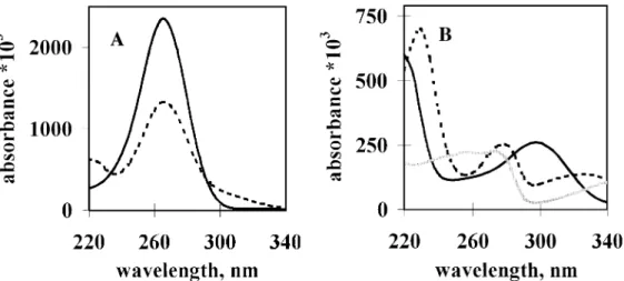
The UV spectrum of ascorbic acid at pH 7.4 exhibited a high absorption peak with a maximum at 265 nm (Figure 1A).
Oxidation of ascorbic acid led to a loss of this absorption band (data not shown), therefore, ascorbic acid oxidation was determined by monitoring the optical density at 265 nm.
The UV absorption spectra of BH4 before and after incubation in Tris–HCl (pH 7.4) at room temperature for 40 min were studied (Figure 1B). The spectrum of BH4 exhibited characteristic changes as BH4 was rapidly auto-
oxidized. Apparently, complete oxidation of BH4to biopterin did not occur, because the absorption spectrum of biopterin is different from the spectrum obtained for BH4after incubation.
The decrease in absorption of BH4at 295 nm during oxidation offered a convenient way to monitor this process.
Inclusion of 25µmol/l BH4 in an ascorbic acid solution of 100 µmol/l resulted in only a slight absorption elevation at
~300 nm (Figure 1A). This elevation was due to the absorption of BH4 and it allowed selective monitoring of BH4 auto- oxidation at 305 nm without substantial interference by light absorption of ascorbic acid. At the same time, oxidation of ascorbic acid could still be quantified by measuring the decrease in absorption at 265 nm, since BH4 and BH2 show a similar quench at 265 nm (Figure 1B) and redox changes of BH4do not interfere at this wavelength.
Oxidation of ascorbic acid and BH4by O2or H2O2dissolved in the medium
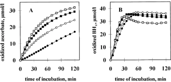
Ascorbic acid (100µmol/l final concentration) underwent fairly rapid auto-oxidation at pH 7.4 with a half-time (T1/2) of 35 min and the rate of this change was only slightly influenced by the presence of 25 µmol/l BH4 (Figure 2A). On the other hand, inclusion of 50µmol/l EDTA in the buffered solution markedly reduced the rate of auto-oxidation (T1/2⬎120 min), indicating that metal contaminants present in the buffer enhance auto- oxidation of ascorbic acid. Under these conditions, BH4exerted a 1.8-fold stimulatory effect on ascorbic acid oxidation (Figure 2B).
The reactivity of ascorbic acid and BH4with H2O2was also studied. Oxidation of ascorbic acid (36µmol/l) was accelerated markedly by low concentrations of H2O2 (Figure 3A) while the effect of H2O2 on BH4 (36 µmol/l) was much smaller (Figure 3B). For instance, 60µmol/l H2O2 resulted in 3-fold and 1.2-fold acceleration in the initial rate of ascorbic acid and BH4 auto-oxidation respectively. Taken together, Figures 2 and 3 clearly demonstrate that: (i) BH4reacts faster with O2 than does ascorbic acid (i.e. BH4is more susceptible to auto- oxidation than is ascorbic acid); (ii) the opposite holds true for H2O2: BH4 oxidation is only slightly increased, whereas ascorbic acid oxidation is significantly enhanced, in response to H2O2.
In order to compare the contribution of H2O2, formed during auto-oxidation of both ascorbic acid and BH4, to the oxidation of these compounds, the effect of catalase on the initial rate of their auto-oxidation was also studied. The oxidation rate of ascorbic acid was inhibited by 8.6 and 17.2µg/ml catalase at 38.7 and 51.9% respectively, in the presence of 50 µmol/l EDTA (Figure 4A). This value definitely exceeded the 25%
catalase-induced decrease in the initial rate of BH4 auto- oxidation measured under the same conditions (Figure 4B).
Evidently, ascorbic acid is more sensitive than BH4 to the H2O2generated during the auto-oxidation of these compounds.
Ascorbic acid protects BH4against oxidation
In order to gain insight into the redox mechanisms between ascorbic acid and BH4, the effect of catalase on the spontaneous and the BH4-promoted ascorbic acid oxidation was studied using the standard experimental conditions (using 100µmol/l
Figure 1.Ultraviolet absorption spectra of ascorbic acid (ASC), tetrahydrobiopterin (BH4) and biopterin. (A) Ultraviolet absorption spectrum of ascorbic acid (ASC) in the presence or absence of BH4: UV absorption of ASC dissolved in 50 mmol/l Tris–HCl, pH 7.4 at 200µmol/l final concentration was monitored between 220 and 340 nm using a Hitachi U-2001 double-beam, automatic spectrophotometer (solid line). Absorption spectrum of the mixture of 100µmol/l ASC and 25µmol/l BH4is shown by the dashed line. (B) Ultraviolet absorption spectra of tetrahydrobiopterin (BH4) and biopterin: UV absorption of BH4dissolved in 50 mmol/l Tris–HCl, pH 7.4, at 25µmol/l final concentration was monitored between 220 and 340 nm before (solid line) and after (dashed line) incubation at 22°C for 40 min. The UV spectrum of biopterin is shown by the grey line.
Figure 2.Effect of tetrahydrobiopterin (BH4) on the oxidation of ascorbic acid (ASC) in the absence (A) or presence (B) of EDTA. ASC (100µmol/l) was incubated in 3.5 ml 50 mmol/l Tris–HCl, pH 7.4 (A) or 50 mmol/l Tris–HCl, pH 7.4, 50µmol/l EDTA (B) solution for the time periods indicated, in the presence (open circles) or absence (solid squares) of 25µmol/l tetrahydrobiopterin (BH4). Oxidation of ASC was monitored by measuring the decrease in optical density at 265 nm. Incubations were performed in triplicates and mean values⫾ SD are presented. In most cases, SD values fall within the size of the symbol. Data from one representative experiment out of three giving similar results are presented.
ascorbic acid, 25 µmol/l BH4 and 50 µmol/l EDTA). BH4
increased 1.8-fold the initial rate of ascorbic acid oxidation.
In the presence of 8.6 µg/ml catalase, the rate of BH4- dependent ascorbic acid oxidation decreased markedly, and some attenuation of the BH4-independent auto-oxidation of ascorbic acid also occurred (data not shown). However, in the presence of 8.6 µg/ml catalase, the rate of ascorbic acid oxidation measured in the presence of BH4was 1.25-fold the rate of ascorbic acid oxidation determined in the absence of BH4. This definite rate difference did not change in the presence of a 2-fold concentration (17.2 µg/ml) of catalase indicating that incomplete removal of H2O2was not responsible for the observed residual oxidation of ascorbic acid (data not shown). These observations seemed to reflect a crucial H2O2- independent interaction between BH4 auto-oxidation and
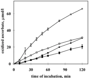
ascorbic acid oxidation. In order to support the validity of this finding, a series of further measurements was performed. With the aim to increase the effect of BH4 on ascorbate oxidation in the presence of catalase, in these experiments the concentra- tions of ascorbic acid, BH4 and EDTA in the incubation medium were enhanced to 143 and 36 µmol and 0.5 mmol/l respectively. In addition, four series of experiments with duplicate incubations each were performed and the results were subjected to statistical analysis. The results indicated an extremely significant ~2-fold BH4-dependent increase in the initial rate of ascorbic acid oxidation in the presence of 27.1 µg/ml catalase (Figure 5). This oxidation could not be caused by incomplete removal of H2O2, because another series of control experiments clearly demonstrated that concentrations of catalase in the range of 17.2–27.1µg/ml (smaller concentra-
Figure 3.Effect of various concentrations of H2O2on the oxidation of ascorbic acid (ASC) (A) and tetrahydrobiopterin (BH4) (B).
ASC or BH4(36µmol/l each) was incubated without H2O2(control, solid circles) or with each of three different concentrations of H2O2: 0.06 mmol/l (open circles), 0.30 mmol/l (solid squares) and 1.20 mmol/l (open squares) in 50 mmol/l Tris–HCl, pH 7.4, 50µmol/l EDTA at 22°C for the time periods indicated. Oxidation of ASC and BH4was followed by the decrease of optical density at 265 and 295 nm respectively. Data from one representative experiment out of three giving similar results are presented.
Figure 4.Effect of catalase on the auto-oxidation of ascorbic acid (ASC) (A) and tetrahydrobiopterin (BH4) (B). ASC or BH4(36µmol/l each) was incubated in 50µmol/l Tris–HCl buffer, pH 7.4, 50µmol/l EDTA in the absence (control, solid circles) or presence of 8.6µg/ml (open squares) or 17.2µg/ml (open triangles) catalase. Oxidation of ASC and BH4was followed by the decrease of optical density at 265 and 295 nm respectively. Each point is a mean value of duplicate incubations. Data from one representative experiment out of three giving similar results are presented.
tions were not studied) completely prevent the oxidation of ascorbate by 60µmol/l H2O2(data not shown). These results indicated that H2O2 released from the auto-oxidation of BH4 is an important, but not the only, factor leading to oxidation of ascorbic acid. Since in the presence of catalase BH4 still accelerated up to 2-fold the oxidation rate of ascorbic acid, we concluded that beside H2O2, BH2(or rather the quininoid- BH2 intermediate of BH4 auto-oxidation) was also able to oxidize ascorbic acid. Because in the absence of H2O2 only BH2 (or qBH2) is present in the system as a redox reaction partner for ascorbic acid, it is reasonable to suggest that ascorbic acid reduced this compound to form BH4.
In order to demonstrate the protective effect of ascorbic acid on net BH4oxidation, BH4stability was measured in the presence of various concentrations of ascorbic acid (Figure 6).
In these measurements, EDTA was not included in the incub- ated mixtures and ascorbic acid solutions at the tested
concentrations were used as photometric blanks (i.e. test solutions and blank solutions contained the same concentration of ascorbic acid). Since in the absence of EDTA there is only negligible difference between the rates of oxidation of ascorbic acid measured either in the presence or absence of 25µmol/l BH4 (Figure 2A), possible interference by optical absorption of ascorbic acid with the BH4absorption measured at 305 nm was largely eliminated. As shown by Figure 6, ascorbic acid inhibits BH4auto-oxidation in a concentration-dependent fashion with 3 mmol/l ascorbic acid providing an almost perfect stabilization of 25µmol/l BH4.
Ascorbic acid stimulates microsomal eNOS activity of the human placenta
Keeping in mind the definite protective action of ascorbic acid on BH4in vitro, the protective effect of ascorbic acid on eNOS activity of placental microsomes was investigated. In these
Figure 5.Effect of catalase on the tetrahydrobiopterin (BH4)- dependent and BH4-independent oxidation of ascorbic acid (ASC).
Oxidation of ASC (143µmol/l) was monitored at 265 nm using 50 mmol/l Tris–HCl, pH 7.4, 0.5 mmol/l EDTA mixture as solvent.
Auto-oxidation of ASC in the absence of BH4and catalase served as control (solid circles). Other incubates contained (in addition to ASC): 36µmol/l BH4(open circles), 27.1µg/ml catalase (solid squares) and 36µmol/l BH4plus 27.1µg/ml catalase (open squares). Results are from four experiments with duplicate incubations each and mean values⫾SEM (n⫽4) are presented.
SEM values are indicated by error bars; where the bar is not visible it is within the size of the symbol. Statistical analysis (ANOVA followed by Bonferroni’st-test) on the basis of the initial rates of ascorbate oxidation:⫹catalase,⫹BH4versus⫹catalase, –BH4: P⬍0.001;⫹catalase,⫹BH4versus –catalase, –BH4:P⬍0.01;
⫹catalase, –BH4versus –catalase, –BH4:P⬎0.05 (not significant); –catalase,⫹BH4versus the other three:P⬍0.001.
Figure 6.Effect of different concentrations of ascorbic acid (ASC) on the auto-oxidation of tetrahydrobiopterin (BH4). BH4 (25µmol/l) was incubated at 22°C in the absence (squares) or in the presence of 0.5 mmol/l (diamonds) or 1.5 mmol/l (triangles) or 3 mmol/l (circles) final concentration of ASC in 50 mmol/l Tris–HCl, pH 7.4 for the time periods indicated and the optical density at 305 nm was recorded. ASC concentrations of blank solutions were exactly identical to that of the test solutions. Mean values⫾SD (n⫽4–6) are presented. Statistical analysis (ANOVA followed by Bonferroni’st-test) on the basis of the initial rates of decomposition: 0.5 mmol/l ASC versus control or 1.5 mmol/l ASC:
P⬎0.05 (not significant); 1.5 mmol/l ASC versus control:
P⬍0.01; 3.0 mmol/l ASC versus control:P⬍0.001; 3.0 mmol/l ASC versus 0.5 or 1.5 mmol/l ASC:P⬍0.01.
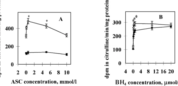
experiments the otherwise routinely used DTT was omitted, since the strong reducing power of this non-physiological dithiol would mask the protective effect of ascorbic acid. In addition, EGTA (100µmol/l final concentration) was included in the incubation mixtures to protect ascorbic acid against metal-catalysed auto-oxidation. Physiological BH4 concentra- tions stimulated eNOS activity 2.5-fold, and ascorbic acid afforded an additional 1.4-fold increase in the BH4-stimulatable eNOS activity (Figure 7). The activity increase was maximal at 500µmol/l ascorbic acid (Figure 7A) and at 200–500 nmol/l BH4 (Figure 7B) concentrations. However, a significant increase of enzyme activity was observed already at 100µmol/l ascorbic acid concentration (Figure 7A).
Ascorbic acid concentrations in the human placenta First trimester and term human placentae contained 117.4 ⫾ 12.5 µmol/kg (mean ⫾ SD, n ⫽ 3 placentae) and 254.5⫾85.2µmol/kg tissue (mean⫾SEM,n⫽7 placentae) ascorbic acid when measured as the dehydroascorbic acid form. Determination of ascorbic acid on the basis of its reducing capacity (314.3 ⫾ 47.5 µmol/kg, mean ⫾ SEM, n ⫽ 7) did not show a statistically significant difference (P⫽0.5513). This finding indicates that most of the ascorbic acid is present in reduced form in placental tissues and in sufficient concentrations to exert a protective effect on auto- oxidative BH4 inactivation.
Discussion
NO is produced during human pregnancy by type III (endo- thelial) NOS (Conradet al., 1993; Garveyet al., 1994; Gude et al., 1994; Kukor and To´th, 1994; To´th et al., 1995) in syncytiotrophoblasts and in the endothelial cells of the umbil- ical and villous blood vessels (Conrad et al., 1993; Myatt et al., 1993a,b; Butteryet al., 1994; Eiset al., 1995). Adequate NO production is considered to be important in the maintenance of feto-placental and materno-placental perfusion and in the adaptation of maternal circulation by vasodilation and blood pressure decrease to the expanded blood volume during preg- nancy (Gudeet al., 1990; Myattet al., 1991, 1992; Chaudhuri et al., 1993; Hull et al., 1994; Rutherford et al., 1995). Our previous results have indicated that eNOS of human placenta is not saturated with BH4, therefore elevation of BH4 levels can stimulate enzyme activity and NO production in placental tissues (Kukor et al., 1996, 2000; Sahin-To´th et al., 1997;
To´th et al., 1997, 1998). eNOS is active as a dimer and, similarly to neuronal NOS (Gorren et al., 1996; Riethmu¨ller et al., 1999) and inducible NOS (Mayeret al., 1997), one of its subunits appears to bind BH4 tightly, while the affinity of the second subunit to BH4 is much lower and the binding concentrations fall within the tissue concentration range of BH4 (To´th et al., 1998; Kukor et al., 2000). Consequently, variations in cellular BH4 concentrations can regulate NO production via the second subunit of eNOS. Decreased availab- ility of BH4 may cause uncoupling of oxygen reduction and arginine oxidation by eNOS and may lead to generation of superoxide anions and subsequently H2O2(Stroeset al., 1998;
Va´squez-Vivar et al., 1998; Xia et al., 1998; Leber et al.,
Figure 7.Effect of ascorbic acid (ASC) on the basal and tetrahydrobiopterin (BH4)-stimulated endothelial nitric oxide synthase (eNOS) activities of placental microsomes. (A) Effect of various concentrations of ASC at constant BH4concentration. Microsomal suspensions (1.4 mg protein) were incubated in duplicates using the standard reaction mixture containing various concentrations of ASC [0.0 (control), 0.1, 0.5, 5.0 and 10.0 mmol/l respectively] in a final volume of 250µl for 15 min at 37°C in the absence (solid circles) or presence (open circles) of 0.5µmol/l BH4. (B) Effect of various concentrations of BH4at constant ASC concentration. Microsomal suspensions (1.8 mg protein) were incubated in duplicates with various concentrations of BH4[0.0 (control), 0.2, 0.5, 1.0, 10.0 and 20.0µmol/l respectively] in the absence (control, solid circles) or presence (open circles) of 5.0 mmol/l ASC using the standard reaction mixture, for 15 min at 37°C.
Mean values from four experiments (n⫽4) with duplicate determinations each⫾SEM (shown by error bars) are presented. Asterisks indicate significant differences (P⬍0.05) from activities obtained with microsomes incubated with BH4in the absence of ASC (A; one-way ANOVA followed by Bonferroni’st-test) or between microsomes incubated in the absence and presence of ASC (B; Wilcoxon’s unpairedt-test).
1999). Thus BH4 deficiency may cause both impaired NO formation and increased production of oxygen radicals. More- over, NO and superoxide can combine with each other to form peroxynitrite (ONOO–), an aggressive oxidant (Beckman and Koppenol, 1996; Cosentino and Lu¨scher, 1998). Coupled with NO inactivation, the formation of peroxynitrite may constitute a serious risk factor for hypertension and atherosclerosis and, in the case of pregnancy, for placental ischaemia, a putative cause of pre-eclampsia (Davidge, 1998; Dekker and Sibai, 1998; Hubel, 1999; Lowe, 2000). We have proposed recently that diminished binding affinity of BH4to the ‘second’ subunit of placental eNOS may play a role in the pathogenesis of pre- eclampsia through promoting the production of abnormally large quantities of O2–and peroxynitrite (Kukoret al., 2000).
Because BH4is sensitive to oxidative agents and can easily react with molecular oxygen even at ambient temperature, a potentially feasible approach to protect BH4 under various conditions of oxidative stress could be the application of natural antioxidants such as ascorbic acid (vitamin C). According to recently reported experimental observations, ascorbic acid treatment of endothelial cells leads to an increase in intracellular BH4 levels and this effect is due to chemical stabilization of the fully reduced form of the pterin (Helleret al., 1999, 2001;
Huanget al., 2000; Bakeret al., 2001). These findings validated the usefulness of vitamin C supplementation for preventing vascular damages and justified the administration of vitamin C in order to help prevent endothelial dysfunction or restore normal endothelial functions (Heitzer et al., 1996; Levine et al., 1996; Ting et al., 1996, 1997; Timimi et al., 1998;
Gokceet al., 1999; Jeserichet al., 1999). In the same context,
ascorbic acid may contribute to the maintenance of satisfactory eNOS activity in the human placenta, thereby reducing the risk of placental dysfunctions or improving placental functions in pathological pregnancies (Gulmezogluet al., 1997; Chappell et al., 1999).
The present findings confirm the BH4-stabilizing antioxidant effect of ascorbic acid and shed some light on its chemical mechanism. Although ascorbic acid itself is an antioxidant, it reacts readily with O2 and produces H2O2, a potent oxidant.
The BH4-protective antioxidant effect of ascorbic acid may result from at least three mechanisms. (i) Elimination of O2 from the solvent. In this respect, competition for O2 as an antioxidant mechanism of ascorbic acid is unlikely, because simultaneous auto-oxidation of 25µmol/l BH4 and 100µmol/l ascorbic acid could proceed in the same solution (Figure 5).
Moreover, O2 had a greater affinity toward BH4 than toward ascorbic acid. (ii) Consumption of H2O2via reduction. Indeed, ascorbic acid reacted with H2O2 readily (Figure 3A), and catalase dramatically decreased oxidation of ascorbic acid incubated with BH4 (Figure 5), suggesting that H2O2 formed by the auto-oxidation of BH4 is consumed by ascorbic acid.
However, in the presence of O2 dissolved in the incubation medium, BH4was relatively insensitive to oxidation by H2O2, as its auto-oxidation was only marginally increased by H2O2 (Figure 3B) and only slightly decreased by catalase (Figure 4B). Therefore, elimination of H2O2by ascorbic acid does not contribute to the chemical stabilization of BH4to any significant extent. (iii) Direct reduction of BH2 to regenerate BH4with the concomitant formation of dehydroascorbic acid. Our results suggest the existence of this mechanism. Thus, incubation of
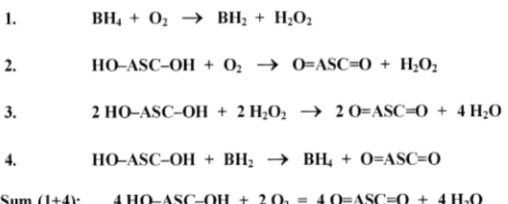
Figure 8.Proposed catalytic effect of tetrahydrobiopterin (BH4) on ascorbic acid (ASC) oxidation. In the mixture of BH4, ASC and O2, an extensive oxidation of ascorbic acid takes place, whereas the concentration of BH4remains relatively unchanged.
ascorbic acid with BH4 markedly increased (~3-fold) the rate of ascorbic acid oxidation and a major part of this increase was abolished by catalase, indicating that this part is mediated by H2O2 (Figure 5). Importantly, in the presence of catalase (which was shown to prevent completely the oxidation of ascorbic acid by H2O2), BH4still stimulated the rate of ascorbic acid oxidation by a factor of ~2 (Figure 5), demonstrating that a portion of the BH4-stimulated ascorbic acid oxidation is not mediated by H2O2. Although BH4 concentrations were not measured in these experiments, this part of increased ascorbic acid oxidation may be accounted for by the direct reduction of BH2(or rather the quininoid-BH2intermediate of the auto- oxidation process) to BH4, since, under the conditions used, BH2 was the only redox reaction partner for ascorbic acid.
Because regenerated BH4is subject to repeated auto-oxidation, we propose that the reaction cycle recurs, continuously produ- cing dehydroascorbic acid from ascorbic acid and O2, while BH4 concentrations remain approximately constant. Chemi- cally, the overall process can be described as ‘BH4-catalysed oxidation of ascorbic acid’ (Figure 8).
An important question is whether or not the in-vitro chemical stabilization of BH4 by ascorbic acid is physiologically rele- vant. In our studies, 3 mmol/l ascorbic acid exerted an almost complete protective effect on 25 µmol/l BH4in the absence of EDTA, indicating that a 120-fold molar excess of ascorbic acid is sufficient for BH4protection. Average tissue concentra- tions of BH4in human placentae from first trimester and term pregnancies are 0.189 and 0.057 µmol/l respectively (Kukor et al., 2000). These values suggest that ascorbic acid concentra- tions as low as 7 µmol/l in term placentae or 23 µmol/l in primordial placentae can have significant protective effects in vivo. Importantly, these ascorbic acid concentrations are well within the range of vitamin C levels we measured in placental tissues, indicating that ascorbic acid may play a physiological role in the regulation of eNOS activity. In this respect, our in- vitro studies with placental eNOS enzyme clearly show that the protective effect of ascorbic acid on BH4 can lead to elevated enzyme activity. In agreement with reports from other laboratories (Heller et al., 1999, 2001; Huang et al., 2000), the ascorbic acid dependent activity increase was not detectable at high, unphysiological BH4concentrations.
Vitamin C is evidently an important daily dietary supplement during pregnancy, and among other effects it may have a beneficial influence on placental and vascular functions. In some of the pre-eclamptic patients, failure to show significant
improvements in response to regular vitamin C administration may stem from the malfunction of their placental eNOS enzyme. In a previous study (Kukor et al., 2000) we have found that the homogenates of seven out of 10 placentae obtained from pre-eclamptic pregnancies contain eNOS enzyme that is resistant to the stimulatory effect of 0.025–
1.00 µmol/l BH4, while the basal activity is sustained. This malfunction could not be corrected by elevated BH4concentra- tions, therefore in these cases increased vitamin C supply cannot alleviate the characteristic pre-eclamptic symptoms. On the other hand, in pre-eclamptic patients with functionally normal BH4-responsive placental eNOS, one may expect beneficial protective and preventive effects from sustained vitamin C supplementation during pregnancy.
Acknowledgements
The help and interest of Miklo´s Sahin-To´th and the technical assistance of Eszter Be´rczi is greatly appreciated. This work was supported by Hungarian Research Fund (OTKA) grant T-29165.
References
Baker, T.A., Milstien, S. and Katusic, Z.S. (2001) Effect of vitamin C on the availability of tetrahydrobiopterin in human endothelial cells.J.Cardiovasc.
Pharmacol.,37, 333–338.
Beckman, J.S. and Koppenol, W.H. (1996) Nitric oxide, superoxide, and peroxynitrite: the good, the bad, and the ugly. Am. J. Physiol., 271, C1424–C1437.
Buttery, L.D.K., McCarthy, A., Springall, D.R., Sullivan, M.H., Elder, M.G., Michel, T. and Polak, J.M.(1994) Endothelial nitric oxide synthase in the human placenta: regional distribution and proposed regulatory role at the feto-maternal interface.Placenta,15, 257–265.
Chappell, L.C., Seed, P.T., Briley, A.L., Kelly, F.J., Lee, R., Hunt, B.J., Parmar, K., Bewley, S.J., Shennan, A.H., Steer, P.J. and Poston, L. (1999) Effect of antioxidants on the occurrence of preeclampsia in women at increased risk: a randomised trial.Lancet,354, 810–816.
Chaudhuri, G., Cuevas, J., Buga, G.M. and Ignarro, L.J. (1993) NO is more potent, than PGI2in maintaining low vascular tone in fetoplacental vessels.
Am.J.Physiol.,265, H2036–H2043.
Conrad, K.P., Vill, M., McGuire, P.G., Dail, W.G. and Davis, A.K. (1993) Expression of nitric oxide synthase by syncytiotrophoblast in human placental villi.FASEB J.,7, 1269–1276.
Cosentino, F. and Lu¨scher, T.F. (1998) Tetrahydrobiopterin and endothelial function.Eur.Heart J.,19(Suppl. G), G3–G8.
Davidge, S.T. (1998) Oxidative stress and altered endothelial cell function in preeclampsia.Semin.Reprod.Endocrinol.,16, 65–73.
Davies, M.B., Austin, J. and Partridge, D.A. (1991)Vitamin C.Its Chemistry and Biochemistry. Thomas Graham House, Science Park, Cambridge, pp.
36 and 117.
Dekker, G.A. and Sibai, B.M. (1998) Etiology and pathogenesis of preeclampsia: current concepts.Am.J.Obstetr.Gynecol.,179, 1359–1375.
Denson, K.W. and Bowers, E.F. (1961) The determination of ascorbic acid in white blood cells. A comparison of W.B.C. ascorbic acid and phenolic acid excretion in elderly patients.Clin.Sci.,21, 157–162.
Eis, A.L.W., Brockman, D.E., Pollock, J.S. and Myatt, L. (1995) Immunohistochemical localization of endothelial nitric oxide synthase in human villous and extravillous trophoblast populations and expression during syncytiotrophoblast formationin vitro.Placenta,16, 113–126.
Enstrom, J.E., Kanim, L.E. and Klein, M.A. (1992) Vitamin C intake and mortality among a sample of the United States population.Epidemiology, 3, 194–202.
Frei, B. (1999) On the role of vitamin C and other antioxidants in atherogenesis and vascular dysfunction.Proc.Soc.Exp.Biol.Med.,222, 196–204.
Gale, C.R., Martyn, C.N., Winter, P.D. and Cooper, C. (1995) Vitamin C and risk of death from stroke and coronary heart disease in cohort of elderly people.Br.Med.J.,310, 1563–1566.
Garvey, E.P., Tuttle, J.V., Covington, K., Merrill, B.M., Wood, E.R., Baylis, S.A. and Charles, I.G. (1994) Purification and characterization of the constitutive nitric oxide synthase from human placenta.Arch.Biochem.
Biophys.,311, 235–241.
Gey, K.F., Stahelin, H.B. and Eichholzer, M. (1993) Poor plasma status of carotene and vitamin C is associated with higher mortality from ischemic heart disease and stroke: Basel Prospective Study.Clin.Invest.,71, 3–6.
Gokce, N., Keaney, J.F. Jr, Frei, B., Holbrook, M., Olesiak, M., Zachariah, B.J., Leeuwenburgh, C., Heinecke, J.W. and Vita, J.A. (1999) Long-term ascorbic acid administration reverses endothelial vasomotor dysfunction in patients with coronary artery disease.Circulation,99, 3234–3240.
Gorren, A.C.F., List, B.M., Schrammel, A., Pitters, E., Hemmens, B., Werner, E.R., Schmidt, K. and Mayer, B. (1996) Tetrahydrobiopterin-free neuronal nitric oxide synthase: evidence for two identical highly anticooperative pteridine binding sites.Biochemistry,35, 16735–16745.
Gude, N.M., King, R.G. and Brennecke, S.P. (1990) Role of endothelium- derived nitric oxide in maintenance of low fetal vascular resistance in placenta.Lancet,336, 1589–1590.
Gude, N.M., DiIulio, J., Brennecke, S.P. and King, R.G. (1994) Human placental villous nitric oxide synthase activity. Pharm. Commun., 4, 163–171.
Gulmezoglu, A.M., Hofmeyr, G.J. and Oosthuisen, M.M. (1997) Antioxidants in the treatment of severe preeclampsia: an explanatory randomised controlled trial.Br.J.Obstetr.Gynecol.,104, 689–696.
Heitzer, T., Just, H. and Munzel, T. (1996) Antioxidant vitamin C improves endothelial dysfunction in chronic smokers.Circulation,94, 6–9.
Heitzer, T., Brockhoff, C., Mayer, B., Warnholtz, A., Mollnau, H., Henne, S., Meinertz, T. and Munzel, T. (2000) Tetrahydrobiopterin improves endothelium-dependent vasodilation in chronic smokers: evidence for a dysfunctional nitric oxide synthase.Circ.Res.,86, E36–E41.
Heller, R., Mu¨nscher-Paulig, F., Grabner, R. and Till, U. (1999)L-Ascorbic acid potentiates nitric oxide synthesis in endothelial cells.J.Biol.Chem., 274, 8254–8260.
Heller, R., Unbehaun, A., Schellenberg, B., Mayer, B., Werner-Felmayer, G.
and Werner, E.R. (2001)L-Ascorbic acid potentiates endothelial nitric oxide synthesis via a chemical stabilization of tetrahydrobiopterin.J.Biol.Chem., 276, 40–47.
Huang, A., Vita, J.A., Venema, R.C. and Keaney, J.F. Jr (2000) Ascorbic acid enhances endothelial nitric-oxide synthase activity by increasing intracellular tetrahydrobiopterin.J.Biol.Chem.,275, 17399–17406.
Hubel, C.A. (1999) Oxidative stress in the pathogenesis of preeclampsia.
Proc.Soc.Exp.Biol.Med.,222, 222–235.
Hubel, C.A., Kagan, V.E., Kisin, E.R., McLaughlin, M.K. and Roberts, J.M.
(1997) Increased ascorbate radical formation and ascorbate depletion in plasma from women with preeclampsia: implications for oxidative stress.
Free Radic.Biol.Med.,23, 597–609.
Hull, A.O., White, C.R. and Pearce, W.J. (1994) Endothelium derived relaxing factor and cyclic GMP-dependent vasorelaxation in human chorionic arteries.Placenta,15, 365–375.
Jeserich, M., Schindler, T., Olschewski, M., Unmussig, M., Just, H. and Solzbach, U. (1999) Vitamin C improves endothelial function of epicardial coronary arteries in patients with hypercholesterolemia or essential hypertension|assessed by cold pressure testing.Eur.Heart J.,22, 1676–1680.
Kharb, S. (2000) Vitamin E and C in preeclampsia.Eur.J.Obstetr.Gynecol.
Reprod.Biol.,93, 37–39.
Kukor, Z. and To´th, M.(1994) Ca2⫹-dependent and Ca2⫹-independent NO- synthesizing activities of human primordial placenta.Acta Physiol.Hung., 82, 313–319.
Kukor, Z., Me´sza´ros, G., Hertelendy, F. and Toth, M. (1996) Calcium- dependent nitric oxide synthase is potently stimulated by tetrahydrobiopterin in human primordial placenta.Placenta,17, 69–73.
Kukor, Z., Valent, S. and To´th, M. (2000) Regulation of nitric oxide synthase activity by tetrahydrobiopterin in human placentae from normal and preeclamptic pregnancies.Placenta,21, 763–772.
Leber, A., Hemmens, B., Klo¨sch, B., Goessler, W., Raber, G., Mayer, B. and Schmidt, K. (1999) Characterization of recombinant human endothelial nitric-oxide synthase purified from the yeastPichia pastoris.J.Biol.Chem., 274, 37658–37664.
Levine, G.N., Frei, B., Koulouris, S.N., Gerhard, M.D., Keaney, J.F. Jr and Vita, J.A. (1996) Ascorbic acid reverses endothelial vasomotor dysfunction in patients with coronary artery disease.Circulation,93, 1107–1113.
Lowe, D.T. (2000) Nitric oxide dysfunction in the pathophysiology of preeclampsia.Nitric Oxide: Biol.Chem.,4, 441–458.
Lowry, O.H., Rosebrough, N.J., Farr, A.L. and Randall, R.J. (1951) Protein measurement with the Folin phenol reagent.J.Biol.Chem.,143, 265–275.
Mayer, B., Wu, C., Gorren, A. C.F., Pfeiffer, S., Schmidt, K., Clark, P., Stuehr, D.J. and Werner, E.R. (1997) Tetrahydrobiopterin binding to macrophage inducible nitric oxide synthase: heme spin shift and dimer stabilization by the potent pterin antagonist 4-amino-tetrahydrobiopterin.Biochemistry,36, 8422–8427.
Mikhail, M.S., Anyaegbunam, A., Garfinkel, D., Palan, P.R., Basu, J. and Romney, S.L. (1994) Preeclampsia and antioxidant nutrients: decreased plasma level of reduced ascorbic acid, alpha-tocopherol and beta-carotene in women with preeclampsia.Am.J.Obstetr.Gynecol.,171, 150–157.
Myatt, L., Brewer, A.S. and Brockman, D.E. (1991) The action of nitric oxide in the perfused human fetal–placental circulation.Am.J.Obstetr.Gynecol., 164, 687–692.
Myatt, L., Brewer, A.S., Langdon, G. and Brockman, D.E. (1992) Attenuation of the vasoconstrictor effects of thromboxane and endothelium by nitric oxide in the human fetal–placental circulation. Am.J. Obstet.Gynecol., 166, 224–230.
Myatt, L., Brockman, D.E., Eis, A.L.W. and Pollock, J.S. (1993a) Immunohistochemical localization of nitric oxide synthase in the human placenta.Placenta,14, 487–495.
Myatt, L., Brockman, D.E., Langdon, G. and Pollock, J.S. (1993b) Constitutive calcium-dependent isoform of nitric oxide synthase in the human placental villous vascular tree.Placenta,14, 373–383.
Ness, A.R., Powles, J.W. and Khaw, K.T. (1996a) Vitamin C and cardiovascular disease: a systematic review.J.Cardiovasc.Risk,3, 513–521.
Ness, A.R., Khaw, K.T., Bingham, S. and Day, N.E. (1996b) Vitamin C status and undiagnosed angina.J.Cardiovasc.Risk,3, 373–377.
Ness, A.R., Khaw, K.T., Bingham, S. and Day, N.E. (1996c) Vitamin C status and blood pressure.J.Hypertens.,14, 503–508.
Ness, A.R., Chee, D. and Elliott, P. (1997) Vitamin C and blood pressure|an overview.J.Hum.Hypertens.,11, 343–350.
Nyyssonen, K., Parviainen, M.T., Salonen, R., Tuomilehto, J. and Salonen, J.T. (1997) Vitamin C deficiency and risk of myocardial infarction:
prospective population study of men from eastern Finland. Br.Med. J., 314, 634–638.
Omaye, S.T., Turnbull, J.D. and Sauberlich, H.E. (1979) Selected methods for the determination of ascorbic acid in animal cells, tissues, and fluids.
Methods Enzymol.,62, 3–11.
Pieper, G.M. (1997) Acute amelioration of diabetic endothelial dysfunction with a derivative of the nitric oxide synthase cofactor, tetrahydrobiopterin.
J.Cardiovasc.Pharmacol.,29, 8–15.
Riemersma, R.A., Wood, D.A., Macintyre, C.C., Elton, R.A., Gey, K.F. and Oliver, M.F. (1991) Risk of angina pectoris and plasma concentrations of vitamins A, C, and E and carotene.Lancet,337, 1–5.
Riethmu¨ller, C., Gorren, A.C.F., Pitters, E., Hemmens, B., Habisch, H.J., Heales, S.J., Schmidt, K., Werner, E.R. and Mayer, B. (1999) Activation of neuronal nitric-oxide synthase by the 5-methyl analog of tetrahydrobiopterin.
Functional evidence against reductive oxygen activation by the pterin cofactor.J.Biol.Chem.,274, 16074–16051.
Rosenkranz-Weiss, P., Sessa, W.C., Milstien, S., Kaufman, S., Watson, C.A. and Pober, J.S. (1994) Regulation of nitric oxide synthesis by proinflammatory cytokines in human umbilical vein endothelial cells.
Elevations in tetrahydrobiopterin levels enhance endothelial nitric oxide synthase specific activity.J.Clin.Invest.,93, 2236–2243.
Rutherford, R.A.D., McCarthy, A., Sullivan, M.H.F., Elder, M.G., Polak, J.M.
and Wharton, J. (1995) Nitric oxide synthase in human placenta and umbilical cord from normal, intrauterine growth-retarded and pre-eclamptic pregnancies.Br.J.Pharmacol.,116, 3099–3109.
Sagol, S., Ozkinay, E. and Ozsener, S. (1999) Impaired antioxidant activity in woman with preeclampsia.Int.J.Gynecol.Obstetr.,64, 121–127.
Sahin-To´th, M., Kukor, Z. and To´th, M. (1997) Tetrahydrobiopterin preferentially stimulates activity and promotes subunit aggregation of membrane-bound calcium-dependent nitric oxide synthase in human placenta.Mol.Hum.Reprod.,3, 293–298.
Schmidt, K., Werner, E.R., Mayer, B., Wachter, H. and Kukovetz, W.R. (1992) Tetrahydrobiopterin-dependent formation of endothelium-derived relaxing factor (nitric oxide) in aortic endothelial cells.Biochem.J.,281, 297–300.
Stroes, E., Kastelein, J., Cosentino, F., Erkelens, W., Wever, R., Koomans, H., Luscher, T. and Rabelink, T. (1997) Tetrahydrobiopterin restores endothelial function in hypercholesterolemia.J.Clin.Invest.,99, 41–46.
Stroes, E., Hijmering, M. and van Zandvoort, R. (1998) Origin of superoxide production by endothelial nitric oxide synthase.FEBS Lett.,438, 161–164.
Tho¨ny, B., Auerbach, G. and Blau, N. (2000) Tetrahydrobiopterin biosynthesis, regeneration and functions.Biochem.J.,347, 1–16.
Tiefenbacher, C.P., Chilian, W.M., Mitchell, M. and DeFily, D.V. (1996) Restoration of endothelium-dependent vasodilation after reperfusion injury by tetrahydrobiopterin.Circulation,94, 1423–1429.
Tiefenbacher, C.P., Bleeke, T., Vahl, C., Amann, K., Vogt, A. and Kubler, W.
(2000) Endothelial dysfunction of coronary resistance arteries is improved by tetrahydrobiopterin in atherosclerosis.Circulation,102, 2172–2179.
Ting, H.H., Timimi, F.K., Boles, K.S., Creager, S.J., Ganz, P. and Creager, M.A. (1996) Vitamin C improves endothelium-dependent vasodilation in patients with non-insulin-dependent diabetes mellitus.J.Clin.Invest.,97, 22–28.
Ting, H.H., Timimi, F.K., Haley, E.A., Roddy, M.A., Ganz, P. and Creager, M.A. (1997) Vitamin C improves endothelium-dependent vasodilation in forearm resistance vessels of humans with hypercholesterolemia.
Circulation,95, 2617–2622.
Timimi, F.K., Ting, H.H., Haley, E.A., Roddy, M.A., Ganz, P. and Creager, M.A. (1998) Vitamin C improves endothelium-dependent vasodilation in patients with insulin-dependent diabetes mellitus.J.Am.Coll.Cardiol.,31, 552–557.
To´th, M., Kukor, Z., Romero, R. and Hertelendy, F. (1995) Nitric oxide synthase in first trimester human placenta: characterization and subcellular distribution.Hypertens.Pregn.,14, 287–300.
To´th, M., Kukor, Z. and Sahin-To´th, M. (1997) Activation and dimerization of type III nitric oxide synthase by submicromolar concentrations of tetrahydrobiopterin in microsomal preparations from human primordial placenta.Placenta,18, 189–196.
To´th, M., Kukor, Z. and Sahin-To´th, M.(1998) Differential response of basal and tetrahydrobiopterin-stimulated activities of placental type III nitric oxide synthase to sodium dodecyl sulphate: relation to dimeric structure.
Mol.Hum.Reprod.,4, 1165–1172.
Ueda, S., Matsuoka, H., Miyazaki, H., Usui, M., Okuda, S. and Imaizumi, T. (2000) Tetrahydrobiopterin restores endothelial function in long-term smokers.J.Am.Coll.Cardiol.,35, 71–75.
Va´squez-Vivar, J., Kalyanaraman, B., Marta´sek, P., Hogg, N., Masters, B.S., Karoui, H., Tordo, P. and Pritchard, K.A. Jr (1998) Superoxide generation by endothelial nitric oxide synthase: the influence of cofactors.Proc.Natl Acad.Sci.USA,95, 9220–9225.
Vita, J.A., Keaney, J.F. Jr, Raby, K.E., Morrow, J.D., Freedman, J.E., Lynch, S., Koulouris, S.N., Hankin, B.R. and Frei, B. (1998) Low plasma ascorbic acid independently predicts the presence of an unstable coronary syndrome.
J.Am.Coll.Cardiol.,31, 980–986.
Werner-Felmayer, G., Werner, E.R., Fuchs, D., Hausen, A, Reibnegger, G., Schmidt, K., Weiss, G. and Wachter, H. (1993) Pteridine biosynthesis in human endothelial cells. Impact on nitric oxide-mediated formation of cyclic GMP.J.Biol.Chem.,268, 1842–1846.
Xia, Y., Tsai, A., Berka, V. and Zweier, J.L. (1998) Superoxide generation from endothelial nitric-oxide synthase. A Ca2⫹/calmodulin-dependent and tetrahydrobiopterin regulatory process.J.Biol.Chem.,273, 25804–25808.
Submitted April 23, 2001; resubmitted August 29, 2001; accepted November 22, 2001