I D E N T I F I C A T I O N A N D I S O L A T I O N OF LICHEN S U B S T A N C E S
JOHAN SANTESSON
I. Identification without Previous Isolation 633
A. Color Tests 633 B. Fluorescence 638 C. Microcrystallization 638 D. Chromatography 639 E. Lichen Mass Spectrometry (LMS) 643
F. Quantitative Determination 644
II. Isolation 646 A. Preparation of Lichen Material for Extraction 646
B. Extraction 647 C. Working-Up Procedures 647
III. Identification after Previous Isolation 648
A. Melting Points 648 B. Spectral Properties 649 C. Chromatographic Comparisons 649
References 650
I. Identification without Previous Isolation
A positive identification of lichen substances usually requires isolation of the compounds and comparison with authentic samples. However, there exist microchemical methods by which more or less reliable identifications can be achieved without the use of too much effort or lichen material.
Color tests and fluorescence analysis give indications of which groups of compounds might be present in a lichen sample, while microcrystallization, chromatography, and lichen mass spectrometry lead to tentative identifica
tions of the compounds.
A. Color Tests
Four color tests for lichen substances are used routinely in lichenology.
The C, K, and KC tests were discovered by Nylander (1866a,b) and the PD 633
634 JOHAN SANTESSON
(or P) test was introduced by Asahina (1934). During the last decades, some other tests have also been suggested. The color tests can be carried out by applying the appropriate reagent to a lichen fragment by means of a pointed glass rod. The color changes are best observed under a binocular lens. Cortex and medulla should be tested separately. If carried out on the same lichen fragment, the PD test should be done before the C test.
Color reactions are often more easily observed if the tests are carried out with the filter paper method (Santesson, 1967a). A lichen fragment is pressed down in the middle of a piece of filter paper, and the lichen substances are extracted by treatment with 10-20 drops of acetone. Each drop of acetone is allowed to evaporate, leaving the extracted substances in a ring around the lichen fragment. The fragment is removed and tests are carried out upon the
"extract ring."
T A B L E I
CO L O R REACTIONS OF LI C H E N SUBSTANCES
Reaction Compound
Pigments
Anthraquinones, bisanthraquinones, terphenylquinones, naphtho
quinones, pyxiferin Xanthones, sordidone Usnic acids
K + red to violet K + , D i m r o t h + , K C + , K + , T i C l3+ , P D + ,
P D + , K +
P D + , K - P D - , K + , C +
P D - , K - , C + red
P D - , K - , C + greenish P D - , K - , C + blueish P D - , K - , C - , K C +
Colorless compounds
Depsides: alectorialic acid, atranorin, baeomycesic acid, barbatolic acid, chloroatranorin, decarboxythamnolic acid, haemathamno- lic acid, nephroarctin, ih&mnoMc&cxd, Depsidones: consticticacid, fumarprotocetraric acid, norstictic acid, physodalic acid, proto- cetraric acid, salazinic acid, stictic acid, virensic acid
Pannarin, psoromic acid, fumarprotocetraric acid, protocetraric acid, virensic acid
Cryptochlorophaeic acid, hiascic acid, hypothamnolic acid (K+
violet), merochlorophaeic acid, paludosic acid, ramalinolic acid, scrobiculin
Anziaic acid, 4-0-demethylbarbatic acid, erythrin, ethyl orsellinate, gyrophoric acid, lecanoric acid, methyl 3,5-dichlorolecanorate, methyl-^-orsellinate, montagnetol, olivetoric acid, siphulin Didymic acid, pannaric acid, porphyrilic acid, strepsilin Diploschistesic acid
Alectoronic acid, α-collatolic acid, glomelliferic acid, lobaric acid, 4-O-methylphysodic acid, microphyllinic acid, norlobaridone, physodic acid, picrolichenic acid
All color test reagents (except C) must be handled with care and should not come into contact with skin, herbarium envelopes, etc. If spilled, PDcanbe (incompletely) removed by washing with very dilute acetic or hydrochloric acid.
Table I summarizes the color-test reactions of some lichen substances.
Color tests are only indicative for groups of compounds, and must be sup
plemented with other tests if positive identification of a compound is sought.
It should be noted that the color reactions of a compound are to a certain extent dependent on concentration and localization in the thallus. Thus, a C+ or K+ red compound might in low concentrations give a C+ orK+ faint orange color. (In the same way, thallus color due to presence of a certain pig
ment might differ considerably from one lichen specimen to another for the same reasons. Pure parietin is red, but Xanthoria parietina might have any shade varying from greenish-yellow to deep orange. Alectoria fremontii is dark brown but still contains yellow vulpinic acid.)
1. THE C TEST
As reagent, a saturated aqueous solution of calcium hypochlorite [bleach
ing powder, Ca(OCl)2] or a dilute aqueous solution of sodium hypochlorite (NaOCl) is employed. Some commercial bleaching fluids, containing "ac
tive chlorine," can be used as substitutes. The Ca(OCl)2 and NaOCl solutions must be prepared daily, since they decompose within 24-48 hours. In sun
light, they are stable for less than 1 hour. The stock chemicals are best stored in a cool dry place. Light, heat, humidity, and carbon dioxide hasten their decomposition.
Aromatic compounds, having two free hydroxyl groups meta to each other, give a positive pink to red C test (Fig. la). If the position between the hydroxyls is substituted by a side chain, as in β-orcinol derivatives, the reac
tion often fails. Halogen substitution (as in methyl 3,5-dichlorolecanorate) does not interfere with the test. Hydroxyl substitution (diploschistesic acid) may change the response to a blue color. The majority of the lichen dibenzo- furanes, having at least one free hydroxyl group, give a positive green Ctest, often difficult to observe.
The colored reaction products obtained in the C test are unstable and easily destroyed by an excess of the reagent. Hypothamnolic acid (albeit sub
stituted at the position between the m-hydroxyls) gives a red color with C which disappears within seconds. The chemistry behind the Ctest has not been elucidated. Possibly a combination of chlorination and oxidation reac
tions leads to monomeric and/or dimeric quinonoid structures. Only one col
ored compound has been isolated from the reaction of orcinol with calcium hypochlorite: the yellow tetrachlorodihydroorcinol (L. Gren and J. Santes
son, unpublished).
6 3 6 JOHAN SANTESSON
FI G . 1. Color reactions of lichen substances. The structural part responsible for the color reaction is drawn in heavy lines, (a) C+ compounds: lecanoric acid (left) and norlichexanthone (right); (b) P D + thamnolic acid reacting with /?-phenylenediamine; (c) KC+ lobaric acid hydrolyzed with KOH to yield a compound with 2 free w-hydroxyls;(d)lichexanthone reacting with Dimroth's reagent to give a fluorescent compound.
2. THE Κ TEST
A 10-25% aqueous solution of potassium hydroxide is used as the reagent.
The solution is stable, but etches glass vessels slowly. Quinonoid lichen pig
ments (anthraquinones, naphthoquinones, terphenylquinones) give a posi
tive dark red to violet Κ test, whereas pulvinic-asid derivatives, xanthones, and usnic acids do not respond. Some depsides (e.g., atranorin, thamnolic acid) and many β-orcinol depsidones exhibit yellow to red colors with K.
The Κ test depends upon salt formation and requires the presence of at least one acidic functional group in the molecule. Thus, fully O-methylated phenolic quinones (not yet found in lichens) would not react. The chemical structures of colored potassium salts of K+ depsides and depsidones are not yet known.
3. THE P D TEST
The commonly used reagent is a 1-5% ethanolic solution of /?-phenyl- enediamine that will keep for about a day. A more stable reagent has been
described by Steiner (1955): 1 gm /?-phenylenediamine, 10 gm sodium sul
fite and 1 ml of a neutral, liquid detergent are dissolved in 100 ml of water.
It can be used for at least a month.
/?-Phenylenediamine reacts with aromatic aldehydes, giving yellow to red Schiff bases (Fig. lb). Most β-orcinol depsidones, as well as some/5-orcinol depsides, give a positive PD test. Aromatic diamines other than /?-phenyl- enediamine also react with aromatic aldehydes. Asahina (1934) found that benzidin gives less intensely colored reaction products thanp-phenylenedia- mine. Santesson (1966) compared some diamine reagents and suggested the use of 0-dianisidine (OD) as a PD substitue. 0-Dianisidine is more sensitive, more stable, and more toxic than PD, but not as corrosive. The color reactions obtained with OD are not always identical with those of PD.
4. THE KC TEST
Κ is first applied to the lichen, then immediately followed by C. The potas
sium hydroxide hydrolyzes depside and depsidone ester bonds, and if the phenolic hydroxyl group released is in a position meta to another hydroxyl, an orange to red color will be obtained as C is applied (Fig. lc). Some meta- depsides (e.g., cryptochlorophaeic acid, scrobiculin), which exhibit a fleet- ingly red C reaction, are KC+ more persistently red, although the freed hydroxyl is located between two free meta hydroxyls. Usnic acids give a deep yellow KC test. If a strong red color is already produced by Κ or C alone, the KC test is meaningless and superfluous.
5. OTHER COLOR TESTS
A 5% aqueous solution of chloramine-T gives a yellow color with usnic acids (Mitsuno, 1953). An 8% aqueous solution of titanium trichloride (TiCl3) produces a yellow-green color with usnic acids (Bendz et al., 1967).
Dimroth's reagent is rather specific for xanthones (Santesson, 1968). It is prepared by adding 10 gm of boric acid to 100 ml hot (100°C) acetic anhyd
ride and allowing the solution to cool. The color test is carried out in UV light, and an intense yellow flourescence, stable for at least 1 minute, suggests the presence of xanthones. The test depends upon the formation of boro- acetates (Fig. Id). Some chromones give a positive test. As a substitute for Dimroth's reagent, an alkaline beryllate solution can be used (Santesson, 1969c).
A methanolic solution of magnesium acetate has been suggested as are- agent for 1-hydroxylated anthraquinones (Shibata et al., 1950). An orange to red color, appearing after a few minutes at 90° C, indicates a positive response. The test is best used in combination with the filter paper method.
Aromatic 0-hydroxyaldehydes (PD + compounds) can be detected by the use of a solution of 5 gm hydrazine sulfate and 10 gm sodium acetate in
638 J O H A N S A N T E S S O N
100 ml water (Feigl and Anger, 1966). A yellow to orange fluorescence (in UV light) appears within a minute.
A solution of 0.2-0.5 gm iodine in 100 ml aqueous 0.5% potassium iodide is often used as a reagent for certain polysaccharides in lichens (the I test).
The reagent is susceptible to air oxidation and should be renewed when the brownish color fades. Isolichenin, but not lichenin, will give a blue color.
The chemistry of the color reaction is probably the same as that for the well- known iodine test for starch. The reaction is reversible (the color disappears upon dilution with water).
Dilute nitric acid (HN03) is sometimes used as a color test reagent in lichenology. It is not known what types of compounds are actually detected in this test.
B. Fluorescence
Many lichen substances fluoresce in long-wave (366 nm) ultraviolet light.
Examination of lichen specimens in UV light can thus provide valuable clues to the presence or absence of certain compounds (Cernhorsky, 1950;
Ozenda, 1951; Hale, 1956a). Anthraquinones appear brick red to vermilion, pulvinic-acid derivatives yellowish and xanthones bright yellow to orange red.
Some depsides and depsidones fluoresce bright white to bluish or greenish white, e.g., alectoronic acid, divaricatic acid, lobaric acid, sphserophorin, and squamatic acid. Addition of a drop of alkali (e.g., Κ reagent) will often change the fluorescence.
C Microcrystallization
In 1936, Asahina introduced microcrystallization as the first generally applicable method for tentative identification of lichen substances on a micro scale (Asahina, 1936, 1937, 1938, 1939, 1940). The method rapidly gained acceptance among lichenologists and has been used extensively for chemical studies in connection with taxonomic work, e.g., on Cladonia
(Evans, 1943; Thomson, 1967), Parmelia (Krog, 1951; Hale, 1965; Hale and Kurokawa, 1964), and Cetrelia and Platismatia (Culberson and Culberson, 1968).
Crystal tests require no specialized equipment. One or a few lichen frag
ments are placed on a microscope slide and the lichen substances present extracted by dropwise treatment of the fragments with a suitable solvent, usually acetone. After evaporation of the solvent, the fragments are re
moved, leaving a more or less crystalline residue of lichen substances on the slide. A drop of a suitable crystallizing solvent mixture is added to the residue and a coverslip added. The slide is heated gently over a tiny flame or on an
electric plate. Upon cooling, substances present may appear in crystalline form and can then be identified from crystal shape and color. The crystals are best observed in polarized light under low magnification (χ 100-χ 400).
The most frequently used crystallizing reagents are: GE, glycerol .acetic acid, 1:3; GAW, glycerohethanol: water, 1:1:1; GAoT, glycerohethanol:
0-toluidine, 2:2:1; GAQ, glycerol:ethanol:quinoline, 2:2:1 (all parts by volume). If kept in stoppered bottles, the solutions are stable for at least a month.
Tests with the GE and GAW solutions are simply recrystallizations, and the volume of the added reagent should be kept at a minimum. The GAoT and GAQ tests depend upon salt formation and—in the case of aromatic aldehydes—possibly also upon condensation reactions. Characteristic crystalline salts are also formed with some inorganic reagents. An aqueous solution of potassium carbonate and potassium hydroxide (10% of both) will precipitate the potassium salt of norstictic acid as red needles. A saturated solution of barium hydroxide produces easily recognized barium salts with many despides, notably atranorin.
Photographs of microcrystals of various compounds are scattered in numerous papers (for references, see Culberson, 1969, 1970a). Hale (1967) illustrates the crystalline appearance of 24 compounds, Taylor (1967) 18 compounds, and Thomson, (1967) 22 compounds of known structure. The latter book also contains an extensive discussion of the microcrystallization methods.
This method is best suited for the identification of depsides, depsidones, and dibenzofuranes. It is less suitable for aliphatic acids and terpenes (except zeorin and ursolic acid) and cannot be used for pigments (except usnic acid).
The sensitivity is high enough to allow the identification of microgram amounts of compounds (Culberson, 1963).
D. Chromatography
Only paper and thin-layer chromatography have been used to any larger extent for the identification of lichen substances. Column and preparative- layer chromatography have been employed for isolation of lichen substances (see below). Gas-liquid chromatography (GLC) has been applied in a few cases. The separation of triterpenes was studied by Ikekawa et al. (1965), Shibata et al. (1965), and Yosioka et al. (1969). Gas-liquid chromatography of the aliphatic lichen acids protolichesterinic acid and lichesterinic acid (as methyl esters) was reported by Bloomer et al. (1970a,b). Published data on high-pressure liquid-liquid chromatography of lichen substances are just beginning to appear (Culberson, 1972).
640 JOHAN SANTESSON 1. PAPER CHROMATOGRAPHY (PC)
Paper chromatography was introduced into lichenology independently by Wachtmeister (1952) and Mitsuno (1953). Further studies have been pub- lished by Mitsuno (1955), Wachtmeister (1956), and Hess (1958). A useful review has also appeared (Wachtmeister, 1959). Most PC analyses, how- ever, can advantageously be replaced by thin-layer chromatographic methods.
In PC of lichen compounds polar solvent systems are used for the most part. Typical examples are A2-butanol:concentrated ammonia (4:1, parts by volume), w-butanol:acetone:water (5:1:2), and rt-butanol:ethanol:water (4:1:5). Better Rvalues and less trailing can sometimes be obtained if the chromatographic papers are buffered with phosphate (Na3P04 o r N a2H P 04) (Wachtmeister, 1956). Depsides and depsidones should be chromatographed both before and after microhydrolysis of the extracts (see below).
2. THIN-LAYER CHROMATOGRAPHY (TLC)
The sensitivity, rapidity, general applicability, and simplicity of equipment needed makes TLC one of the best microchemical methods for the systema- tic botanist. Good texts are available on the general aspects of TLC techniques (e.g., Stahl, 1969; Randerath, 1966; Truter, 1963).
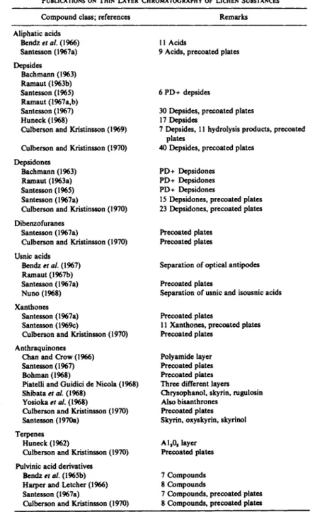
The first TLC separation of lichen substances was reported by Stahl and Schorn (1961). Some papers giving TLC data on one or more groups of lichen substances are listed in Table II. Especially extensive tabulations of fydata are found in the works of Santesson (1967a), Huneck (1968) (a review with many previously unpublished data), and Culberson and Kristinsson(1970).
Numerous publications contain data on TLC of single compounds (for references, see Culberson, 1969, 1970a).
Both "laboratory-made" and precoated TLC plates have been used for separations. The adsorbent is almost always silica gel, although polyamide has been used in TLC of anthraquinones (Chan and Crow, 1966).
Numerous solvent systems have been described. Almost all used for TLC of depsides and depsidones contain an acid (acetic or formic acid) to prevent trailing. Pastuska's mixture (benzene:dioxane:acetic acid, 90:25:4 parts by volume) is especially useful for the separation of acidic aromatic lichen substances.
A number of methods are available for visualizing the spots after chroma- tography. If the adsorbent is impregnated with a fluorescence indicator, most aromatic compounds will be visible in UV light. Even on ordinary adsorbents (e.g., silica gel G) many aromatic compounds will exhibit a characteristic fluorescence in UV light. Nearly all lichen substances can be made visible by spraying the chromatographed plates with a 10% solution of sulfuric acid
TABLE II
PUBLICATIONS ON THIN LAYER CHROMATOGRAPHY OF LICHEN SUBSTANCES
Compound class; references Remarks
Aliphatic acids Bendz et al. (1966) Santesson (1967a) Depsides
Bachmann (1963) Ramaut (1963b) Santesson (1965) Ramaut (1967a,b) Santesson (1967) Huneck (1968)
Culberson and Kristinsson (1969) Culberson and Kristinsson (1970) Depsidones
Bachmann (1963) Ramaut (1963a) Santesson (1965) Santesson (1967a)
Culberson and Kristinsson (1970) Dibenzofuranes
Santesson (1967a)
Culberson and Kristinsson (1970) Usnic acids
Bendz et al. (1967) Ramaut (1967b) Santesson (1967a) Nuno (1968) Xanthones
Santesson (1967a) Santesson (1969c)
Culberson and Kristinsson (1970) Anthraquinones
Chan and Crow (1966) Santesson (1967) Bohman (1968)
Piatelli and Guidici de Nicola (1968) Shibata et al. (1968)
Yosioka et al. (1968)
Culberson and Kristinsson (1970) Santesson (1970a)
Terpenes Huneck (1962)
Culberson and Kristinsson (1970) Pulvinic acid derivatives
Bendz et al. (1965b) Harper and Letcher (1966) Santesson (1967a)
Culberson and Kristinsson (1970)
11 Acids
9 Acids, precoated plates
6 PD+ depsides
30 Depsides, precoated plates 17 Depsides
7 Depsides, 11 hydrolysis products, precoated plates
40 Depsides, precoated plates PD+ Depsidones
PD+ Depsidones PD+ Depsidones
15 Depsidones, precoated plates 23 Depsidones, precoated plates Precoated plates
Precoated plates
Separation of optical antipodes Precoated plates
Separation of usnic and isousnic acids Precoated plates
11 Xanthones, precoated plates Precoated plates
Polyamide layer Precoated plates Precoated plates Three different layers Chrysophanol, skyrin, rugulosin Also bisanthrones
Precoated plates Skyrin, oxyskyrin, skyrinol A120, layer
Precoated plates 7 Compounds 8 Compounds
7 Compounds, precoated plates 8 Compounds, precoated plates
642 JOHAN SANTESSON
and heating at 110°C for a few minutes (e.g., Culberson and Kristinsson, 1970).
Phenolic compounds can be detected by diazonium reagents (also useful in paper chromatography). The most widely used are fe-diazotized benzi
dine, Echtblausalz B, and Echtblausalz BB. The benzidine reagent consists of two solutions (solution A: 5 gm benzidine and 14 ml concentrated hydro
chloric acid in 1000 ml water; solution B: 100 gm sodium nitrite in 1000 ml water) of which equal amounts are mixed just before use. The ready reagent mixture is stable for less than 1 hour. The Echtblausalz reagents can be used as 0.01-0.1% aqueous solutions either alone or followed by a 1% potassium hydroxide solution. Heating the plates for a short time might reveal addi
tional spots.
A solution of 0.5 ml anisaldehyde and 1 ml concentrated sulfuric acid in either 25-50 ml glacial acetic acid or methanol will give colored reaction products with many phenols after 1 to a few minutes at 100°C. Aromatic al
dehydes are most conveniently detected by spraying the plates with a very dilute (0.01-2%) ethanolic solution of /?-phenylenediamine oro-dianisidine.
Triethylamine, used in neat form, will produce intense colors with quinonoid compounds. A saturated solution of antimony pentachloride in chloroform or a 10% solution of chlorosulfonic acid in glacial acetic acid will, for ex
ample, reveal terpenes after heating the plates to 100°-120°C.
Aliphatic acids can be visualized by the use of a 0.04% solution of bromo- cresol green in 0.01 Μ sodium hydroxide or by simply spraying the plates with distilled water (the silica gel is less wetted where aliphatic acids are present).
More complete details on the different reagents have been given by, e.g., Wachtmeister (1959), Santesson (1967a), and Huneck (1968).
The sensitivity of the TLC method is usually higher than that of the PC or microcrystallization methods. Microgram quantities of substance are nearly always sufficient, and under favorable circumstances, less than 100 ng of pulvinic-acid derivatives can be identified (Santesson, 1967b).
An important standardized TLC method for the identification of lichen substances has been described by Culberson and Kristinsson (1970). The chromatography is carried out in three solvent systems (solvent A, Pastuska's mixture; solvent B, hexane.ethyl ether.formic acid, 5:4:1; solvent C,toluene:
acetic acid, 85:15) on precoated plates. Atranorin and norstictic acid are cochromatographed on all plates as controls. The spots of unknowns are as
signed to Rf classes defined by the /fy values of the control substances. Ten
tative identification can then be achieved by checking punched cards con
taining data on Rf classes (and other microchemical properties) for all compounds previously studied. Since "unidentified substances" are suf
ficiently characterized to allow recognition if encountered again, it would be useful if all reports on the occurrence of such compounds in lichens in-
eluded data on Rf classes (and color reactions, etc.) according to the Culber
son and Kristinsson system.
E. Lichen Mass Spectrometry (LMS)
Usually, only a very few secondary metabolites are present in lichens in appreciable quantities. These compounds may sublime if the lichen is heated at very low pressure. This is the basis for lichen mass spectro
metry (Santesson, 1969a).
A small lichen sample is introduced into a mass spectrometer by a direct inlet system. The sample is heated, and many lichen substances sublime readily at the very low pressure (about 10~7 torr) in the mass spectrometer.
Mass spectra of the subliming compounds may then be recorded and used for tentative identification. (For a general introduction to mass spectrometry, see Beynon et al., 1968).
ΙΟΟι
|354 Buellia glaziouana
80H 140° C > 3 %
6 0 Η
4 0 Η
20
311 Ο-^Λτ^τ
150
k
, In',1
,12 0 0 2 5 0 3 0 0 3 5 0 m/e
FI G . 2 . Lichen mass spectrum of Buellia glaziouana and the mass spectrum of 2,7-dichloro-
lichexanthone which occurs in the lichen thallus of this species.
644 J O H A N SANTESSON
The "lichen mass spectra" obtained in this way are as a rule very similar to the spectra of the corresponding pure compounds (Fig. 2). In the low mass region (up to about m/e 150), however, many peaks due to thermal decom- position of the lichen may appear, and hence this region of the lichen mass spectrum may not be very useful for the identification of the vaporized lichen substances.
The method is especially well suited for the study of lichen pigments.
These are generally vaporized at moderate temperatures (100°-150°C) and give very prominent parent ions, thus facilitating the interpretation of spec- tra. Some pigments also give characteristic fragment ions of high intensities,
e.g., usnic acids (m/e 233 and 260) and pulvinic acid derivaties (m/e 145 and 290). Table III lists m/e values for parent ions and important fragment ions of most lichen pigments.
The presence of many types of compounds other than pigments may also be recognized from lichen mass spectra (zeorin and dibenzofuranes: San- tesson, 1969a; atranorin: Santesson, 1969c; picrolichenic acid: Santesson,
1969d).
When several compounds are present in a lichen sample, the spectra obtained are usually a superposition of the spectra of the individual com- pounds (Fig. 3). In some cases a certain amount of fractionation is possible by the recording of spectra at several different temperatures.
Very little plant material is required. Theoretically, less than 50 ng would sometimes suffice. A single apothecium is usually more than enough. In many cases type specimens may be chemically examined by LMS without serious loss of material. The histological distribution of the compounds can also be studied.
Typical applications of LMS are studies of xanthones in Lecanora (San- tesson, 1969c) and Pertusaria (Santesson, 1969d) and a survey of anthraqui- nones in Caloplaca (Santesson, 1970b).
F. Quantitative Determination
Most published quantitative data are based on isolation of the compounds, and thus represent minimum values. Colorimetric and spectrophotometric methods for quantitative determination of a few substances (without isola- tion) have been described. Ramaut et al. (1966) and Rundel (1969) deter- mined usnic acid spectrophotometrically. Laasko and Gustafsson (1952) determined usnic acid as the FeCl3-complex, and Jayasankar and Towers (1968) used the reaction product of usnic acid with Ehrlich's reagent for the determination. Determination methods for parietin (Hill and Woolhouse, 1966; Richardson, 1967) and atranorin (Vainshtein and Ravinskaya, 1971)
T A B L E III
PARENT PEAKS AND CHARACTERISTIC FRAGMENT IONS OF LICHEN PIGMENTS
Parent peak Other (m/e, No. characteristic
of CI) peaks (m/e) Compound
254 Chrysophanol
258 229 Norlichexanthone
270 213 Emodin
284 241,255 Parietin
286 286 Citreorosein
286 243, 257 Lichexanthone
290 Pulvinic dilactone
292 145 Polyporic acid
292, ι α 2-Chloronorlichexanthone
298 297 Fallacinal
300 Teloschistin
300 Xanthorin
300 256 Emodic acid
304 260, 302, 306 Haemoventosin
304, 1 CI 276 1,3,8-Trihydroxy-2-chloro-6-methylanthraquinone
306 161 Calycin
306, 1 CI Vinetorin
308 145, 290 Pulvinic acid
314 284 Parietinic acid
314 284 Endocrocin
318, 1 CI 272, 289, 300 1,3-Dihydroxy-8-methoxy-2-chloro-6- methylanthraquinone
318, 1 CI 275 Fragilin
320, 1 CI Paulosin
320, 1 CI 1,3,5,8-Tetrahydroxy-2-chloro-6-methylanthraquinone 322 145, 290 Vulpinic acid
326, 2 CI 2,4-Dichloronorlichexanthone 326, 2 CI 2,7-Dichloronorlichexanthone 332, 1 CI 286, 303, 314 1 -Hydroxy-3,8-dimethoxy-2-chloro-6-
methylanthraquinone
332, 1 CI 286,300,314 8-Hydroxy-1,3-dimethoxy-2-chloro-6- methylanthraquinone
338, 2 CI 1,3,8-Trihydroxy-2,4-dichloro-6-methylanthraquinone 340, 2 CI 3-0-Methyl-2,5-dichloronorlichexanthone
340, 2 CI Thiophaninic acid
344 233, 2600 Usnic acids 352 145, 175, 320 Leprarinic acid 352 145, 264, 320 Pinastrinic acid
354, 2 CI 311 2,5-Dichlorolichexanthone 354, 2 CI 311 2,7-Dichlorolichexanthone 360, 3 α 325, 331 Arthothelin
360, 3 CI 325, 331 2,5,7-Trichloronorlichexanthone 366 219 Leprarinic acid methyl ether 370 299, 327 Norsolorinic acid
374, 3 CI 3-0-Methyl-2,5,7-trichloronorlichexanthone
374, 3 CI Thuringione
384 313,341 Solorinic acid
394, 4 CI Thiophanic acid
435 145, 290 Epanorin
469 145, 290 Rhizocarpic acid
638 579 Secalonic acid A
646 JOHAN SANTESSON 100
8 0 6 0 4 0
$ 20H
2 3 3
145
0 - Η Λ
217 208
Cetraria pinastri
> 4 % 105° C
260 3 4 4
290 264
320
I322 352
150 2 0 0 250
m/e
3 0 0 350 4 0 0
F I G . 3 . Lichen mass spectrum of Cetraria pinastri. Usnic acid (peaks at m/e 2 3 3 , 2 6 0 , and 344), vulpinic acid (m/e 145, 2 9 0 , and 3 2 2 ) , and pinastric acid (m/e 145, 2 9 0 , 3 2 0 , 3 2 2 , and 3 5 2 ) occur in this lichen.
have also been reported. A polarographic method for determining usnic acid has been described by Hakoila (1970).
II. Isolation
Most lichen substances are stable to air oxidation and ordinary light.
Usually no special precautions such as a nitrogen atmosphere, darkness, or low temperature are necessary during isolation procedures. Carotenes and unsaturated fatty acids constitute the main exceptions.
Isolation of lichen substances without extraction is only rarely possible.
Parietin might be obtained from Xanthoria parietina by microsublimation (Heyl and Kneip, 1913). (-)-16a-Kauranol occurs as moldlike crystals on old herbarium specimens of some Ramalina (Desmaziera) species and can be collected in a very pure state simply by brushing the specimens (Bendz etal., 1965a).
A. Preparation of Lichen Material for Extraction
The lichen material should be free from impurities and homogeneity ascer
tained by inspection in both visible and UV light. If a compound present in large amounts (at least a few percent of the dry weight) and previously known from the species under study is being isolated, small amounts of foreign material (other lichens, mosses, soil, etc.) do not usually interfere and can be tolerated.
The lichens should be dried and pulverized before extraction. Air-drying is almost always adequate, but desiccator-drying and oven-drying have been used for quantitative studies. Only previously air-dried material should be
dried at elevated temperatures. Apothecial pigments are best isolated if detached from the thallus. Basal parts of some fruticose lichens might contain decomposition products which can interfere with purification of lichen substances.
B. Extraction
Only organic solvents which do not react with lichen substances should be used. Methanol and ethanol may cause (trans-)esterification and/or hydrolysis of many compounds, e.g., depsides and pulvinic-acid derivatives (ethanol is often present in chloroform). All solvents must be dry, and ethyl ether should be peroxide-free as well.
The use of a few different solvents in succession will often effect a certain degree of separation of the constituents. The series benzene-ethyl ether- acetone is commonly used. Most lichen substances will appear in benzene and ether extracts, but certain compounds (e.g., erythrin, thamnolic acid, /3-orcinol depsidones) will only be extracted by acetone. Polyols (erythritol, arabinitol, mannitol, etc.) are usually found in the acetone extract, but saccharides (especially polysaccharides) have to be extracted with alcoholic solvents or water after prior removal of other constituents (cf. Lindberg et al., 1953; Lewis and Smith, 1967).
Continuous extraction procedures (Soxhlet extractors) are usually prefer
red. The extraction may be complete after a few to 24 hours, but longer extraction times are sometimes necessary. The "unextractable pigment" of
Mycoblastus sanguinarius (Zopf, 1899) was isolated by a 2 weeks' extrac
tion procedure (Bohman, 1970). In some cases, prolonged extraction might lead to the formation of artifacts. After one week in refluxing acetone, tham
nolic acid may be decarboxylated to the extent of 5-10%.
C. Working-Όρ Procedures
In many cases the main substance will separate out in crystalline form as the extract cools. Separation of carboxylic acids, other rather strong acids (e.g., halogenated phenols), weak acids, and neutral compounds can be achieved by shaking the water-immiscible extract consecutively with aqueous solutions of sodium hydrogen carbonate, sodium carbonate, and sodium hydroxide. In some cases buffer solutions are best used. The aqueous solutions are then acidified and extracted with ether. The operations should be carried out rapidly using ice-cooled solutions. Acetone extracts may be fractionated in the same way if first diluted with 4-5 times their volume of ether.
Many compounds are best isolated by column chromatography. Depsides and depsidones can be eluated from a silica-gel column with benzene-ethyl
648 J O H A N SANTESSON
ether mixtures (Culberson, 1966, 1967, 1970b) or benzene-acetone mixtures (Komiya and Kurokawa, 1970). Elution of a silica gel column with chlo- roform will separate pulvinic acid derivatives (Maas and Neish, 1967).
Anthraquinones have been chromatographed on magnesium carbonate (Murakami, 1956) and on silica gel (Yosioka et al., 1968).
Preparative-layer chromatography has been used in some cases, especially when small quantities of substance are involved. Culberson and Kristinsson (1969) separated some depsides on silica gel plates with Pastuska's mixture.
Piattelli and Guidici de Nicola (1968) isolated anthraquinones, Santesson (1970a) isolated bis-anthraquinones and Santesson (1969b) isolated xan- thones, in all cases on silica-gel plates. Bloomer et al. (1970a,b) reported on preparative-layer chromatography of some aliphatic lichen acids and Aberhart et al. (1970) isolated portentol and its acetate by PLC.
The final purification of isolated lichen substances is usually achieved by recrystallization, but many anthraquinones are purified by high-vacuum sublimation. No special precautions are necessary for storage of purified substances.
III. Identification after Previous Isolation
Only the identification of known compounds will be discussed here.
Structural determination of novel compounds is beyond the scope of this book. Illustrative examples on the use of "classical techniques" are given by Asahina and Shibata (1954). Huneck (1968) reviews the application of spectroscopic methods.
Generally, an isolated lichen substance is identified by comparison of selected physical and chemical properties with recorded data. In many cases, a direct comparison with an authentic sample of the compound is necessary to make positive identification.
A. Melting Points
In most cases melting-point values are of great assistance in the identifica- tion of isolated lichen substances, and a mixed melting point determination may furnish nearly conclusive proof of the identity. However, mp's might be uninformative for some phenolic carboxylic acids.
Many /3-orcinol depsidones discolor and decompose slowly without melt- ing at temperatures above 240°-250° C, the decomposition being dependent upon the heating rate. For physodalic acid a decomposition range of 230°- 260° C has been given, for salazinic acid 260°-280° C, and for fumarpro- tocetraric acid 245°-260°C. Presence of the solvent of crystallization may alter the mp drastically. Alectoronic acid (from benzene) melts at 193°C,
whereas the hydrate (from ethanol-water) melts at 120°-121°C (then re
solidifies at 140° C and remelts at 193° C).
B. Spectral Properties 1. INFRARED (IR) SPECTRA
Very few complete IR spectra of lichen substances have been published, but selected values of absorption frequencies are listed by Huneck (1968) and Culberson (1969,1970a). IfanIR spectrum of an unidentified compound is identical with that of an identified, authentic sample, recorded under the same conditions, this usually constitutes sufficient proof of the identity of the compound. However, optical antipodes of a compound [e.g., (+)- and (-)-usnic acid] will give identical spectra, and the dissimilarities between spectra of pairs of homologue aliphatic acids (e.g., protolichesterinic acid and nephrosterinic acid) are usually too small to be noticed.
2. MASS SPECTRA
An extensive discussion of mass spectra of aromatic lichen substances has been published (Huneck et al., 1968). Identification by comparison of mass spectra is usually only possible if the spectra have been recorded on the same instrument under identical conditions (cf., e.g., Beynon et al., 1968).
The mass spectra of some isomeric pairs of substances (e.g., 2,4-dichloronor- lichexanthone and 2,7-dichloronorlichexanthone) are almost indistinguish
able and thus unsuitable for the purposes of final identification.
3. ULTRAVIOLET (UV) AND NUCLEAR MAGNETIC RESONANCE (NMR) SPECTRA
UV spectra are very useful for a determination of main structural features of an isolated compound but cannot be used for final identification. Brief discussions have been published by Hale (1956b) and Huneck (1968).
Nuclear magnetic resonance spectra can conveniently be used as proofs for the identity of a compound, especially in connection with IR or mass spectra. Extensive data on NMR spectrometry of depsides and depsidones are presented by Huneck and Linscheid (1968), for xanthones by Santesson (1969e). Culberson (1969, 1970a) also lists data on many single compounds.
C Chromatographic Comparisons
Although not a full proof for the identity of a compound, a chromato
graphic comparison of an isolated compound and a sample with known identity can provide very good supplementary evidence. Chromatographic
650 JOHAN SANTESSON
comparisons are best made by TLC in at least two or three solvent systems, where the ify values are in the range 0.2-0.8 and where the compound does not travel with any "secondary solvent front." Preferably the comparisons should be done with cochromatography. Three spots are applied at the start
ing line: the unidentified sample, a known sample, and an equal mixture of the unidentified and the known samples. All the spots should contain approximately equal amounts of material. A depside can sometimes be chro- matographically identified without access to an authentic sample of the compound. Hydrolysis of the depside will give the "acid part" and the
"alcohol part" of the ester (often also the decarboxylated "acid part"). The same parts might be obtained by hydrolysis of other depsides that are avail
able. The depside halves can bechromatographically identified by this means and the identity of the depside deduced.
Hydrolysis is performed ideally by dissolving 0.1-5 mg of the depside in 0.05-1 ml of concentrated sulfuric acid at -10°-O°C, and after 10-30 minutes adding crushed ice. The hydrolysis products are extracted in ether, and the etheral extract can be used directly for chromatography.
For examples of microhydrolyses, see Culberson (1967) and Culberson and Kristinsson (1969). Wachtmeister (1959) discusses both acid and alkaline hydrolyses.
References
Aberhart, D. J., Overton, Κ. H., and Huneck, S. (1970). / . Chem. Soc, London p. 1612.
Asahina, Y. (1934). Acta Phytochem. 8, 47.
Asahina, Y. (1936). J. Jap. Bot. 12, 516 and 859.
Asahina, Y. (1937). J. Jap. Bot. 13, 529 and 855.
Asahina, Y. (1938). J. Jap. Bot. 14, 39, 244, 467, 650, and 767.
Asahina, Y. (1939). J. Jap. Bot. 15, 465.
Asahina, Y. (1940). J. Jap. Bot. 16, 185.
Asahina, Y., and Shibata, S. (1954). "Chemistry of Lichen Substances." Japanese Society for the Promotion of Science, Tokyo.
Bachmann, O. (1963). Oesterr. Bot. Z. 110, 103.
Bendz, G., Santesson, J., and Wachtmeister, C. A. (1965a). Acta Chem. Scand. 19, 1185.
Bendz, G., Santesson, J., and Wachtmeister, C. A. (1965b). Acta Chem. Scand. 19, 1776.
Bendz, G., Santesson, J., and Tibell, L. (1966). Acta Chem. Scand. 20, 1181.
Bendz, G., Bohman, G., and Santesson, J. (1967). Acta Chem. Scand. 21, 1376.
Beynon, J. H., Saunders, R. Α., and Williams, A. E. (1968). "The Mass Spectra of Organic Molecules." Elsevier, Amsterdam.
Bloomer, J. L., Eder, W. R., and Hoffman, W. F. (1970a). Bryologist 73, 586.
Bloomer, J. L., Eder, W. R., and Hoffman, W. F. (1970b). J. Chem. Soc, London p. 1848.
Bohman, G. (1968). Ark. Kemi. 30, 217.
Bohman, G. (1970). Tetrahedron Lett. p. 445.
Cernhorsky, Z. (1950). Stud. Bot. Cech. 11:3, 1.
Chan, A. W. K., and Crow, W. D . (1966). Aust. J. Chem. 19, 1701.
Culberson, C F. (1963). Microchem. J. 7, 159.
Culberson, C F. (1966). Phytochemistry 5, 815.
Culberson, C. F. (1967). Brylogist 70, 397.
Culberson, C. F. (1969). "Chemical and Botanical Guide to Lichen Products." Univ. of North Carolina Press, Chapel Hill.
Culberson, C. F. (1970a). Bryologist 73, 177.
Culberson, C. F. (1970b). Phytochemistry 9, 841.
Culberson, C. F. (1972). T o be published.
Culberson, C. F., and Kristinsson, H. (1969). Bryologist 72, 431.
Culberson, C. F., and Kristinsson, H. (1970). / . Chromatog. 46, 85.
Culberson, W. L., and Culberson, C. F. (1968). Contrib. U.S. Nat. Herb. 34, 449.
Evans, A. W. (1943). Rhodora 45, 417.
Feigl, F., and Anger, V. (1966). "Spot Tests in Organic Analysis," 7th ed. Elsevier, Amsterdam.
Hakoila, E. (1970). Suom. Kemistilenti Β 43, 109.
Hale, Μ. E., Jr. (1956a). Castanea 21, 30.
Hale, Μ. E., Jr. (1956b). Science 123, 671.
Hale, Μ. E., Jr. (1965). Contrib. U.S. Nat. Herb. 36, 193.
Hale, Μ. E., Jr. (1967). "The Biology of Lichens." Arnold, London.
Hale, Μ. E., Jr., and Kurokawa, S. (1964). Contrib. U.S. Nat. Herb. 36, 121.
Harper, S. H., and Letcher, R. M. (1966). Proc. Trans. Rhodesia Sci. Ass. 51, 156.
Hess, D . (1958). Planta 52, 65.
Heyl, G., and Kneip, P. (1913). Apoth. Zg. 28, 982.
Hill, D . J., and Woolhouse, H. W. (1966). Lichenologist 3, 207.
Huneck, S. (1962). / . Chromatog. 7, 561.
Huneck, S. (1968). Prog. Phytochem. 1, 224-346.
Huneck, S., and Linscheid, P. (1968). Z. Naturforsch. Β 23, 717.
Huneck, S., Djerassi, C , Becher, D . , Barber, M , von Ardenne, M., Steinfelder, K., and Tummler, R. (1968). Tetrahedron 24, 2707.
Ikekawa, N., Natori, S., Ageta, H., Iwata, K., and Matsui, M. (1965). Chem. Pharm. Bull. 13, 320.
Jayasankar, N. P., and Towers, G. Η. N. (1968). Anal. Biochem. 25, 565.
Komiya, T., and Kurokawa, S. (1970). Phytochemistry 9, 1139.
Krog, H. (1951). Nytt Mag. Naturv. 88, 57.
Kurokawa, S. (1968). Bull. Nat. Sci. Mus., Tokyo 11, 191.
Laasko, P., and Gustafsson, M. (1952). Suom. Kemistilehti Β 25, 7.
Lewis, D . H., and Smith, D . C. (1967). New Phytol. 66, 185.
Lindberg, B., Misiorny, Α., and Wachtmeister, C. A. (1953). Acta Chem. Scand. 7, 591.
Maas, W. S. G., and Neish, A. C. (1967). Can. J. Bot. 45, 59.
Mitsuno, M. (1953). Pharm. Bull. 1, 170.
Mitsuno, M. (1955). Pharm. Bull. 3, 60.
Murakami. T. (1956). Pharm. Bull. 4, 298.
Nuno, M. (1968). J. Jap. Bot. 43, 359.
Nylander, W. (1866a). Flora {Jena) 49, 198.
Nylander, W. (1866b). Flora (Jena) 49, 233.
Ozenda, P. (1951). C. R. Acad. Sci. 233, 194.
Piattelli, M., and Guidici de Nicola, M. (1968). Phytochemistry 7, 1183.
Ramaut, J. L. (1963a). Bull. Soc. Chim. Belg. 72, 97.
Ramaut, J. L. (1963b). Bull. Soc. Chim. Belg. 72, 316.
Ramaut, J. L. (1967a). J. Chromatogr. 31, 243.
Ramaut, J. L. (1967b). J. Chromatogr. 31, 580.
652 J O H A N SANTESSON Ramaut, J. L., Schuhmacker, R., Lambinon, J., and Baudin, C. (1966). Bull. Jard. Bot. Brux.
36, 399.
Randerath, K. (1966). "Thin-layer Chromatography," 2nd rev. ed. Academic Press, N e w York.
Richardson, D. H. S. (1967). Lichenologist 3, 386.
Rundel, P. W. (1969). Bryologist 72, 40.
Santesson, J. (1965). Acta Chem. Scand. 19, 2254.
Santesson, J. (1966). Lichenologist 3, 215.
Santesson, J. (1967a). Acta Chem. Scand. 21, 1162.
Santesson, J. (1967b). Phytochemistry 6, 685.
Santesson, J. (1968). Acta Chem. Scand. 22, 2393.
Santesson, J. (1969a). Ark. Kemi 30, 363.
Santesson, J. (1969b). Ark. Kemi 30, 449.
Santesson, J. (1969c). Ark. Kemi 31, 57.
Santesson, J. (1969d). Acta Univ. Upps., Abstr. Upps. Diss. Sci. 127, 1.
Santesson, J. (1969e). Ark. Kemi 30, 455.
Santesson, J. (1970a). Acta Chem. Scand. 24, 3331.
Santesson, J. (1970b). Phytochemistry 9, 2149.
Shibata, S., Takito, M., and Tanaka, O. (1950). J. Amer. Chem. Soc. 72, 2789.
Shibata, S., Furuya, T., and Iizuka, H. (1965). Chem. Pharm. Bull. 13, 1254.
Shibata, S., Tanaka, O., Sankawa, U., Ogihara, Y., Takahashi, R., Seo, S., Yang, D.-M., and Iida, Y. (1968). J. Jap. Bot. 43, 335.
Stahl, E. (1969). "Thin Layer Chromatography. A Laboratory Handbook," 2nd ed. Springer- Verlag, Berlin and N e w York.
Stahl, E., and Schorn, P. J. (1961). Hoppe-Seyler's Z. Physiol. Chem. 325, 263.
Steiner, M. (1955). Ber. Deut. Bot. Ges. 68, 35.
Taylor, C. J. (1967). "The Lichens of Ohio. Part I. Foliose Lichens." Ohio State University, Columbus, Ohio.
Thomson, J. W. (1967). "The Lichen Genus Cladonia in North America." Univ. of Toronto Press, Toronto.
Truter, J. (1963). "Thin-layer Chromatography." Wiley (Interscience), N e w York.
Vainshtein, Ε. Α., and Ravinskaya, A. P. (1971). Rast. Resur. 7, 129.
Wachtmeister, C. A. (1952). Acta Chem. Scand. 6, 818.
Wachtmeister, C. A. (1956). Bot. Notis. 109, 313.
Wachtmeister, C. A. (1959). In "Papierchromatographie in der Botanik" (H. F. Linskens,ed.), 2nd ed., pp. 135-141. Springer-Verlag, Berlin and N e w York.
Yosioka, I., Yamauchi, H., Morimoto, K., and Kitagawa, I. (1968). Tetrahedron Lett. p. 3749.
Yosioka, I., Nakanishi, T., and Kitagawa, I. (1969). Chem. Pharm. Bull. 17, 291.
Zopf, W. (1899) Justus Liebigs Ann. Chem. 306, 282.