Histology of Defense in Plants
S. AKAI
Laboratory of Phnt Pathohgy, Kyoto University, Kyoto, Japan
I. Static Anti-Infectious Structures 392 A. Superficial Structure as a Factor in Resistance to Penetration . . 392
1. Structure of Epidermal Cells 392 2. Structure of Stomata and Lenticels 397 B. Mechanical Barricade Tissues as a Factor in Resistance to Invasion . 399
II. Dynamic Defense Reaction 400 A. Histogenic Defense Reaction 401
1. Demarcation of Infected Lesions 402 2. Callus-like Swelling and Callosity 416 B. Defense Originating from Cell Reactions 424
1. Plasmatic Defense Reaction 424 2. Necrogenous Defense Reaction 427 References . 429
Disease resistance in plants is understood to be a condition in which the plant, when attacked by a pathogen, suffers little or no injury. This subject has been treated recently under two headings: resistance to pene- tration, and resistance to disease development and spread (Kawamura and Ono, 1948). The former applies to all structures which oppose the penetration of the pathogen, and the latter to the condition as controlled chiefly by protoplasmic activity in the cell itself (functional resistance) (Tochinai, 1951). In the former case, the mechanical characteristics of superficial layers play a principal role. Resistance in plasma of epidermal cells of a physiological nature is also involved. In the latter case, we are chiefly concerned with the plasmatic defense reaction. Although not com- pletely synonymous, this term is often used in place of immune reaction
(Gaumann, 1950).
In the present chapter, we shall discuss the defensive phenomena in disease incidence from histological and cytological viewpoints. Defense mechanisms will be interpreted here principally as having two meanings:
(1) static anti-infectious structures of superlying layers existing prior to the infection, and acting mainly as a barrier to penetration, and (2)
391
392 S. ΑΚΑΙ
dynamic defense structures that appear in tissues postinfectionally as a response to pathogenic invasion and which impede further spread of the pathogen.
I. STATIC ANTI-INFECTIOUS STRUCTURES
The mode of invasion of the host by the pathogen generally occurs by one of two methods: (1) penetration directly through the cuticle of outer epidermal cell walls, and (2) intrusion through the natural open
ings such as stomata, lenticels, and wounds. Therefore, the quality of epidermal walls, structure of stomata and lenticels, and the characters of cytoplasm in epidermal cells may be enumerated as hindering to infec
tion. The latter, however, will be considered later.
A. Superficial Structure as a Factor in Resistance to Penetration A plant body is passively protected against pathogens by its super
ficial covering layers. These covering layers usually consist of cuticle and epidermis. Epidermis is sometimes covered with a wax layer, and the cell walls of epidermal layers often undergo a suberization or lignifica
tion that acts as a barrier to penetration. In cereals, the silification of epidermal cell walls occurs frequently.
1. Structure of Epidermal Cells
Young blades of cereal plants and fruits of the genus Prunus are usually covered either with a waxy layer or with hairs. By making it more difficult for infection drops to adhere to them, the wax or hairs aid indirectly in resistance to penetration. However, not uncommonly, the removal of wax from leaves has no effect on infection, and hairs do not always make infection difficult.
Cuticle and the outer wall of epidermal cells may directly impede the entrance of pathogens. In this case, the thickness and toughness of cell walls are of importance, although some investigators believe that the fungus does not make its way through cell walls or even cuticle by mechanical means alone. The germinated basidiospores of most rust fungi penetrate through the cuticle into the interior of leaf (cuticular invasion). However, the tough epidermal outer wall and cuticle of leaves make direct penetration by a given pathogen more difficult and serve as barriers against invasion. It is clear from Table I that species of Berberis not susceptible to black rust (Puccinia graminis) resist sporidial invasion, because of the thick cell walls, even when leaves are young (Melander and Craigie, 1927). Thus, plants with tough epidermal walls are not attacked by a given pathogen, and display resistance to penetration.
However, these same plants will display a high degree of susceptibility when wounded if their inner tissues are sensitive to the pathogen.
Some varieties of Japanese pear (Pyrus pyrifolia var. culta) express a high degree of susceptibility to the infection of Alternaria kikuchiana.
Germ tubes of conidia usually penetrate directly through the cuticle and enter the inner tissues of leaves. Under field conditions, incidence of this disease on mature shoots is low, although they are highly susceptible
(Torigata, 1957). The epidermal cell wall of mature leaves is always thick and tough. Possibly, epidermal structures play a minor role in resistance to fungus attack, and resistance to this disease in resistant varieties resides in the cell as a result of plasmatic defense.
TABLE I
AVERAGE THICKNESS OF EPIDERMAL CELL WALLS OF LEAVES OF CERTAIN Berberis SPECIES0
Thickness of outer epidermal wall and cuticle (in microns) Species
Leaves 2-3 days old Mature leaves Highly susceptible
Berberis canadensis 0.88 1.29
Berberis dictyophylla 0.82 1.80
Berberis vulgaris 1.10 1.87
Slightly susceptible
Berberis brachypoda 1.43 2.56
Berberis lycium 1.23 3.41
Berberis pruinosa 1.16 2.20
Not susceptible
Berberis thunbergii 1.57 2.44
Odostemon repens 1.75 3.01
α After Melander and Craigie, 1927.
In susceptible varieties of flax, in general, the epidermis lacks a well- developed cuticle, the individual epidermal cells are rectangular rather than isodiametric, and the hypodermis is usually absent. Consequently, resistance to puncture of the epidermal membrane may be correlated with a well-developed cuticle, shape of the individual epidermal cells, and the presence of hypodermis. Thus, the varieties of flax resistant to rust (Melampsora lint) possess a tougher epidermis than susceptible varieties do (Sharvelle, 1936). The strength of the epidermal membrane in certain varieties may determine the ability of the rust fungus to break
394 S. ΑΚΑΙ
out and liberate its uredospores. Such a condition may reduce the amount of available inoculum produced in the course of a summer.
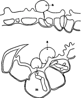
Blast disease causes serious damage to the rice crop every year. Its causal fungus, Tiricularia oryzae, invades directly through the epidermal wall (Fig. 1 ) . The motor cells and the guard cells of stomata are the pathway through which the fungus penetrates most easily. As indicated
FIG. 1. Mode of entrance into the epidermal cells of rice leaves by Piricuhria oryzae: (a) appressorium, (m) motor cell. (After Yoshii, 1 9 3 6 . )
TABLE II
PENETRATING HYPHA FORMATION FROM APPRESSORIA FORMED ON EPIDERMAL CELLS OF KAMEJI RICE No. 3A
Epidermal cell Per cent Per cent
appressoria formed penetrating hypha formed
Motor cells 5 3 . 0 6 3 . 7
Long cells 1 6 . 2 7 . 9
Short cells 1 2 . 8 6 . 1
Hairs 1 . 7
Stomata 6 . 0
Short cells contacted with stoma 7 . 7 1 5 . 1
Midlamella between stoma and
short cell 2 . 6 6 . 1
β After Ito and Shimada, 1937.
in Table II, the entrance of the fungus in more than half of the total invasions is through motor cells. It has been considered that the lignifica- tion of the outer wall of motor cells does not take place rapidly, being kept in a pectocellulosic condition for a long time, while most of the other epidermal cell walls are cellulosic and become lignified sooner (Fig. 2, 1) Yoshii, 1936). Hashioka (1950) confirmed the lignification in epidermal cell wall of rice leaves of differing age by the application of Maule's reaction. According to his results, positive reaction to lignifica- tion in epidermal cell walls was obtained after 60 days of development of the leaf blades. With the exception of motor cells, the histochemical tests of Suzuki et ah (1953) show that chlorogenic acid appears to accu- mulate in walls of epidermal cells usually combined with a certain con-
FIG 2. The cross section of motor cells in leaves of rice plants. (1) lignified cell wall, (p) pectin layer (dotted area), (s) silicic acid deposited layer, (x) cell wall of motor cell, on which silicic acid deposits easily. (After Yoshii, 1936.) stituent of cell membrane. In walls of motor cells, either there is no lignification, or lignin does not deposit sooner, thus at least partially accounting for the more ready penetration by the fungus through motor cells.
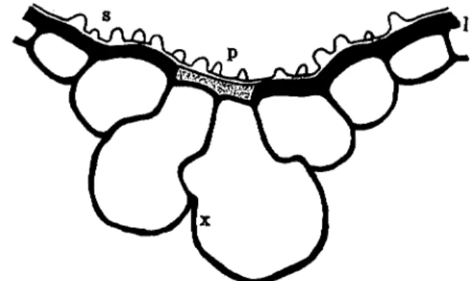
It is a well-known fact that silicic acid content of leaves is inversely related to incidence of diseases of rice. Usually, silicic acid is deposited abundantly on lignocellulosic walls of epidermal cells (Yoshii, 1936) and the deposition increases with the maturity of leaves. Under favorable conditions at maturity, silicic acid is heavily deposited in motor cells, and this increases resistance to fungus penetration (Fig. 3 ) .
Planting rice plants in flooded soil is the usual cultural practice in Japan. This procedure promotes the deposition of silicic acid on cell walls and prevents fungal invasion.
Low air and soil temperature reduce both the thickness of the outer
396 S. ΑΚΑΙ
wall of epidermal cells and the deposit of silicic acid on epidermal cell walls. The thinner walls and the decreased deposit of silicic acid are perhaps the factors which cause severe incidence of blast disease and Helminthosporium blight (Hashioka, 1950; Suzuki, 1951).
In a discussion of mechanical barriers to fungal penetration, the strength of the epidermal membrane to puncture is usually considered.
For this purpose, the Jolly balance is adapted (Hawkins and Harvey, 1919; Melander and Craigie, 1927). As has been cited above, the pres
sure required to puncture the epidermis of leaves and stems of flax varieties varies with their reaction to flax rust. Highly resistant varieties
FIG. 3. Ash-figures of leaves of rice plants. Silicated epidermal cells: (g) guard cell, (k) short cell, (1) long cell, (m) motor cell, (r) rice cell.
require more pressure than the susceptible ones (Sharvelle, 1936). Ito and Sakamoto (1939), measuring the resistance of the epidermis of rice leaves to needle puncture, found that feeding silicic acid to rice plants increased the resistance to puncture. In turn, this resistance to puncture was correlated with a reduction in the incidence of blast dis
ease. Yoshii (1941a), however, aimed to measure the toughness of the cell wall of motor cells, and obtained a result opposite to that of Ito and Sakamoto. His results showed that the resistance to needle puncture decreased with the increase in the supply of silicic acid. In this case, the total amount of silicic acid in the plant increased and the incidence of blast disease decreased (Table I I I ) .
Yoshii (1941b) also tested the toughness of leaf, when the plants
TABLE III
STRENGTH OF EPIDERMAL MEMBRANE TO NEEDLE PUNCTURE IN LEAVES OF RICE PLANTS AND TOTAL AMOUNT OF SILICIC ACID"
Silicic acid given Strength of epidermis (gr./mm.2)
Total amount (mg. per liter) 2nd leaf6 3rd leaf6 4th leaf6 of silicic acid (%)
0 902.3 813.3 777.9 1.8
50 863.3 792.6 766.1 3.5
250 847.8 808.1 755.8 10.2
500 840.1 793.1 753.4 12.9
α After Yoshii, 1941a.
6 From upper level.
TABLE IV
THE TOUGHNESS AND SILICIC ACID CONTENT OF LEAVES OF RICE GROWN UNDER FIELD CONDITIONS AND SUPPLIED WITH
DIFFERENT AMOUNT OF NITROGEN AND SILICIC ACID°
Nitrogen6 supplied (kg. per 0.1
hectare)
Silicic acid supplied (kg. per 0.1
hectare)
Total content of silicic acid (SiOi)
(% dry matter basis)
Toughness of epidermis
(gm./mm.2)
Incidence of blast
disease
18.75 262.5 6.71 719.9 ±
0.0 3.74 938.3
+ +
11.25 262.5 8.90 916.1 ±
0.0 5.97 960.5 ±
3.75 262.5 10.93 941.7 —
0.0 8.46 1055.2 —
a After Yoshii, 1941b.
6 Ammonium sulfate.
were supplied with silicic acid and ammonium sulfate. Evidently nitro
gen affects toughness of the leaf (Table I V ) . However, there is no rela
tionship between silicic acid content and toughness of the leaf.
2. Structure of Stomata and Lenticels
Except for bacterial diseases there is probably no evidence that infec
tion is prevented by the structure of the stomata.
In citrus canker, caused by a bacterium (Pseudomonas citri), the bacteria are not able to attack the dry cutinized or waxy cell walls of citrus. In intact leaves, the bacteria can enter only through the stomatal openings. Szinkum mandarin (Citrus nobilis var. Szinkum) possesses a
398 S. ΑΚΑΙ
resistance to citrus canker, whereas certain kinds of grapefruit (Citrus grandis) are very susceptible. Hence, resistance to penetration must be involved in the structure of stomata (McLean, 1921). Each has stomata of similar size, but they differ mainly in the entrance ridge. In the resist
ant variety, Szinkum mandarin, the stomata have an extremely narrow entrance with broad lips over the stoma, whereas in the Florida grape
fruit the stomata have broad oval lips. In the mandarin this structure may practically exclude water from the stomata, whereas water can more readily enter the stomata of the grapefruit. This exclusion of water is sufficient to account for the resistance of certain citrus varieties to canker.
Possibly in the rusts, the uredospore germ tubes enter plants through stomata. However, as shown by Hart (1929), the stomata of some wheat varieties are closed much of the time and the stem rust, Puccinia gram- inis tritici, cannot usually force its way through closed stomata. She termed this as "functional resistance." The delayed opening in the morn
ing of stomata of resistant wheats, prevents the entry of the stem rust until the moisture on the plant has evaporated, thus exposing the delicate germ tubes to desiccation and death. Closed stomata offer no effective barrier to the entry of leaf rust, Puccinia triticina, which never enters through open stomata because of the prompt stomatal closure in response to appressorium formation (Caldwell and Stone, 1936).
Certain other fungi are not able to push their way through closed stomata. Cercospora beticola can enter sugar beet leaves only through open stomata. The maturity of the leaf is important and infection is closely related to stomatal movement. Young leaves are not infected because stomatal movement is not active. Mature leaves are attacked severely, but old leaves are not invaded because movement is feeble
(Pool and McKay, 1916).
Lenticels, before they become suberized, are sometimes good portals for invasion by pathogenic fungi. Actinomyces scabies (Streptomyces scabies), the cause of common potato scab, enters through lenticels of young tubers and stems (Lutman, 1945), although it can also invade through stomata and wounds. The cells of young lenticels are usually round and loosely arranged with rather large intercellular spaces. When the filaments of the scab organism grow among these cells, the lenticel meristem is stimulated to divide actively and to form closely packed, radially elongated cells. This appears to be an attempt at cork formation.
These cells do not, however, usually constitute a barrier to the further extension of the pathogen because they are not suberized. They do not form a true cork layer, and the scab organisms continue their invasion.
Apparently, the pathogen tends to delay suberization of the cells in the lenticels (Jones, 1931; Longree, 1931; Darling, 1937).
The lenticels of healthy mulberry trees conceal mycelia of various fungi, some 30-60% of which are pathogenic to mulberry trees (Aoki, 1941). Among these are Diaporthe nomurai, the cause of the devastating mulberry blight found in snowy regions, and Gibberella lateritium, the cause of bud blight.
The normal lenticels have a cork cambium at the base which is connected with the periderm of the shoot. The cork cambium divides forming closing layers and complementary cells alternately, and the fungi may be found in the vacuity among these cells.
Under normal conditions when the plants are growing vigorously, the fungi in the lenticels are not able to invade through the cork layers into the interior. However, under unfavorable conditions, the pathogenic fungi accomplish the infection by passing through the cork layers of weakened lenticels. This is why mulberry blight occurs in the heavy snow region, where the plants are buried for a long time under heavy snow cover and lose their vigor.
The fungal components of the lenticels vary with the variety of mulberry, depending on the structure of the lenticels. In general, the rough structured lenticels have more fungi than the closed type. The correlation between resistance to mulberry blight and number of fungi isolated is inverse. A large number of fungi were isolated from the lenticels of resistant varieties, but a few can penetrate the cork cam- bium. On the other hand, only a small number of fungi were isolated from the lenticels of susceptible varieties, but a large percentage of them were able to penetrate the cork cambium of the lenticels (Aoki, 1945).
B. Mechanical Barricade Tissues as a Factor in Resistance to Invasion Mechanisms that interfere with invasion of the host are (1) the static resistance to spread, already present prior to infection, and (2) the dynamic defense reactions which become apparent after infection occurs (Gaumann, 1950). In the present section we shall deal with the former from the histological viewpoint.
Resistance to invasion is sometimes associated with histological char- acteristics of walls. This resistance opposes the progress of pathogens and may be distributed throughout the tissues or localized in certain barricade tissues.
The epidermis of wheat varieties consists of a single layer of cells, the inner walls of which are sometimes lignified. Just beneath the epi-
400 S. ΑΚΑΙ
dermis there is chlorenchymatous collenchyma. This tissue sometimes extends in an almost continuous band around the entire stem, although it usually is interrupted by the strands of sclerenchyma. The collen
chyma cells, then, are aggregated into isolated bundles, the size and num
ber of which vary considerably in different varieties. In Kota, the scle
renchyma fibers divide the collenchyma into distinct areas, while in Little Club the sclerenchyma is much less conspicuous and the collenchyma is practically continuous. Marquis stems have somewhat more scleren
chyma fibers than the Little Club stem; in the stem of Sonem emmer a large amount of sclerenchyma is always developed. The collenchyma areas in this variety are extremely small and the sclerenchyma area is decidedly predominant, making up the major portion of the stem proper.
Less infection with both Puccinia triticina and P. graminis takes place on stems of Sonem emmer than of any other wheat (Hursh, 1924). However, under epidemic conditions, a large number of individual infections may result even on varieties with extensive sclerenchyma. Under such con
ditions, the susceptible Little Club and Marquis varieties are seriously injured. The structure of the stem affects only the extent of the spread of rust fungi and its subsequent rupture of the epidermis. However, re
sistance to stem rust must be considered as being due fundamentally to a plasmatic defense.
Rice leaf smut, caused by the infection of Entyloma oryzae, shows a black, short, linear symptom limiting itself usually between two veins of leaves. Thus, the mechanical tissues affect the extent of the spotted area. Fertilizer affects greatly the development of the mechanical tissues, especially when upland rice plants are cultivated under flooded condi
tions (Shimada, 1957).
II. DY N A M I C DE F E N S E REACTION
In contrast with the defensive structure, the dynamic defense reaction is evoked postinfectionally by response to the stimulus of infection, on the one hand forming histological barricades, and on the other revealing protoplasmic defense in the cell itself. In some cases, these host responses may be considered an inflammatory reaction. However, this discussion will be limited to situations where infection by pathogenic organisms is prevented.
The defense reaction can be classified according to its origin: (1) the autonomous antiparasitic defense reaction, and (2) the induced defense reaction. We shall deal here with the former only. According to Gaumann (1950), the antiparasitic defense reaction (anti-infectious defense reac
tion) is the reaction of cells aimed directly at the pathogen and intended to weaken and destroy it. The existence of this reaction in cells is shown
by the fact that most infectious plant diseases, not systemic, do not spread indiscriminately through the host. The curves in Fig. 4 actually show this. Thus, after a certain time, the disease intrusion comes to a standstill. Infection remains localized, giving to a disease its character
istic symptoms.
We have shown above, that there are some cases where outer epi
dermal layers and mechanical barricade tissues are capable of preventing pathogenic invasion. However, if a pathogenic organism should make its way into the interior of plant tissues by passing through these barricades,
l/37mm.
ο 5 0 a, o> 4 0
CA Ο
.? 30 Η
*S 2 0
£ 10 Η
I 3 4 5 1 2 3 4 5 6 7
Days elapsed after inoculation
FIG. 4. Curves of developing areas in spots revealed in leaves of rice plants due to the infection of Cochliobolus miyabeanus. (1) supplied with a large amount of ammonium sulfate, (2) supplied with normal amount of fertilizers. (After Kuro- saki, 1957.)
the defense reaction against further intrusion by the pathogen may be induced autonomously in the focus of the invasion point. Then, the autonomous defense may appear: on the one hand as a histological bar
rier which acts to demarcate the infected lesion (histogenic defense reac
tion), or, on the other, as a plasmatic activity in the cell itself.
First, we shall deal with the histogenic reaction.
A. Histogenic Defense Reaction
Defense reactions which are exhibited histologically will be consid
ered in this section. Defense of this type may be manifested as a demar
cation of the infected lesions and as a callus-like swelling of the mem
brane. Both serve to prevent further intrusion of the pathogen. Gaumann (1950) has stressed the antitoxic effect in some cases of demarcation.
This involves: (1) demarcation of infected lesions by forming cicatricial layers, abscission cells, tyloses, or gum; (2) callus-like swellings or cal
losities formed on the wall.
402 S. ΑΚΑΙ
1. Demarcation of Infected Lesions
After infection, the invaded tissue is sometimes demarcated histo
logically. In some cases, this results from cork layer formation and in others from gum deposition or from formation of abscission cells.
a. Cicatricial Layers. In some plants, suberized healing tissues de
velop, demarcating the localized lesions of infection. This may lead to scabbing. Metabolic products secreted by the causal organism may stimulate the formation of this cicatricial demarcation. Fungi of the
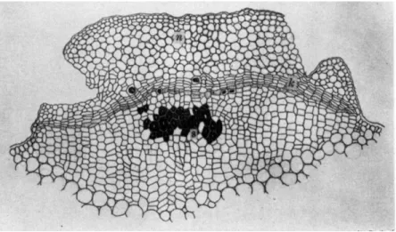
FIG. 5. Renewed meristem formed around the necrotic lesion of fruit of grape invaded by Elsinoe ampelina: (c) conidial sorus, (g) cells showing granular deposits, (n) necrotic cell group, (t) renewed meristem.
genus Sphaceloma are especially able to induce cork cells to form in various plants and give them a characteristic scabby appearance on the surface. As Gaumann (1950) suggested, the cicatrice may cut off the influence of toxic substances of the fungi, which may diffuse from the infected area. He designated this phenomenon as the antitoxic defense reaction. An anthracnose disease of grape infected by Elsinoe ampelina and citrus scab caused by Elsinoe fawcetii are representatives of this type. Sphaceloma ampelinum, the conidial stage of Elsinoe ampelina, attacks every part of the plant. On fruits, the first noticeable symptom is
a minute round speck on the surface. With gradual increase in size of spots, the peripheral region more or less bulges out accompanied by the formation of a depression of the central area. Renewed meristematic activity occurs in cells of the peripheral zone of the spots. From this meristem (Fig. 5,t) cork cells may be formed (Akai, 1951). On soybean pods, a suberized barricade delineates the area infected by Sphaceloma kurozawana. At first, the cytoplasm degenerates and turns brown in the invaded epidermal cells, whereupon the hyphae do not enter into the palisade tissue. Thereafter, the proliferation of palisade cells takes place under the infection focus, thus making a scabby appearance.
FIG. 6. The postinfection cork layers in the lesions of citrus melanose on the stem of grapefruit: (k) renewed phellogen, (n) necrotic cells, (s) stone cells (not a response to this invasion). (From Akai, 1 9 5 0 . )
The melanose disease, caused by Phomopsis citri (Diaporthe citri) develops on leaves, stem, and fruits of citrus, where it causes abundant small black spots. Although there is no corky appearance to the unaided eye, a perfect demarcation of the infected zone is produced by the post- infection cork layers (Fig. 6 ) . On artificially wounded fruits, the path- ogen is forced to express a stem end rot symptom that causes the entire fruit to break down. However, when uninjured fruits are used, typical melanose spots result. The band of cork layers may effectively cut off the growth of the mycelium and prevent further invasion of the causal fungus toward the interior of the fruit. The region bulges out, but not noticeably so, due to hyperplasia of cells. The cells outside the cork layers collapse and turn brown, showing a granular fatty degeneration of the cytoplasm (Bach and Wolf, 1928).
404 S. AKAI
FIG. 7. The infection cushion of Helicobasidium mompa derived from the my- celial strand, passing through the cork layers and dissolving the starchy parenchyma of sweet potato tuber. (After A: Ito, 1949; B: Suzuki et al, 1957.)
The destructive "Murasaki-Mompa" disease caused by Helicobasidium mompa causes rot of underground parts of many plants, some 45 families, 76 genera, and 104 species (Ito, 1949). The basal part of stems and fleshy roots of apple, mulberry trees, and sweet potatoes are among those that are severely attacked. On sweet potatoes, the mycelium of the fungus grows epiphytically for a long time as a purplish, felt-like network of rhizomorphs. During this period, hyphae penetrate into the middle lamellae of the cork layer cells, but not so deeply as to pass through the cork layer (Suzuki et al, 1957).
FIG. 8. Types of reaction in tubers of sweet potato against Helicobasidium mompa: (d) degenerated zone, (p) periderm, (r) postinfectionally renewed cork layers. (After Suzuki, 1 9 5 7 . )
The penetrating hyphae (Fig. 7) gradually develop into a bundle and finally into an infection cushion (Ito, 1949; Suzuki et al, 1957).
Thus, the hyphae of the cushion gain entrance into the starchy pa- renchyma and cause rot of the tubers.
Defense of the tubers against fungal invasion is observed most actively during the period of rapid growth. In response to invasion, the
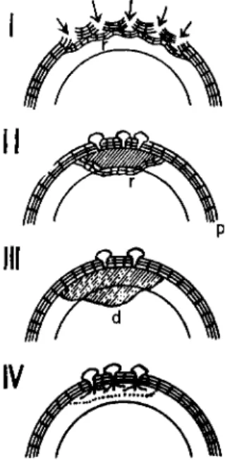
cork cambium develops layers of suberized cells that are more than six layers deeper than normal. Infection in this disease is of the following four types (Suzuki, 1957).
Type 1. The invasion hyphae are inhibited from reaching the starchy
406 S. ΑΚΑΙ
parenchyma by rapid and successive formation of cork layers. This causes sloughing off of the infected zones (Fig. 8-1). Host cells degen
erate, turn brown and enclose the tips of the hyphae. There is no change in appearance of the tuber to the unaided eyes.
Type 2. The starchy parenchyma beneath the infection cushion dis
plays a brown rot appearance, and the post infection cork layers (Fig.
8-II, r) inhibit further invasion of the hyphae by enclosing the brown rotted area. Thus, a complete demarcation of the lesion takes place.
Type 3. A rapid change in light brown color of starchy parenchyma occurs under the infection cushion. This change develops widely, finally causing a soft rot of tubers.
Type 4. Starchy parenchyma is macerated without change in color by intensive pectolytic activity of the fungus.
Type 1 represents the most active defense against fungal attack. If the fungus passes through the barrier, the second type of defense may be induced. The third and fourth types may occur in the susceptible condition of tubers.
Lignification of cells takes place in the periphery directly in contact with the rotted brown zone, and two to three layers away from this peripheral lignification, a second lignification occurs in cells. When the lignification does not occur, the rotting of tissues proceeds rapidly. How
ever, the cork may be most stable against the action of this fungus. There
fore, when the disease proceeds gradually, it is checked almost com
pletely by the newly formed cork cell layers. The lignified cells, however, do not check the disease completely as do the cork cells. Usually, the second type of defensive cork layers develops in the periphery of the necrotic zone, surrounding the lignified cell layers, when tissues are resistant. Cork layers do not form in susceptible tissues.
At the first stage of infection, the middle lamellae of cork layer cells are penetrated by the fungus, as has been shown before. Pectic material in the middle lamellae swells when the hyphae come in contact with it and the pH value is decreased. After passing through the cork layers, the fungus comes into contact with the starchy parenchyma. In suscep
tible varieties, these tissues are then macerated by the action of fungal enzymes. Itaconic acid is isolated from such tissues (Araki et ah 1957).
The decrease of pH in these tissues is mainly due to the accumulation of chlorogenic acid and caffeic acid, and to itaconic acid produced by the fungus. When treated with ruthenium red in the early stage, the pectic substance of the invaded tissues—in contact with the hyphae—is stained yellow, and the tissues beneath the phellogen are stained carmine red. FeCls-potassium ferricyanide solution stains pectic materials in the
cork layers blue. No color reaction occurs in pectic materials produced postinfectionally.
Lignification of the cell membrane is accelerated by infection. The red color reaction of cell membranes treated with phloroglucin-HCl is chiefly due to lignin, although caffeic and galacturonic acids react simi- larly (Suzuki, 1957).
Corky demarcation appears on various other plant diseases. In chest- nut blight, caused by Endothia parasitica, trees often develop a cork barrier (Bramble, 1936; Bazzigher, 1957). Infection by Actinomyces scabies causes a scabby appearance on the surface of developing potato tubers. Similarly, Cladosporium carpophilum causes a corky appearance on the surface of peach fruits. In apples and pears, once the fire blight lesions are corked off, the cork layers and the xylem commonly serve as relatively effective barriers against further invasion of the causal bacteria (Shaw, 1934). In the diseased leaves and petioles of Aralia cor data and Fatsia japonica infected by Elsinoe araliae, cork layers very similar to those found on the diseased stem of grape are formed surrounding ne- crotic spots. If the fungus invades further, after the cork layers are formed, underlying collenchyma cells undergo a rapid lignification. The lignifica- tion of bast fibers beneath the diseased spot takes place more rapidly than in uninfected tissues.
A very definite cicatrice forms at the margin of the necrotic area of leaves of Prunus domestica attacked by Coccomyces prunophorae. The healing tissue of wound periderm consists of several layers of cells. The phellem is made up of a mass of large cells, whose thick walls are not only suberized, but also lignified. The cells lying nearest the necrotic lesion become filled with a dense granular substance resembling tannin, while the remaining cells are apparently devoid of contents. The epi- dermal cells of this layer also have suberized walls. Both phellogen and phelloderm are present in the wound periderm. The cells comprising these two layers are thick walled, but this thickening is entirely cellulosic in nature. Chloroplasts are absent from these cells. The phellogen con- sists of a single layer of cells which are filled with dense protoplasm, while the cells of the phelloderm contain only a peripheral layer. The periderm ties between the two epidermis of the leaf (Fig. 9) and isolates perfectly the diseased portion (Cunningham, 1928).
Cork layers around the necrotic lesions have been noted in leaves of sugar beet infected by Cercospora beticola; in Paulownia twigs infected by Gloeosporium kawakamii; in apple leaves attacked by Physalospora cydoniae; in cankered twigs of apple caused by Valsa malt; in portions of twigs of cherry or peach showing dieback caused by Valsa japonica or
408 S. AKAI
Leucostoma personii; in geranium stem rot caused by Pythium com- plectens. In tobacco plants the rapidity of cork formation beneath the lesions due to the attack of Thielavia basicola is an accurate criterion of its resistance (Conant, 1927). In leaves of Nicotiana glutinosa infected with tobacco mosaic virus, a corky barricade is also formed around the necrotic spots (Yoshii and Kawamura, 1947).
Sweet potatoes have the ability to form cork layers covering wounds when the environmental conditions are favorable (Weimer and Harter, 1921). The wound cork layers may be healing tissue, but they sometimes serve as a defensive mechanism against the invasion of pathogens. In
FIG. 9. Cork layer at the edge of a lesion on a leaf of Prunus domestica caused by Coccomyces prunophorae. (After Cunningham, 1 9 2 8 . )
cut tubers of potatoes, the outer walls of living cells suberize at the cut surface and afterwards wound periderm may be formed. This prelim- inary suberization in cell walls has been termed the pseudocicatrice (Wylie, 1930, 1931). Generally, the first effect is defensive lignification of cells surrounding the wound; and deposition of wound gum takes place. At the inner side of this barricade, a set of cells lose their reserve nutrient and chloroplasts and form a renewed meristem that divides to form cork cells. From the resulting differentiation of cork mother cells, the five or ten layers of cork cells are formed by division on the outer side. These cork barriers are formed most vigorously under conditions ranging between 30°-35° C. and 90-95% relative humidity. Under exces- sive humidity (90-100%) cork formation is retarded, while below 80%
no cork is formed in sweet potato (Yoshii, 1944). In cut tubers of pota- toes the optimum temperature for suberization of superficial cells and
formation of wound periderm is between 21° and 35° C. (Artschwager, 1927).
Soil moisture may be one of the factors influencing cork formation in tubers. Without doubt, water-logged soils favor the growth of the soil-borne pathogens, especially the facultative anaerobes. An excess soil moisture obviously inhibits cork formation and enhances the incidence of the disease, e.g., blackleg of potato. Therefore, the presence of air (oxygen) may be necessary for the formation of cork cells (Jahrmann, 1913; Leach, 1931).
Wound cork layers are found in the mulberry root. This cork is formed during the growing season, as in stems, starting from meristematic cells near the cambium, but not from the lignified bast fibers or woody tissues. These layers, to some extent, prevent the spreading of the dis
ease caused by Roselinia necatrix, but when the mycelial strand reaches the cork layers, it usually ruptures them (Sakurai, 1952). Thus, cork layers are sometimes of no value as a barrier to the invasion of the pathogen. The entrance of Cylindrocarpon ehrenbergi occurs by direct penetration of the cork covering in roots of alfalfa and sweet clover.
The hyphae mass up and push their way between the cork cells in an apparently mechanical manner (Cormack, 1937).
b. Abscission Layers. Whatever the cause may be, the spots pro
duced on leaves of stone fruit plants slough out. Many fungi and bacteria, pathogenic to these plants, produce such a shot-hole effect.
Moreover, the perforation may be formed by wounds or sprays contain
ing a dilute (0.01 Μ) copper sulfate solution. Leaves of peach trees planted under copper wires are very easily perforated by the copper leached from the wires and deposited on the leaves below.
In the leaves of peach trees attacked by Xanthomonas pruni, we see the swelling of one or two layers of cells surrounding the spots. These cells become turgid, thin walled, and meristematic. When their middle lamellae are dissolved, a gap between healthy and necrotic tissues is produced. The swelling takes place mainly in the cells of palisade and spongy parenchyma. These cells become round, ovoid, long ellipsoidal or retort-like in shape. They serve to cut off the necrotic area from the healthy tissues, and this tissue gradually shrivels, dies, and sloughs off.
In this way, the healthy tissues are protected from the damage, caused possibly by the toxins of the pathogen or the products of the dying lesions (Fig. 10).
Perforation of spots in the leaves of Prunus amygdalus takes place as a result of the attack of Cladosporium (Clasterosporium) carpophilum
(Samuel, 1927). According to Samuel, when leaves form the abscission cells, they must be young and active, and well supplied with water.
410 S. AKAI
FIG. 1 0 . Abscission cells produced around the necrotic area in peach leaf (variety Denjuro) induced by Xanthomonas pruni: (p) normal palisade tissue, (t) abscission cells, (v) normal parenchyma cells in the vascular bundle. (From Akai,
1 9 5 1 . )
FIG. 1 1 . Perforation of the diseased spot produced on leaf of Prunus lauro- cerasus infected by Clasterosporium carpophilum. Lignified cells ( 1 ) appear at the periphery of diseased area. The abscission layer (a) is formed outside of the lignified cells. (After Samuel, 1 9 2 7 . )
Therefore, the infected tissues in young leaves are invariably abscissed.
If leaves are old or moisture relations unfavorable, abscission cells are not formed, and the infected tissues do not fall out. In this case the meristem cells become suberized. Thus cutting off of the infected tissue may be caused by a barrier of wound cork, which also serves to check further extension of the fungus into the leaf. Moreover, cells swell in a narrow zone at some distance from the margin of the invaded area and form an abscission layer. Suberization of the walls of cells along the abscission line occurs soon after the cuticle ruptures. Subsequent divisions of the meristematic layer result in the formation of a layer of brick- shaped cells that become suberized and slightly lignified (Fig. 11).
Where abscission does not occur, the initial changes are similar, but in the later stages the walls of the cells on the inner side of the occluded zone lignify and the walls of the meristematically formed cells suberize.
Outside the abscission cell layer similar cork layers are formed in the healthy part of peach leaves around the spot of Xanthomonas pruni. This cicatricial layer may perfectly cut off the tissues from the external world, preventing excessive evaporation from the thin walled abscission cells and keeping back the injurious effect of the secondary invasion of even weakly pathogenic fungi.
On the twigs of cherry (Prunus yedoensis) affected by witches'- broom, small sized leaves are formed. In spring the ascospores of the causal fungus, Taphrina cerasi, are produced on the lower surface of these leaves. This sorus develops in a limited, localized portion of a leaf.
Even in such diseased leaves, the lesion sloughs out, producing abscission cells around the area, after the ascospores mature. When the upper epi- dermal cells begin to collapse, the abscission cells are formed about the margin of the lesions in the same manner as in lesions of peach leaves, even if the functioning of the mesophyll and lower epidermal cells con- tinues. Consequently, it may be assumed that the formation of abscission cells is related to the necrosis of cells, and a hormone-like substance may be considered. On the other hand, Tranzschelia pruni spinosae (rust fungus of peach and plum trees) possesses no such activity and, there- fore, does not result in any delimiting area on leaves.
c. Tylosis and Gummous Deposition. Tylosis is sometimes found in the vessels of the invaded portion of plants. This tylosis may be formed by the stimulation of metabolic products of the fungus. However, litera- ture regarding the fundamental cause of tylosis formation is contra- dictory. Haberlandt (1923) considered that the decomposition products of injured cells may play a decisive role in causing tyloses to form. This theory is based on the fact that tyloses are often found beneath the surface of amputated branches or adjacent to wounded areas. Klein
412 S. AKAI
(1923) supports a different interpretation. He considered the presence of air within the exposed vessels to be the chief cause of tylosis formation.
However, judging from Powers' observation (1954), tyloses and gums in the vessels of diseased stems are caused primarily by toxic effects of decomposition products of invaded cells. These toxic substances are not systemic in nature, but affect primarily a restricted region in the xylem.
Moreover, he considered that these toxic substances are not necessarily products of fungal metabolism since cell decomposition products alone induce a similar reaction. He observed that a severe wilting of tobacco plants, with tylosis and gum formation, developed when excised healthy plants were placed in extracts of either healthy or black shank affected plant tissues. On the contrary, Bazzigher (1957) considered tylosis formation in the diseased portion of chestnut trees invaded by Endothia parasitica to be attributable to the stimulus of diaporthin, a metabolite of this fungus.
Tylosis clogging in vessels impedes the flow of water. Such mechan- ical blocking of conducting elements by tyloses is held as the chief factor responsible for wilting and drying of leaves on infected chestnut trees (Bramble, 1938). In oak trees, infected by Chalara quercina (Endoconi- diophora fagacearum), extensive plugging of the xylem vessels with tyloses and gums precedes the foliage wilt. Tyloses are formed in the large vessels of the spring wood, especially of the last annual ring, but less so in the small vessels of the summer wood (Struckmeyer et al., 1954). In the xylem of diseased sweet potatoes tylosis and vascular dis- coloration usually occur in advance of the invading mycelium of the wilt fungus, Fusarium oxysporum f. batatas (Watanabe, 1939; McClure, 1950). In this case the tylosis-clogged vessels may serve to prevent the invasion of the pathogen. Tyloses have cellulose walls, formed from adjacent living cells by extrusion through half-bordered pits. Often they are so numerous and large that they become closely packed in the vessel lumen, losing their original spherical shape. After staining by Cart- wright's method, the walls of most of the tyloses are blue, except those nearest to the pathogen, which are stained red. In this latter region a substance which appears to be wound gum accumulates in the interstices between tyloses, and between tyloses and their enclosing vessel wall.
Penetration of the tylosis blockage or rupture of tyloses by the wilt fungus has not been observed. Hyphae pass through vessel element aper- tures and through unobstructed pits, but do not directly penetrate cell walls or pits which are covered with wound gum (McClure, 1950).
There seem to be two types of gum materials secreted by plant tis- sues (Yoshii and Kawamura, 1947): one is the gum which appears on stone fruits, and the other a wound gum which shows a lignin-like color
reaction. The former gum appears most often on fruits, branches, or trunks of stone fruit trees, and is also associated with injury from insects or mechanical sources. Valsa disease of peach always produces gummosis if the trees are vigorous. The gum is mainly composed of pentosans as shown by the pectin-like reaction, and is produced by the liquefaction of woody membranes. On the other hand, wound gum deposits occur in the injured portion, where the substance fills the cell lumen and some- times permeates the cell walls, especially on the abnormally swollen walls (Yoshii, 1948).
Gum deposition along the border of diseased lesions often serves as a protective demarcation and constitutes a type of mechanical resist- ance. Surrounding the necrotic lesion on the leaves of Unshu orange produced by Phyllosticta, wound gum deposits demarcate effectively the healthy tissues from the diseased lesion, by causing a marked constric- tion of the necrotic lesions (Yoshii, 1949). In the stems of cherry affected by canker disease, the causal fungus of which is Valsa japonica, similar gum-like deposits are formed in wood vessels. Gum gradually replaces the starch and other contents in the medullary ray cells, wood paren- chyma, and the wood vessels are slowly plugged up by the deposits of the gum (Hemmi, 1916).
In the silver leaf disease caused by Stereum purpureum, under con- ditions favorable for gum formation, the wood of the host produces so much gum in advance of the fungus in a relatively short time, that the fungus becomes completely enclosed by an impassable gum barrier.
Within this barrier, the fungus may continue to live for a considerable time, but eventually it dies. These gum barriers may correspond to the protective wood (Frank, 1895), as developed by the deposition of gum and browning of cell walls. They usually develop within an inch of the wound and require at least two months for their completion (Brooks and Brenchley, 1931). Hesler (1916) has found a similar gum barrier in the diseased part of apple twig, infected by Physalospora cydoniae showing a brown deposit in wood fibers and wood parenchyma cells (Fig. 12).
In noninoculated cotyledons of "Proso," a scab-resistant variety of cucumber, mechanical damage by scratching induced a wound reaction.
This was revealed by the secretion of a granulated yellow substance that almost filled the intercellular spaces between healthy cells in the neigh- borhood of the damaged zone. In addition, the walls of some of these cells become yellowish in color and no longer stained with zinc chloride- iodine. The yellow substance stains with ruthenium red, and reacts posi- tively to the lignin test with phloroglucin-hydrochloric acid. In "Proso,"
3-5 days after inoculation with Cladosporium cucumerinum, the myce- lium seems to stimulate secretion of the yellow substance so that it fills
414 S. ΑΚΑΙ
the interior of the healthy cells as well as the intercellular spaces in an almost uninterrupted zone around the wound. The contents of some of the cells in this zone contain a yellow granular substance, whereas the cell walls are yellow in color and slightly swollen. Within this zone, hyphae are rarely found and beyond it they are entirely absent. Evidently this zone acts as a barrier against further spreading of the causal fungus.
Probably the formation of the yellow granular substance plays an im
portant role in this respect (El-Din Fouad, 1956).
FIG. 1 2 . Gum barrier in the apple twig infected by Physalospora cydoniae.
Mycelium is shown in the xylem ducts. (After Hesler, 1 9 1 6 . )
In the wilt disease of sweet potatoes, wound gum is also produced.
Wound gum, which is golden brown but stains deeply with safranin, is often found in hemispherical masses which protrude into the lumen of the invaded vessel. These gum deposits are usually located on half- bordered pits, and apparently secreted through the pits by the adjacent
living cells. Bordered pits sometimes bear gum deposits, but in every case a living tylosis is contiguous with the other face of the pit (McClure, 1950). Thus, the wood gum formed in half-bordered pits in the vicinity of the pathogen may act, physically or chemically, as a barrier which prevents hyphal penetration of the adjacent living cells. Consequently, wound gum may play a part in preventing both the penetration of the pathogen into tyloses and its intrusion between the vessel wall and tylosis wall.
FIG. 1 3 . The brown change of cells in blast diseased lesion of rice leaves:
(A) Kan-Non-Sen highly resistant, (B) Gin-Nen resistant, (C) Kokuryo no Miyako susceptible. (After Kawamura and Ono, 1 9 4 8 . )
In varieties of rice resistant to blast disease or Helminthosporium leaf spot, similar deposits are formed in the intercellular spaces that aid in restricting the fungus to the area of primary invasion. These deposits are found to be highly developed in Shoemed rice (resistant to helmintho- sporiose), but they also have been found to some extent in susceptible
416 S. ΑΚΑΙ
varieties (Tullis, 1935). In varieties of rice resistant to helminthosporiose and blast disease, even after infection by the causal fungus, necrotic cells are filled with brown wound gum-like substances, but do not show any shrinkage. The failure of cells to shrink may play an important part as a defense reaction together with the antifungal substances extruded from cells (Kawamura and Ono, 1948; Yoshii, 1957) (Fig. 13, Table V ) . 2. Callus-like Swelling and Callosity
a. Swelling of the cell wall. In the infected host, contact with hyphae sometimes results in swelling of cell walls. This is observed frequently in cuticular infection. Before the entry of Botrytis cinerea into pea leaf cells, swelling at the point of penetration is found in the subcuticular layer of cell walls, without causing any change in the cuticle (Blackman and Welsford, 1916). This swelling seems only a softening of the wall, because the fungus is soon able to penetrate it. The actual penetration is effected by pressure exerted on the underlying tissues, accompanied by the development of a fine peg-like growth from the appressorium, which is firmly pressed against the leaf surface. However, some consider that the penetration of cuticular barrier appears to be effected by chemical rather than by mechanical action (Woodward, 1927).
In at least one case, the thickened wall becomes lignified and acts as a principal factor in resistance to penetration. This barrier may exclude the fungus effectively. When the living leaves of tomatoes were artificially inoculated with conidia of Firicularia oryzae, the rice blast fungus, the cells resist penetration. At first, the hyphae form appressoria on the epidermal wall, which reacts by swelling at the contact portion of the appressoria. This swollen portion shows a lignin-like reaction, and is not transparent to light under crossed Nicol prisms. The fungus seems to have difficulty in penetrating such a cell (Fig. 14) (Yoshii, 1948). These abnormal thickenings of cell walls are found in oats attacked by Hel
minthosporium avenae, and in flax attacked by Fusarium lint (Tisdale, 1917). The latter is attributed to the formation of suberin. Subepidermal cell walls of corn roots also become thickened prior to infection by Helicobasidium mompa (Fig. 15) (Ito, 1952). These thickened walls are lignified (perhaps permeated with wound gum substance), and the hyphae are prevented from penetrating the thickened wall.
Cladosporium cucumerinum seemed to enter equally well both the resistant and susceptible varieties of cucumber. However, the progress of the fungus within the tissue was arrested by the host-parasite inter
action which is associated with cell wall thickening and cell necrosis (Pierson and Walker, 1954). This is the mechanism which confines the
Y OF DEFENSE IN PLANTS 417 PRIMARY REACTION OF CELLS IN RICE LEAVES AT THE POINT OF ENTRY OF BLAST FUNGUS AND THEIR SUSCEPTIBILITY*
Primary Resin Constriction Size Number
Variety reaction like of necrotic of of Susceptibility
in cells deposit cells spots infection
Kan Non Sen Rapid Abundant None Minute Numerous Highly resistant
Gin Nen Rapid Considerable Markedly Large Considerable Resistant
Ko Sen Slow Small Markedly Middle A few Resistant
Kameji Slow Considerable Markedly Large Considerable Resistant
Kairyo Shinriki Slow Considerable Considerable Large Considerable Susceptible
Wase Asahi Slow Small Markedly Middle A few Susceptible
β After Kawamura and Ono, 1948.
418 S. AKAI
FIG. 14. Mode of infection of Piricularia oryzae artificially inoculated on the living tomato leaf using hyphae: (a) appressorium. (After Yoshii, 1948.)
FIG. 15. Young root cells of corn infected by Helicobasidium mompa showing the thickened wall of subepidermal cells. Hyphae are constricted more or less when they pass through the wall. (After Ito, 1952.)
disease to a relatively small number of host cells and prevents the forma- tion of large lesions.
b. Callosities. In some cases, when hyphae start to penetrate cell walls, a slight protuberance is formed on the opposite wall. This pro- tuberance elongates at right angles to the wall, directly facing the advancing hyphae (Fig. 16). Sheaths enclosing the invading hyphae were probably described first by De Bary (1863), and these have been called callosities (Young, 1926).
The callus (callosity) formed on epidermal walls usually obstructs the invasion of Olpidium viciae into the cell. The effectiveness of callus
FIG. 16. The penetration of cell walls by penetrating hyphae and ligni-tubers formed about them: (d) ligni-tuber through which the hypha has passed, (h) hypha, (1) ligni-tuber through which the hypha has not passed, (w) cell wall.
(After Fellows, 1928.)
FIG. 17. Infection mode and callosity formation in cells of the rush (Juncus effusus var. decipiens) invaded by Leptosphaeria juncina. (After Ikata and Yoshida, 1940.)
420 S. ΑΚΑΙ
varies according to the plant species. In appropriate hosts, such as Vicia unijuga, V. faba, and Pisum sativum, this parasite is able to enter the cell through the callus. In other hosts such as Impatiens, Taraxacum, Oenothera, etc., callus is more effective in defense. In some nonsuscep- tible plants (Chrysanthemum, Lactuca, Dahlia, etc.), the callus actually blocks out all infectious individuals (Kusano, 1936).
FIG. 18. Mycorrhizal cell of rhizome of Gastrodia elata showing the formation of numerous tubular sheath (t). (After Kusano, 1911.)
FIG. 19. Tubular sheath produced on the wall of cork cells, of Gastrodia callosa and on the hyphae (A) and of Galeoh hydra ( B ) . (After Burgeff, 1932.)
In straws of Japanese rush (Juncus effusus var. decipiens), the pene
trating hyphae of Leptosphaeria juncina form callosities in epidermal cells. The callosity is formed before the hyphae pass through the epi
dermal cell wall. Callosities are generally formed most vigorously in young stems of plants (Fig. 17) (Ikata and Yoshida, 1940).
In the mycorrhizal cells of tubers of Gastrodia elata, a tubular sheath forms on the lignified or unlignified walls. In some cases (Fig. 18) it occurs as an aggregate (Kusano, 1911). Burgeff (1932) also found a tubular sheath in the mycorrhizal cells of Galeola hydra and Gastrodia callosa (Fig. 19).
Evidence has been cited concerning the wart-like protuberances (sheaths) that form on the inner wall opposite the penetration point as if to impede penetration of the haustorium of powdery mildews, downy mildews, and rusts.
Many investigators have stated that callosities or sheaths are pro- duced mainly from the cell walls. Smith (1900) referred to it as a cel- lulose collar, formed by the protruding cell wall, and Corner (1935)
FIG. 20. Callosity (callosity-like body) produced on the epidermal cell wall of stem of the sweet potato seedling by needle pricking. (After Ito. 1949.)
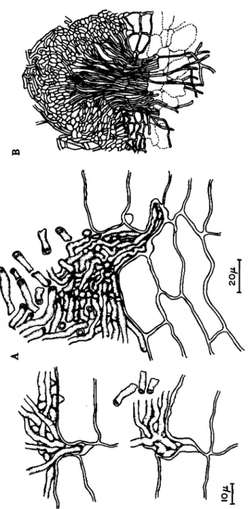
considered it a swelling of wall and called it a "papilla." Kusano (1936), however, has a different opinion: that the sheath of the haustorium is formed directly by the accumulation and aggregation of cytoplasm and is not a deformation of the wall. Aronescu (1934) pointed out that the collar-shaped basal mass surrounding haustoria seems to be composed of materials deposited by the cytoplasm at the same time that the haustorium advances into cells. Ito (1949), however, concluded that at least the callosities found on the cork cells are produced directly from the wall.
The callosities are induced not only by the fungus hyphae, but also by mechanical injuries. Some examples were shown by Ito (1949) in which callosities are formed by pricking the epidermal cell walls of
422 S. ΑΚΑΙ
sweet potato stem with a sterile needle (Fig. 20). Some consider that the callosity functions to heal the wound caused by an invader and not to check the fungus invasion (Kusano, 1939). Accordingly, there is no close connection between the formation of the callosity and the resistance of the plant, a matter possibly recognized in some cases (Iwata, 1940).
FIG. 21. The structure of the callosity produced on phellem cell wall of sweet potato stem (stained with Sudan III and methyl green): (a) outer layer staining light reddish (b) middle layer staining light purplish green (c) basal part (deep purplish green) (d) piercing filament. (After Ito, 1949.)
The callosity on the phellem consists of about three layers (Ito, 1949).
The outer membrane, middle layer and basal part are seen clearly to
gether with a piercing filament in the central portion (Fig. 21). In many cases, the sheath is formed in young vigorous cells (Chu, 1935; Akai, 1950), and also when haustoria become debilitated (Rice, 1927). Haus- toria formed in cells of Brassica infected by Albugo Candida, appeared to
be covered with a wall when they became debilitated (Akai, 1942).
Haustoria of stripe rust (Puccinia glumarum) which are dead or in an early stage of degeneration, are also formed with a heavy sheath (Allen, 1928). Haustoria of Maravalia hyalospora, a rust fungus of Acacia confusa, become covered after maturity. In such a case they are some- times dead (Hirane, 1940).
Almost all hyphae or haustoria are able to grow out through their sheath and enter the lumen of cells and when they do, they immediately increase to normal diameter again. However, some callosities are im- possible to penetrate. Ito (1949) observed no hypha piercing through a callosity formed in the wall of phellem cell.
FIG. 2 2 . Cellulose cushion, in which the hypha of Usttlago zeae is enclosed.
(After Guttenberg, 1 9 0 5 . )
The sheaths may be composed of cellulose (rust and downy mildew), but some are made up of callose (Mangin, 1896). Afterward, in some cases, a gum-like substance (wound gum) permeates them. Protuber- ances formed on the walls of cells in wheat roots infected by Ophiobolus graminis contain no trace of callose, but rather are composed chiefly of lignin (Fellows, 1928; Robertson, 1932). Therefore, in place of the terms callosity or callus used by Stevens (1922) and Young (1926), Fellows has (1928) suggested the name "lignituber."
c. Cellulosic covering of hyphae. Often the cellulose that envelopes the hyphae extends out in cells as a defensive reaction of the host. In
424 S. ΑΚΑΙ
galls of corn smut, Guttenberg (1905) found a cellulose cushion, in which the hyphae of Ustifogo zeae were enclosed (Fig. 22). This may be a defense of the host plant against hyphal intrusion. In addition to the tubular sheath, hyphal branches are covered in the mycorrhizal cells of Gastrodia callosa (Burgeff, 1932).
B. Defense Originating from Cell Reactions
Some defensive cell reactions are of cytoplasmic origin. As a rule, these reactions are unable to prevent infection, thus keeping the parasite at a distance. Infection, therefore, occurs in most cases, but afterwards the antiparasitic defense reaction is evoked and this limits the pathogen to a certain tissue by a necrogenous cell reaction. Consequently, these defense reactions prevent the pathogen from progressing from its initial point of infection to a generalized infection. Thus, the host is protected from suffering serious injury.
As Gaumann (1950) has noted, two aspects of this phenomenon can be considered: (1) plasmatic defense reactions resulting from biochem
ical functioning of living cells and (2) the necrogenous defense reaction resulting from death of cells at the infection point.
1. Plasmatic Defense Reaction
Plasmatic defense is the response of the living plasma of cells against the pathogen. As Gaumann (1950) illustrated, plasmatic defense is observed in the case of weakly pathogenic organisms which remain in contact for a long time with their host cell and evoke a chronic disease.
The relation of the endophytic nitrogen-fixing Bacterium radicicola in the root nodules of leguminous plants, follows the three possible lines depending on the balance of forces between host and parasite: either the host remains uninjured by the parasite, the parasite overcomes the host, or they are about evenly balanced (Schaede, 1932; Gaumann, 1950).
In mycorrhiza, the host-parasite relationship is similar in character to that prevailing between leguminous plants and their nodule bacteria.
This is true in both the endophytic and ectophytic mycorrhizae. The plasmatic defense reaction operates to induce a weakening, localization, and elimination of the endophyte.
Typical mycorrhizal formation results only in the case where fungus and host cells are evenly balanced. They remain in temporary equi
librium. Sometimes, however, the host cytoplasm is consumed by the parasite. Conversely, more centrally situated digestion cells deteriorate the parasite.
The tuberous rhizome of Gastrodia elata forms an endotrophic my-
corrhiza with the mycelial strands of Armillaria mellea (Kusano, 1911).
In the outer region of mycorrhizal cell layers the cytoplasm invests the hyphal clump and the nucleus is stretched, often so much as to be divided into two portions (Fig. 23). When the clump becomes larger, the protoplast disappears entirely. In the inner region (digestion cells), the cytoplasm increases in amount and acquires a granular and dense consistency, while the nucleus undergoes hypertrophy, hyperchroma- tophily, and various deformations by constriction. Prominent bodies appear in the cell which comprise both secretions and excretions of the endophyte. Light yellowish oil-drop-like globules and similar sized vesicles within a hyaline membrane become visible in the cytoplasm.
FIG. 23. Mycorrhizal cells of the outer region in rhizome of Gastrodia elata:
(A) Mycorrhizal cell in the outer region with hyphal clump surrounded by the cytoplasm, nucleus is stretched into two parts. (B) Hyphae in the process of self- disorganization. (After Kusano, 1 9 1 1 . )
These contain yellowish granules. They are consumed later by the host.
In these digestion cells an accumulation of very fine granules is observed around the hyphae previous to their disintegration. Probably this phe- nomenon is connected with the digestive action of the host (Fig. 24). In the digestion cells of Gastrodia javanica, invaded hyphae of the my- corrhizal fungus swell in a ptyosome, which is digested afterwards (Fig. 25) (Burgeff, 1932).
In the acutely infectious diseases, plasmatic defense reactions have the same tendencies to weaken, localize, and eliminate the intruding parasite as have been described above. However, the plasmatic defense reaction does not generally function with high efficiency. As a rule,
426 S. AKAI
Olpidium viciae most readily infects plants the cells of which are attractive to swarm spores and which offer a suitable site for the swarm spores to encyst. On the surface of Oenothera, Taraxacum, Impatiens, and Physalis, swarm spores of Olpidium viciae are attracted to their tissues, and can easily invade them. However, a defensive reac- tion inside the cell may interfere with the development of the parasite.
Death of the parasite occurs in the plant cell. Thus, the defensive action is not always of the same strength (Kusano, 1936).
FIG. 24. Disintegration of hyphae in the mycorrhizal cells of rhizome of Gas- trodia elata: Disintegration of hyphal branches into small granular bodies (a), and into excretion bodies (b) at the end of activity of the mycorrhizal cell. (After Kusano, 1911.)
FIG. 25. Ptyosome (p) formation in mycorrhizal cell of Gastrodia javanica:
(d) digesting ptyosome, (n) nucleus of host cell. (After Burgeff, 1932.)