1172 © 2019 Nigerian Journal of Clinical Practice | Published by Wolters Kluwer ‑ Medknow
A Case of Transient Diplopia and Ophthalmoplegia Following Dental Anesthesia
L Seres, G Petho, Z Peto1, T Peto2
Address for correspondence: Dr. L Seres, Dr. Andras Kocsis Orthodontic Clinic, 6726 Szeged, Marostoi u.
29/A. Hungary.
E‑mail: seresl@yahoo.com
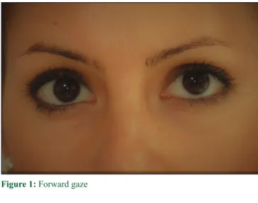
Approximately 5 min after the local anesthetic’s administration, the patient reported double vision when looking left. Otherwise she remained calm, alert, and conscious and she had no other complaints. The pupils were equal and reactive. Limitations of eye movements were confined to abduction of the ipsilateral eye [Figures 1‑3] as a consequence of a complete lateral rectus palsy. There was no evidence of ptosis or proptosis. Blanching of the facial skin or color change was not noted. Adequate anesthesia was achieved in both operating areas. Vital signs were found to be normal.
The patient was assured that this complication was extremely rare and that her symptoms would resolve spontaneously. Possible postponement of surgery was discussed and a decision was made to proceed with the removal of the teeth. These were performed without further complications.
The patient’s diplopia improved slightly by the end of surgery, but she was kept under continuous observation.
Approximately 2 h after the onset of her symptoms, the double vision nearly fully resolved. She was escorted home by a friend and was advised against driving until
Case Report
Introduction
T
he number of local anesthesia carried out by dentists is enormous and is considered to be an extremely safe procedure[1] Ocular complications are unusually rare but can be alarming.[2]Most of the cases remain unreported and/or undocumented and some mild forms may not even be diagnosed.[3] Most ocular complications occur after a posterior superior or an inferior alveolar nerve block.
Case Report
A 26‑year‑old woman was scheduled for the surgical removal of her left‑sided wisdom teeth under local anesthesia. She had undergone an uneventful removal of the right‑sided wisdom teeth 6 weeks before. Her medical history was unremarkable.
A conventional posterior superior alveolar nerve block was administered with 1.5 mL of articaine hydrochloride (40 mg/mL plus adrenaline 0.01 mg/mL) injected into the mucobuccal fold above the third molar.
Greater palatine nerve block was performed with 0.2 mL of the same anesthetic. Inferior dental nerve block (1.5 mL) combined with buccal nerve infiltration (0.2 mL) were also given. A 26‑gauge needle was used for all injections. Aspiration for blood was negative in all cases.
Departments of Oral and Maxillofacial Surgery and
1Emergency Medicine, University of Szeged, Hungary, 2Department of Clinical Ophthalmology, School of Medicine, Dentistry and Biomedical Sciences, Queen’s University Belfast, United Kingdom
Dental anesthesia is one of the most frequently performed medical procedures.
Although the frequency of ocular complications is extremely low, these reactions can be highly alarming and may bring up medicolegal issues when they do occur.
Dentists and oral surgeons should be well‑informed of these adverse reactions and should be aware that both ophthalmologists and emergency physicians might be required to care for these patients.
Keywords: Diplopia, intraoral anesthesia, ocular complications
Abstract
How to cite this article: Seres L, Petho G, Peto Z, Peto T. A case of transient diplopia and ophthalmoplegia following dental anesthesia. Niger J Clin Pract 2019;22:1172-4.
Date of Acceptance:
28-Feb-2019
Access this article online Quick Response Code:
Website: www.njcponline.com DOI: 10.4103/njcp.njcp_660_18 PMID: *******
This is an open access journal, and articles are distributed under the terms of the Creative Commons Attribution‑NonCommercial‑ShareAlike 4.0 License, which allows others to remix, tweak, and build upon the work non‑commercially, as long as appropriate credit is given and the new creations are licensed under the identical terms.
For reprints contact: reprints@medknow.com [Downloaded free from http://www.njcponline.com on Tuesday, February 11, 2020, IP: 160.114.150.250]
Seres, et al.: Transient diplopia following dental anesthesia
Nigerian Journal of Clinical Practice ¦ Volume 22 ¦ Issue 8 ¦ August 2019 1173
her visual function returned to normal. A further 2 h later she reported on the phone that the diplopia had resolved completely.
Discussion
Local anesthetics can paralyze the extraocular muscles directly or through the nervous system. Impairment of the oculomotor (III), trochlear (IV), or abducens (VI) nerves results in different restrictions of the eye
movements. These symptoms are most often attributed to the anesthetic solution reaching the orbit or nearby structures.[4] The most commonly cited theories are the following:
1. The area where the anesthetic is administered is highly vascularized, and unintentional intravascular injections can easily occur. Inadvertent injection into an artery with an intracranial course that anastomoses with the vessels supplying the extraocular muscles may result in limited eye movements[5]
2. Local anesthetic injected into the venous circulation can enter the cavernous sinus through the pterygoid plexus and thus anesthetize the III, IV, or VI nerves[6]
3. Vascular wall trauma of an alveolar artery caused by an injection needle may activate sympathetic fibers, resulting in vasospasm of the vessels supplying the orbital muscles[7]
4. The anesthetic drug may spread directly from the pterygopalatine fossa into the orbit through the inferior orbital fissure following superior alveolar nerve blocks.[3]
The number of documented ophthalmologic complications after intraoral anesthesia has been extremely low, approximately one per year, since it was reported first in the literature.[8] In a review study, diplopia was found to be the most frequently described ophthalmologic complication after intraoral local anesthesia. In 25 (58.1%) of the 43 diplopia patients, diplopia was the only complication, whereas in the other 18 patients (41.9%) it was accompanied with mydriasis and/or ptosis.[9]
The intracranial course of the abducens nerve is long and is vulnerable at many sites, and therefore, the lateral rectus is the most frequently affected muscle.[10] Females are affected more frequently than males; one of the possible explanations is that although the female facial skeleton is generally smaller and thinner, both genders receive the same dose of local anesthetic. Most patients are in their third or fourth decade of life;[9] this may be explained by the fact that most wisdom teeth are removed in this age group. Lidocaine and articaine are the most often involved local anesthetics. As articaine is gaining more popularity as it has excellent diffusion properties, the number of ocular complications may rise.
In nearly all reported cases, aspiration for blood was stated negative.
The most frequent causes of ophthalmologic complications are block anesthesia of the inferior alveolar nerve and of the posterior superior alveolar nerve.[9] The patient in our case report received both, and therefore it cannot be decided which one of them resulted in diplopia.
Figure 1: Forward gaze
Figure 2: Left gaze
Figure 3: Left and upper gaze
[Downloaded free from http://www.njcponline.com on Tuesday, February 11, 2020, IP: 160.114.150.250]
Seres, et al.: Transient diplopia following dental anesthesia
1174 Nigerian Journal of Clinical Practice ¦ Volume 22 ¦ Issue 8 ¦ August 2019
Although there are a few severe and permanent ocular complications reported in the literature, in most cases it is of temporary nature. Diplopia usually resolves within 2 h of onset, occasionally it may take up to 4–5 h, and in extreme cases resolution does not occur up to 24 h.[9] In acute‑onset diplopia, ruling out a stroke or intracranial hemorrhage is an absolute necessity.
First, assessment of the patient’s vital signs (pulse rate, respiratory rate, blood pressure, temperature, and level of consciousness) must be carried out. Symptoms, such as acute headache, nausea, vomiting, and cranial nerve palsy, must be enquired for and tested appropriately, and if any of these are noted, these should be treated as an emergency; the dental procedure should be interrupted immediately and emergency services should be called.
The patient must be managed appropriately while awaiting emergency services, and vital signs must be monitored and documented. If the patient is conscious, reassurance is of major importance.
If vital signs are normal and the patient is agreeable, the planned procedure can be carried out, but postoperative monitoring is necessary until diplopia completely resolves. In any case, the psychological aspect of acute diplopia must be taken seriously. Patients do not expect to have visual complications during dental surgery, and expect the surgical team to deal with such complication competently while providing full explanation. While our case demonstrates that with appropriate management and follow‑up such a case can be brought to successful dental and ophthalmic solution, the potentially life‑threatening implications of acute diplopia are likely, and so require well‑trained staff to deal with it.
Declaration of patient consent
The authors certify that they have obtained all appropriate patient consent forms. In the form the patient(s) has/have given his/her/their consent for his/
her/their images and other clinical information to be
reported in the journal. The patients understand that their names and initials will not be published and due efforts will be made to conceal their identity, but anonymity cannot be guaranteed.
Financial support and sponsorship Nil.
Conflicts of interest
There are no conflicts of interest.
References
1. Malamed SF. Handbook of Local Anesthesia. 4th ed. Mosby;
1997.
2. Patil K, Munoli K, Kumar V, Venkataraghavan K. Intraoral local anesthesia and ocular complications. World J Dent 2013;4:108‑12.
3. Steenen SA, Dubois L, Saeed P, de Lange J. Ophthalmologic complications after intraoral local anesthesia: Case report and review of literature. Oral Surg Oral Med Oral Pathol Oral Radiol 2012;113:1‑5.
4. Boynes SG, Echeverria Z, Abdulwahab M. Ocular complications associated with local anesthesia administration in dentistry. Dent Clin North Am 2010;54:677‑86.
5. Meechan JG. Regional anaesthetic techniques in oral and maxillofacial surgery. In: ShawI, Kumar C, Dodds C, editors.
Oxford Textbook of Anaesthesia for Oral and Maxillofacial Surgery. Oxford University Press; 2010.
6. Freuen ND, Feil BA, Norton NS. The clinical anatomy of complications observed in a posterior superior alveolar nerve block. FASEB J 2007;21:776‑84.
7. Kronman JH, Kabani S. The neuronal basis for diplopia following local anesthetic injections. Oral Surg Oral Med Oral Pathol 1984;58:533‑4.
8. Brain WR. Third nerve palsy following dental extraction. Arch Ophthalmol 1936;5:1164.
9. von Arx T, Lozanoff S, Zinkernagel M. Ophthalmologic complications after intraoral local anesthesia. Swiss Dent J 2014;124:784‑806.
10. Aguado‑Gil JM, Barona‑Dorado C, Lillo‑Rodríguez JC, De la Fuente‑González DS, Martínez‑González JM. Ocular complications following dental local anesthesia. Med Oral Patol Oral Cir Bucal 2011;16:688‑93.
[Downloaded free from http://www.njcponline.com on Tuesday, February 11, 2020, IP: 160.114.150.250]