RESEARCH ARTICLE
Nucleophosmin1 and isocitrate
dehydrogenase 1 and 2 as measurable residual disease markers in acute myeloid leukemia
Petra Ko¨ vy1,2, Zolta´n O˝ rfi1, Andra´s Bors1,3, Andra´s Kozma1, La´szlo´ Gopcsa4, Ja´nos Dolgos4, No´ ra Lovas4, Jo´ zsef Harasztdombi4, Viktor Lakatos4, A´ gnes Kira´ly4, Ga´bor Mikala4, Istva´n Va´lyi-Nagy4, Pe´ter Reme´nyi4, Hajnalka AndrikovicsID1*
1 Laboratory of Molecular Genetics, Central Hospital of Southern Pest National Institute of Hematology and Infectious Diseases, Budapest, Hungary, 2 School of PhD Studies Ra´cz Ka´roly, Semmelweis University, Budapest, Hungary, 3 Department of Transfusiology, Semmelweis University, Budapest, Hungary, 4 Department of Hematology and Stem Cell Transplantation, Central Hospital of Southern Pest National Institute of Hematology and Infectious Diseases, Budapest, Hungary
*andrikovics.hajnalka@dpckorhaz.hu
Abstract
Monitoring measurable residual disease (MRD) in acute myeloid leukemia (AML) plays an important role in predicting relapse and outcome. The applicability of the leukemia-initiating nucleophosmin1 (NPM1) gene mutations in MRD detection is well-established, while that of isocitrate dehydrogenase1/2 (IDH1/2) mutations are matter of debate. The aim of this study was to investigate the stability of NPM1 and IDH1/2 mutations at diagnosis and relapse ret- rospectively in 916 adult AML patients. The prognostic value of MRD was evaluated by droplet digital PCR on the DNA level in a selected subgroup of patients in remission. NPM1 re-emerged at relapse in 91% (72/79), while IDH1/2 in 87% (20/23) of mutation-positive cases at diagnosis. NPM1 mutation did not develop at relapse, on the contrary novel IDH1/2 mutations occurred in 3% (3/93) of previously mutation-negative cases. NPM1 MRD-positiv- ity after induction (n = 116) proved to be an independent, adverse risk factor (MRDpos24- month OS: 39.3±6.2% versus MRDneg: 58.5±7.5%, p = 0.029; HR: 2.16; 95%CI: 1.25–3.74, p = 0.006). In the favorable subgroup of mutated NPM1 without fms-like tyrosine kinase 3 internal tandem duplication (FLT3-ITD) or with low allelic ratio, NPM1 MRD provides a valu- able prognostic biomarker (NPM1 MRDposversus MRDneg24-month OS: 42.9±6.7% versus 66.7±8.6%; p = 0.01). IDH1/2 MRD-positivity after induction (n = 62) was also associated with poor survival (MRDpos24-month OS: 41.3±9.2% versus MRDneg: 62.5±9.0%, p = 0.003; HR 2.81 95%CI 1.09–7.23, p = 0.032). While NPM1 variant allele frequency
decreased below 2.5% in remission in all patients, IDH1/2 mutations (typically IDH2 R140Q) persisted in 24% of cases. Our results support that NPM1 MRD even at DNA level is a reli- able prognostic factor, while IDH1/2 mutations may represent pre-leukemic, founder or sub- clonal drivers.
a1111111111 a1111111111 a1111111111 a1111111111 a1111111111
OPEN ACCESS
Citation: Ko¨vy P, O˝ rfi Z, Bors A, Kozma A, Gopcsa L, Dolgos J, et al. (2021) Nucleophosmin1 and isocitrate dehydrogenase 1 and 2 as measurable residual disease markers in acute myeloid leukemia. PLoS ONE 16(6): e0253386.https://doi.
org/10.1371/journal.pone.0253386 Editor: Chung Hoow Kok, The University of Adelaide, AUSTRALIA
Received: February 19, 2021 Accepted: June 3, 2021 Published: June 21, 2021
Copyright:©2021 Ko¨vy et al. This is an open access article distributed under the terms of the Creative Commons Attribution License, which permits unrestricted use, distribution, and reproduction in any medium, provided the original author and source are credited.
Data Availability Statement: All relevant data are within the manuscript and itsSupporting Informationfiles.
Funding: The study was supported by the Janos Bolyai Research Scholarship of the Hungarian Academy of Sciences (AnB: BO/00809/18/8, https://mta.hu/bolyai-osztondij/bolyaijanos- kutatasi-osztondij-105319), by the New National Excellence Program of the Ministry for Innovation and Technology from the source of the National Research, Development and Innovation Fund (PK:
Introduction
Acute myeloid leukemia (AML) is an aggressive hematological malignancy with a rapidly evolving treatment paradigm. Although the majority of patients remain incurable, long-term remissions can be achieved in roughly one-third of these patients. The identification of prog- nostic markers bears outstanding relevance for optimizing treatment strategy. Measurable residual disease (MRD) after induction therapy and before hematopoietic stem cell transplan- tation is an independent, post-diagnosis prognostic indicator of relapse and survival. The application of molecular genetics and multiparametric flow cytometry are recommended for monitoring. Requirements for a reliable molecular genetic MRD marker are the following: (i) mutation burden fluctuates in parallel with leukemic tumor burden: present at disease onset, disappearing in remission and re-emerging at relapse, (ii) available method with the capability of achieving high sensitivity [1–3].
Nucleophosmin1(NPM1)mutations are among the most frequently detected genetic alter- ations in AML (present in 25–35% of primary AML) defining a separate disease entity.NPM1 frameshift mutations result in altered protein termination, loss of nuclear localization signals, and consequential abnormal cytoplasmic localization of the mutant protein [4–6]. Isocitrate dehydrogenase 1 and 2 (IDH1andIDH2) mutations occur in 7–14% and 8–19% of AML cases respectively. The gain-of-function mutations result in the production of an oncometabolite with consequential hypermethylation, gene expression alterations and impaired hematopoietic differentiation [4,7].
NPM1alterations were reported as definite leukemia-founder mutations and optimal MRD markers. On the other hand contradictory data exist, whetherIDH1andIDH2mutations rep- resent pre-leukemic, or dominant clone mutations, therefore their value in MRD monitoring is not well established [3,8]. In our study, we aimed to correlateNPM1andIDH1andIDH2 mutational variant allele frequencies at diagnosis, remission and relapse to investigate the potential application of these mutations in MRD monitoring.
Material and methods Patients
The study included 916 adult patients (449 males/467 females, median age at diagnosis 54 years; range: 16–94), consecutively diagnosed with AML between January 2001 and June 2020 in our Institute (Department of Hematology and Stem Cell Transplantation, Central Hospital of Southern Pest National Institute of Hematology and Infectious Diseases, Budapest, Hun- gary). In this patient cohort 253 patients wereNPM1, 68IDH1and 94IDH2mutations positive (74 patients carried bothNPM1andIDH1/2mutations). A significant proportion of patients 81% (n = 746/916) received curative treatment, out of which 26% (n = 176/746) was treated by allogeneic hematopoietic stem cell transplantation (HSCT). MRD monitoring was retrospec- tively evaluated in a selected subgroup of 116NPM1(51 male/65 female, median age at diagno- sis 48 years), and 62IDH1/2positive patients (23 male/39 female, median age was 49 years).
The inclusion criteria for the MRD monitored subgroup were the following: (i) curative che- motherapy; (ii) morphologic leukemia-free state (MLFS) after induction [1]; (iii) available DNA sample at diagnosis, after induction, and/or before HSCT. Patients with palliative ther- apy, death in aplasia or death from indeterminate cause, no MLFS after 2 courses of intensive induction treatment; unavailable DNA sample, or patients with rare undetectableNPM1or IDH1/2mutation were excluded from MRD evaluation. MRD was determined after induction and one month before HSCT if DNA samples were available. Data from AML patients diag- nosed between 2001 and 2009 have already been reported in an earlier study [9] andIDH1/2
U´ NKP-20-3-II-SE-79, AnB: U´NKP-19-4-SE-83, http://www.unkp.gov.hu/unkp-rol), by grants from the National Research, Development and Innovation Office (PK, ZO, AnB, AK, LG, JD, NL, JH, GM, IV-N, PR, HA: NVKP_16-1-2016-0005,https://
www.dpckorhaz.hu/informaciok/palyazatok/
projektek/hazai-projektek/kiemelkedo-halalozasi- kockazattal-jaro-betegsegek-gyogyitasanak- eredmenyesseget-lenyegesen-javito-nemzeti- program-nemzeti-innovacios-onkogenomikai-es- precizios-onkoterapias-program-elinditasa-es- kapc/), by the Development of a Translational Medicine Research Center and an Innovative Center for Cellular Therapy at the Central Hospital of Southern Pest – National Institute of
Hematology and Infectious Diseases (ZO, AnB, LG, JD, GM, IV-N, HA: TKP2020-NKA-19,https://nkfih.
gov.hu/palyazoknak/tkp-2020) and by grants of Ministry of Human Capacities (AnB, HA: EMMI IV/219-4/2021/EKF). The funders had no role in study design, data collection and analysis, decision to publish, or preparation of the manuscript.
Competing interests: The authors have declared that no competing interests exist.
data between 2001–2018 were presented in a Hungarian report [10]. Data collection was per- formed retrospectively. Definitions offms-like tyrosine kinase 3 internal tandem duplication (FLT3-ITD) low and high allelic ratio, MLFS, overall survival (OS) and relapse-free survival (RFS) were described by European LeukemiaNet (ELN) 2017 recommendations [1]. The study was in accordance of the Declaration of Helsinki and was approved by the Institutional Review Board of Central Hospital of Southern Pest National Institute of Hematology and Infectious Diseases. Written informed consent was provided by all patients.
Molecular genetic methods
Genomic DNA and RNA were isolated from bone marrow samples drawn at diagnosis, remis- sion and relapse. Screening for hotspot mutations were performed from genomic DNA, at the time points of diagnosis and repeatedly at relapse by fragment analysis in case ofNPM1 (NPM1diagnosis n = 916, relapse n = 161 if DNA was available); [11], and by high-resolution melting (HRM) or allele specific PCR in case ofIDH1/2(diagnosis n = 842, relapse n = 116 if DNA was available) [9]. Positive cases were monitored with droplet digital PCR (ddPCR) after induction therapy (NPM1n = 116;IDH1/2n = 62; double positive = 33), before HSCT (1–30 days before;NPM1n = 38;IDH1/2n = 22), if DNA was available at that time point. Mutant NPM1RNA expression was also tested at diagnosis and after induction therapy (n = 39).
Diagnosis and follow-up samples ofNPM1as well asIDH1andIDH2positive AML patients were investigated by ddPCR. ForNPM1mutation detection primer and probe sequences are summarized inS1 Table[12–14].NPM1type-A (c.860_863dupTCTG, p.Trp288CysfsTer12) specific reverse primer was described by Gorelloet al. [13,15]. A degenerate R primer (type- N) reported by Mencia-Trinchantet al. [14] was applied to detectNPM1mutations at the same position with different nucleotide insertions (c.860_863dupNNNN, p.Trp288Cysf- sTer12, referred asNPM1type-N mutation in this study).GAPDHwas used as the reference gene for the assay for DNA [16], andABL1for RNA [17]. Reactions were performed using Supermix for Probes (no dUTP) (BioRad), 900 nM of each primer, 250 nM of each probe, 100 ng DNA or 240 ng cDNA per well. For genomic DNA, assays were designed by Bio-Rad for the detection of the most commonIDH1/2mutations (IDH1R132C ID: dHsaMDV2010053, R132H ID: dHsaMDV2010055 andIDH2R140Q ID: dHsaMDV2010057, R172K ID:
dHsaMDV2010059). The PCR program started with an initial denaturation at 98˚C for 10 min, 40 cycles of denaturation at 94˚C for 30 sec, annealing at 55˚C (for DNA) and at 60˚C (for RNA) for 60 sec followed by enzyme deactivation at 98˚C for 10 min. The QX200 Droplet Digital PCR System and QuantaSoft Software (Version 1.7.4.0917, BioRad) were used for the evaluation of the results.
The ddPCR measurements were acceptable if: (i) reference copies or total copies>32,000, (ii) total droplet count>15,000; (iii) empty droplets>100. MRD samples were measured in dupli- cate wells to achieve optimally more than 4.5-log sensitivity (at least 32,000 copies of reference gene) [18]. The ddPCR measurements were also performed in 20–35 mutation negative controls to determine the limit of blank (LoB = meannegative samples
+ 1.645x standard deviation [SD]) and to determine the limit of detection (LoD = meannegative samples
+ 3.3×SD) [19,20]. Variant allele fraction (VAF) lower, than 2.5% correspond to<5% (pre)leukemic cells. Samples taken during MLFS (bone marrow blasts<5%; absence of blasts with Auer rods; absence of extramedullary dis- ease) displaying>2.5% VAF were categorized as persisting preleukemic clones.
Statistical analysis
Categorical variables were compared by the Fisher’s exact test, continuous variables by Mann- Whitney tests. Kaplan-Meier method with log-rank statistics were used to calculate OS and
RFS [1]. After induction OS were calculated from the time point of diagnosis, RFS from remis- sion irrespective from performing HSCT. Regarding pre-transplant MRD monitoring, compari- sons of OS and RFS were performed from the time point of HSCT. Following univariate analysis, age, cytogenetics,FLT3-ITD allelic ratio [1],NPM1, white blood cell count (WBC) at diagnosis, and MRD status were included in a Cox proportional hazard model for OS and RFS.
Hazard ratios (HR) and 95% confidence interval (95%CI) values were calculated. In order to identify the cut off discriminating between low and high MRD burden groups, HRs for OS were compared at six different limits (0.05%; 0.1%; 0.2%; 0.5%; 1% and 2%) forNPM1type-A and type-N separately and combined [21]. P values below 0.05 were considered as statistically signifi- cant. For the statistical analysis SPSS Statistics version 22 (Armonk, NY) was applied.
Results
Occurrence ofNPM1,IDH1/2mutations in the total AML cohort
This study included 916 adult AML patients (S2 Table). Cytogenetic results were available for 94% (n = 861) of patients: favorable (n = 136; 16%), intermediate (n = 507; 59%) and adverse (n = 218; 25%) ELN 2017 cytogenetic risk categories were identified.NPM1mutation occurred in 28% (n = 253/916),FLT3-ITD in 25% (n = 226/916);FLT3tyrosine kinase domain muta- tions (FLT3-TKD) in 8% (n = 71/910);IDH1in 8% (n = 68/842) andIDH2in 11% (94/842).
IDH1R132H associated withNPM1positivity more commonly than otherIDH1R132 codon mutations: 90% (n = 27/30) versus 26% (n = 10/38), p<0.0001. AlsoIDH2R140Q co-occurred withNPM1in 49% (n = 37/76), while R172K never associated (p<0.0001).
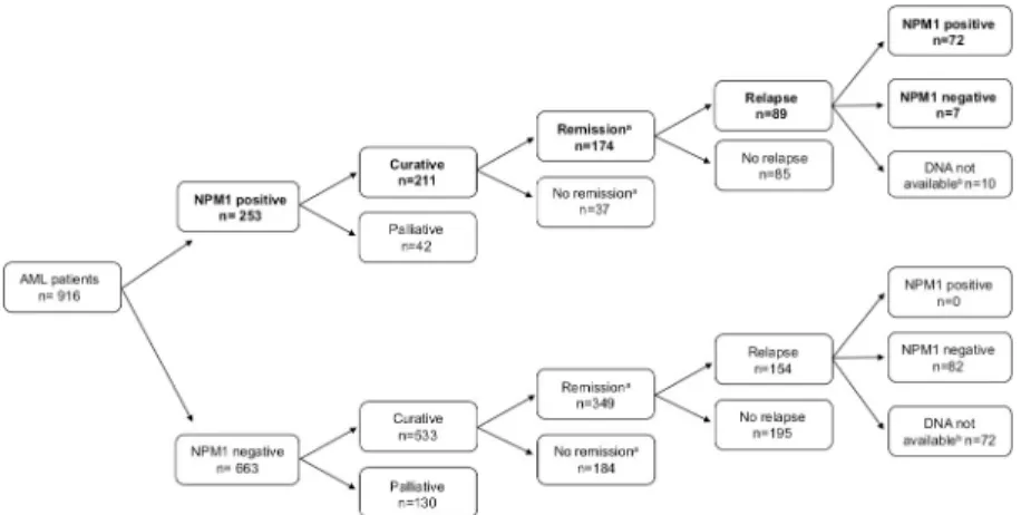
In theNPM1-positive cohort, 211 patients were treated with curative intent, out of which remission (MLFS) was reached in 174 cases (Fig 1). The stability ofNPM1mutation during disease evolution was studied with 79 pairedNPM1mutant samples drawn at diagnosis and relapse. TheNPM1mutation re-emerged at relapse in 91% ofNPM1positive cases (n = 72/79).
Time period from diagnosis till relapse was not significantly longer in cases whereNPM1was undetectable at relapse compared to cases with persistentNPM1mutation at relapse [median 7.1 month (range: 0.1–172.2 month) versus 6.6 month (range: 2.2–152.9 month) respectively, p = 0.46]. All seven patients with clonalNPM1regression had normal karyotype at the time of diagnosis; one patient out of five with karyotyping available at relapse had clonal evolution (tri- somy 8). None of ourNPM1negative AML cases gainedNPM1mutation positivity at relapse,
Fig 1. Clinical characteristics ofNPM1positive AML patients.aRemission was defined as morphologic leukemia- free state (MLFS) after induction.bDNA not available at the time point of relapse.
https://doi.org/10.1371/journal.pone.0253386.g001
among the 82NPM1negative patients, where samples at diagnosis and relapse were available at both time points.
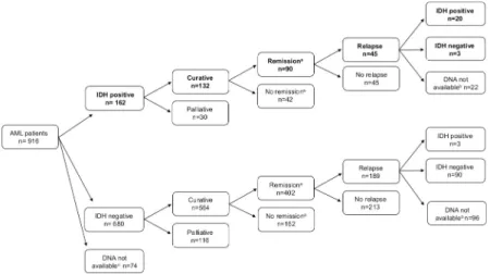
In theIDH1/2-positive cohort (n = 162), 132 patients were treated with curative intent, out of which remission (MLFS) was reached in 90 cases (Fig 2).IDH1/2mutations were undetect- able at relapse in 13% of theIDH1/2-positive cohort with available DNA (n = 3/23, 1IDH1 R132C, 1IDH2R140Q and 1IDH2R172K). Time from diagnosis till relapse was not proven to be significantly longer in cases whereIDH1/2was undetectable at relapse compared to cases with persistentIDH1/2mutation at relapse [median 7.4 month (range: 2.2–11.4 month) versus 8.6 month (range: 0.83–57.2 month) respectively, p = 0.65]. Interestingly in three (IDH1 R132H: n = 1;IDH2R140Q: n = 2) out of 93IDH1/2negative AML cases where diagnosis and relapse samples were both available,IDH1/2mutations appeared only at relapse. These cases were re-evaluated by the more sensitive ddPCR method at diagnosis and VAF (0–0.23%) was under the detection limit of HRM and/or allele specific PCR in each case.
The applicability of ddPCR methods
NPM1-positive patients screened by capillary electrophoresis at diagnosis were retrospectively typed with type-A and type-N primers using ddPCR. Out of 200 AML patients (53 samples were not available) 97% (n = 194) was proved to beNPM1type-A or type-N and not more than 3%
(n = 6) could not be detected with type-N primer [14] (S3 Table).IDH1orIDH2mutations were screened by HRM and allele-specific PCR (S4 Table). Out of the 68IDH1positive AML patients 39% (n = 27) wasIDH1R132C, 46% (n = 30)IDH1R132H, 15% (n = 11)IDH1R132G/L/S/P. In case ofIDH2positive AML, 81% (n = 76) harboredIDH2R140Q and 19% (n = 18)IDH2 R172K. In our patient cohort, 93% (151/162)IDH1/2mutation positive patients carried an IDH1/2variant detectable with ddPCR. Interestingly,IDH1R132H was associated withNPM1 type-A mutation in 48% (n = 13/27), while otherIDH1R132 codon mutants andIDH2R140Q co-occurred withNPM1type-A mutation in 70% (n = 33/47; p = 0.08;S5 Table).
NPM1MRD monitoring
The LoD forNPM1type-A ddPCR was lower than type-N ddPCR both in DNA and RNA set- tings (S6 Table).NPM1mutant VAF values in DNA andNPM1mutant expression levels in
Fig 2. Clinical characteristics ofIDH1andIDH2positive AML patients.aRemission was defined as morphologic leukemia-free state (MLFS) after induction.bDNA not available at relapse.cDNA not available at diagnosis forIDH1/2 testing.
https://doi.org/10.1371/journal.pone.0253386.g002
RNA were considered as MRD negative if below 0.01% (type-A) or below 0.05% (type-N). In case ofNPM1, 174NPM1positive cases reached MLFS after induction, MRD monitoring could not be performed in 5 cases due to technical limitations (NPM1mutation could not be detected by type-A or type-N primers), and in 53 cases due to non-available DNA. Basic char- acteristics such as gender, age at diagnosis, induction therapy, HSCT, and outcome (death in aplasia or in indeterminate cause, remission, relapse, cytogenetic and molecular genetic data) ofNPM1positive and MRD monitored patients were included inS3 Table. In 116NPM1 MRD monitored patients,NPM1mutant VAF was reduced below 2.5% in all patients in MLFS after induction.
We examined the OS and RFS of 90 AML patients who haveNPM1type-A mutation and further 26 patients who were monitored with type-NNPM1ddPCR from DNA. The median NPM1VAF at diagnosis was 45.7% (range: 11.5–49.3%), while after induction therapy was 0.06% (range: 0–2.5%). Out of the 90 patients with type-ANPM1, 35 patients were MRD-nega- tive, and 55 MRD-positive. Favorable outcome measures were observed in MRD negative compared to MRD positive patients forNPM1type-A (24-month OS: 50.2±8.9% for negative versus 27.7±6.5% for positive, p = 0.010; and 24-month RFS: 40.2±8.6% versus 15.8±5.1%
p = 0.009,S1 Fig). MRD-positive patients were further divided into MRD-low and MRD-high burden subgroups. In our patient cohort,NPM1VAF 0.2% was considered as the limit to dis- criminate between low and high burden: 28 patients were classified into the MRD low category (ranging from 0.01 to 0.2%NPM1mutant allele burden), and 27 patients in the MRD high cat- egory (above 0.2%). As expected even within the MRD positive subgroup, high allele burden cases showed a tendency to more adverse outcome measures (24-month OS: 40.6±10.3% for low versus 16.1±7.4% for high MRD, p = 0.088; and 24-month RFS: 19.4±7.8% versus 12
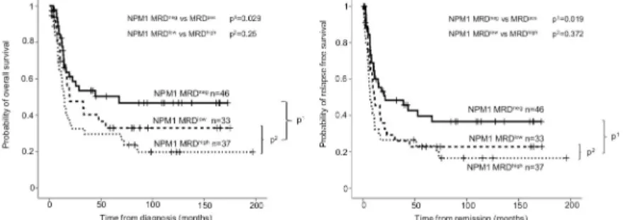
±6.5%; p = 0.107,S1 Fig). Analyses were also performed forNPM1aggregate types-A and -N, and similar results were obtained, (24-month OS: 58.5±7.5% for negative versus 39.3±6.2% for positive, p = 0.029; and 24-month RFS: 48.3±7.5% versus 27.8±5.6%, p = 0.019,Fig 3). The dif- ference did not reach the level of significance within the MRD positive group (24-month OS:
47.6±9.4% for low versus 32.6±8.0% for high, p = 0.250; and 24-month RFS: 29.3±8.2% versus 26.5±7.6%, p = 0.372,Fig 3).NPM1type-A and -N MRD positivity proved to be an indepen- dent risk factor in multivariate analysis beside age at diagnosis, cytogenetics andFLT3-ITD allele burden, and white blood cell (WBC) count above 100.000 per microliter at diagnosis (OS: HR 2.16 95%CI 1.25–3.74, p = 0.006; RFS: HR 2.21 95%CI 1.32–3.68, p = 0.002) (Table 1).
Fig 3. Probability of overall survival and relapse free survival according toNPM1MRD after induction. On both panels (A: overall survival; B: relapse free survival), the outcome ofNPM1MRD-negative (MRDnegVAF<0.01–0.05%
depending onNPM1mutation type) and MRD-positive (MRDposVAF>0.01–0.05%) subgroups are shown with the associated p1value. TheNPM1MRD-positive subgroup was further divided in MRD low-positive (MRDlow VAF = 0.01–0.2%) and MRD high-positive (MRDhighVAF>0.2%) subgroups, and compared with p2values.
https://doi.org/10.1371/journal.pone.0253386.g003
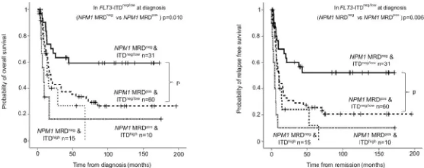
High allelic ratio ofFLT3-ITD at diagnosis (categorized according to the ELN 2017 risk stratification) is a well-documented adverse risk factor in AML. In the favorable subgroup of mutatedNPM1withoutFLT3-ITD (FLT3-ITDneg) or with low allelic ratio (FLT3-ITDlow), the presenceNPM1MRD provided a valuable prognostic biomarker (NPM1MRDnegversus MRDpos24-month OS: 66.7±8.6% versus 42.9±6.7%, p = 0.010; RFS: 60±8.9% versus 31.1
±6.2%, p = 0.006).NPM1MRD did not influence survival in theFLT3-ITDhighsubgroup.
NPM1MRD negative andFLT3-ITD high allele burden resulted similar survival measures to NPM1MRD positive patients (Fig 4).
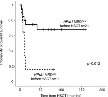
Out of the 38 patients who underwent allo-HSCT for MRD monitoring with theNPM1 mutation type-A (n = 27) and type-N (n = 11), pre-HSCT sample was available in 32 (24 type- A and 8 type-N).NPM1MRD negativity before allo-HSCT proved to be favorable prognostic factor, OS after HSCT was significantly longer in MRD negative compared to positive patients (24-month OS MRDneg: 74.7±9.8% versus MRDpos: 16.2±14.6%, p = 0.012;Fig 5).
Similarly to genomic DNA two or three log reduction was observable in mutantNPM1 RNA expression (NPM1/ABL1, n = 39 patients) [at diagnosis: median 610.8% (range: 124.3–
2882.4%), after induction: 1.0% (range: 0–398%)]. Despite the low number of RNA samples, high mutantNPM1expression after induction correlated with unfavorable outcome (24-month OS mutantNPM1expression<1%: 55.2±12.9% versus mutantNPM1expression
>1% 20.0±11.9%, p = 0.005; and 24-month RFS: 51.6%±12.5% versus 12±7.9% respectively;
Table 1. Multivariate analysis ofNPM1MRD status after induction.
Overall Survival (n = 116) Relapse Free Survival (n = 116)
Hazard ratio (95% CI) P Hazard ratio (95% CI) P
NPM1MRD positivitya 2.16 (1.25–3.74) 0.006 2.21 (1.32–3.68) 0.002
Age (per year) 1.02 (1.00–1.04) 0.019 1.02 (1.00–1.04) 0.053
Cytogeneticsb 1.50 (0.86–2.63) 0.155 1.62 (0.94–2.82) 0.085
FLT3-ITDc 1.75 (1.19–2.56) 0.004 1.74 (1.23–2.46) 0.002
WBC>100.000/μL 0.88 (0.50–1.56) 0.656 0.93 (0.54–1.58) 0.775
aNPM1MRD positivity was defined as VAF>0.01–0.05% depending on mutation type.
bCytogenetics coded as normal karyotype (reference), other intermediate and adverse risk.
cFLT3-ITD coded in three categories as wild type (reference), low and high allelic ratio.
Abbreviations: 95%CI: 95% confidence interval;FLT3-ITD:fms-like tyrosine kinase 3 –internal tandem duplication; MRD: measurable residual disease;NPM1:
nucleophosmin1; WBC: white blood cell count at diagnosis.
https://doi.org/10.1371/journal.pone.0253386.t001
Fig 4. Overall survival and relapse free survival according toNPM1MRD stratified byFLT3-ITD allelic ratio.
Based on the ELN 2017 genetic risk stratification,NPM1positive patients were categorized in to favorable (FLT3- ITDneg/low) and intermediate (FLT3-ITDhigh) subgroups. On both panels (A: overall survival; B: relapse free survival), further subgroups were established according toNPM1MRD after induction.NPM1MRD negativity was defined as VAF<0.01–0.05% depending on mutation type.
https://doi.org/10.1371/journal.pone.0253386.g004
p<0.001). We investigated parallel RNA and DNA-basedNPM1mutddPCR methods from 39 samples after first induction therapy from 39 patient who have both DNA and RNA samples.
The assay sensitivity proved to be higher on RNA samples. Altogether 46% of the RNA samples that displayedNPM1mutexpression (median: 0.1%; range: 0.01–5.1%) were detected as nega- tive in the matching DNA samples (<0.01%). RNA assay (NPM1mutexpression) proved to be more sensitive (median: 1.3-log; range: 0.0–2.78-log) compared to DNA assay (NPM1mut VAF) in samples with concomitant positivity both on RNA and DNA level (S2 Fig).
IDH1/2MRD monitoring
The LoB forIDH1/2mutation detection was 0.06–0.08% and the LoD was 0.09–0.12% (S6 Table). In general, VAF below 0.2% for eachIDH1/2form was considered as negative. In case of 90IDH1/2positive patients in MLFS after induction, MRD monitoring could not be per- formed in 8 cases withIDH1R132G/L/S/P/ and in 20 cases with lacking DNA samples. Basic characteristics ofIDH1/2positive and MRD monitored patients were included inS4 Table.
We observed thatIDH1/2VAF in morphologic leukemia free state was not reduced below 2.5% in 15 out of 62 cases (24%, 10IDH2R140Q, 3IDH2R172K, 1IDH1R132H and 1IDH1 R132C). Seven cases wereNPM1positive (6IDH2R140Q and 1IDH1R132H) at diagnosis but NPM1mutational burden was reduced below 2.5% in remission. Regarding the outcome of patients with persistingIDH1/2mutation: 9 patients relapsed and subsequently died, 2 patients alive after HSCT, 3 patients alive in complete remission after 12 months follow up and 1 patient died without relapse.
In our analyses, the survival ofIDH1orIDH2MRD-negative patients was significantly bet- ter than that of MRD-positive patients (24-month OS MRDneg: 62.5±9% versus MRDpos: 41.3
±9.2%, p = 0.003; 24-month RFS: 45.0±9.3% versus 38.8±9.6% respectively, p = 0.027,Fig 6).
Fig 5. Overall survival according toNPM1MRD measured before HSCT.NPM1MRD negativity before HSCT was defined as VAF<0.01–0.05% depending on mutation type.
https://doi.org/10.1371/journal.pone.0253386.g005
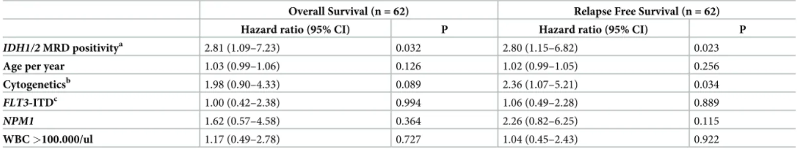
In multivariate analysis,IDH1/2MRD positivity was proved to be an independent risk factor for survival besides age, cytogenetics,FLT3-ITD,NPM1and WBC (OS: HR: 2.81 95%CI: 1.09–
7.23, p = 0.032, RFS: HR: 2.80 95%CI: 1.15–6.82, p = 0.023,Table 2).
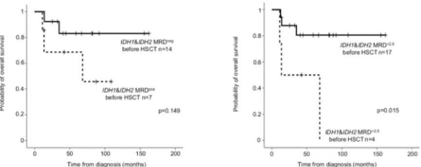
In allo-HSCT cases, pre-HSCT samples were available in 21 out of 22 patients (10IDH1 and 11IDH2).IDH1/2MRD negativity (VAF<0.2%) before allo-HSCT did not reach statisti- cal significance (24-month OS MRDneg: 92.3±7.4% versus MRDpos68.6±18.6%, p = 0.149).
IDH1/2MRD below 2.5% influenced significantly survival (24-month OS: MRD<2.5%87.8
±8.1% versus MRD>2.5%50.0±25.0% before allo HSCT, p = 0.015;Fig 7).
Discussion
The serial acquirement of somatic mutations in myeloid clone(s) was described as the multi- step pathogenesis of AML. Several lines of evidence prove thatNPM1mutations are responsi- ble for the definitive acute leukemic transformation, therefore considered as leukemia founder mutations: (i)NPM1mutations are completely absent in the population without hematological malignancies even at a higher age [22–24]; (ii)NPM1mutations cannot be detected in AML patients months or years before the manifestation of AML [25,26]; (iii)NPM1mutations occur rather rarely (approximately 2–3%) in preleukemic myeloid malignancies such as myelodysplastic syndrome (MDS) or in myelodysplastic/myeloproliferative neoplasm
Fig 6. Overall survival and relapse free survival according toIDH1/2MRD after induction. On both panels (A:
overall survival; B: relapse free survival)IDH1/2MRD negativity defined as VAF<0.2% and MRD positivity as VAF>0.2%.
https://doi.org/10.1371/journal.pone.0253386.g006
Table 2. Multivariate analysis ofIDH1/2MRD status after induction.
Overall Survival (n = 62) Relapse Free Survival (n = 62)
Hazard ratio (95% CI) P Hazard ratio (95% CI) P
IDH1/2MRD positivitya 2.81 (1.09–7.23) 0.032 2.80 (1.15–6.82) 0.023
Age per year 1.03 (0.99–1.06) 0.126 1.02 (0.99–1.05) 0.256
Cytogeneticsb 1.98 (0.90–4.33) 0.089 2.36 (1.07–5.21) 0.034
FLT3-ITDc 1.00 (0.42–2.38) 0.994 1.06 (0.49–2.28) 0.889
NPM1 1.62 (0.57–4.58) 0.364 2.26 (0.82–6.25) 0.115
WBC>100.000/ul 1.17 (0.49–2.78) 0.727 1.04 (0.45–2.43) 0.922
aIDH1/2MRD positivity was defined as VAF>0.2%.
bCytogenetics coded as normal karyotype (reference), other intermediate and adverse risk.
cFLT3-ITD coded in three categories as wild type (reference), low and high allelic ratio.
Abbreviations: 95%CI: 95% confidence interval;IDH1/2: isocitrate dehydrogenase 1/2;FLT3-ITD:fms-like tyrosine kinase 3 –internal tandem duplication; MRD:
measurable residual disease;NPM1: nucleophosmin1; WBC: white blood cell count at diagnosis.
https://doi.org/10.1371/journal.pone.0253386.t002
(MDS/MPN), which are mainly characterized by the progression to overt AML [27]; (iv) NPM1mutations were not present in preleukemic hematopoietic stem cells [28,29]. Our observation thatNPM1VAF decreased below 2.5% in all cases with morphologic leukemia- free state, also proved thatNPM1mutations do not occur in the preleukemic state. (v) A fur- ther proof thatNPM1mutations harbor leukemia-initiating properties is the high reappear- ance rate ofNPM1in relapse, which was demonstrated as high as 86–100% in several clinical observations [30–39]. The long observational period in our study allowed us to detect late AML relapses. In line with previous studies less than 10% of ourNPM1mutation positive cases relapsed as wild typeNPM1AML. The loss ofNPM1mutation in our patient cohort was not associated with longer remission before relapse, which was suggested by several previous studies [31,32]. Although our study did not investigate the spectrum of preleukemic muta- tions, the persistence ofIDH2R140Q mutation was observed in a single case withNPM1- mutation loss relapse 13 month after diagnosis.
Contradictory data exist in the literature, whetherIDH1andIDH2mutations are preleuke- mic or AML founder mutations. Several studies suggestIDH1/2mutations as epigenetic modi- fiers as preleukemic events. (i) In large scale populational screening studies for clonal
hematopoiesis of indeterminate potential (CHIP) mutations,IDH2R140 mutations were extreme rarely detected in elderly individuals (IDH2R140Q/W: 0.014%, four out of 29562 individuals) [22].(ii) IDH1andIDH2mutations were detectable as premalignant, high-risk gene mutations years before the diagnosis of AML, but not in age-matched controls (8%;
n = 15/188; threeIDH1R132C/H/G and 12IDH2R140 positive individuals with a median of 7 years before AML diagnosis) [26]. (iii)IDH1andIDH2mutations are also rarely present in preleukemic myeloid malignancies: 0.8–4% in chronic phase MPN, 4–14% in MDS, but its fre- quency increases up to 20–25% in blast phase transformation [40–43]. (iv)IDH1andIDH2 mutations were detectable in preleukemic hematopoietic stem cells [28,29]. The comparison of VAF values suggested thatIDH1andIDH2mutations were more likely to develop before NPM1mutations [6]. The persistence ofIDH1/2mutations (especiallyIDH2R140Q) in remis- sion was observed in 7–39% of AML cases in the literature, [19,44–46] which is in line with our study (24% ofIDH1orIDH2mutations were detectable in complete remission with a higher than 2.5% VAF, 67% of persisting mutations wasIDH2R140Q). The high mutational load in remission is a direct proof of preleukemic origin of the somatic mutation. This phe- nomenon in caseIDH1/2mutations is not as frequent as in case ofDNMT3A,TET2,ASXL1 gene mutations, where reported rates vary between 51–82% [2,47–49], (v) At relapse both IDH1/2gene mutations showed a relatively high stability (86–88% reported in publications, 87% in our study) similar that ofNPM1mutation [31]. (vi) InIDH1/2mutation negative AML, the emergence ofIDH1orIDH2mutations at relapse was observed in 10% in our study,
Fig 7. Probability of overall survival forIDH1/2MRD before HSCT. On panel A,IDH1/2MRD negativity before HSCT was defined as VAF<0.2%, on panel B as 2.5%.
https://doi.org/10.1371/journal.pone.0253386.g007
which suggests the subclonal, late origin of these mutations. Interestingly, there is a usual mutation order in AML pathogenesis, but some mutations might appear both early and late events [31,50].
A recent meta-analysis proved that lower MRD was consistently associated with improved outcome independently from applied method, sample source or sampling time of the assess- ment [51]. Regarding molecular genetic detection techniques, like quantitative PCR, digital PCR and next generation sequencing are extensively applied for MRD detection. High assay precision and reproducibility make ddPCR particularly suitable for MRD monitoring, which was reported in connection with several oncohematological drivers [52–56]. Individual assay designs make the quantification of multipleNPM1mutations challenging, but the application of degenerated primers allows the simultaneous detection of multipleNPM1mutations affect- ing the same localization (c.860_863dupNNNN) [14]. Our data and other studies also sup- ported, that less than 5% of NPM1 mutations affects nucleotides at different positions [11,38, 57].
Although consensus exists about the importance ofNPM1MRD; broad range of heteroge- neity was displayed concerning thresholds discriminating between low- and high-risk MRD.
Studies comparing mutantNPM1transcript levels parallel in bone marrow (BM) and periph- eral blood (PB) samples identified strong correlation, but an average of 1-log higher sensitivity in BM [21,34,38,57–61]. In line with this observation, 3-log reduction ofNPM1mut/ABL1 transcript level was pointed as favorable prognostic indicator in BM, [21,59,60] but 4-log reduction was required in PB after induction therapy [57,58]. In our study, bone marrow sam- ples were processed. AsNPM1mutexpression is highly abundant, greater sensitivity (median:
1.3 log, range: 0–2.78 log in our study) was achieved on RNA level than on DNA.NPM1mut RNA expression level detection for MRD monitoring is recommended in the literature [13, 38]. Shayegiet al. investigated that 1%NPM1mut/ABL1expression corresponds to 0.016%
NPM1mutVAF or 1 in 32000 cells (1.8 log difference between RNA and DNA levels) [60].
These data suggested thatNPM1mutMRD screening should be performed on RNA expression, but in case of RNA unavailability, highly sensitive DNA methods can substitute. The applied cut-off for MRD negativity in our study (NPM1type-A: 0.01% and type-N: 0.05% on DNA level) corresponds approximately to 1%NPM1mut/ABL1expression level. We were unable to test large number of RNA samples, which is a major limitation of our retrospective study. Ivey et al. [57] demonstrated that RNA-MRD positivity in PB after induction (2 cycles) corre- sponded to higher cumulative incidence of relapse (MRC17 trial 3-year CIR: 82% versus 30%), similarly Balsatet al. [58] (ALFA-0702 trial: 2-year CIR: 55% versus 21%); Hubmannet al. [62]
less than 3log-reduction in BM RNA-MRD (AMLCG 1999, 2004 and 2008 trial: 2-year CIR 77.8% versus 26.4%,); Kapp-Schwoereret al. [34] less than 3-log10BM or PB RNA-MRD (AMLSG 09–09 trial 4-year CIR BM: 60% versus 28.5%; PB: 62.5% versus 33.9%). On the DNA level, we also observed that MRD positivity (less than 3log reduction) was associated with adverse outcome, and DNA-MRD after induction therapy is capable to identify high-risk NPM1mutpatients.
The co-occurrence withFLT3-ITD was recognized as an adverse factor inNPM1mutant AML, due to the highly proliferative nature of the leukemic clone with ITD [38,63]. Although NPM1mutation was referred as favorable or intermediate ELN prognostic categories depend- ing on the presence ofFLT3-ITD with high mutational load [1]. Recently, the reclassification of ELN prognostic criteria identified highFLT3-ITD load as adverse risk irrespective ofNPM1 mutation status [64]. Allogeneic HSCT in first complete remission is not recommended in favorable risk AML, on the other hand relapsedNPM1-positive cases have adverse outcome [65]. We observed that the measurement ofNPM1MRD was capable to identify high risk patients even in the favorable riskNPM1positive AML without high ITD load.NPM1MRD
negativity (NPM1mutVAF<0.01–0.05% after induction) with highFLT3-ITD allele burden at diagnosis showed similarly adverse survival toNPM1MRD positive patients.
Molecular MRD measurements serve not only prognostic, but may influence therapy. In case of persistent MRD, HSCT consolidation improved survival over chemotherapy [66]. In ELN 2017 favorable riskNPM1mutAML subgroup, molecular failure (defined asNPM1mut/
ABL1>0.05% after consolidation orNPM1mutreappearance after molecular response; which
affected 40% ofNPM1mutcases) served as indication for allogeneic HSCT in first complete remission. MRD-guided approach involving early intervention resulted in improved outcome (two-year OS: 85% for HSCT-treated patients with molecular failure and 39% for patients with hematological relapse) [67]. For elderly or unfit patients, azacitidine was reported to prevent or delay hematological relapse in MRD-positive AML [68].
Our data investigating pre HSCTNPM1mutMRD are in good concordance with other stud- ies with similar MRD time-point assessment: pre HSCT MRD negativity predicts favorable outcome after HSCT [21,66,69–71]. Detection of MRD-positivity before HSCT guide thera- peutic choices during conditioning and graft versus host disease prevention, e. g. preferably T- cell repleted versus T-cell depleted transplant [21]; preferably myeloablative versus reduced intensity conditioning [72]. MRD measurements can even guide targetedFLT3-inhibitor ther- apy identifying patients who benefit mostly [73].
The role of MRD-monitoring is well-documented in case ofNPM1, but data are scarce aboutIDH1andIDH2mutations. We applied BioRad-designed mutation detection reagents on BioRad QX200 Droplet Digital PCR System, but interestingly we were not able to reach as high sensitivity as in case ofNPM1. Similar technical limits (LoD: 0.2%) were reported in a previous study applying the same detection [19]. Our data also supported the preleukemic nature ofIDH1/2mutations, but the persistence ofIDH1/2mutations (VAF>2.5%) in com- plete remission was associated with adverse outcome, higher chance of relapse or the develop- ment of myelodysplasia [19,44]. The presence of a preleukemic clone in morphologic
leukemia-free remission was generally reported to associate with inferior survival compared to patients without persisting oncogenic mutations [74,75]. On the other hand, persistent DNMT3A,TET2,ASXL1mutations were not connected with higher relapse rate and several reports described long-term remission even with high VAF [2,47–49]. The frequency of per- sistentIDH1/2mutations in remission was reported as high as 7–39% depending on the VAF cut -off (1–5%) or on the applied chemotherapy [2,19,44,45], which was similar to our obser- vation (24%). In line with previous publications [19,44,45], our data also indicated that per- sistingIDH1/2mutations in remission were associated with adverse prognostic impact.
Currently no guidelines exist whether pre-emptive therapeutic interventions (such as HSCT or IDH1/2inhibitors) could reduce relapse rate or improve survival in case of persistingIDH1/2 mutations in remission. The combination ofIDH1orIDH2inhibitors with intensive chemo- therapy in newly diagnosed AML might improve mutation clearance, although no compara- tive data exist with or without the inhibitors [76].
In summary, we investigated a considerably large number of AML patients systematically over a long time, the limitation of our study is the retrospective study design and the heteroge- neous treatment protocols applied during the observational period. Our results support that NPM1MRD even at DNA level is a reliable prognostic factor. On the other hand,IDH1/2 mutations may represent pre-leukemic, founder or subclonal drivers, stillIDH1/2MRD may also identify high risk AML. As MRD represents a biological continuum, special detailed guidelines are required to establish proper thresholds for the initiation of pre-emptive therapies.
Supporting information
S1 Table. Primers and probes used inNPM1ddPCR.
(XLSX)
S2 Table. Cytogenetic and molecular genetic characteristics of 916 AML patients. Abbrevi- ations forS2–S5Tables: DNR&AraC: standard daunorubicin&cytarabine regimen;FLT3-ITD:
fms-like tyrosine kinase internal tandem duplication,FLT3-TKD:fms-like tyrosine kinase tyro- sine kinase domain, HSCT: hematopoietic stem cell transplantation,IDH: isocitrate dehydro- genase, MLFS: morphologic leukemia-free state, MRD: measurable residual disease,NPM1 mutation type not available�: patients with palliative treatment or with missing DNA samples were not further evaluated forNPM1mutation type.
(XLSX)
S3 Table. Cytogenetic and molecular genetic characteristics ofNPM1positive AML patients.
(XLSX)
S4 Table. Cytogenetic and molecular genetic characteristics ofIDH1andIDH2positive AML patients.
(XLSX)
S5 Table. Baseline characteristics ofNPM1andIDH1/2positive AML patients.
(XLSX)
S6 Table. Descriptives of the applied ddPCR methods. Abbreviations: LoB: limit of blank, LoD: limit of detection.
(XLSX)
S1 Fig. Probability of overall survival and relapse free survival according toNPM1type-A MRD after induction. On both panels (A: overall survival; B: relapse free survival), the out- come ofNPM1type-A MRD-negative (MRDnegVAF<0.01%) and MRD-positive (MRDpos VAF>0.01%) subgroups are shown with the associated p1value. TheNPM1type-A MRD-pos- itive subgroup was further divided in MRD low-positive (MRDlowVAF = 0.01–0.2%) and MRD high-positive (MRDhighVAF>0.2%) subgroups, and compared with p2values.
(TIF)
S2 Fig. Comparison of DNA and RNA basedNPM1mutation MRD detection after induc- tion. DNA based method describes the variant allele frequencies of mutantNPM1(NMP1mut/ GAPDHratio), while RNA method showed theNPM1RNA mutation expression (NPM1mut/ ABL1). RNA samples that displayedNPM1mutexpression and VAF negativity in the are marked with the grey continuous lines (18 samples, 46%). Samples with at least 0.5 log higher RNA expression level with detectable mutantNPM1allele frequency on DNA level are shown with black dashed lines (19 samples, 49%). Only two samples (black continuous lines, 5%) showed equivalentNPM1mutRNA expression and DNA allele burden.
(TIF)
Acknowledgments
The authors wish to thank Brigitta Haluska, Pe´terne´ Petro´ and Alexandra To´th-Zsidai for their technical assistance.
Author Contributions
Conceptualization: Petra Ko¨vy, Ja´nos Dolgos, Ga´bor Mikala, Istva´n Va´lyi-Nagy, Pe´ter Reme´- nyi, Hajnalka Andrikovics.
Data curation: Petra Ko¨vy, La´szlo´ Gopcsa, Ja´nos Dolgos, No´ra Lovas, Jo´zsef Harasztdombi, Viktor Lakatos, A´ gnes Kira´ly, Ga´bor Mikala, Pe´ter Reme´nyi, Hajnalka Andrikovics.
Formal analysis: Petra Ko¨vy, Andra´s Bors, Andra´s Kozma, Hajnalka Andrikovics.
Funding acquisition: Istva´n Va´lyi-Nagy.
Investigation: Petra Ko¨vy, Zolta´n O˝ rfi, Andra´s Bors, Andra´s Kozma, La´szlo´ Gopcsa, No´ra Lovas, Jo´zsef Harasztdombi, Viktor Lakatos, A´ gnes Kira´ly.
Methodology: Petra Ko¨vy, Zolta´n O˝ rfi.
Resources: Istva´n Va´lyi-Nagy.
Software: Zolta´n O˝ rfi.
Supervision: Hajnalka Andrikovics.
Validation: Hajnalka Andrikovics.
Visualization: Petra Ko¨vy, Zolta´n O˝ rfi, Andra´s Bors.
Writing – original draft: Petra Ko¨vy.
Writing – review & editing: Petra Ko¨vy, Hajnalka Andrikovics.
References
1. Do¨hner H, Estey E, Grimwade D, Amadori S, Appelbaum FR, Bu¨chner T, et al. Diagnosis and manage- ment of AML in adults: 2017 ELN recommendations from an international expert panel. Blood. 2017;
129(4):424–47.https://doi.org/10.1182/blood-2016-08-733196PMID:27895058
2. Jongen-Lavrencic M, Grob T, Hanekamp D, Kavelaars FG, Al Hinai A, Zeilemaker A, et al. Molecular Minimal Residual Disease in Acute Myeloid Leukemia. The New England journal of medicine. 2018;
378(13):1189–99.https://doi.org/10.1056/NEJMoa1716863PMID:29601269
3. Schuurhuis GJ, Heuser M, Freeman S, Be´ne´ MC, Buccisano F, Cloos J, et al. Minimal/measurable residual disease in AML: a consensus document from the European LeukemiaNet MRD Working Party.
Blood. 2018; 131(12):1275–91.https://doi.org/10.1182/blood-2017-09-801498PMID:29330221 4. Do¨hner H, Weisdorf DJ, Bloomfield CD. Acute Myeloid Leukemia. The New England journal of medi-
cine. 2015; 373(12):1136–52.https://doi.org/10.1056/NEJMra1406184PMID:26376137
5. Falini B, Brunetti L, Sportoletti P, Martelli MP. NPM1-mutated acute myeloid leukemia: from bench to bedside. Blood. 2020; 136(15):1707–21.https://doi.org/10.1182/blood.2019004226PMID:32609823 6. Papaemmanuil E, Gerstung M, Bullinger L, Gaidzik VI, Paschka P, Roberts ND, et al. Genomic Classifi-
cation and Prognosis in Acute Myeloid Leukemia. The New England journal of medicine. 2016; 374 (23):2209–21.https://doi.org/10.1056/NEJMoa1516192PMID:27276561
7. Figueroa ME, Abdel-Wahab O, Lu C, Ward PS, Patel J, Shih A, et al. Leukemic IDH1 and IDH2 muta- tions result in a hypermethylation phenotype, disrupt TET2 function, and impair hematopoietic differenti- ation. Cancer cell. 2010; 18(6):553–67.https://doi.org/10.1016/j.ccr.2010.11.015PMID:21130701 8. Grimwade D, Ivey A, Huntly BJ. Molecular landscape of acute myeloid leukemia in younger adults and
its clinical relevance. Blood. 2016; 127(1):29–41.https://doi.org/10.1182/blood-2015-07-604496PMID:
26660431
9. Koszarska M, Bors A, Feczko A, Meggyesi N, Batai A, Csomor J, et al. Type and location of isocitrate dehydrogenase mutations influence clinical characteristics and disease outcome of acute myeloid leu- kemia. Leukemia & lymphoma. 2013; 54(5):1028–35.https://doi.org/10.3109/10428194.2012.736981 PMID:23039322
10. Ko¨vy P, Kozma A, Bors A, Meggyesi N, A´ da´m E, Borsy A, et al. U´j tera´pia´s ce´lpont akut myeloid leuke´- mia´ban: izocitra´t dehidrogena´z 1 e´s 2 muta´ cio´k. Hematolo´gia Transzfuziolo´gia. 2019; 152:142–7.
11. Thiede C, Koch S, Creutzig E, Steudel C, Illmer T, Schaich M, et al. Prevalence and prognostic impact of NPM1 mutations in 1485 adult patients with acute myeloid leukemia (AML). Blood. 2006; 107 (10):4011–20.https://doi.org/10.1182/blood-2005-08-3167PMID:16455956
12. Chou WC, Tang JL, Wu SJ, Tsay W, Yao M, Huang SY, et al. Clinical implications of minimal residual disease monitoring by quantitative polymerase chain reaction in acute myeloid leukemia patients bear- ing nucleophosmin (NPM1) mutations. Leukemia. 2007; 21(5):998–1004.https://doi.org/10.1038/sj.leu.
2404637PMID:17361227
13. Gorello P, Cazzaniga G, Alberti F, Dell’Oro MG, Gottardi E, Specchia G, et al. Quantitative assessment of minimal residual disease in acute myeloid leukemia carrying nucleophosmin (NPM1) gene mutations.
Leukemia. 2006; 20(6):1103–8.https://doi.org/10.1038/sj.leu.2404149PMID:16541144
14. Mencia-Trinchant N, Hu Y, Alas MA, Ali F, Wouters BJ, Lee S, et al. Minimal Residual Disease Monitor- ing of Acute Myeloid Leukemia by Massively Multiplex Digital PCR in Patients with NPM1 Mutations.
The Journal of molecular diagnostics: JMD. 2017; 19(4):537–48.https://doi.org/10.1016/j.jmoldx.2017.
03.005PMID:28525762
15. Waterhouse M, Pfeifer D, Duque-Afonso J, Follo M, Duyster J, Depner M, et al. Droplet digital PCR for the simultaneous analysis of minimal residual disease and hematopoietic chimerism after allogeneic cell transplantation. Clin Chem Lab Med. 2019; 57(5):641–7.https://doi.org/10.1515/cclm-2018-0827 PMID:30457973
16. Alizadeh M, Bernard M, Danic B, Dauriac C, Birebent B, Lapart C, et al. Quantitative assessment of hematopoietic chimerism after bone marrow transplantation by real-time quantitative polymerase chain reaction. Blood. 2002; 99(12):4618–25.https://doi.org/10.1182/blood.v99.12.4618PMID:12036896 17. Beillard E, Pallisgaard N, van der Velden VH, Bi W, Dee R, van der Schoot E, et al. Evaluation of candi-
date control genes for diagnosis and residual disease detection in leukemic patients using ’real-time’
quantitative reverse-transcriptase polymerase chain reaction (RQ-PCR)—a Europe against cancer pro- gram. Leukemia. 2003; 17(12):2474–86.https://doi.org/10.1038/sj.leu.2403136PMID:14562124 18. Cross NC, White HE, Colomer D, Ehrencrona H, Foroni L, Gottardi E, et al. Laboratory recommenda-
tions for scoring deep molecular responses following treatment for chronic myeloid leukemia. Leukemia.
2015; 29(5):999–1003.https://doi.org/10.1038/leu.2015.29PMID:25652737
19. Ferret Y, Boissel N, Helevaut N, Madic J, Nibourel O, Marceau-Renaut A, et al. Clinical relevance of IDH1/2 mutant allele burden during follow-up in acute myeloid leukemia. A study by the French ALFA group. Haematologica. 2018; 103(5):822–9.https://doi.org/10.3324/haematol.2017.183525PMID:
29472349
20. Topic E, Nikolac N, Panteghini M, Theodorsson E, Salvagno GL, Miler M, et al. How to assess the qual- ity of your analytical method? Clin Chem Lab Med. 2015; 53(11):1707–18.https://doi.org/10.1515/cclm- 2015-0869PMID:26408611
21. Dillon R, Hills R, Freeman S, Potter N, Jovanovic J, Ivey A, et al. Molecular MRD status and outcome after transplantation in NPM1-mutated AML. Blood. 2020; 135(9):680–8.https://doi.org/10.1182/blood.
2019002959PMID:31932839
22. Genovese G, Ka¨hler AK, Handsaker RE, Lindberg J, Rose SA, Bakhoum SF, et al. Clonal hematopoie- sis and blood-cancer risk inferred from blood DNA sequence. The New England journal of medicine.
2014; 371(26):2477–87.https://doi.org/10.1056/NEJMoa1409405PMID:25426838
23. Jaiswal S, Fontanillas P, Flannick J, Manning A, Grauman PV, Mar BG, et al. Age-related clonal hema- topoiesis associated with adverse outcomes. The New England journal of medicine. 2014; 371 (26):2488–98.https://doi.org/10.1056/NEJMoa1408617PMID:25426837
24. Xie M, Lu C, Wang J, McLellan MD, Johnson KJ, Wendl MC, et al. Age-related mutations associated with clonal hematopoietic expansion and malignancies. Nat Med. 2014; 20(12):1472–8.https://doi.org/
10.1038/nm.3733PMID:25326804
25. Abelson S, Collord G, Ng SWK, Weissbrod O, Mendelson Cohen N, Niemeyer E, et al. Prediction of acute myeloid leukaemia risk in healthy individuals. Nature. 2018; 559(7714):400–4.https://doi.org/10.
1038/s41586-018-0317-6PMID:29988082
26. Desai P, Mencia-Trinchant N, Savenkov O, Simon MS, Cheang G, Lee S, et al. Somatic mutations pre- cede acute myeloid leukemia years before diagnosis. Nat Med. 2018; 24(7):1015–23.https://doi.org/10.
1038/s41591-018-0081-zPMID:29988143
27. Forghieri F, Nasillo V, Paolini A, Bettelli F, Pioli V, Giusti D, et al. NPM1-Mutated Myeloid Neoplasms with<20% Blasts: A Really Distinct Clinico-Pathologic Entity? Int J Mol Sci. 2020; 21(23).
28. Corces-Zimmerman MR, Hong WJ, Weissman IL, Medeiros BC, Majeti R. Preleukemic mutations in human acute myeloid leukemia affect epigenetic regulators and persist in remission. Proc Natl Acad Sci U S A. 2014; 111(7):2548–53.https://doi.org/10.1073/pnas.1324297111PMID:24550281
29. Shlush LI, Zandi S, Mitchell A, Chen WC, Brandwein JM, Gupta V, et al. Identification of pre-leukaemic haematopoietic stem cells in acute leukaemia. Nature. 2014; 506(7488):328–33.https://doi.org/10.
1038/nature13038PMID:24522528
30. Chou WC, Tang JL, Lin LI, Yao M, Tsay W, Chen CY, et al. Nucleophosmin mutations in de novo acute myeloid leukemia: the age-dependent incidences and the stability during disease evolution. Cancer Res. 2006; 66(6):3310–6.https://doi.org/10.1158/0008-5472.CAN-05-4316PMID:16540685 31. Cocciardi S, Dolnik A, Kapp-Schwoerer S, Ru¨cker FG, Lux S, Bla¨tte TJ, et al. Clonal evolution patterns
in acute myeloid leukemia with NPM1 mutation. Nat Commun. 2019; 10(1):2031.https://doi.org/10.
1038/s41467-019-09745-2PMID:31048683
32. Ho¨llein A, Meggendorfer M, Dicker F, Jeromin S, Nadarajah N, Kern W, et al. NPM1 mutated AML can relapse with wild-type NPM1: persistent clonal hematopoiesis can drive relapse. Blood advances. 2018;
2(22):3118–25.https://doi.org/10.1182/bloodadvances.2018023432PMID:30455361
33. Jain P, Kantarjian H, Patel K, Faderl S, Garcia-Manero G, Benjamini O, et al. Mutated NPM1 in patients with acute myeloid leukemia in remission and relapse. Leukemia & lymphoma. 2014; 55(6):1337–44.
https://doi.org/10.3109/10428194.2013.840776PMID:24004182
34. Kapp-Schwoerer S, Weber D, Corbacioglu A, Gaidzik VI, Paschka P, Kro¨nke J, et al. Impact of gemtu- zumab ozogamicin on MRD and relapse risk in patients with NPM1-mutated AML: results from the AMLSG 09–09 trial. Blood. 2020; 136(26):3041–50.https://doi.org/10.1182/blood.2020005998PMID:
33367545
35. Kro¨nke J, Bullinger L, Teleanu V, Tschu¨ rtz F, Gaidzik VI, Ku¨hn MW, et al. Clonal evolution in relapsed NPM1-mutated acute myeloid leukemia. Blood. 2013; 122(1):100–8.https://doi.org/10.1182/blood- 2013-01-479188PMID:23704090
36. Meloni G, Mancini M, Gianfelici V, Martelli MP, Foa R, Falini B. Late relapse of acute myeloid leukemia with mutated NPM1 after eight years: evidence of NPM1 mutation stability. Haematologica. 94. Italy 2009. p. 298–300.https://doi.org/10.3324/haematol.2008.000059PMID:19181793
37. Papadaki C, Dufour A, Seibl M, Schneider S, Bohlander SK, Zellmeier E, et al. Monitoring minimal resid- ual disease in acute myeloid leukaemia with NPM1 mutations by quantitative PCR: clonal evolution is a limiting factor. Br J Haematol. 2009; 144(4):517–23.https://doi.org/10.1111/j.1365-2141.2008.07488.x PMID:19055671
38. Schnittger S, Kern W, Tschulik C, Weiss T, Dicker F, Falini B, et al. Minimal residual disease levels assessed by NPM1 mutation-specific RQ-PCR provide important prognostic information in AML. Blood.
2009; 114(11):2220–31.https://doi.org/10.1182/blood-2009-03-213389PMID:19587375
39. Suzuki T, Kiyoi H, Ozeki K, Tomita A, Yamaji S, Suzuki R, et al. Clinical characteristics and prognostic implications of NPM1 mutations in acute myeloid leukemia. Blood. 2005; 106(8):2854–61.https://doi.
org/10.1182/blood-2005-04-1733PMID:15994285
40. Lin CC, Hou HA, Chou WC, Kuo YY, Liu CY, Chen CY, et al. IDH mutations are closely associated with mutations of DNMT3A, ASXL1 and SRSF2 in patients with myelodysplastic syndromes and are stable during disease evolution. Am J Hematol. 2014; 89(2):137–44.https://doi.org/10.1002/ajh.23596PMID:
24115220
41. Medeiros BC, Fathi AT, DiNardo CD, Pollyea DA, Chan SM, Swords R. Isocitrate dehydrogenase muta- tions in myeloid malignancies. Leukemia. 2017; 31(2):272–81.https://doi.org/10.1038/leu.2016.275 PMID:27721426
42. Tefferi A, Jimma T, Sulai NH, Lasho TL, Finke CM, Knudson RA, et al. IDH mutations in primary myelofi- brosis predict leukemic transformation and shortened survival: clinical evidence for leukemogenic col- laboration with JAK2V617F. Leukemia. 2012; 26(3):475–80.https://doi.org/10.1038/leu.2011.253 PMID:21912393
43. Tefferi A, Lasho TL, Abdel-Wahab O, Guglielmelli P, Patel J, Caramazza D, et al. IDH1 and IDH2 muta- tion studies in 1473 patients with chronic-, fibrotic- or blast-phase essential thrombocythemia, polycy- themia vera or myelofibrosis. Leukemia. 2010; 24(7):1302–9.https://doi.org/10.1038/leu.2010.113 PMID:20508616
44. Debarri H, Lebon D, Roumier C, Cheok M, Marceau-Renaut A, Nibourel O, et al. IDH1/2 but not DNMT3A mutations are suitable targets for minimal residual disease monitoring in acute myeloid leuke- mia patients: a study by the Acute Leukemia French Association. Oncotarget. 2015; 6(39):42345–53.
https://doi.org/10.18632/oncotarget.5645PMID:26486081
45. Ok CY, Loghavi S, Sui D, Wei P, Kanagal-Shamanna R, Yin CC, et al. Persistent IDH1/2 mutations in remission can predict relapse in patients with acute myeloid leukemia. Haematologica. 2019; 104 (2):305–11.https://doi.org/10.3324/haematol.2018.191148PMID:30171025
46. Wiseman DH, Williams EL, Wilks DP, Sun Leong H, Somerville TD, Dennis MW, et al. Frequent recon- stitution of IDH2(R140Q) mutant clonal multilineage hematopoiesis following chemotherapy for acute