Expression of antibodies or antibody fragments in plants is a useful tool for producing active antibody derivatives for diagnostic or pharmaceutical purposes as well as for immunomodulation. We investigat- ed the effect of cellular expression site on the stability and yield of double-stranded RNA (dsRNA)-spe- cific single-chain Fv-fragments (scFv) in transgenic tobacco. Two antibodies (J2 and P6) belonging to the V23(J558) heavy chain variable gene family but differing in the light chain variable domain were used. scFvs were targeted to the cytoplasm – with or without anchoring them in the plasma membrane –, into the endoplasmic reticulum (ER) and to the apoplast. Although high mRNA concentrations were detected in all cases, scFv proteins accumulated only when scFvs were made ER-resident by appropriate signal sequences. When the ER retention signal was removed to allow scFv-secretion to the apoplast, no scFv-proteins were detected. Despite the strong homology of the VH-sequences of J2 and P6 antibodies, only P6 provided a stable scFv scaffold for intracytoplasmic expression. J2-scFv could not be stabilised neither by adding a C-terminal stabilisation signal nor by anchoring the protein at the cytoplasmic side of the plasma membrane (PM). It was found that dsRNA-specific J2-scFvs are active in vivoand enhance Potato Virus Yinduced symptoms in infected tobacco. This is the first report describing the expression and biological effect of RNA-specific antibodies in plants.
Keywords: scFv – plantibody – GFP – protein targeting – dsRNA
INTRODUCTION
Antibodies are complex glycoproteins, that recognize and bind target antigens with great specificity and play a major role in the specific immune response of vertebrates.
In addition to their natural occurrence in vertebrates, a number of recombinant anti-
TARGETING dsRNA-SPECIFIC SINGLE-CHAIN Fv ANTIBODY FRAGMENTS
TO DIFFERENT CELLULAR LOCATIONS IN NICOTIANA TABACUM L.
B. MORGUN,1A. RICHTER,2S. D. DESHMUKH,1V. STEPANYUK,1KATALINKÁLAI,3 G. NAGY,3L. HUFNAGEL4and NOÉMILUKÁCS1,2,3*
1Institute of Plant Biology, Biological Research Centre, Szeged, Hungary,
2Institut für Physikalische Biologie, Heinrich-Heine-Universität Düsseldorf, FRG,
3Department of Plant Physiology and Plant Biochemistry,
4Department of Mathematics and Informatics, Corvinus University of Budapest, Hungary (Received: WWWW 21, 2005; accepted: QQQ 30, 2006)
* Corresponding author; e-mail: noemi.lukacs@uni-corvinus.hu
bodies have also been successfully expressed in transgenic plants or plant cell cul- tures [15]. Antibody expression in plants can be applied widely for research and diagnostic purposes: Recombinant antibodies produced in plants (plantibodies) can be purified and applied ex planta for human diagnosis or therapy [9, 16, 25], or be put to use in plantato immunomodulate enzymes [11] or signal molecules [23], to develop herbicide-tolerance [8], to interfere with cellular metabolism or pathogen infectivity [26]. A survey of recent publications indicates that immunomodulation and protection approaches are harder to perform and consequently rarer than the expression of plantibodies for bulk production purposes. The explanation lies in the difficulties associated with the expression of correctly folded active antibodies or antibody fragments in different cell compartments, especially in the cytoplasm [6].
To avoid at least one step in this complex process, namely the assembly of different antibody chains, in most cases single-chain antibody fragments (scFv) are used.
ScFvs are antibody derivatives composed of the variable domains of the heavy and light immunoglobulin chains joined covalently by a flexible linker, and form a sin- gle polypeptide with one antigen-binding site.
In our laboratory, a set of mouse monoclonal antibodies recognizing double- stranded RNA (dsRNA) was raised several years ago [13, 21]. Immunoglobulins J2 and K1 are strictly dsRNA-specific, i.e. they do not cross-react with DNA or sin- gle/stranded RNAs (ssRNA) including viroid RNA [21]. The P6 monoclonal anti- body, however, binds to structured ssRNA, e.g. to rRNA or viroid RNA as well as to completely base-paired dsRNA molecules [13]. The heavy chains of all three anti- bodies are very similar. Their VH-genes belong to the V23(J558) family, while the light chain sequences are unrelated [1, 12].
The aims of the present research were: i) to construct single-chain antibody frag- ments (scFv) for intracellular expression; ii) to establish strategies for expression of correctly assembled antibodies or antibody fragments in different plant cell com- partments; and iii) to use dsRNA-specific monoclonal antibodies to modulate the biological activity of dsRNAs in plants, in particular to influence virus replication by binding scFv to double-stranded RNA replication intermediates.
MATERIALS AND METHODS scFv construction and detection
scFvs were constructed starting from cloned H- and L-chain sequences of the J2 and K1 monoclonal antibody (mAb) or from poly A+ mRNAs isolated from the P6 hybridoma cell line using the Amersham Biosciences kit [18, 19]. Standard molecu- lar biology protocols were performed essentially according to Sambrook et al. [20].
scFv-expression in E. coli periplasm was detected by Western blotting using E-tag specific monoclonal antibodies.
Vectors, strains and constructs used for plant transformation
The pGEJAE1 vector was designed for transformation of dicotyledonous plants with scFv sequences selected from phage display libraries [5]. Its special feature is the presence of SfiI and NotI restriction sites in the cloning site between the CaMV P35S promoter and 3’ octopine synthase terminator. These restriction sites are very rare in antibody sequences. The vector along with the pRK2013/HB101 helper E. colistrain for Agrobacteriumtransformation and the A. tumefaciensC58C1RifR(pGV2260) [7]
strain for tobacco transformation were kindly provided by Dr. Geert De Jaeger.
Constructs were made in E. coli. All co-integrative vector constructs were trans- formed into A. tumefaciens(C58C1RifR) containing Ti plasmid pGV2260 [7] by tri- parental mating and were then used for leaf disk transformation of Nicotiana tabacumL. cv. Xanthi.
Plasmamembrane targeted scFv- or GFP-constructs were made by attaching the N-terminal myristoylation/palmitoylation signal MGCVQCKDKEA derived from fyn protein tyrosine kinase (PTK) or a modified src-PTK signal [17]. The latter (MGCSKSKPKDPSQRR) contained signals leading to myristoylation, palmitoyla- tion as well as to polybasic interactions with the membrane. Transformation of in vitro cultivated N. tabacum BY-2 cells was performed as described previously [2]
with minor modifications. Untransformed cells were maintained in basic BY-2 liq- uid medium at 150 rpm on a gyratory shaker and were subcultured every week using 4% inoculum. Three days after subculture, 4 ml of plant cells were transferred to 100 mm petri dishes and incubated at 28 °C with 10 ml of fresh overnight culture of A.
tumefaciens containing co-integrative vector. After 48 h of co-cultivation, the bacte- rial cells were washed off and the plant cells were grown on basic BY-2 medium con- taining 0.2 mg/ml 2,4-D, 250 mg/ml augmentin, 250 mg/ml claforan and 200 mg/ml kanamycin. Localisation of GFP was investigated by fluorescent microscopy (Olympus BH 2, Japan).
Analysis of transgenic plants
Antibody expression in regenerated plants was first analysed by Western blotting using a c-myc specific monoclonal antibody, kindly provided by Udo Conrad, for scFv-detection. Synthesis of antibody mRNA was detected by Northern blotting or by RT-PCR [20].
Analysis of activity and of antigen specificity in ELISA
To analyse antigen binding of bacterially expressed scFv periplasmic extracts were prepared from 50 ml cultures inoculated from single colonies. Extracts were dialysed overnight against PBS buffer (10 mM Pi-buffer, pH 7.2, 150 mM NaCl). Plant expressed scFvs were applied as freshly made soluble protein extracts from young
fully developed tobacco leaves in 20 mM Tris-HCl pH 8.0, 150 mM NaCl and 1 mM PMSF without further purification. Extracts were cleared by centrifugation and dilut- ed with 1% BSA in PBS.
Binding activity of scFv was analysed by anti-nucleic acid ELISA according to Schönborn et al. [21] with minor modifications. scFvs were immobilised through their immunological tag (E-tag or c-myc) with tag-specific monoclonal antibodies on ELISA plates. Biotin-labelled yeast L-dsRNA (4.3 kbp) or ribosomal RNA (rRNA) from E. coliin STE buffer (10 mM Tris-HCl pH 7.4, 150 mM NaCl and PMSF) was used as antigen. Antigen binding was detected by alkaline phosphatase conjugated streptavidin. Absorption was measured at 405 nm in MR 5000 Microplate Reader (Dynatech).
Infecting transgenic N. tabacum with Potato Virus Y
The Hungarian isolate of NTN strain of Potato Virus Y,provided by E. Balázs and L.
Palkovics, was used to infect plants. Young 25 cm high plants of different genotypes, grown in the greenhouse under the same conditions were used for tests. Virus stock was diluted with 20 mM potassium phosphate buffer, pH 7.0. The third leaf from the top was powdered with cellite and then 200 µl of freshly diluted virus suspension was evenly spread with a flattened glass spoon. 5–10 plants of each genotype were infect- ed; non-transgenic Xanthi was used as control. Symptom development was moni- tored for three weeks.
Virus concentration was monitored by coat protein-specific DAS-ELISA. Samples were taken at 7 days and 21 days, respectively, after virus inoculation. Protein extrac- tion and ELISA using anti-PVY antibodies from BIORAD were carried out accord- ing to the manufacturers instructions.
RESULTS AND DISCUSSION
Construction of double-stranded RNA-specific scFvs
Single-chain antibody fragments were constructed from cloned genes of the mAbs K1 and J2 or from reverse transcribed mRNA of mAb P6 using the phage display technique [24]. scFv-cDNA was ligated into pCANTAB 5E phagemid vector and proteins were expressed in the periplasm of E. coli HB2151 strain [18]. In case of the J2- and K1-scFvs colonies showing high scFv expression levels and a minimum number of mutations were selected for further use. (Since we used degenerate primers for PCR amplification, some mutations were usually introduced in the primer region.) Selection of P6-expressing colonies was on the basis of scFv activi- ty analysed in ELISA by using biotinylated rRNA as antigen [21].
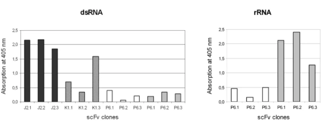
Although we observed quantitative differences in activity of periplasmic extracts, both J2 and K1 scFvs retained the original specificity of the monoclonal antibody, i.e.
they bound exclusively to dsRNA (Fig. 1). The original P6 antibody reacted with structured ssRNA as well as with dsRNA. The selected clones of bacterially expressed P6-scFvs exhibited high ssRNA-binding activity, however, their affinity to dsRNA was much lower than that of mAb P6 (Fig. 1). Sequence analysis of the P6-scFv clones has shown, that the selected clones incorporated some mutations in their LCDR1 (180Ser→180Asn) and LCDR3 (244Cys-His245→244Trp-Ser245 and
249Phe→249Leu) and the modified binding site may influence the fine specificity of the scFv. No such mutations were observed in the J2- and K1-scFv clones.
Targeting scFvs to the cytoplasm and to the plasma membrane of N. tabacum cv. Xanthi
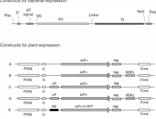
Stability of heterologously expressed proteins depends on their intrinsic properties and the primary sequence. Since it is not possible to predict beforehand which scFv- sequence will allow efficient folding and stability in the cytoplasm, we transformed plants with two antibody fragments: scFv J2 and P6. They were chosen because they differ in specificity as well as in their light chain variable domains. The constructs used to target scFvs to different subcellular compartments are shown in Fig. 2. To achieve cytoplasmic localisation scFvs were expressed without N-terminal signal sequences. We also made constructs with an added C-terminal KDEL tetrapeptide which was shown to enhance scFv stability in the cytoplasm in some cases [22].
Anchoring proteins in the membrane may also help to stabilise and to concentrate proteins in certain membranes. This is why myristoylation and palmitoylation signals were used to anchor scFv J2 on the cytoplasmic side of the plasma membrane [17].
To prove that the signal sequences used by us do indeed direct anchoring to the plas-
Fig. 1. Antigen binding activity of bacterially expressed J2-, K1- and P6-scFvs in ELISA. scFv from periplasmic extracts of different transformed colonies were immobilised by anti-E-tag monoclonal anti- bodies on ELISA plates. Biotinylated dsRNA or ribosomal RNA at 15 ng/well was used as antigen. The figure shows that J2- and K1-scFvs retained their dsRNA-binding activity, while P6-scFvs have a much
higher affinity to rRNA than to dsRNA
ma membrane, GFP (green fluorescent protein) constructs containing the same sig- nals were also used for transformation.
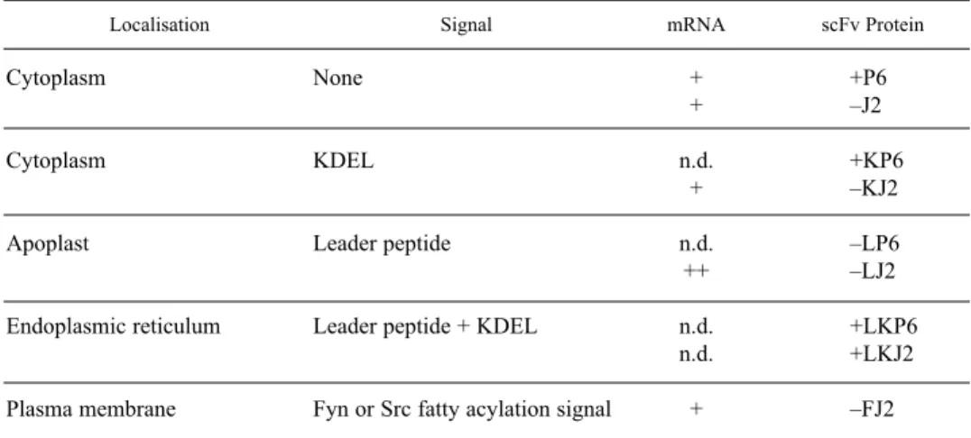
Transgenic plants were obtained after transformation with all three types of con- structs and the synthesis of mRNA was also detected (Table 1). However, only P6- scFv – with or without KDEL added – could be stably expressed in the cytoplasm (Fig. 3, Table 1), while in case of J2 no scFv proteins could be detected (Table 1).
Even in the case of cytoplasmically expressed P6-scFvs it was obvious that the
Fig. 2.Constructs made for scFv-expression in E. coli and in N. tabacumcv. Xanthi. The construct for bacterial expression was made in pCANTAB 5E phagemid vector and contained Plac, lactose promoter;
O, operator; g3 signal, an element of phage protein 3 to secret scFv in periplasma; SfiI and NotI, restric- tion sites to re-clone scFv; VHand VL, variable domain of heavy and light chain, respectively; E-tag, immunological tag for detection of expressed protein. Antibody gene constructs for expression in high- er plants are shown in the lower part of the figure. The basic (A) and KDEL protected constructs (B) were intended for expression in the cytosol. Type C constructs were designed for scFv secretion to the apoplast, type D for retention in the ER and type E for plasma membrane targeting. To investigate the effect of PM-targeting signals independent of scFv-expression, in a second variant of the type E construct the scFv sequence was substituted with that of green fluorescent protein. P35S, 35S promoter of Cauliflower Mosaic Virus;U, 5’ untranslated omega-leader of Tobacco Mosaic Virus; LP, leader peptide of mouse IgG J2 heavy-chain; SS, signal sequence for fatty acylation; scFv, coding sequence of single- chain antibody fragment, GFP, or green fluorescent protein (GFP); tag, c-myc tag; 3’ocs, 3’ end of the
octopine synthase gene; KDEL, carboxy-terminal ER-retention signal
reducing cytoplasmic environment had a negative influence on RNA-binding activi- ty. While the protein concentration was comparable to the ER-resident P6-scFv, the activity of scFvs remained low (see next chapter and Fig. 4). The result indicates that although P6-scFvs adopt a stable structure in the cytoplasm, this structure differs somehow from the native structure of the original P6 antibody.
Targeting proteins to the plasma membrane in BY-2 cells was successful in case of the GFP-constructs and fluorescent signals were clearly observed at the PM. In addi- tion, after cell fractionation GFP protein was found to be associated with the mem- brane fraction (results not shown). Although the signal sequences should have the same effect on scFv, no J2-scFvs were detected in transformants by Western blotting or on dot blots with or without cell fractionation. We presume that because of the inef- ficient folding of J2-scFv the protein became degraded and could not reach detection levels even in the membrane fraction. Taken together the results show that only the P6 antibody provides a scFv sequence, which can be stably folded in the cytoplasm.
Attaching the stabilising KDEL sequence at the C-terminus or targeting the scFv sequence to the PM did not lead to accumulation of detectable amounts of J2-scFv.
Table 1
Summary of single-chain antibody fragment expression of J2 and P6 antibodies in transgenic N. tabacum cv. Xanthi plants. n.d. stands for not determined, K for the presence of KDEL sequence,
L for mouse leader peptide and F for fatty acylation signals
Localisation Signal mRNA scFv Protein
Cytoplasm None + +P6
+ –J2
Cytoplasm KDEL n.d. +KP6
+ –KJ2
Apoplast Leader peptide n.d. –LP6
++ –LJ2
Endoplasmic reticulum Leader peptide + KDEL n.d. +LKP6
n.d. +LKJ2
Plasma membrane Fyn or Src fatty acylation signal + –FJ2
Fig. 3. Western-blot analysis of scFv-expression in transgenic plants expressing P6-scFv in the cyto- plasm or in the ER and J2-scFv in the ER. In all cases shown scFv-protein was clearly detected with
anti-c-myc monoclonal antibody. Non-transgenic tobacco was used as negative control
Targeting scFv to the endoplasmic reticulum (ER)
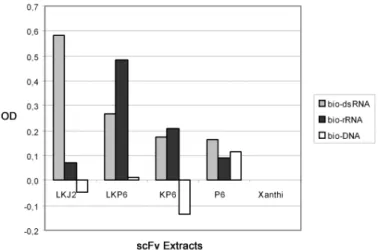
J2- as well as P6-scFvs were targeted to the endoplasmic reticulum (ER) with an N- terminal mouse leader peptide and made ER-resident by adding the ER-retention sig- nal KDEL to the C-terminal end. In this case, very high levels of protein expression were observed for both scFvs (Fig. 3) and both antibodies were found to retain large- ly their original antigen specificity: J2-scFvs have shown the expected dsRNA-speci- ficity, while P6-scFvs reacted with dsRNA as well as with ribosomal RNA (Fig. 4).
The reaction pattern of plant expressed, ER-resident P6 differs from the antigen specificity of bacterially expressed P6-scFv (cf. Fig. 4 and Fig. 1). Since the anti- body-derived sequences are identical in both cases, it may be concluded that the mutated amino acids in LCDR1 and -3 play no major role in determining the fine specificity. The altered fine specificity of the bacterially expressed P6-scFv may either be due to the influence of the attached E-tag sequence or to subtle differences of the folding environment between bacterial periplasma and plant ER.
The results show that the mouse leader sequence efficiently directs scFv into the ER, where it gets properly folded. When however, to achieve secretion into the apoplast we removed the ER-retention signal from scFv, no J2- or P6-scFv proteins could be detected. This finding indicates that despite their correct folding before
Fig. 4.Comparison of antigen binding activity and specificity of J2- and P6-scFvs expressed in tobacco.
scFvs from freshly made plant extracts were diluted 10-fold and immobilised via c-myc specific anti- bodies. 20 ng/well biotinylated dsRNA, rRNA or dsDNA was used as antigen and antigen binding was detected by using alkaline phosphatase conjugated streptavidin. ELISA readings measured 60 min after substrate incubation were corrected with the negative controls (non-transgenic Xanthi). ER-resident J2- and P6-scFv (LKJ2 and LKP6, respectively) both bound to the dsRNA antigen and LKP6 also to rRNA.
Antigen binding of P6 expressed with (KP6) or without (P6) the KDEL-signal in the cytosol was signif- icantly lower than that of the ER-resident scFv. Binding to dsDNA was at background levels in all cases
secretion inside the ER, J2- and P6-scFvs are broken down in the apoplast. In earli- er experiments by our group the J2 antibody was expressed at high concentration as intact IgG molecule in the apoplast [14, 19]. Thus, for targeting to the apoplast full- length antibodies could be the molecule of choice.
Effect of dsRNA-specific antibodies on PVY infection in vivo
scFvs expressed in different compartments of tobacco cells accumulated and folded with different efficiency. To investigate whether they are biologically active at their location in the plant in vivo, we analysed their effect on virus infection. Double- stranded RNA, the antigen recognised by our J2 antibody, commonly occurs in plants infected by RNA viruses. During replication of positive-stranded RNA viruses dsRNAs arise by base pairing between the template and the newly synthesising RNA strand. With respect to virus multiplication viral dsRNAs play different roles: Their formation is essential for virus replication, but to initiate a new replication cycle dsRNAs have to dissociate. In earlier experiments we found that the J2 antibody is able to inhibit virus replication on partially double-stranded template in vitro [4], probably by stabilising the dsRNA and thus counteracting viral helicase activity. On the other hand, viral dsRNAs are the targets of dsRNA-mediated gene silencing, which is initiated by fragmenting the viral dsRNA by host enzymes [3]. dsRNA-spe- cific antibodies may influence this reaction as well, but in this case antibody binding to the dsRNA will probably protect the antigen against the activity of host dicer enzyme.
To find out whether J2-scFv expressed in transgenic tobacco has an effect on virus multiplication, the Hungarian isolate of NTN strain of Potato Virus Ywas used to mechanically inoculate plants and to evaluate, whether there was a difference in the speed of symptom development between transgenic and non-transgenic control plants. PVY is a relatively slow virus, which induces well-defined symptoms and which is known to be subject of dsRNA-mediated gene silencing. We found no agro- nomically relevant protection against PVY infection in our transgenic genotypes, but observed that expression of dsRNA-specific antibodies may alter the virus level, virus distribution and symptoms. Virus concentration was measured by coat protein- specific ELISA using extracts from the third leaf (7 d p.i.) or from the third to sixth leaves (21 d p.i.) above the infected leaf as antigen source. The third leaf was chosen because the first symptoms usually appeared on this leaf. 7 days after infection the PVY coat protein (CP) concentration was found to be lower in We found that plants accumulating ER-targeted J2-scFv or secreted J2-IgG than in non-transgenic Xanthi or in plants expressing P6-scFv in the cytoplasm (Table 2A). By Tukey’s pair wise comparison LKJ2 and HLM differed from Xanthi at p = 0.008 and 0.002, respec- tively.
21 days after infection CP-concentrations were equally high in leaves 5 and 6 of transgenic lines as in non-transgenic Xanthi, while in the third, and some cases also in the fourth leaf large differences between individual plants and between genotypes
were observed (Table 2B). The highest coat protein concentration was detected in LKJ2 plants, while the average concentration in Xanthi and LKP6 plants was much lower. Since the variance within the two latter groups was high, the difference between genotypes is not significant at p < 0.05 level. We feel it is relevant to note, that the physiological state of leaves 3 and 4 was not identical (see below and in Fig.
5 and 6), and this might have influenced the ELISA results.
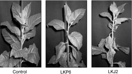
At this late stage of infection (21 days) plants expressing ER-resident J2-scFv were more susceptible to symptom development developed more pronounced symp- toms than the non-transgenic control or the other scFv-expressing genotypes. In LKJ2-plants plants expressing ER-resident J2-scFv symptoms appeared earlier, and- moreover, leaves with viral symptoms towards the plant apex wilted, became necrot- ic and died faster and in larger number than those in other genotypes (Fig. 5). To evaluate the necrotic symptomsdata statistically quantitatively in two series of exper-
Table 2
PVY concentration in transgenic genotypes measured by coat protein specific ELISA A) Samples were taken from the third leaf above the infected leaf 7 days p.i. In each group seven
plants and two parallel samples per plant were analysed A405 nm
Difference from Genotype
non-transgenic Xanthi
average SD
HLM 0.375 0.287 0.432
LKJ2 0.432 0.199 0.378
LKP6 0.652 0.407 0.158
P6 0.685 0.301 0.124
Xanthi 0.810 0.196 0.000
Variance (MQ) calculated by one-way ANOVA was 0.460 between groups and 0.083 within groups, i.e. the variance between different transgenic plants significantly larger (p = 0.0007) than the variance of experimental error. The homogeneity of variance calculated by Levine’s test also showed that at 1%
probability level the groups (genotypes) may be regarded as homogenous.
B) Comparison of CP-concentration in different leaves 21 days p.i.
LKJ2 LKP6 Xanthi
Leaf
A405 nm SD A405 nm SD A405 nm SD
3. 1.079 0.324 0.184 0.412 0.538 0.591
4. 1.176 0.385 0.340 0.525 1.200 0.144
5. 1.162 0.209 0.980 0.469 1.402 0.104
6. 1.013 0.257 1.089 0.232 1.202 0.105
Samples were taken from leaves 3 to 7 above the infected leaf. Because of the inhomogeneity of vari- ances the differences between genotypes can not be regarded as significant.
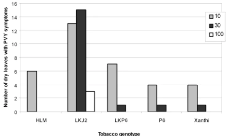
iments we infected 5 or 7 plants of each genotype at 10-, 30- and 100-fold dilutions of the PVY stock and counted the hanging dry leaves 21 days p.i. As shown in Fig.
6 at all three virus dilutions used the number of necrotic leaves was highest in those plants, which expressed J2-scFv in the endoplasmic reticulum. The same effect was not observed in plants expressing ER-resident P6 (see LKP6 in Fig. 5 and 6). It should be emphasised that although P6 binds to dsRNA, it reacts with single-strand- ed RNA (ssRNA) as well, and ssRNA species present at high concentrations in the cell may out compete the viral dsRNAs for binding to the P6-scFv.
The results described above may indicate that although J2-scFv does not inhibit virus replication confer virus resistance, it may influence the time course and the symptoms of virus multiplication. Further experiments are needed to clarify the bio- logical causes of this effect. At present we suspect that bound J2-scFv may protect viral dsRNA and, as a result, interfere interferes with dsRNA-mediated gene silenc- ing by protecting viral dsRNA. The question arises how an ER-resident protein can influence a process taking place outside the ER in the cytosol. Potyviruses are known to replicate in association with large vesicular structures derived from the ER. For tobacco etch virus (TEV) it was shown that on infection with TEV the ER-network collapses into aggregated structures and this process probably begins with the bind- ing of the 6 kDa virus protein to the ER-membrane [10]. We believe that when PVY is replicating in association with the ER, the chances are relatively high that the high- ly concentrated scFv comes in contact with the replication complex. It is also possi- ble, that scFvs leak out the ER into the cytosol. Whichever explanation is correct, our results demonstrate that not only hapten- or protein-specific antibodies but also
Fig. 5.Comparison of PVY symptoms in non-transgenic tobaccos (control) to symptoms in transgenic LKP6 and LKJ2 on the 22nd day post inoculation. LKP6 and LKJ2 express ER-resident P6- and
J2-scFVs, respectively. The dying necrotic leaves on the LKJ2 plant can be clearly seen
nucleic acid specific scFv may efficiently influence in vivo equilibria. Ongoing experiments indicate that they may also induce characteristic morphogenetic changes in non-infected plants.
ACKNOWLEDGEMENTS
The project was supported by OTKA grant T032861. We are indebted to Geert De Jaeger for providing us the plant expression vector pGEJAE1 and the Agrobacterium-mediated plants transformation system and to Ervin Balázs, Zoltán Divéky, László Palkovics for help with virological studies. We thank Péter Kós, István Likó, Gábor Rigó, László Bakó, Taras Pasternak and Ferhan Ayadin for discussions. Special gratitude goes to Erzsébet Bárdosi, Gabriella Fleit and Ildikó Pázmándi for excellent technical support.
REFERENCES
1. Alexin, T. (2001) Comparative sequence analysis of antigen binding sites of RNA-binding antibod- ies. Diploma Thesis, University of Szeged (in Hungarian)
2. An, G. H. (1985) High-efficiency transformation of cultured tobacco cells. Plant Physiol. 79, 568–570.
3. Baulcomb, D. (2004) RNA silencing in plants. Nature 431, 356–363.
4. De Graaf, M., Houwing, C. J., Lukács, N., Jaspars, E. M. J. (1995) RNA duplex unwinding activity of alfalfa mosaic virus RNA-dependent RNA polymerase.FEBS Lett. 371,219–222.
5. De Jaeger, G., Buys, E., Eeckhout, D., De Wilde, C., Jacobs, A., Kapila, J., Angenon, G., Van Montagu, M., Gerats, T., Depicker, A. (1999) High level accumulation of single-chain variable frag- ments in the cytosol of transgenic Petunia hybrida. Eur. J. Biochem. 259, 426–434.
6. De Jaeger, G., De Wilde, C., Eeckhout, D., Fiers, E., Depicker, A. (2000) The plantibody approach:
expression of antibody genes in plants to modulate plant metabolism or to obtain pathogen resistance.
Plant Mol. Biol. 43, 419–428.
Fig. 6.Total number of dry hanging infected leaves 21 days after mechanical inoculation with 10-, 30- or 100-fold diluted PVY inoculum. The number of necrotic leaves in plants expressing ER-resident J2- scFv is significantly higher at all virus dilutions. HLM-plants express native J2-IgG in the apoplast.
Xanthi is the non-transgenic control
7. Deblaere, R., Bytebier, B., De Greve, H., Deboeck, F., Schell, J., Van Montagu, M., Leemans, J.
(1985) Efficient octopine Ti plasmid-derived vectors for Agrobacterium-mediated gene transfer to plants. Nucleic Acids Res. 13, 4777–4788.
8. Eto, J., Suzuki, Y., Ohkawa, H., Yamaguchi, I. (2003) Anti-herbicide single-chain antibody expres- sion confers herbicide tolerance in transgenic plants.FEBS Lett. 27570,1–6.
9. Francisco, J. A., Gawlak, S. L., Miller, M., Bathe, J., Russell, D., Chace, D., Mixan, B., Zhao, L., Fell, H. P., Siegall, C. B. (1997) Expression and characterization of bryodin 1 and a bryodin 1-based single-chain immunotoxin from tobacco cell culture. Bioconjug. Chemistry 8, 708–713.
10. Hull R. (2002) Matthew’s Plant Virology.Fourth edition. Academic Press, London, pp. 293–372.
11. Jobling, S. A., Jarman, C., Teh, M. M., Holmberg, N., Blake, C., Verhoeyen, M. E. (2003) Immunomodulation of enzyme function in plants by single-domain antibody fragments. Nat.
Biotechnol. 21,77–80.
12. Kós, P. B., Oberstrass, J., Richter, A., Likó, I., Morgun, B. V., Lukács, N. (1999) Analysis of bind- ing sites of dsRNA-specific monoclonal antibodies. In: Bajusz, S., Hudecz, F. (ed.) Peptides, Akadémiai Kiadó, Budapest, pp. 600–601.
13. Lukács, N. (1993) Analysis of epitopes recognized by anti-viroid monoclonal antibodies. Biol. Chem.
374, 775.
14. Lukács, N., Richter, A. (1994) Antikörpersynthese in transgenen Pflanzen. Bioscope 4, 29–35.
15. Ma, J. K. C., Drake, P. M. W., Christou, P. (2003) The production of recombinant pharmaceutical pro- teins in plants. Nat. Rev. Genet. 4, 794–805.
16. Ma, J. K. C., Hikmat, B. Y., Wycoff, K., Vine, N. D., Chargelegue, D. M., Yu, L., Hein, M. B., Lehner, T. (1998) Characterization of a recombinant plant monoclonal secretory antibody and pre- ventive immunotherapy in humans. Nat. Med. 4,601–606.
17. McCabe, J. B., Berthiaume, L. G. (1999) Functional roles for fatty acylated amino-terminal domains in subcellular localization. Mol. Biol. Cell 10, 3771–3786.
18.Recombinant Phage Antibody System(RPAS).
http://www.amershambiosciences.com/aptrix/upp01077.nsf/Content/Products?OpenDocument&
ParentId=38835&zone=Proteomics
19. Richter A. (1994) Expression von Doppelstrang-RNA-spezifischen Antikörpern in Pflanzen zur Untersuchung von Replikationsprozessen bei RNA-Niren in vivo. PhD thesis, Universíty of Düsseldorf.
20. Sambrook, J., Fritsch, E. F., Maniatis, T. (1989) Molecular cloning: a laboratory manual. Cold Spring Harbor Press, New York.
21. Schönborn, J., Oberstrass, J., Breyel, E., Tittgen, J., Schumacher, J., Lukács, N. (1991) Monoclonal antibodies to double-stranded RNA as probes of RNA structure in crude nucleic acid extracts.
Nucleic Acids Res. 19,2993–3000.
22. Schouten, A., Roosien, J., van Engelen, F. A., de Jong, G. A., Borst-Vrenssen, A. W., Zilverentant, J.
F., Bosch, D., Stiekema, W. J., Gommers, F. J., Schots, A., Bakker, J. (1996) The C-terminal KDEL sequence increases the expression level of a single-chain antibody designed to be targeted to both the cytosol and the secretory pathway in transgenic tobacco.Plant Mol. Biol. 30,781–793.
23. Strauss, M., Kauder, F., Peisker, M., Sonnewald, U., Conrad, U., Heineke, D. (2001) Expression of an abscisic acid-binding single-chain antibody influences the subcellular distribution of abscisic acid and leads to developmental changes in transgenic potato plants.Planta 213, 361–369.
24. Winter, G., Griffiths, A. D., Hawkins, R. E., Hoogenboom, H. R. (1994) Making antibodies by phage display technology. Annu. Rev. Immunol. 12,433–455.
25. Zeitlin, L., Olmsted, S. S., Moench, T. R., Co, M. S., Martinell, B. J., Paradkar, V. M., Russell, D. R., Queen, C., Cone, R. A., Whaley, K. J. (1998) A humanized monoclonal antibody produced in trans- genic plants for immunoprotection of the vagina against genital herpes. Nat. Biotechnol. 16, 1361–1364.
26. Zimmermann, S., Schillberg, S., Liao, Y. C., Fisher, R. (1998) Intracellular expression of TMV-spe- cific single-chain Fv fragments leads to improved virus resistance in Nicotiana tabacum. Mol.
Breeding 4,369–379.