Evaluation of grading systems in stage I lung adenocarcinomas: a retrospective cohort study
Tamás Zombori,
1József Furák,
2Tibor Nyári,
3Gábor Cserni,
1,4László Tiszlavicz
1To cite: Zombori T, Furák J, Nyári T, et al. J Clin Pathol 2018;71:135–140.
1Department of Pathology, University of Szeged, Szeged, Hungary
2Department of Surgery, University of Szeged, Szeged, Hungary
3Department of Medical Physics and Informatics, University of Szeged, Szeged, Hungary
4Department of Pathology, Bács-Kiskun County Teaching Hospital, Szeged, Hungary Correspondence to Dr Tamás Zombori, Department of Pathology, University of Szeged, Faculty of Medicine Állomás, u. 1. H6725 Szeged, Hungary; zomtam@ gmail. com Received 22 December 2016 Revised 23 May 2017 Accepted 12 June 2017 Published Online First 26 July 2017
AbsTrACT
Aims There is no internationally accepted grading system for lung adenocarcinoma despite the new WHO classification. The architectural grade, the Kadota grade and the Sica score were evaluated and compared with overall (OS) and disease-free survival (DFS).
Methods Comprehensive histological subtyping was used in a series of resected stage I lung adenocarcinoma to identify subtypes of adenocarcinomas, the
architectural grade, the Kadota grade, the Sica grade, the mitotic count, nuclear atypia, the presence of lymphovascular, vascular and airway propagation, necrosis, and micropapillary or solid growth pattern in any percentage. Statistical models fitted included Kaplan- Meier estimates and Cox proportional hazard regression models.
results 261 stage I adenocarcinomas were included.
The 5-year survivals of different subtypes were as follows: lepidic (n=40, OS: 92.5%; DFS 91.6%), acinar (n=54, OS: 81.8%; DFS: 68.6%), papillary (n=49, OS:
73.6%; DFS: 61.0%), solid (n=95, OS: 64.7%; DFS:
57.8%) and micropapillary (n=23, OS: 34.8%; DFS:
33.5%). Concerning the architectural grade, there were significant differences between OS and DFS of low and intermediate (pOS=0.005, pDFS<0.001), low and high (pOS<0.001, pDFS<0.001) and intermediate and high grades (pOS=0.002, pDFS<0.001). Low- grade and intermediate grade tumours did not differ in survival according to Kadota grade and Sica grade. In the multivariable model, architectural grade was found to be an independent prognostic marker. In another model, architectural pattern proved to be superior to architectural grade.
Conclusions Of the three grading systems compared, the architectural grade makes the best distinction between the outcome of low-grade, intermediate-grade and high-grade stage I adenocarcinomas.
bACkgrOund
The WHO introduced a new classification of lung cancers in 2015.1 The new classification follows the suggestions of the International Multidisciplinary Lung Adenocarcinoma Classification proposed by the International Association for the Study of Lung Cancer (IASLC), American Thoracic Society (ATS) and European Respiratory Society (ERS).
The non-invasive growth pattern previously called bronchioloalveolar carcinoma is now called lepidic pattern.2 In situ adenocarcinoma (AIS) and mini- mally invasive adenocarcinoma (MIA) were intro- duced. The former has 100%3–6 and the latter has nearly 100% overall survival (OS).7–9 The subtypes of invasive adenocarcinoma are based on the
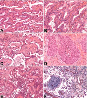
predominant growth pattern, and include lepidic, acinar, papillary, solid and micropapillary carci- nomas (figure 1). Lepidic carcinomas have been correlated with better prognosis,10–12 whereas solid and micropapillary carcinomas are at the other end of the spectrum and have an unfavourable prognosis.13–15
Although grading is an essential part of histo- pathology reports in almost all carcinomas, there is no internationally accepted grading system for lung adenocarcinomas. Architectural grade, which is the most frequently referred one, is based on the predominant growth pattern of invasive carcinoma.
It contains three grades: low (lepidic), intermediate (acinar and papillary) and high grade (solid and micropapillary).16 Despite the fact that most lung adenocarcinomas have mixed growth patterns, the architectural grade of these tumours refers to the predominant growth pattern only, in keeping with the IASLC/ATS/ERS and WHO classifications. A lepidic carcinoma (named after its predominant pattern) may have solid or micropapillary parts too, and this combination could result in a more aggres- sive biological behaviour.
The Sica grade is similar to the Gleason score of prostatic adenocarcinoma. The advantage of this system is that it takes both the predominant and the second most common growth patterns into account. Both are scored and the sum of these points refer to the clinical behaviour.17 18 The orig- inal grading system does not exactly match the new IASLC/ATS/ERS classification because of the use of the term bronchioloalveolar carcinoma and its mucinous variant.18 Therefore, we used the modi- fied scores19 based on the new classification, and these are referred to as Sica grade throughout this work. Lepidic pattern gets 1, acinar and papillary 2, and solid and micropapillary 3 points. AIS, with its 2 points, means the low-grade group. Carcinomas with 3–4 points make up the intermediate-grade and those with 5–6 points form the high grade group.
Kadota and coworkers20 suggested a grading system that combines the predominant growth pattern and the mitotic count in 10 high-power fields (HPF). In this system low grade includes lepidic invasive carcinoma along with acinar and papillary carcinomas with low mitotic counts (0–1 mitosis/10 HPFs).
Our aim was to evaluate and compare the prog- nostic value of architectural grade, the Sica grade and the Kadota grade, and look for the one that may best reflect prognosis in early-stage adenocar- cinomas where adjuvant systemic therapy is gener- ally not given to patients having clear margins.21
on 22 June 2018 by guest. Protected by copyright.http://jcp.bmj.com/J Clin Pathol: first published as 10.1136/jclinpath-2016-204302 on 26 July 2017. Downloaded from
MeThOds
Patients with a diagnosis of primary pulmonary adenocarcinoma in stage IA or IB according to the 7th Edition of Tumour, Node, Metastasis Classification1 who were operated on at the Divi- sion of Thoracic Surgery, Department of Surgery, University of Szeged, between 2004 and 2013 were selected for the cohort to be analysed. Accordingly, these tumours were not larger than 5 cm, localised only in the lung, had no pleural involvement and lymph node, or distant metastasis. Cases with multicentric, metachronous or metastatic disease, positive surgical margin, lung cancer surgery in the preceding 2 years, preoperative chemotherapy, unavailable H&E-stained slides, perioperative death (within 30 days following surgery) and vascular inva- sion were excluded. Clinical information concerning gender, age, smoking habits, tumour localisation, type of surgery and follow-up data was collected from medical charts. The follow-up of patients consisted of three monthly chest X-ray examina- tions in the first 2 years, then six monthly until the fifth year.
Chest CT was performed every 6 months for the first 2 years.
In case of any progression, chest CT and abdominal ultraso- nography were included. The follow-up period ended on 31 August 2015.
Surgically resected specimens had originally been fixed in neutral buffered formalin for at least 24 hours. As a routine, 3–4 μm thick sections made from the formalin-fixed and paraffin-embedded tissue blocks were stained with H&E. As a special stain, orcein was used for highlighting elastic fibres17 22 to assess intact stromal architecture in lepidic growth pattern.
All tumour-containing slides were reviewed by two patholo- gists (TZ and LT) who were blinded to patients’ outcomes.
The number of slides per case depended on tumour size as one
section had been generally taken for each centimetre of largest tumour dimension. Any discrepancies between the evaluation of the two pathologists were resolved via consensus using a multiheaded microscope. The histological identification of lung adenocarcinoma was based on the current WHO classi- fication.1 23 The most frequent and the second most frequent pattern components were both defined. The subtype of adeno- carcinoma was determined by the growth pattern with the highest percentage. Tumours were grouped by architectural grade as of low (lepidic), intermediate (acinar and papillary) or high (solid, micropapillary) grade. The Sica scores were calculated by evaluation of the predominant and the second most common growth pattern. The Kadota grade was derived from the observation of the predominant growth pattern and the mitotic count in 10 HPFs. For each case, 30 HPFs were counted for mitotic activity using an Olympus BX43 (Olympus, Tokyo, Japan) microscope at 400× magnification (objective 40×, field area=0.237 mm2), and the average rounded number of mitoses per 10 HPFs was recorded. The Kadota and Sica grading systems include AIS and MIA, but the architectural grade refers only to the invasive carcinomas and not the prein- vasive or minimally invasive ones. In order to overcome this discrepancy and make the comparison of the grading systems limited to the same set of early-stage tumours, AIS and MIA were excluded from the study. As among the Sica grades, the low grade is composed exclusively of cases we excluded; we used the scores rather than the grades for verifying differences in prognosis.
Besides the grading systems, the following histological factors were re-evaluated: nuclear atypia, presence of lymphatic inva- sion, airway spread and necrosis. Nuclear atypia was graded as low (monomorphic nucleus in size and shape) or high (large differences in the diameter of nuclei, multinucleated tumour cells). Lymphatic invasion was deemed present when at least one cluster of tumour cells was seen in an endothelium-lined, preformed lumen without red blood cells. Airway spread was detected if clusters of tumour cells were seen in the peritumorous alveoli, in bronchioli or bronchi. Tumour size was assessed by standard gross measurement.
The presence of a solid or a micropapillary component was always noted, regardless of the predominant growth pattern and its percentage. Variants of adenocarcinoma like invasive muci- nous, mixed invasive mucinous/non-mucinous, colloid, fetal and enteric were excluded from the present study due to the distinctly different morphology and rarity.
OS and disease-free survival (DFS) were assessed using Kaplan-Meier estimates. The patients were followed from the date of surgery until the time of recurrence or tumour-related death. The events considered were therefore manifestations of a recurrence and death of the patient due to lung cancer, respec- tively. Patients alive without recurrence and patients dying from other causes were censored at the time of the last follow-up and death, respectively. The receiver operating characteristic (ROC) curve model was used to identify a cut-off value between low and high mitotic counts. Mann-Whitney U model was applied for seeking difference between the mitotic rate of surviving patients and patients dying of tumour progression. The log-rank test was used for pairwise comparisons. All statistical tests were two-sided, and p<0.05 values were considered statistically significant. The parameters found significant in the univari- able model were entered in multivariable Cox proportional hazard model to identify factors of independent prognostic significance. Statistical models were fitted using SPSS Statistics V.22.0 software.
Figure 1 Architectural patterns of pulmonary adenocarcinoma:
(A) lepidic growth pattern (HE, 20×); (B) acinar carcinoma (HE, 20×);
(C) papillary carcinoma (HE, 20×); (D) solid carcinoma (HE, 20×); (E) micropapillary carcinoma (HE, 20×); and (F) papillary carcinoma with micropapillary features (orcein-haematoxylin stain, 20×).
on 22 June 2018 by guest. Protected by copyright.http://jcp.bmj.com/J Clin Pathol: first published as 10.1136/jclinpath-2016-204302 on 26 July 2017. Downloaded from
This retrospective study was approved by the institutional ethical committee of the Albert Szent-Györgyi Clinical Centre of the University of Szeged.
resulTs
Altogether, 327 patients matching the stage, type and subtype criteria described in the methods were operated on at the Depart- ment of Surgery, University of Szeged, between 2004 and 2013.
There was no clinical information about 35 surviving patients and about 31 deceased patients. In three cases, slides were not available. Patients with these missing data were excluded from the model. Accordingly, 261 resected adenocarcinomas were examined in this retrospective study: 159 cases were of stage IA and 102 of stage IB.
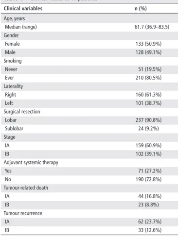
The clinical features of all 261 patients are shown in table 1.
The patients’ median age was 61.7 years (range: 36–83). The gender distribution was close to equal (50.9% vs 49.1%). There were 90.8% (n=237) lobar or greater resections and 9.2%
(n=24) sublobar resections. Lung adenocarcinoma was more frequently right-sided (61.3%; n=160). More than two-thirds of the patients had history of smoking in the clinical anamnesis (80.5%; n=210).
All the available H&E-stained slides were reviewed (median:
3 tumour slides; range: 2–10 tumour slides per patient).
The distribution of subtypes of adenocarcinomas and the results of predominant and second most common growth patterns are summarised in table 2.
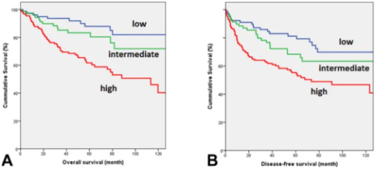
The median follow-up was 52.3 months (range: 1.5–150.3 months). The 5-year OS and DFS of adenocarcinoma subtypes are shown in table 3. We evaluated the architectural grade by using Kaplan-Meier curves (figure 2). There were significant differences between the OS estimates of low-grade (lepidic) and intermediate-grade (acinar and papillary), low-grade and high- grade (solid and micropapillary), and intermediate-grade and high-grade adenocarcinomas. Similarly, there were significant differences in DFS between low-grade and intermediate-grade, low-grade and high-grade, and intermediate-grade and high- grade adenocarcinomas. Among cases with a high architectural grade, a significant difference in both OS and DFS was also detectable between solid and micropapillary carcinomas, the latter being the worst.
According to the fourth edition of the WHO classification of lung tumours, the cribriform pattern is a subgroup of the acinar growth pattern. In our study eight cribriform adenocarcinomas were identified, with equal OS and DFS estimates of 75%.
With the Kadota grade (figure 3), a significant difference in both OS and DFS was seen between low-grade and high-grade, and between intermediate-grade and high-grade adenocarci- nomas. Low-grade and intermediate-grade tumours showed no such difference. Regarding the Sica scores, significant differences were detected in OS and DFS (figure 4): there were significant differences between survival estimates of scores of 3 and 5, 3 and 6, 4 and 5, and 4 and 6, but there was no such difference between scores of 3 and 4 and 5 and 6.
By using the mitotic rate per 10 HPFs, we attempted to identify a group of tumours with a high mitotic rate that would be associ- ated with poor prognosis and a group with a low mitotic rate asso- ciated with better prognosis. We used the ROC curve model for the determination of an optimal cut-off value for mitotic count. Low sensitivity (52.0%–74.0%) and specificity (44.7%–57.5%) values were found regardless of the mitotic cut-off value. The Mann- Whitney U model revealed a significant difference between the Table 1 Clinical features of patients
Clinical variables n (%)
Age, years
Median (range) 61.7 (36.9–83.5)
Gender
Female 133 (50.9%)
Male 128 (49.1%)
Smoking
Never 51 (19.5%)
Ever 210 (80.5%)
Laterality
Right 160 (61.3%)
Left 101 (38.7%)
Surgical resection
Lobar 237 (90.8%)
Sublobar 24 (9.2%)
Stage
IA 159 (60.9%)
IB 102 (39.1%)
Adjuvant systemic therapy
Yes 71 (27.2%)
No 190 (72.8%)
Tumour-related death
IA 44 (16.8%)
IB 23 (8.8%)
Tumour recurrence
IA 62 (23.7%)
IB 33 (12.6%)
Table 2 Distribution of primary and secondary growth patterns of the series Total Predominant growth pattern
n (%) lepidic (%) Acinar (%) Papillary (%) solid (%) Micropapillary (%)
261 40 (15.3) 54 (20.7) 49 (18.7) 95 (36.5) 23 (8.8)
Second most common growth pattern (%)
None 94 (36.2) – 19 (7.2) 14 (5.3) 58 (22.2) 3 (1.1)
Lepidic 25 (9.5) – 5 (1.9) 14 (5.3) 1 (0.3) 5 (1.9)
Acinar 64 (24.5) 21 (8.0) – 13 (5.0) 24 (9.2) 6 (2.2)
Papillary 48 (18.4) 16 (6.1) 19 (7.2) – 5 (1.9) 8 (3.0)
Solid 11 (4.2) 0 (0) 8 (3.0) 2 (0.6) – 1 (0.3)
Micropapillary 19 (7.2) 3 (1.1) 3 (1.1) 6 (2.2) 7 (2.7) –
on 22 June 2018 by guest. Protected by copyright.http://jcp.bmj.com/J Clin Pathol: first published as 10.1136/jclinpath-2016-204302 on 26 July 2017. Downloaded from
mitotic count of surviving patients and patients dying from tumour progression (p=0.042). The median mitotic counts of these two groups of patients were 6 and 8, respectively.
Significant 5-year OS differences were identified (table 3) according to low or high nuclear atypia (p=0.04), mitotic
rate with 5 mitosis/10 HPFs cut-off value (p=0.049) and solid growth pattern in any percentage (p=0.03).
In stage IA, a significant difference was observed between OS of tumours with presence or absence of airway spread (p=0.038) and micropapillary pattern in any percentage (p=0.027).
Airway spread was detected in 98 cases, including eight tumours removed by sublobectomies, and six patients with this feature received adjuvant systemic chemotherapy. Survival was not influ- enced by necrosis.
A multivariable Cox proportional hazard model was applied for OS, including architectural grade, Kadota grade and Sica score.
Architectural grade was found to be the only independent prog- nostic marker (pOS=0.002, HR: 7.21, 95% CI 2.04 to 25.43;
pDFS=0.004, HR: 3.62, 95% CI 1.51 to 8.67). We also explored a Cox model for OS, including architectural grade and subtype.
Regarding OS, subtype was identified as an independent prog- nostic marker (pOS=0.039, HR: 1.9, 95% CI 1.03 to 3.53).
disCussiOn
Although grade is a generally recognised prognostic factor of malignant tumours and grading is a standard component of their pathology reports, there is no internationally accepted, well-studied Table 3 Morphological variables and their relation to overall survival
(OS) and disease-free survival (DFS) Morphological
variables n %
5-year
Os p
5-year
dFs p
Nuclear atypia 0.04 0.022
Low 22 8.4 90.1 90.9
High 239 91.6 71.5 61.4
Mitotic rate (cut-off=5 M/10 high-power field)
0.049 0.158
Low 123 47.1 80.2 69.2
High 138 52.9 68.1 61.7
Necrosis 0.352 0.223
Presence 125 47.8 68.5 60.7
Absence 136 52.2 78.6 69.4
Lymphatic invasion 0.088 0.372
Presence 61 23.3 64.4 60.1
Absence 200 76.7 75.6 67.2
Airway spread* 0.157 0.207
Presence 98 37.5 65.7 59.4
Absence 163 62.5 77.7 68.0
Micropapillary pattern* 0.214 0.408
Presence 90 34.5 68.1 58.7
Absence 171 65.5 76.8 67.8
Solid pattern 0.03 0.178
Presence 128 49.0 69.3 59.3
Absence 133 51.0 78.1 70.2
Histological subtype <0.001 <0.001
Lepidic 40 15,3 92.5 91.6
Acinar 54 20.7 81.8 68.6
Papillary 49 18.7 73.6 61.0
Solid 95 36.5 64.7 57.8
Micropapillary 23 8.8 34.8 33.5
*Significant OS differences were found between tumours with and without airway spread (p=0.038) and micropapillary pattern in any percentage (p=0.027) in stage IA.
Figure 2 (A) Overall survival (OS) and (B) disease-free survival (DFS) estimates of architectural grade: low grade (n=40; OS=92.5%, DFS=95.2%), intermediate grade (n=99; OS=78.5%, DFS=65.3%) and high grade (n=122; OS=60.3%, DFS=54.1%). Significant differences were found between low and intermediate grade (pOS=0.005;
pDFS <0.001), low and high grade (pOS <0.001; pDFS <0.001) and intermediate and high grade (pOS=0.002; pDFS <0.001).
Figure 3 (A) Overall survival (OS) and (B) disease-free survival (DFS) estimates of Kadota grade: low grade (n=75; OS=87.9%, DFS=79.3%), intermediate grade (n=64; OS=80.3%, DFS=65.9%) and high grade (n=122; OS=61.7%, DFS=54.1%). Significant differences were found between low and high grade (pOS<0.001; pDFS<0.001) and intermediate and high grade (pOS=0.009; pDFS=0.031), but not such a difference was observed between low and intermediate grade (pOS<0.187; pDFS<0.104).
Figure 4 (A) Overall survival (OS) and (B) disease-free survival (DFS) estimates of Sica scores: score of 3 (n=61; OS=82.4%, DFS=78.1%), score of 4 (n=66; OS=85.1%, DFS=74.1%), score of 5 (n=62; OS=67.3%, DFS=57.5%) and score of 6 (n=72; OS=59.6%, DFS=51.0%). There were significant differences between survival estimates of scores of 3 and 5 (pOS=0.025; pDFS=0.030), 3 and 6 (pOS=0.002; pDFS=0.002), 4 and 5 (pOS=0.020; pDFS=0.001), and 4 and 6 (pOS=0.002; pDFS=0.008), but there was no such difference between scores of 3 and 4 (pOS=0.923; pDFS=0.734) and 5 and 6 (pOS=0.42; pDFS=0.29).
on 22 June 2018 by guest. Protected by copyright.http://jcp.bmj.com/J Clin Pathol: first published as 10.1136/jclinpath-2016-204302 on 26 July 2017. Downloaded from
grading system for lung adenocarcinomas. The aim of this study was to evaluate and compare the prognostic value of accessible grading systems for stage IA and IB lung adenocarcinomas.
For OS as endpoint, the prognostic variables found significant in the univariable model were the following: subtype of adeno- carcinoma, architectural grade, Kadota grade, Sica grade, nuclear atypia, mitotic count and solid phenotype in any percentage. In cases of stage IA, airway spread and micropapillary pattern in any percentage were found to be significant too (table 3).
As concerns the architectural grade, significant differences were found between the OS and DFS of patients between each grade, both in stage IA and stage IB. In the high-grade group, solid and micropapillary adenocarcinomas also demonstrated significantly different OS and DFS.
Kadota and coworkers launched a grading system combining architectural features and the mitotic count in 10 HPFs20. In this novel grading system, adenocarcinomas with an intermediate architectural grade (acinar or papillary) with a low (<3/10 HPFs) mitotic count are classified as of low grade. The low grade also includes AIS, MIA and lepidic carcinoma, which do not fully coincide with the criteria of invasive adenocarcinoma. In order to maintain comparability between the grades, AIS and MIA were excluded from the study. Consistent with the results of Kadota and coworkers, we found no difference in the survival of low-grade and intermediate-grade groups either in stage IA or stage IB.
A special focus was given to mitotic counts with emphasis on finding a cut-off value distinguishing between tumours with a low mitotic count and a better prognosis versus tumours with a high mitotic count and a worse prognosis. The ROC curve model showed that the specificity and the sensitivity of mitotic count are low, and there is no optimal cut-off value for the mitotic count in 10 HPFs. A high mitotic rate does not necessarily confer bad prognosis. The Mann-Whitney U model however revealed a significant difference in the mitotic count between patients alive at the last follow-up and patients dead of disease. The median mitotic rate of the latter was higher. The literature about the prognostic role of mitotic activity is controversial. Kadota et al found that only mitotic count had a significant association with the risk of recurrence in a multivariable model including nuclear diameter, prominence of nucleoli and mitotic count.
After adjusting for clinicopathological factors including gender, stage, pleural/lymphovascular invasion, necrosis and architec- tural/mitotic grade (ie, the Kadota grade), mitotic count was not an independent predictor of recurrence.19 Duhig et al24 have identified mitotic index as the only independent prognostic marker, which was more closely associated with outcome than either the pathological T stage or the IASLC/ATS/ERS architec- ture-based classification. Consistent with our results, Barletta and coworkers25 could not identify mitotic count as a significant prognostic variable. To assess mitotic activity, Kadota et al as well as Duhig et al had evaluated 50 HPFs, Barletta et al counted between 30 and 50 HPFs, and the present study used 30 HPFs to derive the average number of mitoses in 10 HPFs. Although there was a difference in the number of HPFs assessed in these studies, and this might have contributed to the differences in their conclusions, it is unlikely that this factor has played a major role in the discrepancy of the results.
Sica et al introduced a grading system based on predom- inant and second most common growth pattern. The impor- tance of a secondary growth pattern is clear if there is a lepidic tumour with micropapillary or solid features. Recent studies have revealed that the presence of a micropapillary or a solid pattern in any percentage is associated with poor overall and recurrence-free survival, and this is in keeping with our results
too.13 14 26 27 As the Sica grade also covers AIS and MIA, and the low grade is composed exclusively of cases we excluded for maintaining comparability by restricting the evaluation to invasive adenocarcinomas, we used the scores rather than the grades. In the present study on stage I invasive adenocarci- nomas, a significant difference in OS and DFS was identified between survival estimates of Sica scores of 3 and 5, 3 and 6, 4 and 5, and 4 and 6, but there was no such difference between scores of 3 and 4 as well as 5 and 6, which supports the stage grouping of these scores into intermediate and high grades, respectively. (In a previous analysis, where the low-grade cases including AIS and MIA were also considered (data not shown), there was no significant difference in OS and DFS between low and intermediate Sica grade tumours, such a difference mani- fested only for low-grade vs high-grade, and intermediate-grade vs high-grade tumours.)
By using a multivariable model including architectural grade, Kadota grade and Sica score, the architectural grade was identified as an independent prognostic factor. A multivariable Cox regres- sion model including architectural grade and subtypes of adeno- carcinoma revealed that the subtype is superior to architectural grade. Consistent with our result, Tsuta and coworkers have found that subtyping of adenocarcinoma is a better prognostic factor than histological grouping28. Our findings suggest that the Kadota and the Sica grading systems cannot divide stage I adenocarcinomas into three groups with different outcomes. According to our results, the architectural grade is the one that best describes the prognosis of adenocarcinomas, at least in stage I.
We also pointed out that in the high-grade group, there is a remarkable difference between solid adenocarcinoma with a poor prognosis and micropapillary carcinoma with the worst prognosis both in stages IA and IB. The reason for this could be the fact that solid and especially micropapillary carcinomas show a greater tendency to give lymph node and distant metas- tasis.26 We also found a worsening of the survival data in other subtypes of lung adenocarcinoma whenever solid or micropapil- lary features were present.
Finally, a few limitations of this study should be mentioned. The subjectivity in estimating the predominant pattern of pulmonary adenocarcinomas could bias the architectural grade itself. With training, this categorisation has been reported to improve.29 Such a subjectivity and less than perfect reproducibility is the hallmark of several grading systems, including the histological grading of breast carcinomas30 or the Gleason grading of prostate cancer,31 both of which are established prognosticators of the relevant malignancies.
Although subjectivity in establishing the architectural grade may diminish its prognostic strength, our data, in keeping with other publications,16–19 also suggest that this prognostic role is neverthe- less present. The architectural grade refers to invasive adenocar- cinoma only, and the Kadota grade and Sica score refer to AIS, MIA and invasive adenocarcinoma together. In order to limit the comparisons to the same set of tumours, and avoid a bias from the influence of stage 0 on grade, we excluded AIS and MIA, which could be considered a further limitation of our work, although we consider this one of its strengths. Regarding nuclear atypia, only the nuclear size and shape uniformity were evaluated in this work.
However Nakazato and coworkers32 proposed a nuclear grading based on measuring nuclear area and dimension.33 The propor- tions of growth patterns can be diagnosed only on resected spec- imens; therefore, only resected specimens were included in this study, and some tumours were therefore even not considered for analysis. This is due to the fact that biopsy specimens are generally too small for an accurate assessment of the predominant pattern and therefore grading.
on 22 June 2018 by guest. Protected by copyright.http://jcp.bmj.com/J Clin Pathol: first published as 10.1136/jclinpath-2016-204302 on 26 July 2017. Downloaded from
In summary, we have evaluated three grading systems in stage I pulmonary adenocarcinomas — the architectural grade, the Kadota grade and the Sica grade — and have compared them for their ability to reflect OS and DFS. To our knowledge, these grading systems have never been compared according to their prognostic value. Tumours with a low or intermediate grade according to the Kadota and Sica grades showed no differ- ence in survival. Despite the supposed accuracy of Kadota and Sica grades, only the architectural grade could separate the outcome of low-grade, intermediate-grade and high-grade inva- sive adenocarcinomas. Tumour differentiation as reflected by their grade is an important prognosticator in many cancers. On the basis of the presented results suggesting the superiority of the architectural grade over the others, we propose its use in routine histopathology reports of lung adenocarcinomas in keeping with the current WHO recommendation.
Take home messages
► Of available grading systems of pulmonary adenocarcinomas, the architectural grade based on the predominant growth pattern is the best for characterising disease outcome in early stage, that is, stage I.
► We propose its use in routine histopathology reports of lung adenocarcinomas in keeping with the current WHO recommendation.
► The two alternative grading systems of Kadota and Sica failed to make a difference between overall and disease- free survival of low-grade and intermediate-grade adenocarcinomas.
handling editor Cheok Soon Lee.
Acknowledgements We gratefully acknowledge the assistance of Tibor Géczi MD, Aurél Ottlakán MD, Balázs Pécsy MD, Dániel Urbán and Réka Némedi in collecting clinical data of the patients.
Contributors Conceptualisation: TZ, GC, LT and JF. Methodology: TZ. Investigation:
TZ and LT. Formal analysis: TZ and TN. Writing — original draft: TZ and GC. Writing
— review and editing: TZ, GC, LT and JF. Resources (data): JF. Supervision: GC and LT.
Revision: TZ, TN and GC. Approval of final version: All authors.
Funding The study was funded by the National Research, Development and Innovation Office grant GINOP-2.3.2-15-2016-00020.
Competing interests None declared.
ethics approval Regional Research Ethics Board.
Provenance and peer review Not commissioned; externally peer reviewed.
© Article author(s) (or their employer(s) unless otherwise stated in the text of the article) 2018. All rights reserved. No commercial use is permitted unless otherwise expressly granted.
RefeRences
1 Travis WD, Brambilla E. B, et al. WHO classification of Tumours of the lung, Pleura, thymus and Heart. Fourth edition. France: LyonInternational Agency for Research on Cancer, 2015:26–50.
2 Travis WD, Brambilla E, Noguchi M, et al. International association for the study of lung Cancer/american thoracic society/european respiratory society international multidisciplinary classification of lung adenocarcinoma. J Thorac Oncol 2011;6:244–85.
3 Sakurai H, Maeshima A, Watanabe S, et al. Grade of stromal invasion in small adenocarcinoma of the lung: histopathological minimal invasion and prognosis. Am J Surg Pathol 2004;28:198–206.
4 Noguchi M, Morikawa A, Kawasaki M, et al. Small adenocarcinoma of the lung.
histologic characteristics and prognosis. Cancer 1995;75:2844–52.
5 Vazquez M, Carter D, Brambilla E, et al. Solitary and multiple resected
adenocarcinomas after CT screening for lung Cancer: histopathologic features and their prognostic implications. Lung Cancer 2009;64:148–54.
6 Koike T, Togashi K, Shirato T, et al. Limited resection for noninvasive bronchioloalveolar carcinoma diagnosed by intraoperative pathologic examination. Ann Thorac Surg 2009;88:1106–11.
7 Borczuk AC, Qian F, Kazeros A, et al. Invasive size is an independent predictor of survival in pulmonary adenocarcinoma. Am J Surg Pathol 2009;33:462–9.
8 Yim J, Zhu LC, Chiriboga L, et al. Histologic features are important prognostic indicators in early stages lung adenocarcinomas. Mod Pathol 2007;20:233–41.
9 Maeshima AM, Tochigi N, Yoshida A, et al. Histological scoring for small lung adenocarcinomas 2 cm or less in diameter: a reliable prognostic Indicator. J Thorac Oncol 2010;5:333–9.
10 Lee HY, Han J, Lee KS, et al. Lung adenocarcinoma as a solitary pulmonary nodule:
prognostic determinants of CT, PET, and histopathologic findings. Lung Cancer 2009;66:379–85.
11 Yokose T, Suzuki K, Nagai K, et al. Favorable and unfavorable morphological prognostic factors in peripheral adenocarcinoma of the lung 3 cm or less in diameter.
Lung Cancer 2000;29:179–88.
12 Lin DM, Ma Y, Zheng S, et al. Prognostic value of bronchioloalveolar carcinoma component in lung adenocarcinoma. Histol Histopathol 2006;21:627–32.
13 Lee MC, Buitrago DH, Kadota K, et al. Recent advances and clinical implications of the micropapillary histological subtype in lung adenocarcinomas. Lung Cancer Manag 2014;3:245–53.
14 Nitadori J, Bograd AJ, Kadota K, et al. Impact of micropapillary histologic subtype in selecting limited resection vs lobectomy for lung adenocarcinoma of 2cm or smaller. J Natl Cancer Inst 2013;105:1212–20.
15 Ujiie H, Kadota K, Chaft JE, et al. Solid predominant histologic subtype in Resected Stage I lung adenocarcinoma is an Independent Predictor of Early, Extrathoracic, Multisite Recurrence and of Poor Postrecurrence Survival. J Clin Oncol 2015;33:2877–84.
16 Yoshizawa A, Motoi N, Riely GJ, et al. Impact of proposed IASLC/ATS/ERS classification of lung adenocarcinoma: prognostic subgroups and implications for further revision of staging based on analysis of 514 stage I cases. Mod Pathol 2011;24:653–64.
17 Zhao ZR, Xi SY, Li W, et al. Prognostic impact of pattern-based grading system by the New IASLC/ATS/ERS classification in Asian patients with stage I lung adenocarcinoma.
Lung Cancer 2015;90:604–9.
18 Sica G, Yoshizawa A, Sima CS, et al. A grading system of lung adenocarcinomas based on histologic pattern is predictive of disease recurrence in stage I tumors. Am J Surg Pathol 2010;34:1155–62.
19 Ambrosini-Spaltro A, Ruiu A, Seebacher C, et al. Impact of the IASLC/ATS/ERS classification in pN0 pulmonary adenocarcinomas: a study with radiological- pathological comparisons and survival analyses. Pathol Res Pract 2014;210:40–6.
20 Kadota K, Suzuki K, Kachala SS, et al. A grading system combining architectural features and mitotic count predicts recurrence in stage I lung adenocarcinoma. Mod Pathol 2012;25:1117–27.
21 National Comprehensive Cancer Network. NCCN clinical practice guidelines in oncology Non-small cell lung carcinoma. 2017;v1 https://www. nccn. org.
22 Henwood A. Current applications of orcein in Histochemistry. A brief review with some new observations concerning influence of dye batch variation and aging of dye solutions on staining. Biotech Histochem 2003;78:303–8.
23 Travis WD, Brambilla E, Nicholson AG, et al. The 2015 World Health Organization classification of lung tumors: impact of genetic, clinical and radiologic advances since the 2004 classification. J Thorac Oncol 2015;10:1243–60.
24 Duhig EE, Dettrick A, Godbolt DB, et al. Mitosis trumps T stage and proposed international association for the study of lung Cancer/american thoracic society/
european respiratory society classification for prognostic value in resected stage 1 lung adenocarcinoma. J Thorac Oncol 2015;10:673–81.
25 Barletta JA, Yeap BY, Chirieac LR. Prognostic significance of grading in lung adenocarcinoma. Cancer 2010;116:659–69.
26 Zhao Y, Wang R, Shen X, et al. Minor Components of Micropapillary and solid subtypes in lung adenocarcinoma are Predictors of Lymph Node Metastasis and Poor Prognosis. Ann Surg Oncol 2016;23:2099–105.
27 Hirano H, Maeda H, Takeuchi Y, et al. Lymphatic invasion of micropapillary Cancer cells is associated with a poor prognosis of pathological stage IA lung adenocarcinomas.
Oncol Lett 2014;8:1107–11.
28 Tsuta K, Kawago M, Inoue E, et al. The utility of the proposed IASLC/ATS/ERS lung adenocarcinoma subtypes for disease prognosis and correlation of driver gene alterations. Lung Cancer 2013;81:371–6.
29 Warth A, Stenzinger A, von Brünneck AC, Cortis J, Fink L, et al. Interobserver variability in the application of the Novel IASLC/ATS/ERS classification for Pulmonary adenocarcinomas. Eur Respir J 2012;40:1221–7.
30 Sloane JP, Amendoeira I, Apostolikas N, et al. Consistency achieved by 23 european pathologists from 12 countries in diagnosing breast disease and reporting prognostic features of carcinomas. european Commission Working Group on breast screening pathology. Virchows Arch 1999;434:3–10.
31 Ozkan TA, Eruyar AT, Cebeci OO, et al. Interobserver variability in Gleason histological grading of prostate Cancer. Scand J Urol 2016;50:420–4.
32 Gu J, Lu C, Guo J, et al. Prognostic significance of the IASLC/ATS/ERS classification in chinese patients-A single institution retrospective study of 292 lung adenocarcinoma.
J Surg Oncol 2013;107:474–80.
33 Nakazato Y, Maeshima AM, Ishikawa Y, et al. Interobserver agreement in the nuclear grading of primary pulmonary adenocarcinoma. J Thorac Oncol 2013;8:736–43.
on 22 June 2018 by guest. Protected by copyright.http://jcp.bmj.com/J Clin Pathol: first published as 10.1136/jclinpath-2016-204302 on 26 July 2017. Downloaded from