PLASMA DISPOSITION OF CEFOPERAZONE AFTER SINGLE INTRAVENOUS AND INTRAMUSCULAR ADMINISTRATIONS
IN CAMELS (CAMELUS DROMEDARIUS)
Mohamed ABOUBAKR1*, Ahmed SOLIMAN2, Kamil UNEY3 and Muammer ELMAS3 1Pharmacology Department, Faculty of Veterinary Medicine, Benha University, 13736 Moshtohor, Toukh, Qalioubeya, Egypt; 2Pharmacology Department, Faculty of Veterinary Medicine, Cairo University, Giza, Egypt; 3Department of Pharmacology and
Toxicology, Faculty of Veterinary Medicine, University of Selcuk, Konya, Turkey (Received 18 March 2018; accepted 25 July 2018)
The plasma disposition of cefoperazone was investigated after intravenous (IV) and intramuscular (IM) administrations of 20 mg/kg as a single dose in six camels (Camelus dromedarius) in a crossover design. Blood plasma samples were analysed by high-performance liquid chromatography (HPLC). After IV admin- istration, elimination half-life (t1/2β), volume of distribution at steady state (Vdss), total body clearance (Cltot) and mean residence time (MRT) of cefoperazone were 1.95 h, 0.38 L/kg, 0.17 L/h/kg and 2.16 h, respectively. After IM administration of cefoperazone, peak plasma concentration (Cmax) was 21.95 μg/mL and it was obtained at (tmax) 1.23 h. Absorption half-life (t1/2ab), elimination half-life and mean absorption time were 0.45 h, 2.84 h and 2.07 h, respectively. The bioavaila- bility of cefoperazone was 89.42%. The lack of local reaction or any other ad- verse effects and the very good bioavailability following IM administration indi- cate that cefoperazone might be a promising alternative treatment for a variety of infectious diseases in camels.
Key words: Cefoperazone, concentrations, bioavailability, camels
Cefoperazone is a safe third-generation cephalosporin with few adverse ef- fects, having broad-spectrum activity against Gram-positive, Gram-negative and anaerobic bacteria. Cefoperazone is very active against most strains of the family Enterobacteriaceae and Pseudomonas aeruginosa (Gupta et al., 2008).
Pharmacokinetic studies of cefoperazone had been conducted in different animals such as sheep (Guerrini et al., 1985), calves (Carli et al., 1986), rabbits (Marino et al., 1987), unweaned calves (Soback and Ziv, 1989), horses (Soraci et al., 1996), dogs (Montesissa et al., 2003), buffalo calves (Goyal et al., 2005), cross-bred calves (Gupta et al., 2008), and goats (Attia et al., 2015).
*Corresponding author; E-mail: mohamed.aboubakr@fvtm.bu.edu.eg;
Phone: 0020-1099874671; Fax: 0020-132463074
No data on the pharmacokinetics of cefoperazone had been reported in camels. This study investigated the plasma disposition profile of cefoperazone after a single IV and IM administration of 20 mg/kg body weight in camels.
Materials and methods
Animals
Six clinically healthy male camels (body weight: 400–500 kg, age: 6–7 years) were used. Camels had not received any drug for at least two months be- fore the study and were fed concentrates and hay, with water provided freely.
The Ethics Committee of the Faculty of Veterinary Medicine, Cairo University, approved the use of camels and study protocols.
Experimental design
A crossover design (3×3) was used in this study; cefoperazone (20 mg/kg) was administered IV (3 camels; right jugular vein) or IM (3 camels; left neck muscles) to each camel as assigned. After a 2-week ‘washout’ period, camels that had been dosed IM with cefoperazone were injected IV, and vice versa.
Cefoperazone was obtained as a sterile injectable powder (Cefobid®, cefopera- zone sodium powder produced by SmithKline Beecham for Pfizer, Egypt) and dissolved in sterile saline immediately prior to administration.
A 10-mL blood sample was collected from each camel by jugular veni- puncture into tubes containing heparin from the left jugular vein at 0 min and at 0.08, 0.16, 0.25, 0.5, 1, 2, 4, 6, 8, 12 and 24 h after administration. Blood sam- ples were centrifuged at 3000 g for 15 min and the plasma was stored at –20 °C until analysis by high-performance liquid chromatography (HPLC).
Analytical method
Plasma cefoperazone concentrations were determined by HPLC according to Haghgoo et al. (1995). An HPLC system (Shimadzu Corporation, Kyoto, Japan) with a reverse-phase C18 analytical column (Whatman®, MA, USA, 4.60 × 100 mm internal diameter, particle size 5 μm) at room temperature was used for separa- tion and reading at 266 nm wavelength using an UV detector (Shimadzu, SPD- 10A UV detector, Shimadzu Corporation). The mobile phase was 30 mM KH2PO4 buffer and methanol (70:30, v/v) at a pH of 5.0. The mobile phase was filtered through 0.45 µm filters and was pumped into the column at a flow rate of 1.0 mL/
min at ambient temperature. Plasma samples (50 µl) were deproteinised by add- ing a solution containing 50 µl of 10% perchloric acid and 50 µl of a mixture of acetonitrile (50%) and methanol (50%), and were mixed in a clean centrifuge tube by shaking on a vortex mixer for 1 min; after centrifugation at 10,000 g at
5 °C for 5 min, the clear supernatant was transferred to an autosampler vial and a 20 μl aliquot was injected into the HPLC system.
The calibration curves were performed between 0.5 and 200 µg/mL using blank camel plasma. The standard curve of cefoperazone was linear with a corre- lation coefficient of 0.99. The retention time of cefoperazone in camel plasma was approximately 9.7 min. LOD was 0.1 μg/mL, LOQ was 0.5 μg/mL, and CV was < 15%. The mean percentage recovery values for cefoperazone in plasma samples spiked at concentrations of 1, 10 and 20 μg/mL were 98.37 ± 4.28%, 97.19 ± 3.53% and 94.06 ± 3.41%, respectively. Intra-assay CV values ranged from 2.62 to 3.08% (n = 5, 3 times). The inter-assay CV varied from 3.49 to 4.73% (n = 5, 3 times, 3 days).
Pharmacokinetic analysis
Plasma cefoperazone concentrations after IV and IM administrations were subjected to analysis using a computerised WinNonlin 6.3 program (Pharsight, Mountain View CA, USA). For IV and IM data, the appropriate pharmacokinetic model was determined by visual examination of individual plasma concentration versus time curves and by application of the Akaike information criterion (Yamaoka et al., 1978) resulting in the following two-compartmental model be- ing chosen for data analysis. Half-lives were calculated using of the following equations:
t1/2ab = ln(2)/kab
t1/2α = ln(2)/α t1/2β = ln(2)/β,
where kab, α, and β are the absorption, distribution and elimination rate constants, respectively. AUC and AUMC were calculated using the method of trapezoids with extrapolation to infinity (Gibaldi and Perrier, 1982).
Absolute bioavailability (F) = AUCIM/AUCIV × 100; mean absorption time (MAT) = MRTIM – MRTIV. For IV data, Vdss was estimated as follows: Vdss = dose × AUMC/AUC2 and CL = dose/AUC.
Statistical analysis
The results obtained were displayed as mean ± SE. The paired t test was used to test for differences between the two administration routes. The statistical software SPSS version 16 (SPSS Inc., Chicago, USA) was used for statistical analysis and values of P < 0.05 were considered significant.
Results
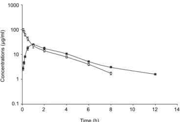
No side effects occurred after either of the two administrations routes in camels. Plasma concentration–time profiles of cefoperazone (20 mg/kg body weight) after single IV and IM administrations are presented in Fig. 1. The val- ues of pharmacokinetic parameters estimated from the curve fitting are shown in Table 1. Following IV administration, t1/2α and t1/2β were 0.18 and 1.95 h, respec- tively. Following IM administration the corresponding pharmacokinetic variables were t1/2ab 0.45 h, Cmax 21.95 µg/mL at tmax 1.23 h and t1/2β 2.84 h, while bioavail- ability was 89.42%.
Fig. 1. Semi-logarithmic graph depicting the time-concentration of cefoperazone in the plasma of camels after a single IV (○) and IM (■) administration of 20 mg/kg body weight (n = 6)
Discussion
Following IV administration, the disposition of cefoperazone followed a two-compartment open model, which is consistent with previous reports on cefoperazone in horses (Soraci et al., 1996) and dogs (Montesissa et al., 2003), and it was similar to the disposition of other cephalosporins in camels such as ceftiofur (Goudah, 2007), ceftriaxone (Goudah, 2008), cefepime (Goudah et al., 2009), and ceftazidime (Goudah and Hasabelnaby, 2013).
Cefoperazone has a short distribution half-life (t1/2α = 0.18 h). Similar find- ings have been recorded in calves (0.15 h; Carli et al., 1986), dogs (Montesissa et al., 2003), longer than cefoperazone in horses (0.07 h; Soraci et al., 1996) and shorter than other cephalosporins in camels such as ceftiofur (0.3 h; Goudah, 2007), ceftriaxone (0.27 h; Goudah, 2008), cefepime (0.3 h; Goudah et al., 2009), and ceftazidime (0.3 h; Goudah and Hasabelnaby, 2013).
Time (h)
0 2 4 6 8 10 12 14 1000
100
10
1
0.1
Concentrations (µg/ml)
Table 1
Plasma pharmacokinetic parameters (mean ± SE) of cefoperazone in camels following IV and IM administration of 20 mg/kg body weight (n = 6)
Parameters Unit IV IM
t1/2ab h – 0.45 ± 0.05
t1/2α h 0.18 ± 0.02 0.54 ± 0.04**
t1/2β h 1.95 ± 0.05 2.84 ± 0.13***
AUC μg mL–1 h–1 116.13 ± 6.31 103.65 ± 5.23***
AUMC μg mL–1 h–2 251.24 ± 12.01 444.49 ± 28.63***
MRT h 2.16 ± 0.03 4.26 ± 0.08***
MAT h – 2.07 ± 0.09
Vdss L kg–1 0.38 ± 0.02 –
Cltot L kg–1 h–1 0.174 ± 0.009 –
Cmax μg mL–1 – 21.95 ± 0.68
tmax h – 1.23 ± 0.04
F % – 89.42 ± 1.74
**P < 0.01;***P < 0.001; –: not calculated; t1/2ab: absorption half-life after IM administration;
t1/2α: distribution half-life; t1/2β: elimination half-life; AUC: area under plasma concentration- time curve; AUMC: area under moment curve; MRT: mean residence time; MAT: mean ab- sorption time; Vdss: volume of distribution at steady state; Cltot: total body clearance; Cmax: maximum plasma concentration; tmax: time to peak plasma concentration; F: bioavailability
The elimination half-life in camels (t1/2β) was 1.95 h. Similar findings were recorded for cefoperazone in dogs (1.40 h; Montesissa et al., 2003), for cefepime in camels (2 h; Goudah et al., 2009), longer than cefoperazone in calves (0.89 h;
Carli et al., 1986), horses (0.77 h; Soraci et al., 1996) and shorter than other cephalosporins in camels such as ceftiofur (3.18 h; Goudah, 2007) and ceftazidime (2.85 h; Goudah and Hasabelnaby, 2013).
The Vdss of cefoperazone was small (0.38 L/kg); this value is close to those reported for ceftriaxone in camels (0.32 L/kg; Goudah, 2008), higher than data reported for cefoperazone in sheep (0.16 L/kg; Guerrini et al., 1985), ceftio- fur and cefepime in camels (0.13 L/kg and 0.1 L/kg, respectively; Goudah, 2007;
Goudah et al., 2009), and lower than cefoperazone in horses (0.68 L/kg; Soraci et al., 1996) and unweaned calves (0.71 L/kg; Soback and Ziv, 1989).
The total body clearance of cefoperazone was 0.17 L/h/kg. Similar find- ings were reported for cefoperazone in sheep (0.11 L/h/kg; Guerrini et al., 1985).
In contrast, a higher clearance of cefoperazone was recorded in horses (0.72 L/
h/kg; Soraci et al., 1996) and a lower clearance was recorded for ceftiofur and cefepime in camels (0.03 and 0.04 L/h/kg, respectively; Goudah, 2007; Goudah et al., 2009), and these differences could be correlated to interspecies differences in drug elimination and metabolism.
The higher value of clearance of cefoperazone in camels suggests that there is negligible tubular reabsorption and cefoperazone is primarily excreted
through glomerular filtration. This finding is in agreement with the results re- ported in humans (Barbhaiya et al., 1990).
Species differences in pharmacokinetic parameters are relatively common and are frequently related to inter-species variation, the length of period between blood samplings, the assay method used, as well as the age and the health status of the animal (Haddad et al., 1985).
After a single IM administration, cefoperazone was rapidly and efficiently absorbed in camels, as the t1/2ab was 0.45 h. This result was reasonably similar to that reported for cefoperazone in dogs (0.48 h; Montesissa et al., 2003) and long- er than that reported for cefoperazone in horses (0.04 h; Soraci et al., 1996) and for ceftiofur and ceftriaxone in camels (0.34 and 0.29 h, respectively; Goudah, 2007, 2008). The t1/2β of cefoperazone in camels was 2.84 h, which is longer than that reported in calves (0.79 h; Carli et al., 1986), horses (1.52 h; Soraci et al., 1996), longer than that reported for ceftriaxone in camels (1.76 h; Goudah, 2008) and shorter than that reported for ceftiofur in camels (3.29 h; Goudah, 2007). The t1/2β after IM administration (2.84 h) was significantly longer than that obtained after IV administration (1.95 h). This difference is probably the result of contin- ued absorption of cefoperazone from the IM injection site during the elimination phase, thereby prolonging the t1/2β of cefoperazone.
The Cmax of cefoperazone after IM administration in camels was 21.95 μg/
mL, very similar to that observed for cefoperazone in dogs (24.5 μg/mL; Mon- tesissa et al., 2003) and higher than that reported for cefoperazone in calves and horses (7.91 and 7.98 μg/mL, respectively; Carli et al., 1986; Soraci et al., 1996), and lower than that reported for ceftazidime in camels (32.43 μg/mL; Goudah and Hasabelnaby, 2013). The absorption process was rapid with a tmax of 1.23 h, which was similar to that of ceftiofur (1.22 h) and ceftazidime (1.21 h) in camels (Goudah, 2007; Goudah and Hasabelnaby, 2013). The MRT was longer after IM administration (4.26 h) than after IV dosing (2.16 h), as the MRT after IM ad- ministration depends on both the disposition and absorption rates.
The IM bioavailability was 89.42%, which is very similar to that of cefepime in camels (91.7%), higher than that of cefoperazone in calves (44.15%;
Carli et al., 1986), horses (42%; Soraci et al., 1996), dogs (41.4%; Montesissa et al., 2003) and lower than that of ceftiofur in camels (97.4%; Goudah, 2007).
The pharmacokinetics of many drugs has been found to be different in camels than in other animals. The species differences might be multifactorial and might depend on the metabolic rate of drugs (Ali et al., 1996).
In conclusion, cefoperazone might be a promising alternative treatment for a variety of infectious diseases in camels due to its favourable pharmacokinetic profile. Further studies on tissue distribution and specific determination of the minimum inhibitory concentration (MIC) of cefoperazone for the major bacteria responsible for diseases in camels should be performed in order to obtain more complete efficacy data of cefoperazone in camels.
References
Ali, B., Oukessou, M. and Bashir, A. (1996): Pharmacokinetic considerations in the camel (Camelus dromedarius): a review. Comp. Biochem. Physiol. C Pharmacol. Toxicol. Endo- crinol. 115, 1–9.
Attia, T., El-Komy, A., El-Hewaity, M., El Latif, A. A. and El-Hanbally, S. (2015): Comparative pharmacokinetics of cefoperazone following intravenous and intramuscular administration in goats. Int. J. Vet. Sci. Med 3, 21–25.
Barbhaiya, R. H., Forgue, S. T., Gleason, C. A., Knupp, C. A., Pittman, K. A., Weidler, H. and Martia, R. R. (1990): Safety, tolerance and pharmacokinetic after multiple intravenous ad- ministrations in healthy subjects. Antimicrob. Agents Chemother. 34, 1118–1122.
Carli, S., Montesissa, C., Sonzogni, O. and Madonna, M. (1986): Pharmacokinetic of sodium cefoperazone in calves. Pharmacol. Res. Commun. 18, 481–490.
Gibaldi, M. and Perrier, D. (1982): Noncompartmental analysis based on statistical moment theory.
In: Swarbrick, J. (ed.) Pharmacokinetics. 2nd edition. Marcel Dekker Inc., New York. pp.
409–417.
Goudah, A. (2007): Pharmacokinetics of ceftiofur after single intravenous and intramuscular ad- ministration in camels (Camelus dromedarius). J. Vet. Pharmacol. Ther. 30, 371–374. Goudah, A. (2008): Pharmacokinetic parameters of ceftriaxone after single intravenous and intra-
muscular administration in camels (Camelus dromedarius). Res. Vet. Sci. 84, 483–489.
Goudah, A. M. and Hasabelnaby, S. M. (2013): Pharmacokinetics and distribution ofceftazidime to milk after intravenous and intramuscular administration to lactating female dromedary camels (Camelus dromedarius). J. Am. Vet. Med. Assoc. 243, 424–429.
Goudah, A., Shin, H. C., Kim, J. S., Chang, B. J., Shim, J. H. and Abd El‐Aty, A. (2009): Evalua- tion of single‐dose pharmacokinetics of cefepime in healthy bull camels (Camelus drome- darius). J. Vet. Pharmacol. Ther. 32, 393–396.
Goyal, S., Chaudhary, R. and Srivastava, A. (2005): Pharmacokinetics following intramuscular admin- istration and dosage regimen for cefoperazone in buffalo calves. Indian J. Anim. Sci. 75, 31–32.
Guerrini, V. H., Filippich, L. J., Cao, G. R., English, P. B. and Bourne, D. W. (1985): Pharmacokinetics of cefaronide, ceftriaxone and cefoperazone in sheep. J. Vet. Pharmacol. Ther. 8, 120–127. Gupta, J. K., Chaudhary, R. K. and Dumka, V. K. (2008): Pharmacokinetics after single intramus-
cular administration and in vitro plasma protein binding of cefoperazone in cross bred calves. Vet. Arhiv 78, 441–448.
Haddad, N. S., Pedersoli, W. M., Ravis, W. R., Fazeli, M. H. and Carson, R. L. (1985): Combined pharmacokinetics of gentamicin in pony mares after a single intravenous and intramuscular administration. Am. J. Vet. Res. 46, 2004–2007.
Haghgoo, S., Hasegawa, T., Nadai, M., Wang, L., Nabeshima, T. and Kato, N. (1995): Effect of a bacterial lipopolysaccharide on biliary excretion of a beta-lactam antibiotic, cefoperazone, in rats. Antimicrob. Agents Chemother. 39, 2258–2261.
Marino, E. L., Fernandez Lastra, C., Gonzalez Alonso, I. and Dominguez-Gil, A. (1987): Disposi- tion and excretion of cefoperazone in rabbits. Arzneimittelforschung 37, 359–349.
Montesissa, C., Villa, R., Anfossi, P., Zanoni, R. and Carli, S. (2003): Pharmacodynamics and pharmacokinetics of cefoperazone and cefamandole in dogs following single dose intrave- nous and intramuscular administration. Vet. J. 166, 170–176.
Soback, S. and Ziv, G. (1989): Pharmacokinetics of single doses of cefoperazone given by the in- travenous and intramuscular routes to unweaned calves. Res. Vet.Sci. 47, 158–163. Soraci, A. L., Mestorino, O. N. and Errecalde, J. O. (1996): Pharmacokinetics of cefoperazone in
horses. J. Vet. Pharmacol. Ther. 19, 39–43.
Yamaoka, K., Nakagawa, T. and Uno, T. (1978): Statistical moments in pharmacokinetics. J.
Pharmacokinet. Biopharm. 6, 547–558.