NERVOUS SYSTEM
Ph.D Thesis
Seyed Farzad Hashemi Dolatabadi
Doctoral School of Pharmaceutical Sciences Semmelweis University
Supervisor: Kornélia Tekes, Pharm.D, Ph.D, D.Sc Official reviewers: Béla Juhász, Ph.D
Borbála Dalmadi Kiss, Ph.D
Head of Final Examination Committee: Éva Lemberkovics, Ph.D
Members of Final Examination Committee: Mahmoud Al-Khrasani, Ph.D István Zupkó, Ph.D
Budapest, 2015
1. List of content ... 2
2. Abbreviations ... 5
3. Introduction ... 8
3.1. Oxytocin as a hormonal imprinter ... 8
3.2. Pharmacology of organophosphate-intoxication ... 9
4. Hormonal imprinting ... 11
4.1. Oxytocin ... 12
4.1.1. Oxytocin structure ... 12
4.1.2. Oxytocin central pathways ... 12
4.1.3. Oxytocin receptor ... 15
4.1.4. Oxytocin effects in the body ... 16
4.2. Relation of hormonal imprinting to catecholamines and serotonin ... 17
4.2.1. Catecholamines ... 17
4.2.2. Serotonin ... 22
5. Acetylcholinesterase inhibiton and reactivation ... 25
5.1. Acetylcholinesterase inhibitors (AChEI) ... 27
5.1.1. Organophosphate type inhibitors ... 28
5.1.2. General structure of organophosphates ... 28
5.1.3. Mechanism of toxicity ... 30
5.1.4. Application of organophosphates ... 33
5.2. Acetylcholinesterase reactivation : Oximes ... 33
5.2.1. Mechanism of acetylcholinesterase reactivation ... 38
5.2.2. Application of acetylcholinesterase reactivators ... 39
5.2.3. K-compounds ... 40
7.1.1. Experimental animals ... 43
7.1.2. Chemicals ... 43
7.1.3. Instrumentation and chromatographic conditions ... 44
7.2. Methodes ... 45
7.2.1. Animals treatment ... 45
7.2.2. Samples preparation ... 48
7.2.3. Statistical evaluation ... 48
8. Results ... 49
8.1. Calibration curve determination ... 49
8.1.1. Calibration curve determination for oxytocin imprinted rats ... 49
8.1.2. Calibration curve determination for K-203 experiment ... 61
8.2. Oxytocin hormonal imprinting measurement ... 64
8.3. K-203 treated rats ... 74
9. Discussion ... 81
9.1. Effect of the oxytocin hormonal imprinting on the biogenic amine levels ... 81
9.2. Effect of K-203 on the biogenic amine levels in rat CNS ... 85
10. Conclusion ... 89
11. Summary ... 90
11.1. Összefoglalás...91
12. References ... 92
13. Publications ... 107
13.1. Publications related to the thesis ... 107
13.2. Other publications ... 107
14. Acknowledgement ... 108
15. List of figures and tables...109
15.1. List of figures...109
15.2. List of tables...111
AChE = Acetylcholinesterase AChR = Acetylcholine receptor
AChERs = Cholinesterase Enzyme Reactivators AChEI = Acetylcholinesterase Inhibitors
AMP = Adenosine Monophosphate CB = Cerebellum
CNS = Central Nervous System
COMT = Catechol-O-Methyl-Transferase CSF = Cerebrospinal Fluid
DA = Dopamine
DOPA = Dihydroxyphenylalanine
DOPAC = 3, 4-Dihydroxyphenylacetic Acid FC = Frontopolar Cortex
GPCRs = G-protein Coupled Receptors HC = Hippocampus
5-HIAA = 5-Hydroxyindoleacetic Acid
HPLC = High Performance Liquid Chromatography HT = Hypothalamus
5-HT = Serotonin or 5-hydroxytryptamine 5-HTOL = 5-Hydroxytryptophol
HVA = Homovanillic Acid
LD50 = Lethal Dose
LOQ = Limit Of Quantification MAO = Monoamine Oxidase MO = Medulla Oblongata MPOA = Medial Preoptic Area 3-MT = 3-MethoxyTyramine
Na2HPO4 = Disodium Hydrogen Phosphate
Na2EDTA = Ethylenediaminotetraacetic Acid Disodium NA = Noradrenaline
Np1 = Neurophysin 1 Ops = Organophosphates OXT = Oxytocin
OXT-R = Oxytocin Receptor OSA = Octane Sulfonic Acid 2-PAM = Pralidoxime PCA = Perchloric Acid PVN = Paraventricular Nuclei
RP-HPLC = Reversed Phase High Performance Liquid Chromatography SC = Spinal Cord
SD = Standard Deviation SON = Supraoptic Nuclei ST = Striatum
TC = Truncus Cerebri TMB-4 = Trimedoxime
3. Introduction
3.1. Oxytocin as a hormonal imprinter
Oxytocin (OXT) is a mammalian neurohypophysial hormone, which was discovered by British pharmacologist Sir Henry Dale in 1906 (Dale 1906; Mitchell and Schmid 2001).
Its name is translated directly from the Greek language, and literally means ’fast birth’
(Dale 1906). The nonapeptide oxytocin was isolated and synthesised for the first time by the US Chemist Vincent du Vigneaud (Du Vigneaud et al. 1953). It was the first ever neuropeptide to be decoded and artificially reproduced again. Already in year 1960, oxytocin was available on the pharmaceutical market. Oxytocin is produced in the paraventricular nuclei of the hypothalamus and stored in the posterior part of the neurohypophysis, but also in neurons projecting from the paraventricular nucleus and surrounding structures to extra hypothalamic brain areas (i.e., the septum, the ventral tegmental area, the hippocampus, the amygdala, the medulla oblongata and the spinal cord) (Arias 2000; Mitchell and Schmid 2001). Oxytocin once thought to be limited to female smooth muscle reproductive physiology and neurotransmitter acting on its oxytocin receptor but recent studies have begun to investigate oxytocin’s role in various behaviors, including numerous central functions such as sexual, maternal behaviour, social recognition, anxiety, memory, learning, stress and social behaviors. Most of oxytocin’s roles are due to the cooperation with biogenic amines in different brain region (Melis et al. 1986; Pedersen et al. 1994; Arletti et al. 1995; Insel et al. 1997;
Melis and; Waldherr and Neumann 2007; Wsol et al. 2008; Argiolas 2011; Tekes et al.
2011).
In psychiatric patients, oxytocin is emerging as one particular neural substance that may be influenced by the altered dopamine levels subserving neuropathologic related behavioral diseases (Carter 2007; Harony and Wagner 2010). It has emerged that disturbance in peripheral and central oxytocin levels have been detected in some patients with dopamine dependent disorders (Baskerville and Douglas 2008 and 2010).
Thus, oxytocin is proposed to be a key neural substance that interacts with central dopamine systems (Mackenzie 2006). In addition oxytocin has recently been implicated in mediating mesolimbic dopamine pathways during drug addiction and withdrawal (Elliott et al. 2001; Arletti et al. 1993; Johns et al. 2004 and 2010).
release of serotonin (Uher and McGuffin 2008), also stimulation of the hypothalamus by serotonin has been shown to lead to release of oxytocin (Lee et al. 2003). Galfi et al.
in 2005 found that in rat oxytocin secretion was influenced directly by the serotonergic system (Jorgensen et al. 2003). The assessment of central biogenic amines functioning is a critical important in a wide range of neurochemical studies. Investigation of biogenic amines roles in the mechanism of drug action, their relation with oxytocin, their relations with the developing hormone receptors (hormonal imprinting), and their possible alteration in neuropsychiatric disorders all require an accurate determination of biogenic amines. The present study is concerned with the effect of oxytocin on the biogenic amine levels of the adult rat brain.
3.2. Pharmacology of organophosphate-intoxication
Organophosphates (OPs) are widely used all over the world in agriculture (pesticides, insecticides, acaricides) and in chemical industry (softeners, additives to lubricants) (Jeyaratnam 1990). In the terrorist attack were used as an organophosphate warfare agent at Tokyo metro station (Okumura et al. 1996) and during the Iraq-Iran war (McCauley et al. 2001), therefore organophosphate poisoning is a constant danger in the agriculture, giving hundreds of thousands of fatal cases in each year.
Organophosphates are esters, amides or thiol derivatives of phosphoric, phosphonic, phosphinic acids, and phosphorothioic or phosphonothioic acid. The phosphonic acids derivatives are more toxic than the phosphinic acids. They are very lipophilic agents and acute exposure results in acute cholinergic crisis at muscarinic and nicotinic acetylcholine receptors (AChR) both in the central and the peripheral nervous systems.
Organophosphates cause irreversible inhibition of cholinesterases via a covalent reaction with the serine in the active center of the enzyme (Bajgar 2004; Bajgar et al.
2007; Kuca et al. 2006; Thiermann et al. 2007).
The therapy is known by the acronym “AFLOP” (atropine, fluid, oxygen and pralidoxime) (Petroianu and Kalasz 2007).
Pyridinium aldoximes such as pralidoxime and obidoxime are the only clinically available cholinesterase enzyme reactivators (AChERs) applied to organophoshates poisoned persons (Buckley et al. 2005; Eddleston at al. 2002). In the Department of Toxicology at the Faculty of Military Health Sciences, Defence University, Hradec Kralove, Czech Republic more than 500 asymmetric pyridinium aldoxime compounds (K-compounds, kukoximes) were synthesized. Following preliminary toxicological and in vitro effectiveness studies some of them were shown to be used as acetylcholinesterase reactivators for the treatment of intoxication following exposure to tabun, soman and certain organophosphate pesticides (Berend et al. 2008). Oximes are well known to reactivate the inhibited/phosphylated acetylcholinesterase but after so many years of the discovery, no oximes found to be a broad spectrum and effective against different groups of organophosphorus anticholinesterases therefore requesting more investigation. The present study is concerned with the effect of K-compounds on the biogenic amine levels of the adult rat brain but for the better understanding AChE, organophosphorus compounds and oximes are being discussed here.
developmentally critical periods, animals or their cells memorize normally or pathologically this first encounter with a given hormone or related structures, and this determines the receptor’s later binding capacity as well as the reaction of the imprinted cell to the hormone for life (Csaba 1980, 1984, 2000 and 2008).
Hormonal imprinting can be observed already at a unicellular level, when the signal molecule and the ligand-binding membrane structure (to be receptor) meet each other and because of this meeting, the binding specificity and the response of the cell is altered. As a consequence of imprinting, receptor memory develops, which is transmitted to hundreds of progeny generations (Csaba 1980).
The process may have had evolutionary importance, since it helps to select the best molecules for being signals (hormones) and best protein configurations for being receptors. In addition, it helps to maintain the cell population (species), providing advantage by easier recognition and discrimination of useful or harmful materials (Csaba 1980).
Hormonal imprinting also takes place in mammals during the perinatal critical periods.
Genetically determined hormone receptors are maturing in the presence of hormones, reaching binding capacity characteristics to the adult age. This process determines the lifelong binding capacity of the hormone receptor and, as a consequence, the physiological response of the cell (Csaba 2000).
During the time of physiological imprinting, molecules similar to the adequate hormone (members of the same hormone family, hormone analogues and environmental pollutants) can falsely imprint the receptors, resulting in disturbed hormone binding capacity, abnormal morphology and altered response (Csaba 2000).
4.1. Oxytocin
4.1.1. Oxytocin structure
In humans, oxytocin is transcribed from a single copy gene on chromosome 20p13, composed of 3 exons and 2 introns. Exon 1 encodes the promoter region, a signal peptide, oxytocin, a tripeptide, and the first 9 amino acids of neurophysin 1 (Np1).
Exons 2 and 3 encode the remaining conserved region and carboxyl terminal of Np1 respectively. The oxytocin protein is a 12.8kDa nonapeptide with a disulphide bridge between cysteines 1 and 6 forming a cyclical region, with a 3 amino acid carboxyl tail (Gimpl and Fahrenholz 2001) (Figure 1).
Figure 1. Oxytocin structure 4.1.2. Oxytocin central pathways
The oxytocin central pathways:
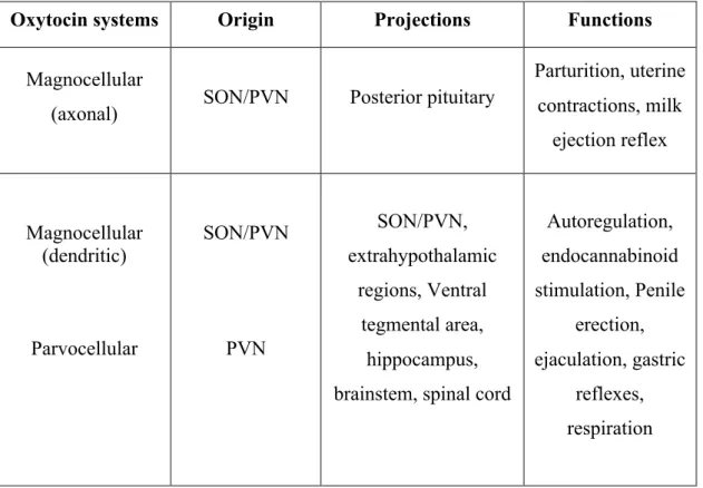
[1] The magnocellular oxytocin system originating in the supraoptic nuclei (SON) and hypothalamic paraventricular nuclei (PVN) can be further subdivided by its release characteristics in to axonal release (into the posterior pituitary), which regulates reproductive behavior and dendritic release (within the SON and PVN and may diffuse to other distant sites) to mediate oxytocin autoregulation (Baskerville and Douglas 2010) (Table 1 and Figure 2).
respectively.
Table 1. Oxytocin central pathways
Abbreviation: SON, supraoptic nuclei; PVN, paraventricular nuclei.
Oxytocin systems Origin Projections Functions
Magnocellular
(axonal) SON/PVN Posterior pituitary
Parturition, uterine contractions, milk
ejection reflex
Magnocellular (dendritic)
Parvocellular
SON/PVN
PVN
SON/PVN, extrahypothalamic
regions, Ventral tegmental area,
hippocampus, brainstem, spinal cord
Autoregulation, endocannabinoid stimulation, Penile
erection, ejaculation, gastric
reflexes, respiration
Oxytocin release from axon terminals occurs in the classical manner where axonal terminal release is preceded by an influx of calcium into axonal terminals in response to an invading action potential. However, as first demonstrated by Moos et al. (Moos et al.
1984), oxytocin can also be released somatodendritically from magnocellular oxytocin neurons in the hypothalamic PVN and SON to regulate its own release. This finding was further substantiated in numerous in vivo studies using microdialysis to quantitatively measure oxytocin release in the plasma and the brain of parturient and lactating rats (Moos et al. 1989; Neumann et al. 1993).
Unlike axonal release of oxytocin, dendritic release of oxytocin is triggered by release of calcium from intracellular stores and is generally electrically independent (Ludwig and Leng 2006; Ludwig et al. 2002).
Central (axon terminal) and peripheral (via hypophyseal secretion into circulation) oxytocin release from magnocellular cells can act synergistically to influence behavioral consequences. During various paradigms like suckling, there is a concomitant release of oxytocin into the bloodstream, SON and PVN (Moos et al. 1989; Neumann et al.
1993a).
Figure 2. Oxytocinergic projections in the rodent brain
Abbreviation: PVN, paraventricular nucleus of hypothalamus; SON, supraoptic nucleus of hypothalamus; VTA, ventral tegmental area; NAc, nucleus accumbens;
AMY, amygdala; MPOA, medial preoptic area of the hypothalamus (Paxinos and Watson 1998)
Such synergy between the central and peripheral oxytocin systems does not always exist and there can be an apparent dissociation between the two as seen during a psychosocial stressor such as social defeat (Bosch et al. 2004; Neumann et al. 2001).
Engelmann et al. demonstrated that whilst intra SON oxytocin release increased in response to social defeat, peripheral oxytocin release remained unaffected. Thus, it can be seen that during certain neuroendocrine mediated behaviors, centrally acting and peripherally acting oxytocin may act in union or independently to exert their behaviorally specific effects (Engelmann et al. 1999).
paraventricular nucleus, the ventromedial hypothalamic nucleus, and the dorsal motor nucleus of the vagus nerve. Thus, in the adult brain, there was a high correlation between the localization of OXT-R mRNA and that of OXT-binding sites. This localization of mRNA and the binding site coincided well with the functions of oxytocin described above in memory, maternal and sexual behavior, social perception and autonomic function (Shapiro and Insel 1989; Tribollet et al. 1989; Melchers et al. 2015).
The developmental expression profile of OTX-R mRNA could be divided into two types: transient type and constant type. During brain development, OXT-binding sites were observed more abundantly in the early postnatal rather than in the adult brain (Shapiro and Insel 1989; Tribollet et al. 1989).
Moreover, OXT-binding sites appeared as early as at day 14 of pregnancy, although the appearance of OXT-immunoreactive fibers was not observed until after birth (Buijs et al. 1980; Buijs 1992). The OTX-R is tied to Phospholipase C. Its genetic sequence was decoded in 1992 (Kimura et al. 1992). For the hormone to bond, magnesium (Pliska and Kohlhauf 1991) and cholesterol are necessary (Gimpl et al. 2000).
The encoded oxytocin receptor is a 389-amino acid polypeptide with seven transmembrane domains and is thus part of the G protein-coupled receptor family.
When oxytocin binds to its receptor it initiates a cascade of intracellular events that culminate in a range of cellular responses including an increase in neuronal firing, neurotransmitter release, smooth muscle contraction and protein phosphorylation. In rats, peripheral expression of oxytocin receptors is concentrated (but not exclusively) in the male and female reproductive tract and in myoepithelial cells in mammary tissue (Gimpl and Fahrenholz 2001; Zhang et al. 2005).
Oxytocin receptors are found in both the brain and the periphery (Adan et al. 1995). In addition, oxytocin receptors are also abundantly expressed throughout the CNS and often exist in the same regions containing oxytocin fibers. In addition to their expression in the SON and PVN, oxytocin receptors are also found in the regions of the cortex,
hippocampus, limbic system, basal ganglia, MPOA, olfactory bulbs, amygdala, and the brain stem (Freundmercier et al. 1994; Yoshimura et al. 1993).
There is widespread distribution of oxytocin receptors in the thoracic and lumbosacral segments of the spinal cord, with the dorsal horn, dorsal gray commissure, intermediolateral cell column all possessing oxytocin receptors (Veronneau-Longueville et al. 1999). However, some brain areas show a distinct mismatch between oxytocin fiber distribution and oxytocin receptor expression, such as seen in the amygdala and olfactory bulbs where there is a significantly greater proportion of oxytocin receptors compared to oxytocin fibers that innervate these nuclei (Ferguson et al. 2001; Huber et al. 2005; Terenzi and Ingram 2005).
Such an anatomical mismatch gives rise to the possibility that centrally released oxytocin can diffuse to distant sites within the brain to exert its effects. Therefore, oxytocin in the brain is described as a neuromodulator and appears to have broad permissive actions. OXT-R is expressed in OXT-target tissues and is a class G-protein coupled receptors (GPCRs), which primarily activates the G-protein αq/11 upon oxytocin binding (Sanborn et al. 1995).
4.1.4. Oxytocin effects in the body
Oxytocin has a short half-life in the blood. It has between 3.5 (Fuchs 1984) and 15 minutes (Gonser 1995).
Oxytocin has many different effects. It seems to play a large part in the masculine sexuality. Oxytocin has a function in the ejaculation, in some mammals, changes the contractility in the tubuli seminiferi (Insel et al. 1997).
Oxytocin injections were able to release erections in animals (Melis et al. 1986).
Oxytocin also modulates our experience of pain (Arletti et al. 1993).
Memory and mood are also influenced through its release (Arletti et al. 1995).
Intranasal oxytocin can be positively affected on hippocampal learning and memory loss due to chronic restraint stress (Dayi et al. 2015).
Oxytocin has an important role in digestion and nutrient absorption. Oxytocin release activates the vagal nerve, which increases activity of the gut hormones (Uvnas-Moberg 1989 and 2003). Oxytocin actions also promote anabolic metabolism and release insulin (Uvnas-Moberg 1989 and 2003).
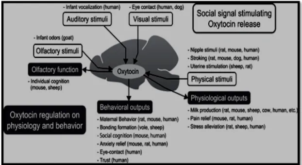
In particular, physiological stimuli are known to induce oxytocin system activation in mammals. When oxytocin release is increased in the central nervous system, many sensory, physiological, and behavioral functions are enhanced. Maternal as well as affiliative behaviors are enhanced by oxytocin.
Additionally, negative responses, such as pain, stress endocrine, and anxiety behaviors are diminished by oxytocin (Yoshimura et al. 1993) (Figure 3).
Figure 3. Summary of the role of the oxytocin system in reciprocal communication (Reproduced from Yoshimura et al. 1993)
4.2. Relation of hormonal imprinting to catecholamines and serotonin
4.2.1. CatecholaminesBiogenic amine neurotransmitters are biogenic substances with one or more amine groups (Purves et al. 2001). The amino acid tyrosine is the precursor for all three catecholamine neurotransmitters (Figure 5).
Dopamine is produced in brain cells and adrenal cells by the action of DOPA decarboxylase and the enzyme aromatic L-amino acid decarboxylase on L-DOPA in the presynaptic terminals and after transported to synaptic sites and packaged into vesicles for release. After release, free dopamine is either reabsorbed into the presynaptic terminal for reuse or it is converted to DOPAC and 3-MT via the enzymes COMT and MAO. Homovanillic acid (HVA) is a result of the further degraded of DOPAC and 3- MT by COMT and MAO enzymes (Moron et al. 2002; Yavich et al. 2007) (Figure 4).
Dopamine does not readily cross the blood-brain barrier (BBB). Dopamine acts by activating GPCRs, which exert their effects via complex second messenger systems (Purves et al. 2001). Dopamine receptors in mammals can be divided into two families, known as D1-like and D2-like. The effect of D1-like receptors (D1 and D5) can be excitation via opening of sodium channels or inhibition via opening of potassium channels and increase intracellular levels of cyclic AMP by activating adenylate cyclase. The effect of D2-like receptors (D2, D3, and D4) is usually inhibition of the target neuron and decrease intracellular levels of cyclic AMP by inhibiting adenylate cyclase (Grace 1991).
Figure 4. Dopamine metabolic pathway
Figure 5. The biosynthetic pathway for the catecholamine neurotransmitters
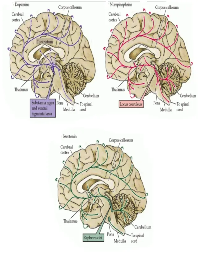
Dopamine is also believed to be involved in motivation, reward, cognition, arousal and reinforcement, also plays a poorly understood role in lactation, sexual activity, and nausea (Purves et al. 2001). The major dopamine area of the brain is the substantia nigra (SN), a small midbrain area that forms a component of the basal ganglia (Figure 7) and their most important projections go to the striatum, globus pallidus, and subthalamic nucleus, which play important roles in motor control. Another large group of dopaminergic neurons are located in the VTA, which is a group of neurons located in the midbrain area (Björklund and Dunnett 2007). Central neurons system dopamine pathways, projections and functions are summarized in the Table 2 (Ben-Jonathan and Hnasko 2001; Sanghera et al. 1991; Bitran et al. 1988; Eriksson et al. 1996; Skoog and Noga 1995; Holstege et al. 1996).
Table 2. Central nervous system dopamine pathways
Dopamine system Origine Projections Functions
Nigrostriatal SN (substantia nigra) Striatum Motility
Mesocortical VTA (ventral tegmental area) Cortex Emotionality
Mesolimbic VTA (ventral tegmental area) Nucleus accumbens
Reward desired Tuberoinfundibular Arcuate Nucleus Median
eminence
Regulation of prolactine
release
Incertohypothalamic Zona incerta, Periventricular region
Various hypothalamic
nuclei, Thalamus
Sexual arousal and
copulation
Diencephalospinal Hypothalamus Spinal cord
Afferent stretch reflex, contraction
of penile, striated muscle
have been detected in some patients with dopamine-dependent disorders therefore stimulation of central dopamine and oxytocin pathways are known to have similar effects on certain social behavioral paradigms and associated disorders such as sexual dysfunction, autism, anorexia and depression (Baskerville and Douglas 2008; Waldherr and Neumann 2007; Mackenzie 2006).
Norepinephrine (or noradrenaline in Europe) is produced by the action of dopamine β- hydroxylase on dopamine (Purves et al. 2001) (Figure 5). It is released from the adrenal medulla into the blood as a hormone, and is also a neurotransmitter in the CNS and the sympathetic nervous system (Purves et al. 2001).
Norepinephrine is also the transmitter used by the locus coeruleus, a brainstem nucleus that projects diffusely to a variety of forebrain targets including the cerebral cortex, limbic system, and the spinal cord, where it influences sleep and wakefulness, attention, and feeding behavior (Purves et al. 2001) (Figure 7).
Norepineprine and epinephrine performs their actions on the target cell by binding to and activating α- and -β adrenergic receptors that are G-protein-coupled (Dale Purves et al. 2001).
Norepinephrine plays a role in arousal, attention, fear, anxiety, learning, memory, (Barnes and Pompeiano 1991) and in aggressive behavior of males (Matsumoto et al.
1991) therefore alternation in the oxytocin system and norepinephrine has been associated with increased aggression in humans (Chichinadze et al. 2010; Bosch et al.
2005; Consiglio et al. 2005).
Epinephrine or adrenaline is produced by the action of phenylethanolamine-N- methyltransferase (PNMT) on norepinephrine, which utilizes S-adenosylmethionine as the methyl donor (Figure 5). Epinephrine-containing neurons in CNS are found in two groups in the rostral medulla (Figure 7). Epinephrine acts on the target cell by binding to and activating α- and -β adrenergic receptors (Purves et al. 2001).
Stress is a risk factor for a variety of illnesses, ranging from metabolic and cardiovascular disorders to mental illness. Stress induced increases in sympathoadrenal release of adrenaline and noradrenaline (Caldji et al. 2000), which increases heart and respiratory rate (Sabyasachi Sircar 2007).
Oxytocin administered cause reduces the rate and force of cardiac cells intrinsic contractions causing them to beat more slowly and contract less forcefully (Mukaddam- Daher 2001) therefore the connection between the catecholamines and oxytocin was always important (Wsol et al. 2008; Uvnas-Moberg and Petersson 2004).
4.2.2. Serotonin
Serotonin or 5-hydroxytryptamine (5-HT) is a monoamine neurotransmitter, synthesized from the amino acid L-tryptophan by tryptophan hydroxylase and aromatic L-amino acid decarboxylase (Young 2007; Purves et al. 2001) (Figure 6).
Seven types of 5-HT receptors are known (5-HT1, 5-HT2, 5-HT3, 5-HT4, 5-HT5, 5-HT6
and 5-HT7). Most 5-HT receptors are metabotropic (G-protein coupled that activate an intracellular second messenger cascade), only the 5-HT3 is a ligand-gated ion channel.
Serotonin has been implicated in behaviors, including the emotions, circadian rhythms, motor behaviors, and state of mental arousal. Impairments in the function of these receptors have been implicated in numerous psychiatric disorders, such as depression, anxiety disorders, and schizophrenia (Purves et al. 2001, Veenstra-VanderWeele et al.
2000).
Serotonin is located in groups of neurons in the raphe region of the pons and upper brainstem, which have widespread projections to the forebrain and have been implicated in the regulation of sleep and wakefulness (Hutson et al. 1986) (Figure 7). The serotonergic system may be important in this regard through, its potential influence on the release of oxytocin. Stimulation of the hypothalamus by serotonin has been shown to lead to release of oxytocin as a precursor molecule (Lee et al. 2003).
Galfi et al. (2005) and Jorgensen et al. (2003) reported that in the rats, oxytocin secretion was influenced directly by the serotonergic system (Galfi et al. 2005;
Jorgensen et al. 2003) and also administration of 5-HT antagonists blocked stress- induced increases in oxytocin secretion (Jorgensen et al. 2002).
action, their roles in a variety of normal processes, their relation with oxytocin, their relations with the developing hormone receptors (hormonal imprinting), and their possible alteration in neuropsychiatric disorders all require an accurate determination of biogenic amine neurotransmitters.
Figure 6. Serotonin biosynthesis and metabolism
Figure 7. The distribution in the human brain of neurons and their projections (arrows) containing biogenic amine neurotransmitters (Purves D, Augustine GJ, Fitzpatrick D, Katz LC, LaMantia AS, McNamara JO and Williams SM. (2001) The Biogenic Amines. Neuroscience, 2nd edition)
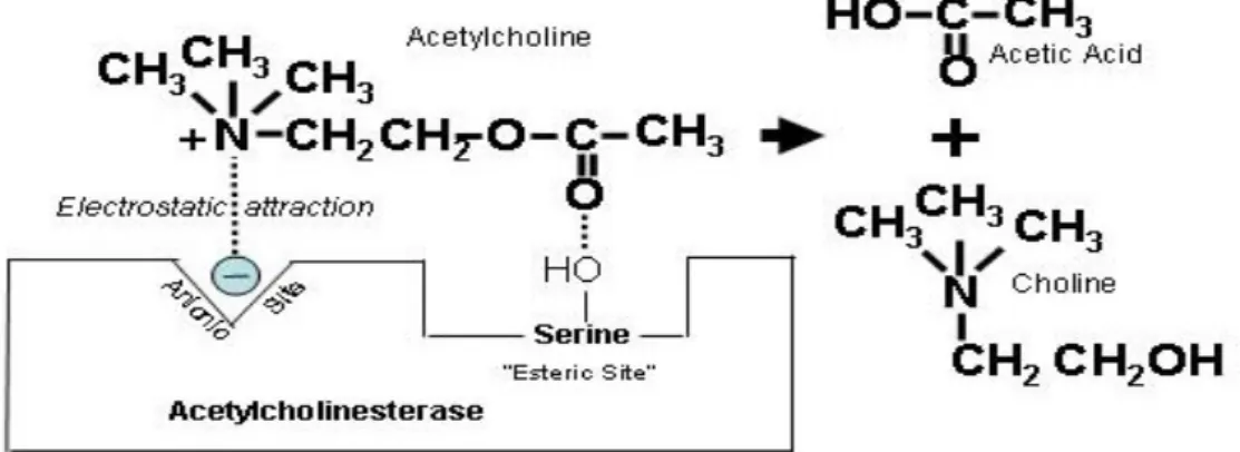
hydrolysis of the neurotransmitter ACh. There are different types of cholinesterases in the human body. The principle ones are AChE (EC 3.1.1.7, AChE) found in the nervous system and also present in the outer membrane of red blood cells and other one is plasma cholinesterase (EC 3.1.1.8, BuChE). The acute toxicity of organophosphorus compound is due to inhibition of AChE (EC 3.1.1.7, AChE), which belongs to serine esterase family (Miroslav et al. 2013).
Figure 8. The mechanism of action of acetylcholinesterase (Introduction to autonomic pharmacology. In: Basic and clinical pharmacology, Katzung 2001)
26
Acetylcholine mediates messages between the nerves, which is responsible for muscle contraction. When ACh is released from the nerve into the synaptic cleft, it got recognised by its receptors present on the postsynaptic membrane, which further transmits signal. Along with the acetylcholine receptors, AChE is also present on the postsynaptic membrane, which helps in the termination of the signal transmission by hydrolyzing ACh. On hydrolysis, ACh split into two products one is choline and the other is acetic acid. Choline and acetic acid are recycled by the body to form again ACh to maintain the reserves of the neurotransmitter therefore they can be used by the body again during the time of need (Boublik et al. 2002) (Figure 8).
Acetylcholinesterase contains one catalytic center which has two active sites: anionic and esteratic. The anionic site, positions the ACh in the active site through an electrostatic interaction with the quaternary nitrogen in choline. The hydroxyl group forms a serine residue in the esteratic site then covalently binds to the ester carbonyl group. This forms an unstable tetrahedral intermediate, which rapidly decomposes, liberating choline and leaving the enzyme covalently bound to acetate. A water molecule spontaneously hydrolyzes this bond, regenerating the free enzyme (Wiener et al. 2004).
Figure 9 shows the positively charged nitrogen in the ACh molecule that is attracted to the ionic site on AChE, and hydrolysis is catalyzed at the esteric site to form choline and acetic acid (Wiener et al. 2004).
Agency for Toxic Substances and Disease Registry Case Studies in Environmental Medicine
Cholinesterase Inhibitors: Including Pesticides & Chemical Warfare Nerve Agents
Part 2: What are cholinesterase inhibitors?
Learning
Objectives Upon completion of this portion of the case study, you should be able to:
Identify the chemical responsible for the acute pathology in cholinesterase inhibitor poisoning.
Describe how cholinesterase inhibitors, including organophosphorus compounds (e.g., pesticides, nerve agents) and carbamates block the ability of acetylcholinesterase to break down acetylcholine.
The Primary Toxic Effect of Cholinesterase Inhibitors
Acetylcholinesterase inhibitors (which, for brevity, we will refer to as cholinesterase inhibitors) are chemicals whose primary toxic effect is to block the normal breakdown of the neurotransmitter, acetylcholine.
This normal breakdown is shown in Figure 1 below.
Breakdown of Acetylcholine (Optional Reading)
Figure 1. Breakdown of acetylcholine.
Breakdown of Acetylcholine (Optional Reading)
They do this by occupying and blocking the site where the neurotransmitter, acetylcholine, attaches to the enzyme, acetylcholinesterase. If you are interested in the details at the chemical level, see the Optional Reading below.
How
Acetylcholine is Blocked (Optional Reading)
Figure 2 below shows how a cholinesterase inhibitor (in this case, a nerve agent) attaches to the serine hydroxyl group on acetyl-
cholinesterase. This prevents acetylcholine from interacting with the cholinesterase enzyme and being broken down.
+ Indicates that phosphorus is partially electropositive.
– Indicates that oxygen is partially electronegative.
Diagrams modified from Wiener, S. W., and R. S. Hoffman. "Nerve Agents: A Comprehensive Review." Journal of Intensive Care Medicine 19, no. 1 (2004): 22-37.
Figure 9. Break down of acetylcholine (Wiener et al. 2004)
resulting in over stimulation of nicotinic and muscarinic ACh receptors, which cause acute poisoning with AChEI (Erdman 2004; Costa 2008) (Table 3).
Table 3. The signs and symptoms of acetylcholinesterase inhibitors (AChEI) poisoning (Erdman 2004; Costa 2008)
Site Affected Cholinergic
Receptor Manifestation
1 Bladder Muscarinic Urinary frequency, urinary incontinence 2 Cardiovascular
system Muscarinic Bradycardia, bradydisrhythmias, hypotension 3 Eyes Muscarinic Blurred vision, lacrimation, miosis 4 Exocrine glands Muscarinic Increased salivation, perspiration 5 Gastrointestinal
tract Muscarinic Abdominal cramps, diarrhea, nausea, vomiting 6 Respiratory
system Muscarinic Bronchoconstriction, increased bronchial secretion, rhinorrhea
7 Central nervous system
Muscarinic and/or nicotinic
Agitation, anxiety, coma, confusion, convulsion depression of respiratory and circulatory centers,
dizziness, fatigue, hallucination, headache, lethargy, seizures, somnolence 8 Cardiovascular
system Nicotinic Tachycardia, transient hypertension 9 Skeletal muscle Nicotinic Cramps, flaccid paralysis, generalized weakness,
muscle fasciculation, twitching
According to the mode of action, acetylcholinestrase inhibitors can be divided into two groups:
1. Reversible AChEI play an important role in pharmacological manipulation of the enzyme activity. These inhibitors include compounds with different functional groups (carbamate, quaternary or tertiary ammonium group), and have been applied in the diagnostic and/or treatment of various diseases such as: myasthenia gravis, Alzheimer’s disease, post-operative ileus, bladder distention, glaucoma, as well as antidote to anticholinergic overdose (Aldridge and Davison 1953; Boublik et al. 2002).
2. Irreversible AChEI (e.g. the insecticide: carbofuran, nerve agents: sarin, soman, tabun, OPs and VX) inhibit AchE by alkyl phosphorylation of a serine hydroxyl group at the esteratic site of the enzyme. The inactive phosphorylated enzyme is very stable and AChEI eventually loses an alkyl side chain and the stability of the enzyme-nerve agent complex is enhanced, thereby increasing both the level and duration of the neurotransmitter acetylcholine at nicotinic, muscarinic and central nervous synapses.
This process is known as ageing which varies in half time (time for half of involved cholinesterase to age) from 2 min for soman, to > 40 h for VX and tabun (Aldridge and Davison 1953; Boublik et al. 2002).
5.1.1. Organophosphate type inhibitors
An organophosphate or phosphate ester is the general name for esters of phosphoric acid. Phosphates are probably the most pervasive organophosphorus compounds. The first synthesized organophosphorus compound was a mono ester named tetraethylpyrophosphate (TEPP) and the process was first published in 1854 by Philippe de Clermont (Antonijevic and Stojilkovic 2007; Karczmar 1970 ).
organophosphates cause irreversible inhibition of cholinesterases via a covalent reaction with the serine in the active center of the enzyme (Bajgar 2004; Bajgar et al. 2007; Kuca et al. 2006; Thiermann et al. 2007).
5.1.2. General structure of organophosphates
Organophosphates are esters or thiols derived from phosphoric, phosphonic, phosphinic or phosphoramidic acid. The basic chemical structure of all OPs is described by Schrader’s formula (Figure 10).
Figure 10. General structure of organophosphates (OPs)
R1 and R2 are aryl or alkyl groups that are bond to the phosphorus atom either directly (forming phosphinates), or through an oxygen or sulphur atom (forming phosphates or thiophosphates). In some cases, R1 is directly bonded to the phosphorus atom, and R2 is bonded to an oxygen or sulphur atom (forming phosphonates or thiophosphonates). In phosphoramidates, at least one of these groups is –NH2 (un-, mono- or bi-substituted), and the atom double-bonded with phosphorus is either oxygen or sulphur (Bajgar 2004;
Sogorb and Vilanova 2002; Smulders et al. 2004).
The –XR3 group, also binding to the phosphorus atom through oxygen or sulphur atom, may belong to a wide range of halogen, aliphatic, aromatic or heterocyclic groups. This leaving group is released from the phosphorus atom when the organophosphate is hydrolyzed by phosphotriesterases or upon interaction with protein targets (Bajgar 2004; Sogorb and Vilanova 2002; Smulders et al. 2004).
Phosphorothionate esters (P=S) are generally poor anticholinesterases. The poor AChE activity of P=S esters is explained on the basis of their relatively low reactivity, attributed to the smaller extent to which the P=S bond is polarized compared to the P=O, owing to the lower electronegativity of sulfur compared to oxygen. Polarization of the P=O linkage results in a more electropositive phosphorus atom, which facilitates attack on phosphorus by nucleophilic agents, e.g. the serine hydroxyl of AChE.
Organophosphorus esters containing the P=S moiety are less reactive and more stable to hydrolytic degradation than the corresponding P=O esters. Investigations on the metabolism and mode of action of organophosphorus compounds revealed that the toxicity of a P=S ester is attributed to the corresponding P=O esters, formed by metabolic oxidation of P=S to P=O (Gage 1953; Dauterman 1971).
5.1.3. Mechanism of toxicity
There are various groups of organophosphorus compounds, which are structurally and toxicologically different.
The OPs and their active metabolites are electrophilic molecules with moderate to high potency for phosphylation (denotes both phosphorylation and phosphonylation) of the serine hydroxyl group located at the active site of AChE. This phosphylation occurs by the loss of the leaving group and the establishment of a covalent bond with AChE through the serine hydroxyl. The resultant phosphylated AChE is typically very stable and is only slowly regenerated by spontaneous hydrolysis of the phosphate ester. While the AChE remains phosphylated, its enzyme activity is inhibited and therefore ACh accumulates in the synaptic clefts of muscles and nerves, leading to over-stimulation of cholinergic receptors that is essentially poisoning by endogenous ACh (Marrs 1993;
Pope 1999).
Additionally, OPs may also interact (inhibit) with other serine esterases, (Casida and Quistad 2004 and 2005) may have a direct action on muscarinic and nicotinic receptors binding to (with high/low affinity) and modulating the function of these receptors, (Bakry et al. 1988) and may induce specific organ lesions (Cao et al. 1999).
Propyl, isopropyl, butyl, and higher alkyl phosphates and phosphonates are more likely than methyl or ethyl analogs to act with secondary targets (Cao et al. 1999).
The concentrations of organophosphorus compounds required to act directly on nicotinic receptors are much higher than those on muscarinic receptors, suggesting that muscarinic receptors are more important as secondary targets in organophosphorus compound action. Combination of possible interactions will produce resultant toxic effect(s) for the particular organophosphorus compound. The spectrum of effects is further modulated by various toxicokinetic factors (Karalliedde et al. 2003).
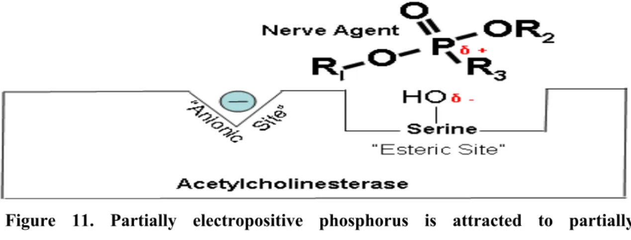
There are four stages of interaction of OPs with AChE:
Stage 1: Partially electropositive phosphorus of the organophosphorus compounds (ex.
nerve agent) is attracted to partially electronegative serine hydroxyl group on AChE and a complex forms (Figure 11). In the transition state showing which bonds break and which ones form (Johnson et al. 2000; Wiener and Hoffman 2004) (Figure 12).
Figure 11. Partially electropositive phosphorus is attracted to partially electronegative serine. δ + Indicates that phosphorus is partially electropositive. δ – Indicates that oxygen is partially electronegative (Wiener and Hoffman 2004).
Figure 12. Transition state showing which bonds break and which ones form (Wiener and Hoffman 2004)
Stage 2: The formation of a stable covalant bond between organophosphate compound and serine group of AChE. In this step organophosphorylation of the enzyme (esterase) takes place (Johnson et al. 2000; Wiener and Hoffman 2004) (Figure 13).
Figure 13. Organophosphates attached to acetylcholinesterase preventing the attachment of acetylcholine (Wiener and Hoffman 2004)
Stage 3: Cholinesterase is blocked, but it can hydrolyze to original state (slow) or regenerate with an oxime (fast) or ageing can appear which cannot regenerate (Johnson 2000; Wiener and Hoffman 2004) (Figure 14).
Figure 14. Blocking of cholinesterase by organophosphates (Wiener and Hoffman 2004)
Stage 4: Ageing. Over time, a functional group leaves the nerve agent, strengthening the bond and making it permanent. When this occurs, the enzyme-nerve agent complex is said to have aged (Johnson 2000; Wiener and Hoffman 2004) (Figure 15).
Figure 15. The “aged” bond (Wiener and Hoffman 2004)
VX) that pose potential neurotoxic threat to both military and civilian populations, as evidenced in recent terrorist attacks (Okumura et al. 1996).
The developing and production of these extremely toxic nerve agents started in the 1930s, and later used in wars and by terrorists on several occasions. As chemical weapons, they are classified as weapons of mass destruction by the United Nations, and their production and stockpiling was outlawed by the chemical weapons convention. In 1994, sarin was used in terrorist attacks in Japan, indicating that these compounds constitute a clear terrorist threat (Weiner and Hoffman 2004).
Several Middle-Eastern nations are believed to have developed and stockpiled nerve agents, notably Libya and Iraq. In 1984, it was confirmed that Iraq had used nerve agents in its war against Iran (Sidell and Borak 1992; McCauley et al. 2001).
In 1949, Dr. Ranajit Ghosh, a chemist working in England, was working on insecticide synthesis and discovered an extremely potent nerve agent that later came to be known as VX (Somani et al. 1992). Based on the acute toxicity, VX is the most toxic compound among all the nerve agents (Gupta 2006).
According to the WHO statistics more than 2 million pesticide poisonings occur every year and the rate of death is alarming mainly in the developing countries.
Organophosphate compounds may be used in the therapy of neurological damages such as Alzheimer’s disease. The example is trichlorfon (metrifonate) that used to be applied as a pesticide, and has medicine implementation analogous to the carbamate rivastigmine (Cummings et al. 2001).
5.2. Acetylcholinesterase reactivation : Oximes
The current treatment of organophosphate compounds poisoning includes three stages:
1. The use of an anticholinergic drug (e.g., atropine) 2. Cholinesterase-reactivating agents (e.g., oximes) 3. Anticonvulsant drugs (e.g., benzodiazepines)
Atropine, a parasympatholytic alkaloid isolated from Atropa belladonna by the German pharmacist Heinrich F. G. Mein in 1831, first successfully used by Thomas Richard Fraser, as an antidote for SLUDGE symptoms (salivation, lacrimation, urination, diaphoresis, gastrointestinal motility, emesis) caused by organophosphate poisoning (Karczmar 1970). The most important anticonvulsant is diazepam (Lipp 1972; Sellström 1992) and the combination of atropine and diazepam is more effective than atropine or oxime alone in reducing mortality (McDonough et al. 1989). In the cholinergic nervous system, diazepam probably decreases the synaptic release of ACh (Shih 1991), therefore diazepam has benefit in organophosphate poisoned patients by reducing anxiety, restlessness and muscle fasciculations, terminating convulsions, reducing morbidity and mortality when used in conjunction with atropine and oxime. Diazepam should be given to patients poisoned with organophosphorus compound whenever convulsions or pronounced muscle fasciculation are present. In severe poisoning, diazepam administration should be considered even before these complications develop (Marrs 2003). Oximes (cholinesterase-reactivating agents) are the nitrogen containing organic compounds drived from ketones or aldehydes by condensing them with hydroxylamine, with the general formula RR'C= NOH, where R is an organic side chain and R' may be hydrogen, forming an aldoxime (Figure 16), or another organic group, forming a ketoxime (Dawson 1994) (Figure 16).
Figure 16. structure of oximes (aldoxime, ketoxime)
Oximes are strong nucleophilic type compounds, comprised oxime moiety attached to a quaternary nitrogen pyridinium ring or imidazolium ring or quinuclidinium ring or other modified structure with basic oxime moiety to enhance the nucleophilicity.
Pralidoxime (pyridinium-2-aldoxime or 2-PAM) was synthesised by Wilson and Ginsburg in the USA and Childs et al. in the UK in 1955 (Wilson and Ginsburg 1955).
Chemically, pralidoxime is a (2-hydroxyimino-methyl-1-methylpyridinium chloride).
As a quaternary pyridinium salt, 2-PAM does not penetrate the blood-brain barrier (BBB), and because of that pro-2-PAM was synthesised as a pro-drug of 2-PAM that can access to the CNS. Unexpectedly, pro-2-PAM turned out to be even less effective than 2-PAM against experimental poisoning with paraoxon (Boškovic et al. 1980).
However, it appears that in organophosphate compounds poisoning 2-PAM can pass BBB at higher concentrations when given with atropine. There are four salts of pralidoxime namely 2-PAM Cl, methiodide, methysulphate and mesylate, that were investigated and introduced into practice which 2-PAM Cl is used all over the world where mesylate is used in UK only (Bismuth et al. 1992). 2-PAM is very efficient in reactivating AChE inhibited with sarin or VX (Johnson and Stewart 1970; Nozaki and Aikawa 1995), but is not successful in reactivation with tabun or soman (Inns and Leadbeater 1983; Koplovitz and Stewart 1994).
Trimedoxime
Trimedoxime (TMB-4 Cl2) was synthesised in the USA in 1957. Chemically, trimedoxime is a 1,3-bis (4-hydroxyimino-methyl-1-pyridinium) propane-dibromide (Poziomek et al. 1958). It is the only of the major bispyridinium oximes with a propylene bridge between the two-pyridinium rings. Experiments have shown that TMB-4 is a more potent reactivator of the AChE than 2-PAM (Hobbiger and Sadler 1958) and LüH-6 in case of tabun inhibition (Hobbiger and Vojvodic 1966).
Trimedoxime was the first oxime that was efficient in the treatment of animals intoxicated with tabun (Schoene and Oldiges 1973) and at the same time, shown in mice that its median lethal dose (LD50) is 3, 4 and 8 times lower than respective LD50 of LüH- 6, 2-PAM and HI-6 (Clement 1981) therefore TMB-4 is the most toxic oxime among the others (Bokonjic et al. 1993).
Obidoxime
Obidoxime or LüH-6 Cl2 was named in honour of Lüttringhaus and Hagedorn who synthesised it in Germany and introduced into medical practice in 1964. Chemically, obidoxime is a [1,1-bis (4-hydroxyimino-methyl-1-pyridinium)-2-oxapropane]
dichloride (Lüttringhaus and Hagedorn 1964). Obidoxime showed a significant potential, as antidote in organophosphorus compound poisoning and up to now, it is one of the most active reactivator (Erdmann and Engelhard 1964). Obidoxime was more efficient than TMB-4 as antidote against tabun poisoning (Heilbronn and Tolagen 1965).
HI-6
The HI-6 was synthesised in 1966 and given the code name HI-6, after the last name of Ilse Hagedorn and the first name of her student Irmo Stark, chemists who synthesised it in Freiburg, Germany in 1966 (Schoene 1967; Hagedorn and Gündel 1967). Chemically it is [1-(2-hydroxyimino-methyl-1-pyridinium)-3-(4-carbamoyl-1-pyridinium)-2- oxapropane] dichloride. It was the first oxime that could reactivate soman-inhibited AChE and it was more potent than obidoxim against poisoning with sarin and VX (Clement 1982; Inns and Leadbeater 1983). The only drawback of HI-6 was that this oxime could not reactivate tabun-inhibited AChE (Clement 1982, Cetkovic 1984). The intrinsic toxicity of HI-6 is lowest among the aforementioned oximes, with the LD50
values as high as 781.3 mg/kg in rats (Dawson 1994; Rousseaux and Dua 1998).
(Å2)
Pralidoxime
(2-PAM) N+ N
OH CH3
-2.38 36.47
Trimedoxime
N+ O N
H
N+
N OH
-2.65 72.94
Obidoxime
-3.04 82.17
HI-6 N
O H
N+ O N+
NH2
O -3.16 92.6
5.2.1. Mechanism of acetylcholinesterase reactivation
Cholinesterases have two binding sites: (1) catalytic site (anionic and esteratic) and (2) allosteric site. The allosteric site is catalytically inactive. Oximes bind to cholinesterases either at the catalytic site or at the allosteric site, or at both sites of the enzymes (Primozic et al. 2004).
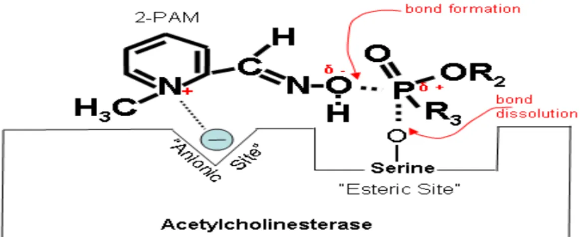
Figures 17 and 18 below explain that electropositive nitrogen on the oxime (2-PAM) is attracted to electronegative anionic site on cholinesterase. Then the oxime is oriented proximally to exert a nucleophilic attack on the phosphorus and form the enzyme- inhibitor complex.
The enzyme-inhibitor-oxime complex is then split off, leaving the regenerated enzyme.
However, this cannot happen after aging has occurred (de Jong and Ceulen 1978).
Phosphylated oximes formed during the reactivation process might be potent inhibitors of cholinesterases, which could cause re-inhibition of the previously reactivated enzyme. Re-inhibition of AChE can be faster than reactivation in case when a phosphylated oxime inhibits the enzyme at a rate higher than that of its elimination or decay to non-toxic products (Thiermann et al. 1999).
Figure 17. Partially electropositive nitrogen on the oxime (2-PAM) is attracted to electronegative anionic site on cholinesterase. δ + Indicates that phosphorus is partially electropositive. δ – Indicates that oxygen is partially electronegative (Wiener and Hoffman 2004)
Figure 18. Regenerated cholinesterase (Wiener and Hoffman 2004)
5.2.2. Application of acetylcholinesterase reactivators
Clinical view shows of oximes as adjuncts in the therapy of organophosphates poisoning. There are many factors that influence the beneficial role and efficacy of oximes for therapy therefore there would be very limited basis for choosing an effective oxime.
1. An oxime may be effective against a specific organophosphate and ineffective for others (Skrinjaric et al. 1973). For example 2-PAM is very efficient in reactivating AChE inhibited with sarin or VX (Nozaki and Aikawa 1995) but is inefficient in the reactivation of the tabun-inhibited and soman-inhibited enzyme. TMB-4 is a powerful reactivator and was the first oxime efficient in the treatment of animals intoxicated with tabun (Bokonjic et al. 1993) also effective against sarin or VX poisoning but is totally ineffective against soman (Inns and Leadbeater 1983).
2. Oximes are not equally effective and rank order of effectiveness changes with the organophosphate compound involved. Even if the same dosage regimen of an oxime is administered, due to interindividual variations, different plasma concentrations will be obtained therefore dosing and time of treatment plays also vital role for successful treatment (Skrinjaric et al. 1973). HI-6 is that this oxime cannot reactivate tabun- inhibited AChE (Clement 1982).
3. Acetylcholinesterase inhibited by OPs may undergo a secondary reaction in the different time period, i.e., spontaneous dealkylation through alkyl-oxygen bond scission (aging), resulting in an irreversibly inactivated enzyme and cannot be reactivated by oximes (Skrinjaric et al. 1973).
5.2.3. K-compounds
In the Department of Toxicology at the Faculty of Military Health Sciences, Defence University, Czech Republic several promising asymmetric pyridinium aldoxime compounds were synthesized by Kamil Kuca and Kamil Musilek and these compound were named K-oximes (Kuca et al. 2003b; Musilek et al. 2005).
These AChE reactivators are nucleophilic compounds and react with the nerve agent after it has been bound to the cholinesterase. K-oximes were basically targeted for tabun intoxication. More than 500 structurally different K-oximes have been synthesized since 2003 (Kassa et al. 2008).
The reactivating potency of the newly synthesized pyridinium aldoximes (K-027 and K- 203), were compared with that of the classic mono- and bis-aldoximes. The oximes K- 075 and K-203 showed higher potency to reduce tabun-induced acute lethal toxicity than obidoxime or HI-6 (Kassa et al. 2008; Musilek et al. 2007c).
The most promising among K-compounds are K-027, K-048 and K-203, which worked well against pesticide poisoning as well. The efficiency of treatment decreases in the following order: K-027 > K-048 > K-203 > methoxime > BI-6 > pralidoxime (Petroianu et al. 2007).
Structurally all the K-oximes are either asymmetrical or symmetrical bispyridinium aldoximes with changes in the position of functional aldoxime as well as in some cases changes in linker chain. Pyridinium aldoximes are polar organic compounds with large negative lipophilicity (logP) values and it has been widely demonstrated that they are highly hydrophilic, therefore can hardly penetrate in the BBB (Kalasz et al. 2015).
The chemical structure and values of lipophilicity of novel pyridinium aldoximes synthesized by Kuca’s group: K-027 (Kuca et al. 2003a); K-048 (Kuca et al. 2003b); K- 074 and K-075 (Kuca et al. 2005); and K-203 (Musilek et al. 2007a, 2007b, 2007c) are shown in Table 5.
1 K-027 -3.03 83.44
2 K-048 -3.02 83.44
3 K-074 -2.60 72.94
4 K-075 -2.71 72.94
5 K-203 -3.11 83.44
6. Aims and objectives
6.1. Optimize a sensitive bioanalytical method according to the validation requirements for determination of the biogenic amines and their metabolites by HPLC-EC
6.2. Determine the possible hormonal imprinting effect of oxytocin and examine it’s effect on the biogenic amine and their metabolite levels of the adult rat brain
6.3. Determine the effect of K-203 (AChER), a potential antidote in OPs intoxication on the biogenic amine and their metabolite levels of the adult rat brain
During all the experiments, the guiding principles in the care of and use of laboratory animals have been observed. All experimental procedures conformed to 86/509/EEC regulation on the well-being of experimental animals, and the experimental protocol was approved by the local ethical committee (permission No: 1806/007/2004 ANTSZ, Budapest, Hungary).
Wistar rats of our Charles River originated closed breeding colony (Toxicoop, Budapest, Hungary) were housed in polypropylene cages (43 x 22.5 x 20.5 cm) at room temperature (22-24 °C), humidity (55±6%), and 12h light-dark cycle with light on at 7.00 am. Standard laboratory food and tap water were available ad libitum. Male Wistar rat’s average weights in the K-203 experiments were 196±1.9g (Toxicoop, Budapest, Hungary).
7.1.2. Chemicals
Oxytocin acetate salt hydrate (C43H66N12O12S2), dopamine hydrochloride (3,4- dihydroxyphenethylamine), DOPAC (3-4-dihydroxyphenylacetic acid), homovanillic acid (HVA), serotonin hydrochloride (5-HT), 5-hydroxy-3-indole acetic acid (5- HIAA), 5-hydroxytriptophol (5-HTOL) and phosphoric acid (H3PO4), disodium hydrogen phosphate dihydrate (Na2HPO4·12H2O), citric acid monohydrate, l-octane sulfonic acid sodium salt, ethylenediaminotetraacetic acid disodium salt dihydrate (Na2EDTA) and perchloric acid 70% (PCA) in the best available quality for HPLC were from Sigma-Aldrich (Steinheim, Germany). Acetonitrile was from Merck (Darmstadt, Germany).
K-203 (E-1-(4-carbamoylpyridinium)-4-(4-hydroxyiminomethylpyridinium)-but-2-ene dibromide) was synthesized and kindly donated by K. Kuca (Department of Toxicology, Faculty of Military Health Sciences, Defence University, Hradec Kralove, Czech Republic).
7.1.3. Instrumentation and chromatographic conditions
Samples were analyzed by reversed phase high-performance liquid chromatography with amperometric/electrochemical detection (HPLC–EC) consisting from a Jasco pump (PU1580, Tokyo, Japan) equipped with a DG-2080-54 four-line degasser, an AS 2057 Plus Automatic injector and connected to an Intro digital amperometric (Antec, Leyden, Zoeter-woude, Netherlands) detector operated at Eox=+0.65 V with a sensitivity of 1-10 nA/V with a time filter of 1.0 sec, and JMBS Hercule 2000 Chromatography Interface (Le Fontanil, France). Standard temperature of the column was 25±0.15 °C.
Chromatograms were electronically stored and evaluated using Borwin 1.50 Chromatography Software (JMBS, Le Fontanil, France). The separations were done using a Zorbax RX-C18 4.6x12.5 mm (5-µm) pre-column and a Zorbax RX-C18 4.6x250 mm, (5-µm) octadecyl silica column (Agilent Technologies, supplied by Kromat Kft, Budapest, Hungary).
For the serial determinations of the biogenic amines the mobile phase contained 56.2 mmol/L Na2HPO4, 47.9 mmol/L citric acid, 0.027 mmol/L Na2EDTA, 0.925 mmol/L octane sulfonic acid sodium and 75:925 mL acetonitrile/phosphate buffer for hormonal imprinting oxytocin treatment and 65:935 mL acetonitrile/phosphate buffer for K-203 treatment. The pH was adjusted to 3.7 with 85% phosphoric acid (H3PO4) (inoLab pH Level 2, WTW GmbH, Germany). The flow rate of the mobile phase was 1 mL/min.
During the optimization we fully adapted the guideline for bioanalytical method validation of the FDA (Food and Drug Administration, Guidance for Industry - Bioanalytical Method Validation, 2001).
During the validation process, the following parameters were determined:
1) Calibration curve and Linearity: the calibration sequences from stock solutions were prepared at a concentration of at least 8 and measurements were replicated 3 times.
Linearity was determined by calculation of a regression line from the peak area versus concentrations of standard solutions using the equation y = mx + c. A value of ≥0.98 would be considered acceptable for the correlation coefficient.
different animals. The limit of quantitation (LOQ) was determined at a signal to noise ratio of 10.The limit of detection (LOD) was determined at a signal to noise ratio of 3.
7.2. Methodes
7.2.1 Animals treatment
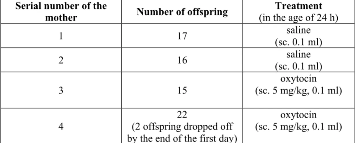
1) Oxytocin hormonal imprinting treatment: offspring of four females and two males were used in the experiment. Three females (185±12 g) were co-housed with one male for 8 days for mating and following this period females were housed separatedly during the 21 day period of pregnancy (number of offspring and treatment is shown in Table 6). Offsprings were treated subcutaneously (sc.) with a single dose of 5 mg/kg oxytocin (dissolved in saline) when they were one day old. At the end of the suckling period (in age of 21 days) offspring was housed separatedly from the mother. The 21 days old rats were selected according to their sex and were grown in standard cages.
Table 6. Number of offspring and treatment of rats Abbreviation: (sc., subcutaneously)
Serial number of the
mother Number of offspring Treatment
(in the age of 24 h)
1 17 saline
(sc. 0.1 ml)
2 16 saline
(sc. 0.1 ml)
3 15
oxytocin (sc. 5 mg/kg, 0.1 ml)
4
22
(2 offspring dropped off by the end of the first day)
oxytocin (sc. 5 mg/kg, 0.1 ml)
No death of the treated rats was observed during the 4 months of the experiment.
Weight gain of the rats was recorded weekly. The numbers of rats were 68, divided to four groups (14 control females, 19 control males, 17 oxytocin treated females and 18 oxytocin treated males).
When the offspring were fourth months old, they were exsanguinated through the canthus under ether anesthesia. Data are summarized in Table 7. The eight regions of the brain (hippocampus, HC; hypothalamus, HT; medulla oblongata, MO; spinal cord, SC; frontal cortex, FC; cerebellum, CB; striatum, ST; truncus cerebri, TC) were dissected on 0 ◦C aluminum surface according to the method of Paxinos G. and Watson C. (1998) and Palkovits M. (2001). The samples were kept frozen at -80 ◦C.
2) K-203 treatment: rats were injected in 0.2 mL volume intramuscularly (i.m) with 50 µmol of K-203 freshly dissolved in saline. The control group received an equal volume (0.2 mL) of solvent treatment. Five rats were used for each data point. Rats were sacrificed by decapitation 15 or 60 min following treatment. Seven regions of the brain (FC, HT, HC, ST, MO, CB and SC) were dissected and immediately placed on an ice- cold aluminum surface according to the method of Paxinos G. and Watson C. (1998) and Palkovits M. (2001). All samples were kept frozen at -80 °C until direct analysis.
Weight (g± SD)
Control
male (n=19)
55
±0.8 81
±0.7 114
±0.9 138
±1.0 198
±1.9 234
±1.4 256
±3.1 female
(n=14) 54
±0.5 78
±0.8 100
±0.9 119
±2.1 129
±2.3 136
±3.1 141
±3.9
Oxytocin imprinted
male (n=18)
55
±0.6 79
±0.8 113
±1.4 137
±1.6 199
±2.3 231
±2.6 258
±3.0 female
(n=17) 55
±0.5 80
±0.7 101
±1.1 120
±1.2 131
±1.6 135
±2.3 139
±2.3
Group of rats
Age (weeks)
10 11 12 13 14 15 16
Weight (g± SD)
Control
male (n=19)
271
±2.3 320
±5.2 329
±4.6 332
±4.1 349
±5.8 342
±6.9 359
±7.4 female
(n=14) 156
±5.1 174
±4.8 228
±5.3 238
±5.2 236
±4.9 239
±6.0 254
±5.6
Oxytocin imprinted
male (n=18)
272
±3.5 322
±3.2 331
±4.1 334
±4.2 348
±3.9 356
±6.0 358
±7.4 female
(n=17) 152
±2.7 168
±2.9 219
±3.2 229
±2.9 241
±4.6 243
±4.5 258
±6.9