Factors influencing pre-receptor cortisol activation
PhD thesis outline
Péter Szelényi
Molecular Medicine Doctoral School Semmelweis University
Supervisor: Dr. Miklós Csala Ph.D.
Official reviewers: Dr. Nikolette Szücs PhD.
Dr. Balázs Veres Ph.D.
Head of the Final Examination Committee:
Dr. László Tretter Ph.D.
Members of the Final Examination Committee:
Dr. Pál Gróf Ph.D.
Dr. Katalin Monostory Ph.D.
Budapest 2014
1
Introduction
The metabolic syndrome
The metabolic syndrome is a major public health issue worldwide, and its preva- lence is increasing at a high rate. This complex metabolic disorder stands between obesity and type 2 diabetes. It refers to a constellation of at least three of the symptoms including abdominal obesity, elevated blood pressure, elevated serum triglycerides, low HDL- cholesterol and insulin resistance. It is also associated with an increased risk of numerous diseases (e.g. cardiovascular diseases) and is characterized by progression towards diabe- tes. Because of the rapid spreading of the metabolic syndrome, there is a growing need for efficient treatment and prevention. Although the risk factors are well known, the un- derlying molecular mechanisms leading to the metabolic syndrome are not fully charac- terized yet.
Nutritional factors
Development and progress of the metabolic syndrome are mainly influenced not only by genetic factors and low physical activity, but nutritional factors as well. It has been well demonstrated that the elevated sugar consumption involved in western diet highly increases the risk of the metabolic syndrome. Besides the amount of ingested car- bohydrates, their monosaccharide composition also deserves attention. Fructose content for instance is a substantial factor from the aspect of metabolic disorders.
Fructose absorption is quick, does not need ATP and is separated from Na+ absorp- tion. It enhances de novo lipogenesis and glucogenesis of the intestinal cells. Most of the fructose absorbed into portal circulation is metabolized by the liver. This process is un- regulated and quicker than glucose metabolism. It escapes insulin-control, citrate- and ATP-mediated feedback as it bypasses the rate-limiting phosphofructokinase-1 step of glycolysis. By the continuous formation of glycerol-3-phosphate and acetyl-coA, fructose metabolism also enhances lipogenesis and VLDL-formation in the liver.
2
In contrast, consumption of green tea, or its main catechin-component, EGCG is known by its protective effect against metabolic disorders, such as obesity, metabolic syndrome and type 2 diabetes. Mainly in vivo studies have been reported in this field, and most of the molecular mechanisms remain to be clarified.
Cushing’s syndrome
Metabolic syndrome shares multiple symptoms with Cushing’s syndrome, a com- plex metabolic disorder based on cortisol overproduction. Therefore, in spite of normal blood cortisol levels, an impaired glucocorticoid metabolism has been implicated in the pathomechanism of the metabolic syndrome, too.
Pre-receptor cortisol metabolism
Cortisol has got a tissue-specific pre-receptor metabolism that can modulate local cortisol levels without influencing the plasma concentrations. Cortisol-cortisone conver- sion (inactivation) takes place mainly in the kidneys and subcutaneous adipose tissue, while it is mostly reactivated in the liver and splanchnic adipose tissue. Besides circulat- ing cortisol levels, pre-receptor glucocorticoid activation in the target tissues is a major determinant of the cortisol effect. It has been demonstrated that the amount of active cor- tisol that originates from pre-receptor activation is as much as 25% of the hormone newly synthetized in the adrenal glands. Liver is responsible for roughly one third and visceral adipose tissue for nearly two thirds of this local production.
11β-hydroxysteroid dehydrogenases
Interconversions of cortisol and the prohormone cortisone are catalyzed by two isoforms of 11β-hydroxysteroid dehydrogenase enzyme (11βHSD) in the target cells.
The cytosolic NAD+ dependent 11βHSD2 is expressed in mineralocorticoid target tissues, such as kidney, colon, and salivary glands. It oxidizes cortisol to cortisone and thereby confers aldosterone selectivity on inherently nonselective mineralocorticoid re- ceptors.
3
In contrast, type 1 isoenzyme (11βHSD1) is expressed in glucocorticoid target tis- sues, such as liver, skeletal muscle and adipose tissue and generally catalyzes the reaction in the other direction (except for subcutaneous adipose tissue, where inactivation is cata- lyzed). This isoenzyme is localized in the lumen of the endoplasmic reticulum (ER) and uses NADP+ or NADPH cofactor, depending on the direction of the reaction. Because of the impermeability of the ER membrane to pyridine nucleotides, the enzyme can only use the luminal pyridine nucleotide pool of the organelle. NADPH requirement of the reac- tion is met by the activity of hexose 6-phosphate dehydrogenase (H6PD), which is not only functionally coupled but also physically associated to 11βHSD1. H6PD uses glucose 6-phosphate (G6P), which enters the ER lumen through glucose 6-phosphate translocase (G6PT).
Pre-receptorial cortisol production and the metabolic syndrome
The distribution of reductase and dehydrogenase activities of 11βHSDs in different tissues contributes to an active recycling between cortisol and cortisone in various human tissues without influencing plasma cortisol level. Mounting evidence support the role of local cortisol production in the metabolic syndrome. All or most symptoms appear in transgenic mice overexpressing 11βHSD1, while 11βHSD1 knockout mice seem to be protected from the disease.
Local cortisol production as a potential drug target
Elucidation of the role of pre-receptor cortisol production in the metabolic syn- drome launched the development of 11βHSD1 inhibitors as anti-diabetic drug-candidates.
Compounds that inhibit both reductase and dehydrogenase activities of 11βHSD1 possess a limited effectiveness in decreasing the overall glucocorticoid activity, which might ex- plain their limited success in phase II clinical trials. Therapeutic efficacy would be largely improved if the selective 11βHSD1 inhibitor could specifically inhibit the reductase ac- tivity and, ideally, also increase the dehydrogenase activity of the enzyme, features that seem to be hard to achieve.
4
Objectives
We aimed to investigate the effects of the above diabetogenic (fructose consump- tion) and antidiabetic (catechin ingestion) nutrition factors. We hypothesized that F6P, the common metabolite of fructose and glucose metabolism, enhances, while the green tea cathecin EGCG reduces cortisol production by the microsomal G6PT-H6PD- 11βHSD1 catalytic triad. The specific questions to answer were:
- How does F6P infuence cortisol production in the microsomal vesicles?
- Can F6P contribute to NADPH formation of liver and adipose microsomes?
- Can F6P enter the lumen of the endoplasmic reticulum?
- Does F6P act in our model through isomerization to G6P?
- How does EGCG influence cortisol production in microsomal vesicles?
- Does EGCG affect any of the protein components of the G6PT-H6PD-11βHSD1 catalytic triad specifically?
- How does EGCG influence the ER luminal redox state?
- Does EGCG have any specific redox interactions with our system or acts only as a general anti- or pro-oxidant?
Methods
Preparation of microsomes: Microsomes were prepared from the livers and visceral adi- pose tissue of male Wistar rats using fractional centrifugation. Microsomes resuspended in MOPS-KCl buffer were rapidly frozen and stored in liquid N2 until used. Intactness of microsomal vesicles was checked by measuring the latency of mannose 6-phosphatase activity, which was greater than 92% in all the preparations used.
Glucose production: Microsomes were incubated in KCl-MOPS buffer at 37°C in the presence of 2 mM G6P or F6P. The reaction was stopped with heat denaturation (100°C, 5 min). After centrifugation, glucose content of the supernatants was measured by using glucose (GO) assay kit according to the manufacturer’s instructions.
5
Microsomal cortisone-cortisol conversion: G6P-driven reduction of cortisone to cortisol was measured by incubating intact microsomes in MOPS-KCl buffer with 5 μM cortisone and 50 μM G6P for 30 min. The reaction catalyzed by 11βHSD1 was assessed directly in similar conditions by measuring NADP+-driven cortisol oxidation after pretreatment of the microsomes with pore-forming alamethicin. These permeabilized vesicles were incu- bated in MOPS-KCl buffer containing 5 μM cortisol and 50 μM NADP+ for 30 min. In either case, the reaction was terminated with ice-cold methanol, and the samples were stored at -20°C until HPLC analysis of cortisone and cortisol contents.
H6P isomerase activity: The isomerase activity was indirectly evaluated by incubating washed microsomes or cytosolic fraction in KCl-MOPS buffer at 22°C. The formation of G6P upon the addition of F6P was measured enzymatically with G6PD. For this assay, we used G6PD isolated from L. mesenteroides, which is NAD+ dependent, so that we could distinguish isomerase activity from H6PD activity (which is prevalently NADP de- pendent). The production of NADH by the G6P-dependent dehydrogenase reaction was monitored fluorometrically at 350-nm excitation and 460-nm emission wavelengths.
Microsomal F6P transport: The microsomal uptake of F6P and G6P was evaluated by a rapid filtration technique.
Immunoblot: Total protein amounts of microsomal and cytosolic fractions corresponding to comparable PGI activity of HEK-293 cells and rat liver were examined by Western blot technique.
Liquid chromatography: The methanol-treated samples were centrifuged and the protein- free supernatants were analyzed to measure cortisone and cortisol simultaneously. Com- pounds were separated by HPLC in a Nucleosil 100 C18 column with isocratic methanol- water at 0.7 mL/min flow rate; and the absorbance in the eluate was detected at 245 nm wavelength. Retention times of cortisone and cortisol were determined by injecting 10 μM standards.
Fluorescent detection of dehydrogenase activities: NADPH formation was monitored in spectrophotometer at 350-nm excitation and 500-nm emission wavelengths.
6
Endogenous Reducing/Oxidizing Capacity : Intrinsic cortisone reducing or cortisol oxi- dizing capacity of the microsomes was analyzed by incubating intact microsomal vesicles in MOPS-KCl buffer at 37°C for 2 h in the presence of 10 μM cortisone or cortisol, re- spectively. The cortisone and cortisol contents were determined by HPLC analysis.
Lipid peroxidation: Oxidative damage of the membrane lipids was tested by measuring the formation of thiobarbituric acid reactive substances (TBARS).
Statistical analysis: Experiments were performed in triplicates, with each of the values of a single set of experiments corresponding to the mean of a minimum of two to three de- terminations. Data are presented as mean ± SEM. The results were analyzed by oneway ANOVA and Tukey-Kramer Multiple Comparison Test using GraphPad Prism® soft- ware. A p value below 0.05, 0.01, or 0.005 was considered as a significant difference.
Results
F6P metabolism in the endoplasmic reticulum
F6P-dependent glucose production in liver microsomes
G6P stimulates reduction of cortisone to cortisol in the ER lumen via the G6PT- H6PD-11βHSD1 triad. A more recent observation that microsomal cortisone reduction can be enhanced also by F6P indicates that this hexose-phosphate can somehow contrib- ute to luminal NADPH generation. However, the transport processes and enzymatic reac- tions involved are unknown.
Whether F6P can be isomerized to G6P in the ER lumen was first measured by means of measuring glucose production after F6P addition in rat liver microsomes, taking advantage of the presence of glucose-6-phosphatase enzyme in the lumen. F6P proved to be nearly as good a source of glucose production as G6P, strongly suggesting isomeriza- tion of F6P to G6P in this system. The isomerase activity may be due to the cytoplasmic enzyme PGI. This protein can be also secreted by an unconventional (i.e. ER/Golgi- independent) mechanism and has an ER membrane-bound receptor named autocrine mo- tility factor receptor/ubiquitin ligase 3. Alternatively, the observed isomerization might
7
be catalyzed by an enzyme localized in the ER lumen. This question was first addressed by washing the microsomal vesicles by multiple sedimentation and buffer replacement to eliminate the membrane-adherent cytosolic proteins. The first such cleansing resulted in a 9-fold decrease in the rate of glucose production from F6P in intact microsomes, alt- hough, as expected, it modestly affected glucose production from G6P, i.e. glucose-6- phosphatase activity. This suggests that a major part of the total hexose-6-phosphate iso- merase activity, probably corresponding to PGI, is loosely associated with the outer sur- face of the microsomal vesicles. However, the remaining capacity of the microsomes to use F6P as a glucose precursor could not be eliminated by subsequent washing steps and hence seems to be based on a tightly membrane-associated or luminal hexose-6- phosphate isomerase activity. The higher latency of glucose production in the case of F6P (approximately 85%) with respect to the case of G6P (approximately 40%) might be due to the lower rate of entry of F6P compared with that of G6P into liver microsomal vesi- cles.
F6P-dependent cortisone reduction in liver microsomes
Efficiency of F6P (and of G6P for comparison) to stimulate the conversion of cor- tisone to cortisol was investigated in washed microsomes. In this case, the first washing step caused only a moderate decrease in cortisone reduction (32 and 11% when F6P and G6P was used, respectively), and the subsequent washing steps did not affect the rate of cortisone reduction significantly. It can be concluded that an irremovable intrinsic hex- ose-6-phosphate isomerase activity of the microsomes can provide the dehydrogenase enzymes with G6P at a sufficient rate. Thus, only washed microsomes were used in all further experiments, to avoid any interference of the extravesicular (cytosolic) isomerase activity.
8
F6P-dependent NADPH generation and 6-phosphogluconate production in hepatic and adipose tissue microsomes
F6P acts like G6P in liver microsomes, i.e. stimulates cortisol formation. Further evi- dence was collected that its microsomal isomerization provides G6P, which feeds NADPH generation by H6PD, both in hepatic and adipose tissue microsomes. Due to the luminal localization of H6PD and the poor permeability of microsomal membrane to NADP+, when microsomes are incubated in the presence of NADP+ and G6P, NADPH generation cannot be detected until the membrane barrier is eliminated. Once the lipid bilayer is permeabilized with a detergent, the linear increase in fluorescence indicates the progress of the redox reaction both in hepatic and adipose tissue microsomes. F6P proved to be also efficient in stimulating NADPH generation in this system. Fructose-1,6- bisphosphate (F1,6BP), a known inhibitor of the cytoplasmic PGI, also significantly re- duced the rate of F6P-driven NADPH generation, although it did not interfere with the process in microsomes using G6P. Besides functioning as a dehydrogenase, H6PD pos- sesses lactonase activity, converting G6P ultimately to 6-phosphogluconate. Formation of 6-phosphogluconate from F6P was demonstrated in our experimental model by measur- ing further NADPH generation in the presence of exogenous 6-phosphogluconate dehy- drogenase to the hepatic or adipose tissue microsomes. Because F6P-driven NADPH generation was inhibited by F1,6BP and accompanied by 6-phosphogluconate formation, we concluded that F6P is indeed isomerized to G6P, providing H6PD with substrate.
Microsomal hexose-6-phosphate isomerase
Although our findings convincingly demonstrated the formation of G6P from F6P in he- patic and adipose tissue microsomes, it still remained to be elucidated whether this con- version occurs on the outer surface (i.e. representing the cytoplasmic side) of the vesicular membrane or inside the vesicles. To answer this question, formation of G6P was compared in intact and permeabilized microsomes; i.e. the latency of the hexose-6- phosphate isomerase was investigated. The experimental conditions were similar to those in the previous set of measurements, but NADP+ was substituted by NAD+. In prelimi-
9
nary experiments, a slow reduction of NAD+ to NADH was observed in the presence of G6P in permeabilized microsomes, indicating, under our experimental conditions, a lower (approximately one tenth) preference of H6PD for NAD+. Accordingly, in the presence of NAD+ and F6P, the permeabilization of hepatic or adipose tissue microsomes resulted in a very slow NADH generation. The subsequent addition of the NAD+-specific L. mesen- teroides G6PD to the samples greatly increased the rate of NADH generation, indicating the efficient isomerization of F6P to G6P in the permeabilized microsomes. F1,6BP re- markably repressed this process both in hepatic and adipose tissue microsomes (Fig. 1).
The addition of F6P to intact (nonpermeabilized) microsomes caused no NADH genera- tion, because the membrane is impermeable to pyridine nucleotides. Most importantly, the subsequent addition of the L. mesenteroides G6PD poorly stimulated NADH genera- tion, indicating a negligible isomerization of F6P on the outer surface of intact micro- somes. These findings clearly demonstrate the luminal localization of hexose- 6- phosphate isomerase activity.
Figure 1 F6P-G6P isomerization in permeabilized liver (A), and adipose (B) microsomes, in the absence (−) or presence (+) of F1,6BP
Activities: A: 10.9 ± 1.3 (−), 4.7 ± 0.5* (+5 mM) and 1.5 ± 0.4* (+10 mM) B: 12.2 ± 2.0 (−) and 0.7 ± 0.4* (+5 mM) nmol/min/mg protein (*p < 0.01 vs. control)
10
Transport of F6P across the microsomal membrane
Uptake of F6P and G6P was investigated in hepatic and adipose tissue microsomes by using a rapid filtration transport assay. The initial rate of transport was determined at 30 sec; to minimize the contribution of the intravesicular accumulation of metabolites of G6P. The transport activity was found to be concentration dependent in both hepatic and adipose tissue microsomes, for both F6P and G6P, in the investigated range of extrave- sicular hexose-phosphate concentrations. The rate of influx of F6P was approximately 1.7-fold higher in adipose tissue compared with liver microsomes, whereas G6P was transported at a higher rate (approximately 3.7-fold) in liver than in adipose tissue micro- somes. This latter observation is consistent with our previous results and is very likely due to the higher representation of G6PT in liver microsomes. On the other hand, in adi- pose tissue microsomes, G6P was transported at a higher rate (approximately 1.4-fold) than F6P. The possibility that F6P permeates the microsomal vesicles through G6PT was investigated in liver microsomes by using the selective inhibitor of the transporter S3483.
The G6PT inhibitor markedly reduced the transport of G6P but did not affect the transport of F6P, suggesting different routes of entry. Moreover, the transport activity for each phosphohexose were unaffected by the other phosphohexose at a high (competitive) concentration, which strengthens the hypothesis that the phosphoesters do not share the same transporter. Furthermore, S3483 did not inhibit F6P transport in adipose tissue mi- crosomes, and we previously reported that the inhibitor reduces G6P entry in adipose tis- sue microsomes by approximately 70%. The possibility that the route of entry of F6P is via the nonspecific glucose-phosphate transporter (GPT) previously described in human fibrocytes was also tested by measuring the transport of F6P in the presence of competi- tive concentrations of G1P, a known substrate of GPT. G1P inhibited the transport of F6P by approximately 50%, but did not affect G6P transport. The results are consistent with the role of GPT in F6P transport. The apparent lack of effect on G6P transport, which is also mediated by GPT, is very likely due to the high representation of G6PT in liver mi- crosomes that allows a massive entry of G6P.
11
Examination of microsomal hexose-phosphate isomerase in HEK-293 cells F6P does not serve as substrate for H6PD
To prove the assumption that H6PD cannot use F6P as a substrate, myc-tagged human enzyme was overexpressed in HEK-293 cells followed by subsequent affinity pu- rification. Cells transfected with the empty pcDNA3.1 vector were used as control. After SDS-PAGE analysis and Coomassie Blue staining, a single protein band at approximately 90 kDa, corresponding to H6PD was detected. The identity of H6PD was further con- firmed by immunoblot analysis using a rabbit primary antibody raised against the myc peptide. Next, NADPH generation was measured by the purified enzyme upon supplying G6P or F6P and NADP+ as substrates. NADPH was produced in the presence of G6P but not of F6P. This result confirms our assumption that F6P is not directly used by H6PD to generate NADPH.
H6PD does not catalyze the isomerization of F6P and G6P
To investigate whether H6PD possesses intrinsic isomerase activity and can interconvert F6P and G6P, we used G6PD from L. mesenteroides as an indicator enzyme to measure NADH production from NAD+ with G6P or F6P as substrates. G6PD could generate- NADH from G6P only, independent of whether it was supplied with or without F1,6BP, an inhibitor of the isomerase activity. H6PD was not able to generate G6P from F6P.
These results indicate that human H6PD itself cannot function as a hexose-phosphate isomerase.
Intrinsic microsomal hexose-phosphate isomerase enzyme
To rule out that the cytoplasmic PGI was responsible for the conversion of F6P to G6P in microsomes, total proteins were subjected that correspond to comparable hexose- phosphate isomerase activity, i.e. 30μg of washed microsomes or 3μg of cytosolic frac- tion from HEK-293 cells or 100 μg of washed microsomes or 2 μg of cytosolic fraction from rat liver to SDS-PAGE and subsequent immunoblot analysis using an antibody
12
raised against the cytosolic PGI. A single protein band was detected at 63 kDa in the cy- tosolic but not in the microsomal fraction of HEK-293 cells and rat liver preparations, even if 10 or 50 times more microsomal protein was loaded, respectively, indicating the absence of the cytosolic PGI in the lumen of the microsomes. The hexose-phosphate iso- merase activity of the two protein preparations were practically the same. To further strengthen this evidence, microsomes (30 μg protein) were incubated as well as the cyto- solic fraction (3 μg protein) of HEK-293 cells in the presence of erythrose-4-phosphate (E4P), a known potent inhibitor of the human cytosolic PGI, followed by measurement of NADH production by the G6PD from L. mesenteroides. Without the addition of E4P, both incubations showed comparable, time-dependent activity. The microsomal hexose- phosphate isomerase activity, however, was more efficiently inhibited by E4P than the cytosolic PGI. We obtained similar results using 2μg of rat liver cytoplasmic and 100 μg of microsomal protein with F1,6BP as an inhibitor. Upon incubation with F1,6BP instead of E4P, we detected similar differences between the cytosolic and microsomal enzymes in HEK- 293 cells. These results indicate that the microsomal phosphohexose isomerase possesses kinetic properties distinct from the cytosolic PGI.
pH-sesitivity
It was also investigated whether the enzymes in the two subcellular fractions might differ in their sensitivity toward changes in the pH of the reaction buffer. The microsomal enzyme was less sensitive to pH changes and showed approximately 2-fold higher cata- lytic activity at low pH compared with the cytoplasmic enzyme, suggesting different pH sensitivities of the cytoplasmic and the luminal phosphohexose isomerases.
EGCG
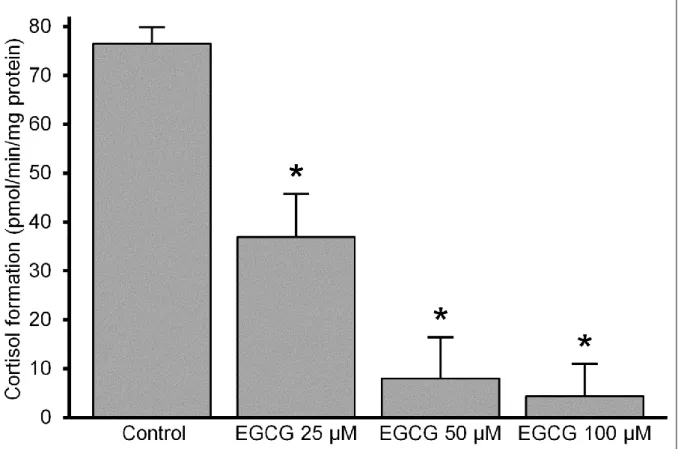
Concentration-dependent inhibition of microsomal cortisol production by EGCG The possible interference of EGCG with the overall activity of the G6PT-H6PD- 11βHSD1 triad (i.e., the cortisone reducing machinery of the ER) was tested first. Intact rat liver microsomal vesicles (0.5 mg/mL) were incubated in the presence of cortisone (5
13
μM) and G6P (50 μM) substrates for 30 min, and the amount of cortisol was measured with HPLC. Cortisol production was significantly inhibited by EGCG at 25 μM level; and the effect increased in a concentration-dependent manner. The rate of cortisone-cortisol conversion was less than half of the control value at 25 μM and hardly detectable above 50 μM concentrations of the flavanol (Fig. 2).
Figure 2 Effect of EGCG on cortisol production in intact microsomes (mean + SEM; n = 3; **p < 0,01 vs. control)
Effect of EGCG on individual protein components of the G6PT-H6PD-11βHSD1 catalytic triad
Hindrance of G6P-driven cortisol production observed in intact microsomes can be due to either direct inhibition of 11βHSD1 or decreased luminal NADPH generation. In other words, all the three members of the G6PT-H6PD-11βHSD1 triad are potential tar- gets of EGCG action. However, attenuation of G6P uptake into rat liver microsomal vesi- cles, that is, inhibition of G6PT by EGCG was ruled out in our previous study but the two luminal dehydrogenases remain to be investigated. The pore forming alamethicin (0.1
14
mg/mg protein) uncouples H6PD and 11βHSD1 activities by providing free access of the enzymes to NADP(H) cofactors added into the incubation medium. Therefore, the two enzymes can be separately studied in permeabilized microsomes. H6PD and 11bHSD1 activities were assessed by incubation of rat liver microsomes in the presence of NADP+ and realtime fluorescent detection of G6P-dependent or cortisol-dependent NADPH pro- duction, respectively. Both enzyme activities proved to be completely latent, that is, they only started upon membrane permeabilization due to luminal localization of the enzymes and impermeability of the ER membrane to pyridine nucleotides. This also approves membrane integrity and excludes the interference of extravesicular activities with our measurements. The linear increment of the fluorescent signal corresponds to H6PD or 11βHSD1-mediated NADPH generation. H6PD activity was not affected by EGCG even at 100 μM concentration of the flavanol (5.76 ± 0.65 nmol/mg vs. 5.77 ± 0.41 nmol/mg control value, mean ± SEM; n = 3); whereas 11βHSD1 activity was only slightly inhibit- ed (0.68 ± 0.05 nmol/mg vs. 0.82 ± 0.03 nmol/mg control value, mean ± SEM; n = 3; p <
0.05). This effect of EGCG on 11βHSD1 activity was also tested by HPLC measurement of cortisol-cortisone conversion in permeabilized microsomes. In line with the fluorimet- ric experiments, thealternative approach also revealed only a small inhibition. Statistical- ly significant, ≈30% reduction of 11bHSD1 activity was found only at the highest (100 μM) EGCG concentration (0.34 ± 0.02 nmol/mg vs. 0.51 ± 0.04 nmol/mg control value, mean ± SEM; n = 3; p < 0.05). It can be concluded that the remarkable suppression of microsomal cortisol production observed in intact microsomes (Fig. 2) cannot be attribut- ed to direct inhibition of either protein component involved in the process.
Luminal Redox Shift in EGCG-Treated Microsomes
The above results show that intact membrane barrier and retained compartmentation are prerequisites for an efficient inhibition of the cortisol producing machinery by EGCG.
This strongly suggests that the existence of a separate NADP+-NADPH pool plays a key role in the mechanism of action, and oxidation of luminal NADPH might underlie the at- tenuation of cortisone reduction, as it was reported earlier in case of metyrapone. Altera-
15
tion in the redox state of luminal NADP+-NADPH pool was first studied by estimating the endogenous reducing/oxidizing capacity of the microsomes, that is, their intrinsic ability to convert cortisone to cortisol and vice versa. The relevant enzyme activities were essentially not affected by EGCG; therefore, they could be reliably utilized in such an indirect analysis. Intact microsomes were incubated with either cortisone or cortisol and the rate of cortisol or cortisone production (driven merely by endogenous reducing or ox- idizing power) was measured by HPLC. Cortisone reducing power of the microsomes (control activity: 0.55 ± 0.04 pmol/min/mg protein; mean ± SEM; n = 3) was remarkably suppressed by EGCG in a concentration-dependent manner (Fig. 3). The effect was al- ready significant at 25 μM level, and cortisol production was not detectable in the pres- ence of the flavanol at 50 μM concentration or above. At the same time, cortisol oxidation (control activity: 2.37 ± 0.31 pmol/min/mg protein; mean ± SEM; n = 3) was largely enhanced by EGCG treatment (Fig. 3).
Figure 3 Effect of EGCG on the endogenous cortisone reducing / cortisol oxidizing capacity of the liver microsomes (mean + SEM, n = 3, *p < 0.05, **p < 0.01 vs. control)
ND: not detectable (below 1% of control)
16
The effect was saturable: the rate of cortisone production could be more than doubled up- on addition of 50μM EGCG and did not further increase at higher concentrations of the flavanol. Shift in the endogenous cortisol-cortisone conversion toward cortisone produc- tion is likely due to oxidation of luminal pyridine nucleotides. Therefore, changes in NADPH content of the ER vesicles were also assessed directly by fluorimetry. Luminal NADPH can be detected as stable baseline fluorescence in intact microsomal vesicles.
Addition of EGCG caused a remarkable decrement of this signal, which could be partial- ly reverted by the addition of G6P (Fig. 4). This phenomenon corresponds to an EGCG- induced conversion of luminal NADPH to NADP+ and a concomitant G6P-driven regen- eration of NADPH. Similar redox shift was detected upon the addition of metyrapone or cortisone in accordance with several studies reported earlier. When NADPH was added to a protein free EGCG solution (100 μM) in the same experimental condition, a sustained increase of the fluorescent signal was recorded, which rules out both non enzymatic oxi- dation of NADPH and fluorescence quenching by EGCG.
Figure 4 Effect of EGCG on the luminal NADPH pool in liver microsomes
17
Effect of EGCG on microsomal lipid peroxidation
The observed oxidative shift in the luminal NADP+-NADPH pool could be just part of a general oxidative stress involving peroxidation. Therefore, the possible effect of EGCG on lipid peroxidation was also tested and compared to ascorbate/Fe2+ combination by measuring the amount of TBARS in rat liver microsomes. According to the basically antioxidant features of tea flavanols, EGCG slightly (although not significantly) de- creased rather than increased peroxidation of membrane lipids, which strongly contradicts the role of a massive oxidative challenge in our system.
Conclusions
Two common nutritional factors exerting opposite in vivo effects on obesity-related metabolic disorders were examined during my Ph.D. work. It was investigated, whether they influence the pre-receptor activation of cortisol, and if so, what are their direct mo- lecular targets. Our results led to the following conclusions:
1. F6P promotes NADPH formation in the endoplasmic reticulum, and by this effect stimulates pre-receptor activation of cortisol as well.
2. The precondition of NADPH-generating and cortisol-reducing effect of F6P is its conversion to G6P, which is also catalyzed in the endoplasmic reticulum by an isomerase enzyme, different from the cytosolic PGI.
3. F6P enters the lumen of the endoplasmic reticulum via a protein mediated transport, and the yet-unidentified transporter differs from the G6PT protein.
4. The green tea flavanol EGCG inhibits cortisone-cortisol conversion in intact mi- crosomal vesicles. EGCG treatment reduces the endogenous cortison-reducing ca- pacity while increases cortisol-oxidizing capacity of liver microsomes.
5. EGCG-mediated inhibition of cortisol production is not due to interference with any member of the catalytic triad, but to oxidation of luminal NADPH.
18
6. The observed NADPH-oxidizing effect of EGCG is specific and enzymatic as it cannot be detected in the absence of microsomes, and is not accompanied by in- creased lipid peroxidation.
It can be concluded that the harmful effects of high fructose diet and positive health effects of tea flavanol consumption are related with their influence on pre-receptor corti- sol metabolism in the endoplasmic reticulum. This also provides evidence, that nutrient sensing in the organelle affects the metabolic state of the whole body. According to our results, the luminal pyridine nucleotide pool of the endoplasmic reticulum is a promising drug target for the treatment of obesity-related metabolic diseases.
Bibliography
Publications related to the theme of the Ph.D. thesis
1. Szelényi P, Révész K, Konta L, Tuttő A, Mandl J, Kereszturi É, Csala M. (2013) Inhibition of microsomal cortisol production by (-)-epigallocatechin-3-gallate through a redox shift in the endoplasmic reticulum-A potential new target for treat- ing obesity-related diseases. Biofactors, 39(5):534-41.; IF: 3,088
2. Senesi S, Legeza B, Balázs Z, Csala M, Marcolongo P, Kereszturi É, Szelényi P, Egger C, Fulceri R, Mandl J, Giunti R, Odermatt A, Bánhegyi G, Benedetti A.
(2010) Contribution of fructose-6-phosphate to glucocorticoid activation in the en- doplasmic reticulum: possible implication in the metabolic syndrome. Endocrinol- ogy, 151(10):4830-4839.; IF: 4,717
Publications not related to the theme of the PhD thesis
1. Révész K, Tuttő A, Szelényi P, Konta L. (2011) Tea flavan-3-ols as modulating factors in endoplasmic reticulum function. Nutrition research, 31(10):731-740.; IF:
2,142
2. Zámbó V, Simon-Szabó L, Szelényi P, Kereszturi E, Bánhegyi G, Csala M. (2013) Lipotoxicity in the liver. World Journal of Hepatology, 5(10):550-557.