Inhibition Due to Radiation
R. Goutier and Ζ. M. Bacq
I. Introduction 631 II. Effects in vitro 632
A. Direct and Indirect Action 632 B. Sulfhydryl Enzymes 634 C. Protection by Added Substances 635
III. Differences between in vitro and in vivo Effects 635
A . Activation of Enzymic Activities 635 B. Differences in Relative Biological Efficiency ( R B E ) 637
IV. Inhibitions in vivo 637 A. Inhibition of D N A Synthesis 637
B. Inhibition of Auxin Synthesis 641 C. Inhibition of Oxidative and Glycolytic Processes 642
D. Inhibition of Phosphorylation 643 E . Inhibition of Various Enzymes 645
V. Conclusions 646 References 647
I. INTRODUCTION
The most conspicuous effect of ionizing radiation on living matter is the inhibition of mitotic activity. It was discovered 54 years ago and clearly depicted by Bergonie and Tribondeau (1), who noticed that the higher the rate of mitotic activity of living tissue, the higher the sensi
tivity to radiation.
In the last 20 years, a large number of publications appeared dealing with the action of radiation on enzymes involved in the diverse aspects of cell metabolism. Although we shall coneern ourselves here mainly with the inhibitory action of radiation on enzymes, we must not forget that activation is by no means a rare event in vivo and in some cases leads to reinforcement of the deleterious effect of the inhibition of other enzyme systems on the celPs life.
631
II. EFFECTS
IN VITROActivation of enzymes is essentially an in vivo effect of radiation, the reasons of which will be given in the following sections.
On the other hand, in vitro irradiation of purified enzymes always results in a decrease of enzyme activity. There is no exception to this rule. In Table I are listed a few examples of in vitro inhibition of enzymes by X - and γ-rays.
A. Direct a n d Indirect Action
The inhibition observed in vitro when dilute aqueous solutions of pure enzymes are irradiated is mainly due to indirect effects, that is to say, to the interaction of free radicals produced by ionization of water with the enzyme molecule. The direct hit of radiation on the enzyme molecule represents an increasing percentage of the total effect with increasing enzyme concentration, but still remains less important than the indirect effect. This has been shown by Dale (9) for carboxypeptidase.
Useful discussions of the relative importance of direct and indirect action of ionizing radiation on enzymes in vitro may be found in Bacq and Alexander's textbook (10) and in Dale's lecture (11). An exam
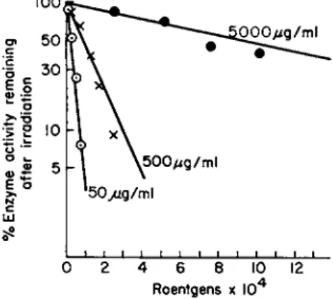
ple of enzyme inhibition in vitro is given in Fig. 1, where the decreas-
100
Ο Ο Ο μ ς / m l
4 6 8
Roentgens χ 10"*
FIG. 1. Effect of X-rays on D N a s e activity. Each point on the curve repre
sents the average of 5 experiments with a standard deviation from the mean of ± 10%. From Okada (6).
ing inhibitory effect with increasing enzyme concentration points to an indirect effect of radiation by way of free radicals originating from
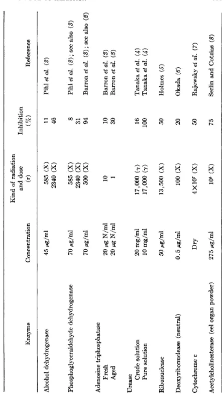
TABLE I INHIBITION OF PURIFIED ENZYMES BY RADIATION Enzyme Concentration
Kind of radiation and dose (r) Inhibition (%) Reference Alcohol dehydrogenase 45 Mg/ml 585 (X) 2340 (X) 11 46 Pihl et al. (2) Phosphoglyceraldehyde dehydrogenase 70 Mg/ml 70 Mg/ml 585 (X) 2340 (X) 500 (X)
8 31 94
Pihl et al. (2); see also (3) Barron et al. (3); see also (2) Adenosine triphosphatase Fresh Aged 20 Mg N/ml 20 Mg N/ml 10 1 10 30 Barron et al. (3) Barron et al. (3) Urease Crude solution Pure solution 20 mg/ml 10 mg/ml 17,000 (7) 17,000 (7) 16 100 Tanaka et al. (4) Tanaka et al. (4) Ribonuclease 50 Mg/ml 13,500 (X) 50 Holmes (δ) Deoxyribonuclease (neutral) 0.5 Mg/ml 100 (X) 20 Okada (6) Cytochrome c Dry 4X10⁷ (X) 50 Rajewsky et al. (J) Acetylcholinesterase (eel organ powder) 275 Mg/ml 106 (X) 75 Serlin and Cotzias (8)
radiolysis of water. Indeed, at a given radiation dose the number of free radicals produced in the solvent is always the same, regardless of the concentration of the solute. Accordingly, the number of enzyme molecules attacked by these radicals will also be the same at any con
centration, and therefore the percentage of inactivation of the enzyme decreases with increasing concentration (10).
B. Sulfhydryl Enzymes
The production of oxidizing radicals ( Ο Η · and H 02 #) and hydrogen peroxide in irradiated water is liable to oxidize the sulfhydryl groups and to inactivate such enzymes as phosphoglyceraldehyde dehydro
genase, alcohol dehydrogenase, hexokinase, and adenosinetriphosphatase in which free SH groups are essential for enzymic activity. Barron (8, 12) performed experiments which pointed to a definitely greater sensitivity of SH enzymes to radiation in vitro. Two examples from his work are quoted in Table I ; the greater sensitivity of aged solutions of adenosinetriphosphatase might be due to a decreased amount of free SH groups prior to irradiation. Unfortunately, these facts could not be confirmed by Pihl et al. (2), who observed that SH enzymes are inhibited by X-rays in vitro with the same ionic yield as non-SH enzymes
(Table I ) . According to them, the ionic yield for the inhibition of phos
phoglyceraldehyde dehydrogenase is 0.02, similar to that for non-SH enzymes, whereas Barron found an ionic yield equal to 1. They likewise did not confirm the reactivation of irradiated SH enzymes (phosphogly
ceraldehyde dehydrogenase) by addition of glutathione, although gluta
thione protects urease (another SH enzyme) against inactivation by γ-rays (4), i.e., decreases the number of altered molecules, provided that glutathione is introduced into the system before irradiation.
Evidence, recently presented, shows that the inactivation of dry ribonuclease by irradiation is associated with the rupture of the S—S bond (12a).
At the present time, the idea of a preferential inhibition of SH enzymes by irradiation in vitro has been abandoned.
In vivo, inactivation of SH enzymes and coenzymes does not even seem to occur, unless at high doses after a long latent period.
Coenzyme A is not inactivated in the first hours following total body irradiation, as proved by the normal rate of acetylation of p-amino- benzoic acid (13). Other examples are presented and discussed by Errera (H), e.g., the lack of inhibition, or the weak inhibition of suc
cinic dehydrogenase and hexokinase in tissues. Therefore, oxidation of
SH groups, even if it occurs to some extent in vivo, does not play a leading role in the eruption and the evolution of the radiolesion (10, 15).
C. Protection b y A d d e d Substances
B y adding to a purified enzyme solution other substances, the mole
cules of which will compete with the enzyme molecules for the free radi
cals produced during irradiation, one decreases the probability of re
action between free radicals and enzyme molecules. Many examples are quoted in a recent review by Marples and Glew (16). The more purified an enzyme, the higher its sensitivity to radiation. Braams (17) found that different purification methods of invertase brought about different sensitivities of the enzyme to α-rays and protons.
Neutral deoxyribonuclease (DNase) can be protected against X-irra- diation by addition of various compounds (18), but it is thought provoking that the best protection is afforded by deoxyribonucleic acid
( D N A ) , the substrate of the enzyme, which is, indeed, the most likely substance to combine with the active sites of the enzyme molecule.
Cellulose, Dowex, silicagel, and mitochondria all protect neutral D N a s e in vitro. The behavior of the dry enzyme is quite different; dry neutral D N a s e is more radiosensitive than wet D N a s e (the water molecules would seem to protect the enzyme), and adsorption of the dry enzyme on a solid support leads to an increased radiosensitivity, contrary to what happens to the wet enzyme. But, wet or dry, D N a s e is protected against radiation by complexing with its substrate, D N A (19, 20).
III. DIFFERENCES BETWEEN
IN VITROA N D
IN VIVOEFFECTS
A. Activation of Enzymic Activities
Two main factors determine the wide differences observed between the effects of irradiation on a given enzyme in solution and on the same enzyme in a living tissue: the presence of many other molecules in a cell and the possible adsorption of the enzyme on the cell substructure.
Both factors afford protection; enzymes are much more resistant when irradiated in vivo than when purified and irradiated in vitro.
As we have already said, enzymic activity is often increased by irra
diation of a living organism. This is, indeed, the main difference between in vitro and in vivo effects of irradiation. I t deserves a short comment.
The example of neutral D N a s e is striking in this respect; D N a s e is
inhibited in vitro (see Table I) by doses which produce activation in vivo [21). In vivo, D N a s e (acid DNase, especially) always displays a sharp increase of activity in various tissues after irradiation [for references, see the review by Goutier (22).] Other enzymes, such as cathepsin (28) and ribonuclease (24), also undergo activation after in vivo irradiation. Together with acid DNase, they are located in the lysosomes (25). The activation of these enzymes may originate from the rupture of the granules by irradiation and the release of their con
tents into the cell sap. This has been shown to be the case for acid D N a s e (26) and ribonuclease (27) and has been considered by Bacq and Errera (28) as one of the main primary mechanisms by which radiation impairs cell life. This is the "enzyme release theory," accord
ing to which more enzyme comes into contact with its substrate. Acid DNase, for example, is concentrated in the lysosomes at a distance from any substrate. The distance, on the molecular scale, between this DNase and D N A in the nucleus is enormous. DNase, in order to be active, must (a) be released from its links in the lysosomes, (b) travel through submicroscopic structures in the cytoplasm, and (c) cross the nuclear membrane.
Other examples of increase of enzyme activity after irradiation of an animal may be found in Bacq and Alexander (10). Besides rupture of granules or lysosomes, activation of enzyme activity after in vivo irradiation may be due to the destruction of an inhibitor occurring naturally in normal tissues, as is the case for carboxypeptidase (28) and neutral D N a s e (29), to an endocrine reaction, such as the suprarenal cortex reaction inducing an increased synthesis of the tryptophan peroxidase in rat liver (80), or to an increase in cell membrane permea
bility, which is probably responsible for the increase of glutamic- oxalacetic transaminase activity in rabbit serum (31, 82).
The complexity of a living cell makes it difficult to predict the behavior of a particular enzymic activity after in vivo irradiation. For instance, it would be incorrect to assume that irradiation always inhibits syntheses and activates hydrolytic mechanisms. Although this is roughly true under certain conditions for D N A metabolism, it does not hold for hemin and globin syntheses, which are stimulated by in vivo irradiation, as found when bone marrow cells or reticulocytes were tested 30 minutes after irradiation (38, 84) · Cholesterol synthesis, measured by incorpora
tion of acetate-l-C1 4 and tritiated water into intact animals and into tissue slices, has also been shown to be enhanced to two times normal after 2400 r total body X-irradiation of rats (85). Cathepsin also seems to be synthesized at a higher rate in the thymus of irradiated rats; this
increase, however, is associated with phagocytosis and may represent enzyme induction rather than enzyme activation (35a). Nevertheless, many syntheses, some of which are most important for cell mitosis and growth, are indeed inhibited.
B. Differences in Relative Biological Efficiency (RBE)
Fast neutrons and α-particles (densely ionizing radiations) kill cells more effectively than hard X-rays and γ-rays (sparsely ionizing radia
tions). If cell death originates from alteration of any particular chemical or enzymic reaction, one would expect this reaction to display in vitro the same relative sensitivities to different radiations as in vivo. But, in fact, for most radiochemical and enzymic in vitro reactions known so far, the R B E is higher for hard X-rays and γ-rays than for α-particles, contrary to what is observed in vivo.
On these grounds, the inactivation of enzymes observed in vitro can hardly be accepted as a possible primary lesion (36, 37).
The only case where the RBE's are qualitatively the same in vitro as in vivo for different radiations is the production of double breaks in the D N A molecule. The alteration of the D N A molecule might therefore be an acceptable candidate for the primary lesion. But, as pointed out by Bacq and Alexander (38), cells without D N A (red cells) or anucleated fragments of cells (Amoeba, Acetabularia mediterranaea) show typical radiolesions.
IV. INHIBITIONS IN VIVO
In the review by Errera (14) are listed the enzymes inhibited after in vivo irradiation (see also 38a). We want to discuss some examples of inhibition of biologically important systems in which the inhibited enzymes or enzymic reactions have been clearly identified.
A. Inhibition of DNA Synthesis
A large number of publications describe the decrease in the incorpora
tion of labeled precursors into D N A after in vivo irradiation. Recent reviews will provide the reader with the references: Bacq and Alexander
(10), Errera (14), Howard (39), Kelly (40), Holmes (41), Stocken (42),
and Goutier (22). The biological importance of this effect of radiation does not need to be emphasized.
Recent investigations have shown that in tissue cultures (42a) and in regenerating rat liver (42b), irradiation results in an inhibition of D N A synthesis in all cells in the synthetic phase and not in a decrease in the number of cells in the same phase.
Considering only the enzymological point of view, the radiolesion seems to be located at the end of the long sequence of events leading to the biosynthesis of D N A , when the nucleosides are converted to nucleo
side triphosphates by specific kinases and then linked together to form the polynucleotide chain as a result of the intervention of a specific polymerase.
Since nucleosides accumulate in the tissues of an irradiated animal, it may be concluded that irradiation does not inhibit the nucleoside syn
thesis (43-49).
The first lesion that has been detected in the reaction sequence is a decreased rate of formation of the nucleoside triphosphates. Creasy and Stocken (50) observed that within 1 hour, the formation of high energy phosphorus compounds by nuclear suspensions of thymus tissue was suppressed by 100 r; it was only 20-50% of normal after 25 r total body irradiation. The inhibition can already be detected 3-5 minutes after
100 r. This is shown in Table II. Potter (49) observed that within a few
T A B L E I I
INHIBITION OF THE FORMATION OF LABILE PHOSPHATE IN NUCLEI ISOLATED FROM TISSUES OF RATS EXPOSED TO A WHOLE BODY X-IRRADIATION 1 HOUR PREVIOUSLY0
Tissue
Rate of phosphorylation for various doses of X-irradiation as percentage of control values
Tissue
2 5 r 5 0 r 1 0 0 r
Spleen 1 8 , 3 0 0, 0, 0 0, 0, 0
Thymus gland 4 5 , 7 7 1 4 , 2 3 0, 0, 0, 0, 0
Bone marrow 0 , 0 0 0
Lymph node 0 , 6 0 0
α From Creasey and Stocken ( 5 0 ) .
hours, in vivo irradiation with 100-3200 rep gamma rays inhibited the incorporation of thymidine-C1 4 into thymine nucleotides and, to a still greater extent, into D N A .
That inhibition of the polymerization of nucleotides also occurs has
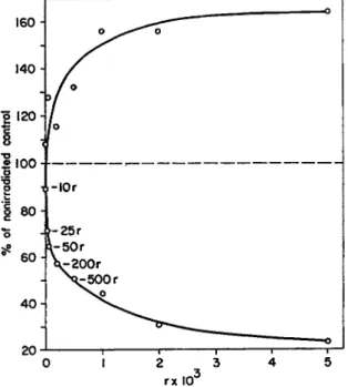
been shown by Okada and Hempelmann (51) and by Bollum et al. (52) in regenerating livers of irradiated rats. The latter authors determined the activity of thymidine kinase (converting thymidine to thymidine mono- phosphate), of thymidylic kinase (converting thymidine monophosphate to thymidine triphosphate), and of polymerase (converting deoxyribo- nucleoside triphosphates into D N A ) . These enzymes are known to appear in the supernatant fraction of homogenates of regenerating liver in increasing amounts at the time that D N A synthesis begins in vivo, between 16 and 24 hours after partial hepatectomy (58). When the animals are irradiated 6 hours after hepatectomy, when D N A synthesis has not yet started, the appearance of the enzymes is delayed, and their activity is lower (depending on the radiation dose), as can be seen in Fig. 2. Since the inhibition of the over-all incorporation of thymidine-H3
X-Ray dose (Roentgen)
FIG. 2. Effect of X-ray dose given 6 hours after partial hepatectomy on the kinase and polymerase enzymes and on the over-all incorporation of thymi- dine-H3 into D N A , measured by in vitro assays 24 hours after the operation.
The data are not corrected for the levels occurring in the tissues in the period 6-16 hours after the operation. Data on the X-ray inhibition of the in vivo incorporation of orotic a c i d - C1 4 into D N A of regenerating rat liver are shown for comparison. The latter data are from Beltz, Van Lancker, and Potter.
O.A. = orotic acid; T D R = thymidine. From Bollum et al. (52).
into D N A closely parallels the inhibition of the kinase, it is clear that the kinase is the rate-limiting step. On the other hand, irradiation given 16 hours after hepatectomy does not slow down the ultimate increase in the enzyme activities which are normally present at that time, although incorporation of thymidine into D N A is still much depressed. In other
words, in regenerating liver, irradiation prevents the synthesis of the enzymes rather than inhibits the activities of the enzymes themselves.
This is an important point to note.
Van Lancker (53a), working with isolated nuclei of regenerating liver, came to the same conclusion. A similar delay has been found in the appearance of deoxycytidine deaminase activity in irradiated regener
ating liver (53b, 53c).
As to our understanding of the inhibition of D N A synthesis by radia
tion, the experiments of Bollum et al. (52, 53) also show that, in liver at least, if the depressed synthesis of D N A may be explained by an inhibition of the synthetic enzymes in the early stages of liver regenera
tion, it must be ascribed to another still unknown cause when irradiation is given at later times, since the liver tissue is then incapable of syn
thesizing D N A in vivo despite the presence in the in vitro tests of a normal activity of synthetic enzymes and despite a slight decrease in D N a s e activity (51, 54).
The progressive inhibition of D N A synthesis observed in vivo when irradiation is given to rats 24 hours after partial hepatectomy is ascribed to a slowing down of the mitotic cycle because no change is observed in the capacity of isolated cells from irradiated livers to incorporate thymidine in vitro in D N A (54a). In rat spleen and small intestine, the radiosensitivity of the nucleotide polymerization process even seems doubtful, and the inhibition of the nucleotide-phosphorylating kinases is too small to account for the in vivo inhibition of D N A synthesis (54b).
In these cases, losses of ions such as Κ or Ca by the nuclei of irradiated tissues (54c) perhaps cause important damage to the process of D N A synthesis (54b).
Other parameters, more recently investigated, may also be involved in the effect of irradiation on D N A synthesis.
Factors inhibiting the DNA-synthesizing enzymes have been described in the supernatant (54d) and microsomal fractions (54c) of normal rat liver homogenates. Induced regeneration of the liver leads to a decrease of the microsomal inhibiting factor; irradiation at lethal X-ray doses prior to partial hepatectomy prevents the normal decrease of the inhibit
ing factor (54c).
Macromolecules such as D N A , RNA, and proteins penetrate into cells in tissue culture (54], 54g) and are translocated within plant tissues (54h) when added to the external medium. Although it is not known whether translocation of the plant's own D N A and R N A molecules also occurs under physiological conditions, irradiation depresses the trans
location of added D N A and R N A in barley seedlings (54h).
B. Inhibition of Auxin Synthesis
In plant tissues, inhibition of D N A synthesis also occurs after irradia
tion (55, 56) and can reach 40% in terms of P3 2 incorporation, after moderate doses of 50-200 r X-rays (55). The same doses affect to an almost similar degree the synthesis of auxin which is also indispensable for plant growth.
The formation of auxin has been studied by Gordon (57). Tryptophan is the starting material, and, in the last step, oxidation of indoleacetalde- hyde yields the indoleacetic acid which exerts the hormonal action.
According to Gordon (58), it is this last conversion of acetaldehyde to the acid form that is inhibited by irradiation.
Figure 3, taken from Gordon (58), shows the accumulation of indole-
20 J 1 1 1 1 r
0 1 2 3 4 5
rx ΙΟ3
FIG. 3. Conversion of tryptophan to auxin by cell-free homogenates of X-irradiated mung bean seedlings. The lower curve gives the relative amount of indoleacetic acid formed; the upper curve, indoleacetaldehyde. From Gor
don (58).
acetaldehyde with a corresponding decrease of indoleacetic acid in irradiated mung bean seedlings. It must be pointed out that this reaction is sensitive to very low doses of X-rays and that the inhibition can already be detected a few minutes after irradiation.
C. Inhibition of Oxidative a n d Glycolytic Processes
Here, again, much difference is observed between the effects of in vivo and in vitro irradiation. In isolated rat liver mitochondria, Fritz-Niggli
(59) produced a 10% inhibition of the oxidation of citrate after in vitro X-irradiation by 50 r, when the mitochondria were isolated in 5.75%
mannitol, and 60% inhibition, when they were isolated in 2.88% man- nitol (the second medium is kept isotonic by means of phosphate buffer).
The oxygen uptake by isolated mitochondria seems to be very radio
sensitive, since, according to Fritz-Niggli (60), it displayed 60 and 74%
inhibition % and 1 hour, respectively, after in vitro irradiation with 0.1 r X-rays. The experiments of Fritz-Niggli have never been confirmed, a fact which suggests that an unknown important factor is introduced during the preparation of the mitochondria.
But, when oxidase activities are individually measured in irradiated liver slices, hardly any inhibition of succinic dehydrogenase, cytochrome oxidase, and succinoxidase is observed for X-ray doses below 106 r (61).
In order to explain this discrepancy, one might think that the isolated mitochondria used by Fritz-Niggli had already been damaged by the isolation procedure and were, for this reason, more sensitive to external attack, whereas in the liver slices, used by Rajewsky, the mitochondria were still in a more physiological state.
The same phenomenon is observed with Tetrahymena. Whole cells given 3 χ 105-6 Χ 105 r retain an almost normal level of oxidizing enzymes, whereas in irradiated cell-free homogenates, the oxidase activi
ties are greatly depressed (62, 68). Here, too, the preservation of intact cell structures protects the enzymes against inactivation by irradiation.
After total body irradiation, the effects observed depend both on the radiation dose and the time lag between radiation and observation. The immediate effects were studied by Smith and Thomson (64), who irradi
ated rats, mice, hamsters, bats, fowls, and frogs continuously at 500 r/min until death; they did not observe any significant changes in aerobic and anaerobic glycolysis, oxidative phosphorylation, succinic dehydrogenase, cytochrome oxidase, adenosinetriphosphatase, and per
oxidase in various tissues. In rats killed immediately after increasing doses of X-rays, Belokonsky and Rusev (65) found an increased 02 demand, with an increase of cytochrome oxidase and a reduction of succinic dehydrogenase in various tissues after doses up to 500-1000 r.
Higher doses (1000-20,000 r) produce a depression of oxidative processes with a reactivation of succinic dehydrogenase in the resistant tissues only.
Observations made several hours after irradiation do not always give concordant results. Ryser et al. (66) do not find any inhibition of suc
cinic dehydrogenase in rat liver 1 - 3 days after 1 0 0 0 r total body irradia
tion. In the rat thymus, likewise, negligible changes in succinic dehydro
genase, malic dehydrogenase, cytochrome oxidase, and adenosinetriphos- phatase occur after 8 0 0 r X-rays (67). On the other hand, according to Di Bella (68), succinic dehydrogenase is inhibited by 5 0 % in the liver and by 7 5 % in the bone marrow 1 - 2 days after 8 0 0 r total body X-irra- diation of rats.
Internal irradiation of rats by injection of 5 μο P3 2/ g m produces a 6 0 % inhibition of anaerobic glycolysis in liver slices after 2 4 hours despite an increase in glycogen content. N o change in succinic dehydro
genase, cytochrome oxidase, fumarase, and phosphorylase is observed (69).
Anaerobic glycolysis in Krebs ascites cells is 5 0 % inhibited after 1 0 0 0 r in vitro irradiation (70, 71). This effect can be mimicked by addition of H202 to the cell suspension. Addition of catalase protects the cells against inhibition of glycolysis by X-rays. Catalase itself is inhibited by total body irradiation of mice, where 8 5 0 r produce a 2 0 % fall in liver catalase activity after 2 4 hours (72).
High doses of X-rays ( > 2 0 kr) given to ascites tumor cells in vitro produce an increase in 02 consumption and in aerobic glycolysis and a depression in anaerobic glycolysis (72a) which is due to a dose- dependent decrease of A T P and D P N (72b). Inhibition of aerobic glycolysis seems, however, to occur in hepatoma cells after in vitro irradiation by 1 0 - 1 0 0 kr (72c).
In conclusion, despite the lack of consistency in the literature, oxida
tive enzymes and glycolytic processes seem to be less radiosensitive in vivo than other processes, such as D N A synthesis and nuclear phos
phorylation.
The delayed inactivation by ionizing radiation of enzymes in vivo may also be interpreted by the enzyme-release theory. Enzymes linked to organelles are protected against inactivation and do not leak into all sap or circulating fluids; when they are set free by irradiation, they leak into the surrounding fluid and may be eliminated or become substrates for inactivating factors.
D. Inhibition of Phosphorylation 1. I N M I T O C H O N D R I A
Decreased P / O ratios were found by Potter and Bethell (73) in rat
spleen mitochondria 1 hour after 8 0 0 r. This was confirmed by Hickman and Ashwell (74).
Van Bekkum et al. (75) showed that 4 hours after 1 1 0 0 r total body irradiation, the phosphorylation, the oxygen uptake, and the P / O ratio were significantly decreased. In thymus, 5 0 r is able to inhibit oxidative phosphorylation. The mitochondrial damage brings about a release of cytochrome c, since cytochrome c added to mitochondria isolated after irradiation considerably enhances the P / O ratio as well as the phos
phorylation and the oxygen uptake (76). Whether nuclear damage, which develops at the same time, has a bearing on the mitochondrial damage, is not known (77). When irradiated in vitro, isolated mitochon
dria possess normal levels of oxidative phosphorylation even after
2 0 , 0 0 0 r (76).
The inhibition of oxidative phosphorylation in rat spleen and liver 2 4 hours after 8 0 0 r total body irradiation is thought by Benjamin and Yost (78) to be an indirect effect of radiation, the inactivation being due in the spleen to an overproduction of thyroxine and in the liver to an overproduction of corticosteroids.
From a comparison between two mammary tumors, Goldfeder (78a) concludes that cells having a smaller number of mitochondria and, therefore, a lower level of phosphorylations are more radiosensitive.
2 . I N T H E N U C L E U S
Oxidative phosphorylation is an energy source common to all cells.
Since some tissues are sensitive to radiation and others are not, it was interesting to look for a biochemical property which would be both sensi
tive to radiation and specific for radiosensitive tissues.
Creasy and Stocken (50) found that the formation of high energy phosphorus compounds by nuclear suspensions occurred in radiosensi
tive organs such as thymus, spleen, intestinal mucosa, and bone marrow and was lacking in liver, brain, kidney, and pancreas. The phosphoryla
tion reaction is suppressed in the radiosensitive organ 1 hour after 5 0 r total body X-irradiation. N o labile phosphate is formed in suspensions of spleen cell nuclei after 4 4 r γ-irradiation given to the nuclear suspen
sion in vitro (see Table I I ) . This seems to be a very sensitive test for early radiation damage. It has recently been confirmed and extended by Ord and Stocken (78b).
Maass (79) observed a decrease of A T P with a simultaneous increase of A M P in various tissues of rats irradiated with 8 0 0 r. He found the same phenomenon in the livers of dormice (Glis glis), provided the animals were irradiated awake. When irradiation was given during
hibernation, no modification of A M P or A T P occurred. This is another indication that phosphorylation of nucleotides is related to the radio- sensitivity of a tissue.
E. Inhibition of Various Enzymes
1. C I T R I C A C I D S Y N T H E S I S
Injection of fluoroacetate into animals brings about an accumulation of citric acid (80, 81) due to inhibition of the oxidation of citrate by conversion of fluoroacetate to fluorocitrate (82). After 800 r X-irradia- tion (83) the accumulation of citric acid after fluoroacetate injection of rats is inhibited in spleen, thymus, ileum, pancreas, and testes; no effect is observed in heart and brain. Sublethal doses prevent the accumulation in spleen and thymus; this effect is reversible after sublethal doses only.
Normal male rats, injected with fluoroacetate, do not accumulate citrate in liver, in contrast to female rats. After irradiation, even injected male rats accumulate citrate in liver; sexual hormones may play a role in this effect of radiation on liver.
It is difficult to detect the specific cause of the inhibition by irradiation of citric acid accumulation in animals injected with fluoroacetate. As Potter (84) pointed out, the cause may be very remote from a direct inter- ference with a reaction in the Krebs cycle. In any case, an inhibition of SH enzymes and coenzymes does not seem to be worth considering;
succinic dehydrogenase is hardly inhibited by the X-ray doses used by D u Bois [see Thomson et al. (85)]. The inhibition of coenzyme A is a rather late phenomenon; a 37% inhibition has been observed in pigeon liver 6-7 days after doses of 3000 r (86).
2. C H O L I N E S T E R A S E
In intestinal loops of irradiated rats, Burn et al. (87) found that pseudocholinesterase is 50% inhibited 48 hours after 1000 r total body irradiation, whereas acetylcholinesterase (the "true" enzyme) is not.
The changes observed 24 hours after irradiation are small (despite more important changes in intestinal motility).
One can only speculate about the mechanism of pseudocholinesterase inhibition. Since pseudocholinesterase is synthesized in liver, is the liver primarily affected by irradiation? Or if the synthesis in liver is con- trolled by the anterior pituitary gland, is the inhibition of pseudocholin- esterase a part of the stress syndrome?
Conard (88) observed a significant depression of pseudocholinesterase
in rat small intestine 20 hours after 500 r total body irradiation; on the fourth day, the inhibition was 60%. Nevertheless, according to Conard, these changes cannot be related to the changes observed in the motility of the intestine. In other organs, however, the activity of cholinesterases seems to be affected differently by irradiation. According to Lundin and Clemedson (88a), irradiation of the guinea pig by 400 r does not produce any change in liver pseudocholinesterase activity, whereas the enzyme activity is decreased in the plasma and increased in the bone marrow.
V . C O N C L U S I O N S
The earliest detected biochemical lesion in many irradiated tissues is the disturbance of nucleic acid metabolism and, especially, the inhibition of D N A synthesis, where polymerization rate and formation of nucleoside triphosphates are both decreased. This, however, does not account for the antimitotic action of radiation, since it has been possible in many in
stances to inhibit mitoses without inhibiting the D N A synthesis to the same extent (a phenomenon which leads to the formation of giant cells (89-91)). Some cases are known where mitosis occurs without D N A synthesis, as in Tetrahymena which can be forced to synthesize D N A without dividing and then to go through several mitoses in succession
(92) without intermediate periods of D N A synthesis.
In plant tissues, irradiation may lead to increased cell growth or elon
gation without any mitotic activity present (93). At the present time, despite the broad insight gained on enzyme attack by radiation, we are still unable to offer a completely satisfactory explanation of the first observed effect of radiation, the mitotic block.
The mixture of inhibitions and activations of enzymes in irradiated tissues which we describe in this chapter reflects the complexity of the reaction of such a highly organized body as a living cell when energy is deposited by ionizing radiation.
Ionizing radiations, like other physical agents, do not inactivate a particular enzyme or series of enzymes in the tissues. After a short period of general metabolic stimulation, irradiation causes many aberrations in biochemical organization which lead to (1) mitotic arrest, (2) growth inhibition generally, and (3), if the dose has been large enough, cell death. The practical use of ionizing radiation in medicine is not re
stricted to cancer therapy, and many clinical observations about appar
ently contradictory effects of irradiation are now better understood.
Progress in fundamental radiobiology is critically linked with progress
in cell biochemistry and biophysics, and an urgent need is felt for a deeper understanding of the metabolic relations between the nucleus and the cytoplasm.
REFERENCES
1. J. Bergonie and L. Tribondeau, Compt. rend. acad. sci. 143, 983 (1906).
2. A. Pihl, R. Lange, and L. Eldjarn, Nature 182,1732 (1958).
3. E. S. G. Barron, S. Dickman, J. A. Muntz, and T. P. Singer, J. Gen.
Physiol. 32, 537 (1949).
4. S. Tanaka, H. Hatano, and S. Ganno, J. Biochem. 46,485 (1959).
5. B. Holmes, Nature 165, 266 (1950).
6. S. Okada, Arch. Biochem. Biophys. 67, 102 (1957).
7. B. Rajewsky, G. Gerber, and H. Pauly, in "Advances in Radiobiology"
(G. C. de Hevesy, A. Forssberg and J. D. Abbatt, eds.), p. 25. C. C.
Thomas, Springfield, Illinois, 1956.
8. I. Serlin and G. C. Cotzias, Radiation Research 6, 55 (1957).
9. W. M. Dale, Brit. J. Radiol., Suppl. 1, 46 (1947).
10. Ζ. M. Bacq and P. Alexander, "Fundamentals of Radiobiology," 2nd rev.
ed. Pergamon Press, N e w York, 1961,
11. W. M. Dale, in "Ionizing Radiation and Cell Metabolism," Ciba Founda
tion Symposium (G. E . W. Wolstenholme and C. M. O'Connor, eds.), p. 25. Little, Brown, Boston, Massachusetts, 1956.
12. E . S. G. Barron, Radiation Research 1, 109 (1954).
12a. D. K. Ray, F. Hutchinson, and J. Morowitz, Nature 186, 312 (1960).
13. J. F. Thomson and Ε . T. Mikuta, Proc. Soc. Exptl. Biol. Med. 86, 487 (1954).
14. M. Errera, in "Protoplasmatologia" (L. V. Heilbrunn and F. Weber, eds.), Vol. 10, 3. Springer, Berlin, 1957.
15. M. Errera, Am. Naturalist 94, 111 (1950).
16. A. Marples and G. Glew, Atomic Energy Research Establishment Rept.
A E R E I / R 2726 (1958).
17. R. Braams, Radiation Research 12, 113 (1960).
18. S. Okada, Arch. Biochem. Biophys. 67,113 (1957).
19. G. Fletcher and S. Okada, Radiation Research 11, 291 (1959).
20. S. Okada and G. L. Fletcher, Radiation Research 13, 92 (1960).
21. P. P. Weymouth, Radiation Research 8, 307 (1958).
22. R. Goutier, Progr. in Biophys. 11, 53 (1961).
23. R. N. Feinstein and J. C. Ballin, Proc. Soc. Exptl. Biol. Med. 53, 6 (1953).
24. J. S. Roth, Arch. Biochem. Biophys. 60, 7 (1956).
25. C. de Duve, B. C. Pressman, R. Gianetto, R. Wattiaux, and F. Appelmans, Biochem. J. 60, 604 (1955).
26. M. Goutier-Pirotte and A. Thonnard, Biochim. et Biophys. Acta 22, 396 (1956).
27. J. S. Roth, Radiation Research 9,173 (1958).
28. Ζ. M. Bacq and M. Errera, Preliminary Report to the U . N . Scientific Committee on the Effects of Atomic Radiation. Document A (AC-82), 1210 (1958).
29. Ν. B. Kurnick, B. W. Massey, and G. Sandeen, Radiation Research 11, 101 (1959).
30. J. E . Thomson and Ε. T. Mikuta, Argonne Natl. Lab., ANL-4794, p. 140;
ANL-4840, p. 80; ANL-4932, p. 59 (1952).
31. R. L. Brent, Μ. M. McLaughlin, and J. N. Stabile, Radiation Research 9, 24 (1958).
32. H. G. Albaum, Radiation Research 12, 186 (1960).
33. J. E. Richmond, Κ. I. Altman, and K. Salomon, J. Biol Chem. 190, 817 (1951).
34. A. Nizet, S. Lambert, and Ζ. M. Bacq, Arch, intern, physiol 62, 129 (1954).
35. R. G. Gould, Los Alamos Sci. Lab., LA-2179 (1958).
35a. U. Hagen, Strahlentherapie 117, 201 (1962).
36. P. Alexander, J. T. Lett, H. Moroson, and Κ. E . Stacey, Intern. J. Rad.
Biol Suppl. 1, 47 (1960).
37. P. Alexander, Proc. 8th Intern. Congr. Radiol, Miinchen, 1959, publ. G.
Thieme (1960).
38. Ζ. M. Bacq and P. Alexander, "The Initial Effects of Ionizing Radiations on Cells" (R. J. C. Harris, ed.), p. 301. Academic Press, N e w York, 1961.
38a. G. B. Gerber, Klin. Physiol. 1, 244 ( I 9 6 0 ) .
39. A. Howard, in "Ionizing Radiation and Cell Metabolism," Ciba Foundation Symposium (G. E. W. Wolstenholme and C. M. O'Connor, eds.), p. 196.
Little, Brown, Boston, Massachusetts, 1956.
40. L. S. Kelly, Progr. in Biophys. 8,144 (1957).
41. Β. E. Holmes, Ann. Rev. Nuclear Sci. 7, 89 (1957).
42. L. A. Stocken, Radiation Research Suppl. 1, 53 (1959).
42a. C. L. Smith, IAEA Symposium on Tritium in the Physical and Biological Sciences 2, SSI (1962).
42b. W. B. Looney, R. C. Campbell, and Β. E. Holmes, Proc. Natl Acad. Sci.
U.S.46, 698 (1960).
43. C. W. Bishop and J. N. Davidson, Brit. J. Radiol. 30, 367 (1957).
44. M. G. Ord and L. A. Stocken, Biochim. et Biophys. Acta 29, 201 (1958).
45. H. Maass and G. Schubert, Proc. 2nd U.N. Intern. Conf. Peaceful Uses of Atomic Energy, Geneva, 1958 22, 449 (1959).
46. L. Cima, G. Fassina, and F. Pozza, Exptl. Cell Research 17, 1 (1959).
47. P. Mandel and P. Chambon, in "Symposium on the Immediate and Low Level Effects of Ionizing Radiations, Venice, 1959" Intern. J. Radiation Biol Suppl. 1,71 (1960).
48. J. J. Jaffa, L. G. Lajtha, G. Lascelles, M. G. Ord, and L. A. Stocken, Intern. J. Radiation Biol. 1, 241 (1953).
49. R. L. Potter, Federation Proc. 18 (1959).
50. W. A. Creasey and L. A. Stocken, Biochem. J. 72, 519 (1959).
51. S. Okada and L. H. Hempelmann, Intern. J. Radiation Biol 1, 305 (1959).
52. F. J. Bollum, J. W. Anderegg, A. B. McElya, and V. R. Potter, Cancer Research, 20, 138 (1960).
53. F. J. Bollum and V. R. Potter, Cancer Research 19, 561 (1959).
53a. J. L. van Lancker, Biochim. et Biophys. Acta 45, 63 (1960).
53b. D. K. Myers, C. A. Hemphill, and C. M. Townsend, Can. J. Biochem. 39, 1043 (1961).
53c. L. Stevens and L. A. Stocken, Biochem. Biophys. Research Communs. 7, 315 (1962).
54. R. Goutier, M. Goutier-Pirotte, and P. Ciccarone, in "Symposium on the Immediate and Low Level Effects of Ionizing Radiations, Venice, 1959"
Intern. J. Radiation Biol. Suppl. 1, 93 (1960).
54a. S. M. Lehnert and S. Okada, Intern. J. Rad. Biol. 5, 323 (1962).
54b. O. F . Nygaard and R. L. Potter, Radiation Research 16, 243 (1962).
5 4 c W. A. Creasey, Biochim. et Biophys. Acta 38,181 (1960).
54d. E . D. Gray, S. M. Weissman, V. Richards, D. Bell, Η. M. Keir, R. M. S.
Smellie, and J. N . Davidson, Biochim. et Biophys. Acta 45, 111 (1960).
54e. R. Goutier and I. Bologna, IAEA Symposium on the Biological Effects of Ionizing Radiations at the Molecular Level, Brno, 1962, p. 287.
54f. E . Borenfreund and A. Bendich, J. Biophys. Biochem. Cytol. 9, 81 (1961).
54g. E . R. M. Kay, Nature 191, 387 (1961).
54h. L. Ledoux, IAEA Symposium on the Biological Effects of Ionizing Radi
ations at the Molecular Level, Brno, 1962, p. 175.
55. S. R. Pelc and A. Howard, Radiation Research 3,135 (1955).
56. A. M. Kuzin and V. I. Tokarskaia-Merenova, Biofizika 4, 446 (1959).
57. S. A. Gordon, in "The Chemistry and Mode of Action of Plant Growth Substances" (R. L. Wain and F . Wightman, eds.), p. 65. Academic Press, N e w York, 1956.
58. S. A. Gordon, Quart. Rev. Biol. 32, 3 (1957).
59. H. Fritz-Niggli, Naturwissenschaften 43, 113 (1956).
60. H. Fritz-Niggli, Naturwissenschaften 43, 425 (1956).
61. B. Rajewsky, G. Gerber, and H. Pauly, Strahlentherapie 102, 517 (1957).
62. H. J. Eichel and J. S. Roth, Biol. Bull. 104, 351 (1953).
63. H. J. Eichel and J. S. Roth, Biol. Bull. 105, 373 (1953).
64. D. E. Smith and J. F. Thomson, Radiation Research 11, 198 (1959).
65. I. Belokonsky and G. Rusev, Biofizika 4, 204 (1959).
66. H. Ryser, H. Aebi, and A. Zuppinger, Experientia 10, 304 (1954).
67. J. F. Thomson, W. W. Tourtelotte, and M. S. Carttar, Proc. Soc. Exptl.
Biol. Med. 80, 268 (1952).
68. S. di Bella, Boll. soc. ital. biol. sper. 29, 1147 (1953).
69. C. C. Irving and J. D. Perkinson, Radiation Research 12, 597 (1960).
70. O. Warburg, K. Gawehn, A. W. Geissler, W. Schroder, H. S. Gewitz, and W. Volker, Arch. Biochem. Biophys. 78, 573 (1958).
71. O. Warburg, W. Schroder, H. S. Gewitz, and W. Volker, Z. Naturforsch.
13b, 591 (1958).
72. R. N. Feinstein, C. L. Butler, and D. D. Hendley, Science 111, 149 (1950).
72a. A. Caputo and B. Giovanelli, Radiation Research 13, 809 (1960).
72b. K. Dose and U. Dose, Intern. J. Rad. Biol. 4, 85 (1961).
72c. P. Cammarano, Nature 194, 591 (1962).
73. R. L. Potter and F. H. Bethell, Federation Proc. 11, 270 (1952).
74. J. Hickman and G. Ashwell, J. Biol. Chem. 205, 651 (1953).
75. D. W. van Bekkum, H. J. Jongepier, Η. Τ. M. Nieuwerkerk, and J. A.
Cohen, Brit. J. Radiol. 27,127 (1954).
76. D. W. van Bekkum, in "Ionizing Radiation and Cell Metabolism," Ciba Foundation Symposium (G. E. Wolstenholme and C. M. O'Connor, eds.), p. 77. Little, Brown, Boston, Massachusetts, 1956.
77. D. W. van Bekkum and 0 . Vos, Brit. J. Exptl Pathol. 36, 432 (1955).
78. T. L. Benjamin and Η. T. Yost, Radiation Research 12, 613 (1960).
78a. A. Goldfeder, Intern. J. Rad. Biol 3, 155 (1961).
78b. M. G. Ord and L. A. Stocken, Biochem. J. 84, 600 (1962).
79. H. Maass, Strahlentherapie 112, 79 (1960).
80. P. Buff a and R. A. Peters, Nature 163, 914 (1949).
81. P. Buff a and R. A. Peters, J. Physiol (London) 110, 488 (1950).
82. C. Liebecq and R. A. Peters, Biochim. et Biophys. Acta 3, 215 (1949).
83. K. P. Du Bois, K. W. Cochran, and J. Doull, Proc. Soc. Exptl Biol Med.
76, 422 (1951).
84. V. R. Potter, Proc. Soc. Exptl. Biol Med. 76, 41 (1951).
85. J. F. Thomson, W. W. Tourtelotte, and M. G. Carttar, Proc. Soc. Exptl Biol. Med. 80, 268 (1952).
86. V. N. Philippova and I. F. Seitz, Biokhimiya 23, 119 (1958).
87. J. H. Burn, P. Kordik, and R. H. Mole, Brit. J. Pharmacol 7, 58 (1952).
88. R. A. Conard, Am. J. Physiol. 170, 418 (1952).
88a. J. Lundin and C. J. Clemedson, Biochim. et Biophys. Acta 42, 528 (1960).
89. Τ. T. Puck and P. I. Marcus, J. Exptl Med. 103, 653 (1956).
90. M. Dickson, J. Paul, and J. N . Davidson, Biochem. J. 70,18P (1958).
91. A. H. W. Nias and J. Paul, Intern J. Radiation Biol 3, 431 (1961).
92. H. S. Ducoff, Exptl. Cell Research 11, 218 (1956).
93. J. Moutschen, Experientia 13,240 (1957).