Influence of heme oxygenase metabolites and endocannabinoids on the cerebrocortical blood
flow under physiological conditions and during hypoxia and hypercapnia
Ph.D. Thesis
Mirjam Leszl-Ishiguro, M.D.
Semmelweis University
Doctoral School of Basic Medicine
Supervisor: Zoltán Benyó, M.D., D.Sc.
Reviewers: Ferenc Bari, M.D., D.Sc.
Violetta Kékesi, M.D., Ph.D.
Members of the examination board: Viktor Bérczi, M.D., D.Sc.
Rudolf Urbanics, M.D., Ph.D.
Ákos Zsembery, M.D., Ph.D.
Budapest
2014
2
T
ABLE OFC
ONTENTS1. LIST OF ABBREVIATIONS 5
2. INTRODUCTION 7
2.1. Main regulatory mechanisms of the cerebral circulation 7
2.1.1 Local regulatory mechanisms 7
2.1.1.1 Myogenic autoregulation 7
2.1.1.2 Flow-metabolism coupling 7
2.1.1.2.1 Cellular activity and metabolism 8 2.1.1.2.2 The role of potassium channels in the cell signaling 10 2.1.1.2.3 The role of protein kinase C in the cell signaling 12
2.1.1.3 Neural regulation 13
2.1.2 Adaptation to hypoxia and hypercapnia: Local and global regulatory
mechanisms 15
2.1.2.1 The role of nitric oxide 16
2.1.2.2 The role of prostanoids 21
2.1.2.3 The role of adenosine 22
2.1.2.4 The role of sympathetic control 23
2.2. Influence of the heme oxygenase pathway on the cerebral circulation 25
2.2.1 Heme oxygenase enzymes and the brain 25
2.2.2 The heme oxygenase pathway in the brain and cerebral vasculature 27 2.2.3 The interaction of HO-NO pathways in the brain tissue and cerebral
vasculature 29
2.3. Endocannabinoids and the cerebral circulation 30
2.3.1 N-arachydonylethanolamine (anandamide) 30
2.3.2 2-arachydonylglycerol (2-AG) 33
2.3.3 Cannabinoid receptors 35
2.3.3.1 The CB1 receptor 35
2.3.3.2 The CB2 receptor 37
2.3.3.3 The putative CB3 receptor (GPR55 receptor) 38
3
2.3.3.4 The VR1 receptor 39
2.3.3.5 A putative novel "endothelial anandamide" receptor 39
2.3.3.6 Other putative cannabinoid receptors 41
2.3.4 Cerebrovascular and hemodynamic effects of endocannabinoids 41
3. THE AIMS OF OUR INVESTIGATIONS 46
4. MATERIALS AND METHODS 48
4.1. Experimental animals, anaesthesia and surgical procedures 48
4.2. Routinely recorded physiological variables 48
4.3. Measurement of the cerebral blood flow 48
4.4. Experimental Protocols 49
4.4.1 The influence of heme oxygenase pathway on
cerebrocortical blood flow 49
4.4.1.1 First experimental protocol: The influence of heme oxygenase pathway on cerebrocortical blood flow under normoxic
normocapnic conditions 49
4.4.1.2 Second experimental protocol: The effect of heme oxygenase blockade on the cerebrocortical blood flow rise to hypoxia
and hypercapnia 50
4.4.2 The influence of endocannabinoids on cerebral circulation 51 4.4.2.1 Third experimental protocol: The influence of CB1 receptor on the
cerebrocortical blood flow under normoxic normocapnic conditions 51 4.4.2.2 Fourth experimental protocol: The effect of CB1 receptor blockade
on cerebrocortical blood flow rise to hypoxia and hypercapnia 51
4.5. Drugs and chemicals 52
4.6. Statistical analysis 52
5.RESULTS 52
5.1. Effect of heme oxygenase inhibition on the cerebrocortical blood flow
under resting conditions 52
5.2. Effect of heme oxygenase inhibition on the cerebrocortical blood flow
during hypoxia and hypercapnia 56
5.3. Influence of endocannabinoids on the cerebrocortical blood flow under
resting conditions 58
4
5.4. Effects of cannabionoid receptor blockade on the cerebrocortical
circulation during hypoxia and hypercapnia 60
6.DISCUSSION 62
6.1. Role of heme oxygenase metabolites in the regulation of the cerebral circulation during resting conditions and during hypoxia and hypercapnia 62
6.2. Role of endocannabinoids in the regulation of the cerebral circulation during resting conditions and during hypoxia and hypercapnia 64
7.CONCLUSIONS 66
8. SUMMARY 67
9. ÖSSZEFOGLALÁS 68
10.LIST OF REFERENCES 69
11.LIST OF PUBLICATIONS 103
12.ACKNOWLEDGEMENTS 104
5
1. LIST OF ABBREVIATIONS
AA arachidonic acid
ABP arterial blood pressure ACh acetylcholine
2-AG 2-arachydonyl-glycerol ANA anandamide
AT anandamide transporter AU arbitrary unit
Ba2+ barium Ca2+ calcium cAMP cyclic AMP CBF cerebral blood flow CB1 cannabinoid 1 receptors CB2 cannabinoid 2 receptors cGMP cyclic GMP
CGRP calcitonin gene-related peptide CO carbon monoxide
CO2 carbon dioxide COX cyclooxygenase
CrMP chromium mesoporphyrin
DPCPX 8-cyclopentyl-1,3-dipropylxanthine EC endocannabinoid
EDHF endothelium-derived hyperpolarizing factor EDRF endothelium-derived relaxing factor
ET-1 endothelin-1
FAAH fatty-acid amide hydrolase, anandamide amidohydrolase H/H hypoxia hypercapnia
HO heme oxygenase ip. intraperitoneally K+ potassium
KATP ATP-sensitive K+ channels
6 KCa Ca2+ sensitive K+ channels
Kir inwardly-rectifying K+ channels Kv voltage-gated K+ channels
L-NAME nitro-L-arginine methyl ester L-NMMA N-mono-methyl-L-arginine LPI Iysophosphatidylinositol
mACh muscarinic acetylcholine receptor Mg2+ magnesium
NE norepinephrine
NMDA N-methyl-D-aspartate NO nitric oxide
NOS NO synthase PGE2 prostaglandin E2
PGH prostaglandin G/H synthase PGI2 prostacyclin
PLC phospholipase C PLA phospholipase A
sGC soluble guanylyl cyclase SMC smooth muscle cell SnMPIX tin mesoporphyrin IX SNP sodium nitroprusside
8-SPT 8-p-sulfophenyl-theophylline SUR sulphonylurea receptor
TEA tetraethylammonium ion TxA2 thromboxane A2
VR1 vanilloid 1 receptors
YC-1 3-(5’-hydroxymethyl-2’-furyl)-1-benyilindazol ZnDPBG zinc deuteroporphyrin 2,4-bis glycol ZnPPIX zinc protoporphyrin IX
7
2. INTRODUCTION
2.1. Main regulatory mechanisms of the cerebral circulation 2.1.1. Local regulatory mechanisms
Cerebral autoregulation is the ability of cerebral blood flow (CBF) to remain constant in spite of changes in the systemic arterial blood pressure (ABP) within the range of 60- 150 mmHg, thereby ensuring a steady, optimal level of blood supply to the brain. If ABP falls outside this range, either at its lower or upper limit, this autoregulation becomes inadequate (Lavi et al., 2003). Cerebral autoregulation involves the transient responses of the ABP-CBF relationship that can be observed during spontaneous fluctuations in ABP or sudden changes in ABP, such as following changes in posture (Panerai, 2007). Mechanisms that may be involved in the cerebral autoregulation are not completely understood. Most likely, local regulatory mechanisms of the cerebral blood flow include myogenic, metabolic, endothelium-mediated, and neuronal regulations.
2.1.1.1. Myogenic autoregulation
The myogenic hypothesis is based on the Bayliss effect. It was shown in rat cerebral arterioles that at elevated intravascular pressures, vasoconstriction occurred, while at decreased intravascular pressures, vasodilation occurred (Bohlen and Harper, 1984).
Increased intraluminal arterial pressure results in an increased transmural pressure, which opens mechanosensitive cation channels (Wu and Davis, 2001). The Na+influx into the vascular smooth muscle results in depolarization and as a consequence, voltage- sensitive Ca2+ channels open and the Ca2+ influx causes cerebral vasoconstriction. The activation of voltage-sensitive Ca2+ channels is essential for myogenic contraction in rat cerebral arteries (McCarron et al., 1997).
2.1.1.2. Flow-metabolism coupling
Roy and Sherrington hypothesized in the late 1800s that the brain possesses an intrinsic mechanism by which its vascular supply can be varied locally via vasodilatory properties of the chemical products of cerebral metabolism, released in correspondence to local variations of functional neuronal activity (Roy CS and Sherrington CS, 1890).
The Roy-Sherrington principle suggests that CBF changes are a function of a tight
8
coupling between cellular energy requirements and the supplies of glucose and oxygen.
Increased neuronal activity in the brain is accompanied by a rise in the local CBF, which serves to satisfy enhanced glucose and oxygen demand (Filosa et al., 2006).
Moment-to-moment regulation of vascular smooth muscle tone involves various mechanisms that ultimately determine the concentration of free Ca2+ within the vascular SMCs (Brayden, 2002).
2.1.1.2.1 Cellular activity and metabolism
There is less knowledge of the role of local metabolites in the cerebral autoregulation, although metabolites released as a result of hypoxia, hypercapnia, pH alterations (such as adenosine and K+), may have a role. The coupling of increased local CBF to neuronal activity, the functional hyperaemia, is facilitated by the proximity of sites of neuronal electrical activity and neurotransmitter release to the cerebral vasculature (Nguyen et al., 2000). These mechanisms include synaptic, metabolic and ionic signals that interact together.
Earlier researchers favored the idea of a negative feed-back system, in which decreased O2-tension or increased CO2-tension in the brain, as a consequence of insufficient CBF, would initiate the release of vasodilator compounds (adenosine, H+, lactate, K+) in order to reset the balance between metabolic demand and energy supply. Recent advances in functional neuroimaging, called our attention to the pivotal role of neuronal mechanisms in the coupling of the CBF to the nutrient demand of the brain. Most notably it has been demonstrated that during neuronal activation the CBF changes precede the reduction of the O2-tension and the increase of the CO2-tension in the brain tissue, and a close interplay between neurons, astrocytes and microvessels (i.e. the
“neurovascular unit”) is responsible for the effectiveness of this regulation (Hamel 2006, Schwedt and Dodick 2009, Figley and Stroman 2011). Within this concept, increased glutamate release during enhanced synaptic activity would activate NMDA and metabotropic glutamate receptors in postsynaptic neurons and neighboring astrocytes, and these cells would release arachidonic acid metabolites, NO and K+ leading to relaxation of the cerebrovascular smooth muscle (Attwell et al. 2010, Koehler et al. 2009).
9
Metabolic regulators of CBF that participate in the flow-metabolism coupling, involve substances released in the abluminal space during periods of increased metabolic demand, as a result of immediate changes in tissue energy balance following neuronal activity and synaptic transmission (Nguyen et al., 2000). An example is the ATP- derived neurotransmitter and vasodilator adenosine, which may be released by neurons during metabolic deprivation induced by neuronal firing (Nguyen et al., 2000).
Adenosine matches metabolic activity to CBF in conditions such as neuronal activity, hypoxia and hypercapnia (see later), with a major role attributed to the adenosine receptor A2A receptor (Phillis, 2004). The primary effect of receptor signaling is the activation of KATP channels with consequent smooth muscle relaxation and elevated CBF (Phillis, 2004). ATP, also an important metabolic regulator, takes part in glial cell control of cerebral arteriolar diameter (Koehler et al. 2006).
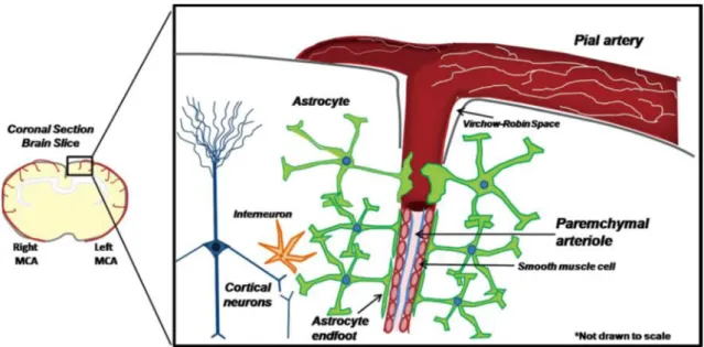
In addition to neurons, cortical astrocytes play a role in the neurovascular coupling. It is known that astrocytes send processes called ‘endfeet’ to synapses and to arterioles and are capable of sending signals to pial arterioles, and communicate with other astrocytes through gap junctions (Dunn and Nelson, 2010). Pial arteries, extrinsically innervated by sympathetic nerves, run on the surface of the brain, then penetrate into the cortical parenchyma and become parenchymal arterioles (see Diagram 1, Dunn and Nelson, 2010). In the cortical parenchyma beyond the Virchow-Robin space, extrinsic innervation is lost and the arteriole becomes surrounded by astrocytic endfeet (Dunn and Nelson, 2010). Information from active neurons is integrated by astrocytes, which generate increases in endfoot Ca2+ as a result of astrocytic metabotropic glutamate receptor activation and inositol 1,4,5-trisphosphate production (Filosa et al., 2006). This Ca2+ signal in the endfoot may activate neuronal NOS as well as Ca2+-sensitive phospholipase A2 (PLA2), thereby releasing arachidonic acid (AA). AA dilates cerebral arterioles and microvessels through a COX-dependent mechanism, with prostaglandin E2 (PGE2), a COX metabolite, mediating a significant component of vasodilation (Filosa et al., 2006). Neuronal NOS-derived NO dilates cerebral vessels via activation of soluble guanylyl cyclase in the vascular smooth muscle.
10
Figure 1: Cortical parenchymal arterioles and the surrounding astrocyte network (Dunn and Nelson, 2010)
2.1.1.2.2 The role of potassium channels in the cell signaling
Functional hyperaemia may also be directly linked to electric current flow through excitable membranes, which involves potassium ions (K+) lost during action potential repolarization. K+ released by activated neurons to regions of lower K+ near the microvasculature act as an important regulator, coupling increases in cerebral metabolism and blood flow (Nguyen et al., 2000). The elevation of astrocytic endfoot Ca2+ accompanying neuronal activation activates astrocytic Ca2+-activated K+ channels (KCa), which also release K+ into the restricted space between the endfoot and the SMC of the arteriole (Filosa et al., 2006).
Vascular K+ channels are a diverse group of transmembrane proteins that regulate membrane potential, thereby modulating Ca2+ entry and cell signalling (Brayden 2002).
The following K+ channels are functionally important in the vasculature, located on both endothelium and vascular SMCs: Ca2+-activated K+ channels (KCa), voltage- dependent K+ channels (KV), ATP-sensitive K+ channels (KATP) and inwardly rectifying K+ channels (Kir) (Dunn and Nelson, 2010).
In large cerebral arteries AA causes vasodilation through activation of Ca2+-activated K+ (KCa) channels, as AA-induced vasodilatation was markedly attenuated by inhibitors of
11
such channels, namely 1 mM tetraethylammonium ion (TEA) and 50 nM iberiotoxin (Faraci et al., 2001). The basilar artery produced relatively few prostanoids when treated with AA. COX does not play a major role in the AA-induced relaxation of the basilar artery (Faraci et al., 2001). It was shown, instead, that AA is metabolized in the basilar artery largely via the lipoxygenase enzyme. This pathway mediates vasodilatation by activation of KCa channels in the basilar artery (Faraci et al., 2001).
The Kir channels have been extensively studied in the cerebral circulation, and were found to be important in coupling cerebral metabolism and CBF (Chrissobolis and Sobey, 2003). The extracellular concentration of K+ in the brain increases from 3 mmol/L to 7 mmol/L during neuronal activity (Chrissobolis and Sobey, 2004). In this concentration range, K+ activates SMC Kir channels, elicits marked SMC membrane potential hyperpolarization, which closes voltage-dependent Ca2+ channels, decreasing intracellular Ca2+, and thus leading to dilatations of cerebral arteries and arterioles. In pial arteries, the disruption of the gene for the Kir channel, abolished K+ induced vasodilations (Dunn and Nelson, 2010).
In isolated basilar arteries 30 µmol barium (Ba2+), which, at that particular concentration, is a selective inhibitor of Kir channels, abolished the hyperpolarization caused by K+, suggesting that such channels exclusively mediate K+-induced vascular hyperpolarization (Chrissobolis et al., 2000). Findings from in vivo studies indicated that only about one-half of the vasodilator response to K+ was inhibited by the same concentration of Ba2+ that abolished K+-induced hyperpolarization in vitro. In addition, this Ba2+-resistant component of the response to K+ in vivo is insensitive to inhibitors of KATP channels, KCa channels and Na+-K+-ATPase, therefore, it is probable that the entire response is indeed mediated by Kir channels, however, Ba2+ cannot fully access and block Kir channels when applied topically in vivo (Chrissobolis et al., 2004).
The KATP channels are present in neurons, cerebral vascular SMCs, and brain microvascular endothelial cells, and are known to play an important role in the regulation of vascular tone, depending on metabolic need and energy supply (Jansen- Olesen et al., 2005). KATP channels exist as an octameric complex containing two distinct types of protein subunits. The channels consist of four inwardly rectifying potassium channel subunits (Kir6.1 or Kir6.2), with each Kir subunit being associated
12
with a larger regulatory sulphonylurea receptor (SUR) (Brayden 2002). Intracellular ATP at micromolar concentrations binds to the Kir subunit and inhibits, while following dissociation of ATP, the ADP, a nucleotide diphosphate, in the presence of magnesium (Mg2+) interacts with the SUR subunit and activates KATP channels (Brayden 2002).
This activation of KATP channels causes hyperpolarization of SMCs, which prevents the opening of depolarization-activated Ca2+ channels, thus blocking Ca2+ entry to the cell, resulting in vasodilation in cerebral arteries (Jansen-Olesen et al., 2005).
Hyperpolarization of the endothelial cells during impaired energy supply elevates the concentration of intracellular Ca2+ via the transmembrane Ca2+ influx due to the membrane potential gradient (Luckhoff and Busse, 1990) and thereby promotes the Ca2+-dependent formation of the vasorelaxant NO (Jansen-Olesen et al., 2005).
Via myo-endothelial communication, hyperpolarization may be transmitted electrically from SMCs to endothelium through gap junctions, which have been described in rat basilar arteries (Ploug et al., 2006). The significance of SMC hyperpolarization spreading to the endothelial cells may be that the production of NO by the endothelial cells may increase the effet of vasodilation.
There is some regional heterogeneity with respect to the apparent distribution of KATP
channels in different sized cerebrovasculatures. Ploug et al. showed that compared to that measured in the aorta, there is a higher expression level of Kir6.1 and SUR2B
proteins in the basilar and middle cerebral arteries. The differential expression seen at the protein level suggests a more significant role for the Kir6.1∕SUR2B KATP channel complex in the basilar and middle cerebral arteries, and thus in the regulation of CBF (Ploug et al., 2006).
2.1.1.2.3 The role of protein kinase C in the cell signaling
The enzyme protein kinase C (PKC) exerts numerous cellular effects, including mediation of responses to vasoconstrictor agonists in cerebral arteries (Chrissobolis and Sobey 2002). The Kir channels and the KATP channels are structurally related, and in the basilar artery, vasodilator responses mediated by activation of both Kir and KATP channels are inhibited by the activity of the PKC. Furthuremore, it has been reported that PKC exerts inhibitory actions on Kv and KCa channels as well, and so PKC-induced inhibition of K+ channels may normally modulate basal cerebral artery tone and may contribute to pathophysiological vascular conditions such as cerebral vasospasm
13
(Chrissobolis and Sobey 2002). PKC phosphorylates ryanodine receptors located on the sarcoplasmic reticulum, which modulates calcium influx into the cell cytoplasm, termed calcium sparks (Liu QH et al., 2009). Ca2+ sparks can generate hyperpolarizing spontaneous transient outward currents in cerebral arterial SMCs, which results in the inhibition of voltage-dependent Ca2+ channels, prevention of Ca2+ influx, and thus cause cerebral vasorelaxation (Nelson et al., 1995). As PKC reduces the frequency of Ca2+
sparks in cerebral arterial SMCs by phosphorylating ryanodine receptors, it acts in the direction of cerebral vasoconstriction (Bonev et al., 1997).
2.1.1.3 Neural regulation
CBF may increase out of proportion to metabolic demands, may increase without significant change in local metabolism, and may increase much faster than the accumulation of the metabolic end products. Therefore, the 120 year old metabolic hypothesis of Roy and Sherrington cannot fully explain the increases of CBF during increased functional activity of the central neurons (Sandor, 1999). Neurogenic stimuli via perivascular nerve endings may act as rapid initiators, to induce a moment-to- moment dynamic adjustment of CBF to the metabolic demands, and further maintenance of these adjusted parameters is ensured by the metabolic and chemical factors (Sandor, 1999).
Nerve fibers that make up perivascular neuronal networks surround not only extracortical, pial vessels, but also follow both intracortical arteries and veins into the cerebral parenchyma (Sandor, 1999). These nerve fibers contain sympathetic adrenergic nerves of superior cervical ganglionic origin, parasympathetic cholinergic, trigeminovascular and sensory nerves. The number of nerve fibers in the walls of the vessels becomes smaller, as the diameter of the vessels decreases (Lee, 2002).
Parenchymal arterioles lack the extrinsic innervation of larger pial arteries (Dunn and Nelson, 2010). Sympathetic nerve stimulation releases norepinephrine (NE) from perivascular nerves, although synaptic concentration of NE upon maximum transmural nerve stimulation is too low to directly affect vascular smooth muscle tone (Lee, 2002).
Furthermore, α-adrenoceptor antagonists were unable to alter pial arteriolar diameter in species such as cats, piglets and lambs, and transsection of sympathetic nerves in resting conditions resulted in minimal effect on CBF in several different species (Sandor,
14
1999). Similarly, isolated human pial arteries were shown to have poor innervation and demonstrated weak responsiveness to adrenoceptor agonists (Lavi et al., 2003). These results suggest that NE is not the major transmitter for cerebral vasoregulation in large cerebral arteries. Instead, NE may act on presynaptic receptors of neighboring nerve terminals and modulate the release of transmitter substances from these nerves. For example, Lee and colleagues showed that in pig cerebral arteries NE acts on β2
adrenoceptors located on nitrergic nerve terminals, and releases NO, causing vasodilation (Lee et al., 2000).
Similar to NE, acetylcholine (ACh) is not the main dilatory transmitter at terminal synapses (Lee, 2002). It is co-released with NO, a potent nonadrenergic, noncholinergic vasodilator transmitter substance in cerebral blood vessels. Choline acetyltransferase, which synthesizes and releases ACh, coexists with NOS in the parasympathetic ganglia and perivascular nerves in cerebral blood vessels.
ACh-induced relaxation depends on the intact functioning of the endothelium (Dong et al., 2000). In guinea-pig mesenteric and cerebral arteries, ACh induced relaxation, neither mediated by NO nor mediated by prostanoids, was abolished completely whith removal of the endothelium as well as when arterial segments were preconstricted with high K+ concentrations, which is known to cause membrane depolarization. Therefore, it seems that membrane hyperpolarization is essential in the vasorelaxation process involving the yet unknown endothelium derived hyperpolarizing factor (EDHF).
The cellular mechanism of EDHF action shows tissue and species variability, and in guinea pig basilar arteries EDHF-mediated relaxation possibly involves the activation of Kir and Kv channels (Dong et al., 2000). The toxins apamin and charybdotoxin inhibit KCa and Kv channels, respectively, both of which inhibit EDHF-mediated relaxation. In addition, ciclazindol and glibenclamide, inhibitors of Kv and KATP channels, have been reported to suppress EDHF-mediated relaxation in guinea-pig basilar artery and ACh- induced smooth muscle hyperpolarization in rabbit middle cerebral artery (Petersson et al., 1997).
The source of endogenous ACh is presumably the cholinergic neurons that innervate the basilar artery, while its target M2 mACh receptors are expressed in neurons, endothelial cells and vascular SMCs (Chrissobolis et al., 2004). Since endothelial denudation has no
15
effect on K+-induced cerebral vasorelaxation in rats, it seems unlikely that endothelial M2 mACh receptors are involved in K+-induced responses.
Another type of nerve fibers, the sensory nerve fibers arrive either via somatosensory pathways or from the sensory organs, and the axonterminals form synapses in the thalamus or in the geniculate bodies (Sandor, 1999). The neurotransmitters involved in the transmissions of the signals may influence the tone of the cerebral resistance vessels in the relay nuclei along the sensory pathways and in the appropriate cerebrocortical regions. Perivascular C-type afferent sensory neurons release substance-P and calcitonin gene-related peptide (CGRP), both of which cause vasodilation in pial arterioles (Sandor, 1999). Substance-P acts via an endothelium-derived hyperpolarizing factor (EDHF) and NO pathway, while CGRP has endothelium-independent actions, by involving activation of smooth muscle adenylate cyclase, with a subsequent vasodilation (Edvinsson et al., 1992).
In the guinea-pig basilar artery, ANA induces vasorelaxation via activation of capsaicin- sensitive sensory nerves (Zygmunt et al., 1999). Moreover, the vasodilatory responses of isolated arteries exposed to ANA were shown to be mediated through the vanilloid TRPV1 receptor and involve the release of CGRP from perivascular sensory nerves (Golech et al., 2004).
2.1.2 Adaptation to hypoxia and hypercapnia: Local and global regulatory mechanisms It is known that CBF is increased by hypoxia, hypercapnia and concomitant hypoxia and hypercapnia (Spicuzza et al. 2005). Hypoxic and hypercapnic stimuli can act on cerebral blood vessels by releasing various local metabolites. Both endothelial and neural processes take part in the CBF regulation in hypoxia and hypercapnia. The endothelial cells of the cerebral blood vessels are not passive barriers, but rather play an important role in the regulation of the cerebral circulation. Both the NO and the prostanoid pathways are considered to be important endothelium-derived relaxing factors (EDRFs), with the prostanoids being the dominating EDRF mediating cerebrovascular responses in the neonatal period, and NO being the dominating EDRF in the juvenile and adult life (Willis and Leffler, 2001). The mechanisms involved in the endothelial dependent dilator responses in newborns and in juveniles are not the same.
In cultured endothelial vascular cells from adult pigs, Parfenova et al. found higher
16
endothelial NOS expression and activity, compared to cells from newborn pigs while finding no differences in the COX expression and activity between the adult and newborn pigs (Parfenova et al., 2000).
2.1.2.1 The role of nitric oxide
Figure 2: Synthesis of nitric oxide in the endothelium
(Source: http://www.kumc.edu/research/medicine/biochemistry/bioc800/sig02-11.htm)
Nitric oxide (NO) is a highly potent dilator of cerebral arteries and arterioles (Sandor, 1999). The endothelium synthesizes NO via the constitutive eNOS (see Diagram 2).
The shear force acting on the endothelial membrane, such as in response to an increase in CBF velocity, as well as substances like histamine, bradykinin, epinephrine, NE, ACh, substance-P, ADP, serotonin and thrombin, stimulate phospholipase C (PLC), thereby increasing the intracellular calcium (Ca2+) level, and with it the Ca2+-calmodulin complex level, which activates NOS in the endothelium (Faraci et al., 1994). Neurons,
17
besides releasing glutamate, adenosine and other neurotransmitters that may act as vasoactive signals, also release NO. The neuronal type of the constitutive form of nitric oxide synthase (nNOS) is related to the endothelial eNOS. The increase in the Ca2+
levels in the neurons activates nNOS, and the NO produced acts not only as a cotransmitter in different neuronal functions, but also activates soluble guanylyl cyclase (sGC) in the vascular smooth muscle cells, increasing the cGMP level and causing vasodilation. This mechanism of vasodilation may play an important role in the flow- metabolism coupling (Edvinsson et al., 2001).
The variation of the CBF during metabolic perturbations partly involves a chemoregulatory mechanism, independent of ABP fluctuations. The CBF increases by 50% during hypercapnia and decreases by 35% during hyperventilation, without major changes in ABP (Lavi et al., 2003). Several studies indicate that NO is necessary to maintain carbon dioxide (CO2)-mediated CBF. The studies of Sandor et al (1994) provided clear evidence that NO plays a major role in the mediation of regional CO2- responsiveness of the cerebral and spinal cord vessels of the cat: flow response of 11 brain and spinal cord regions to CO2 was abolished by 95% after NOS blockade by NOS inhibitor N-nitro-L-arginine methyl ester (L-NAME). This corresponds well to Lavi et al.’s investigation in which another NOS inhibitor, N-mono-methyl-L-arginine (L-NMMA), was shown to attenuate the vasodilatory CBF response to hypercapnia in rats (Lavi et al., 2003).
There are different results from studies carried out on different species regarding the importance of NO as a mediator in hypercapnic vasodilation. Goadsby et. al. showed in cats that NO is not the sole determinant of hypercapnic vasodilation in the cerebrovascular bed (Goadsby 1994). Lavi et al. showed in dogs that hypocapnia did not cause a significant change in CBF during infusion of the NO donor sodium nitroprusside (SNP) (Lavi et al. 2003).
The size of the blood vessel also seems to matter, concerning the contribution of NO pathway. In larger cerebral arteries as well as arterioles, NO plays a role in cerebral vasodilation in response to hypercapnia, while in the cerebral parenchymal arteriole and microvessels, NO derived from endothelial as well as neuronal enzymes does not seem to mediate vasodilation induced by hypercapnia (Nakahata et al. 2003).
18
Another way to view the varying results is that not the endothelium but perivascular NO-releasing nerves from extracerebral origin are the main source of basal NO in large cerebral arteries originating from the anterior portion of the circle of Willis (Iadecola et al., 1993 and Lindauer et al. 2001). Significantly lower perivascular NO concentrations may be present at arterioles which are not surrounded by NO-producing tissue and lack perivascular nerve endings from extracerebral origin. NOS inhibition nearly abolished the vasodilation response to extraluminal acidic buffer solution of rat MCA, while in cat pial arterioles, there seemed to be no permissive effect of NO donors under NOS inhibitor application during hypercapnia (Lindauer et al. 2003).
Several mechanisms might be involved in the molecular relation between CO2 and NO pathway. NO effect may possibly be mediated by cGMP because cGMP application during NOS inhibition or during soluble guanylyl cyclise inhibition restored pH- reactivity (Lindauer et al. 2003). In the vascular smooth muscle cell, hypo- and hyperpolarizing K+ channel activity may possibly be modulated by NO or cGMP and by intracellular proton concentration (Lindauer et al. 2003). In the rat MCA, both KATP and KCa channels were shown to mediate vasodilation to acidosis, and the functions of both channels were dependent on basal perivascular NO concentration, since NOS inhibition reduced vasodilation to specific K+ channel openers (Lindauer et al. 2003).
Vasodilation of cerebral arteries during elevation of proton concentrations, producing pH values of 6.5, is highly dependent on the basal perivascular NO level, whereas at even lower extraluminal pH (6.0) vasodilation becomes independent of NO and is probably mediated by different mechanisms (Lindauer et al. 2003). Extracellular acidosis is associated with increased K+ conductance causing hyperpolarization that, in turn, leads to closure of voltage-gated Ca2+ channels and smooth muscle relaxation (Lindauer et al. 2003).
Other authors provided evidence against a permissive action of NO/cGMP on K+ channels involved in vasodilation to acidosis. For instance, it was shown that arginine analogues inhibit hypercapnic vasodilation by blocking KATP channels independently of NO or cGMP, and that KATP channels may have an arginine site that influences their function instead of the proposed specific role of NO or cGMP. In rat pial arterioles, NOS inhibition had no effect on vasodilation to specific openers of KATP channels
19
(Lindauer et al. 2003). The diminished functional role of basal perivascular NO in pial and larger penetrating arterioles compared with larger cerebral arteries of the anterior part of the circle of Willis may possibly explain the observation that in vessel segments adapted to lower perivascular NO concentrations, KATP channel function may occur less dependent of NO, whereas in vessels with higher perivascular NO concentration, at least part of the channel activity may be specifically influenced by NO (Lindauer et al. 2003).
Both hypoxia and hypercapnia may be accompanied by extracellular and/or intracellular acidosis and acidosis itself produces marked dilatation of the cerebral arteries (Santa et al. 2003). Also, the fast changes in pH that occur during hypercapnia are important modulators of NOS (Lavi et al. 2003). It has been proposed that the mechanisms by which acidosis produces dilataion of the cerebral arteries may be quite different between hypoxia and hypercapnia, and that extracellular pH rather than intracellular pH may be the major determinant of hypercapnia-induced, NO-dependent relaxation of the cerebral arteries in vitro (Santa et al. 2003).
It seems that in the case of the cerebral arteriole, the extracellular change of proton levels and not the CO2 molecule levels is responsible for vasodilation in hypercapnia, which is supported by the observation that under the condition of normal pH, applied CO2 produced no vasodilation of the parenchymal arterioles (Nakahata et al. 2003).
Results concerning contribution of KATP channels in the vasodilation of cerebral arteries to acidosis are inconclusive, although pharmacological manipulations of KATP channel activation were found to be possibly involved in the modulation of CO2-NO cerebral vasomotor reactivity (Faraci et al. 1994). A study of rabbits demonstrated that glibenclamide, a selective KATP channel antagonist, is partly capable of reducing dilation of pial arterioles induced by hypercapnia, suggesting that KATP channels may marginally contribute to cerebral vasodilation during hypercapnia (Faraci et al. 1994).
In addition, study of the canine basilar artery demonstrated that extracellular acidosis that accompanies hypercapnia causes vasorelaxation partly via KATP channels. In humans it was shown that in larger cerebral arterioles, mild hypercapnia-induced vasodilation was only partly mediated by KATP channels. In cerebral parenchymal arterioles, mild hypercapnia-induced vasodilation was completely abolished by glibenclamide. These data suggest that KATP channels, expressed in microvessels of
20
cerebral cortex are very sensitive to the mild increase in levels of CO2, and that these channels are important mediators of hypercapnia-induced vasodilation in the rat cerebral parenchymal microvessels (Nakahata et al. 2003).
As in the case of hypercapnia, NO may contribute to the increase of CBF evoked by hypoxia. Endothelial contribution to hypoxic vasodilatation increases throughout early postnatal life and becomes prominent in adult cerebral arteries (Pearce 2006). A local role for NO in the fetal cortical vasculature is strongly supported by the finding that the release of cGMP from the brain increased in response to hypoxia (Hunter et al. 2003).
Hypoxic increases in cortical cGMP production were completely eliminated by NOS inhibition, strongly suggesting a direct role for NO in hypoxic cortical relaxation. NO produces vascular relaxation by interacting with the heme group of soluble guanylate cyclase (sGC), the enzyme that synthesizes the second messenger cGMP, which in turn promotes vasodilation in many vascular beds, including cerebral arteries. The tonic release of NO is governed by continuous activation of endothelial cells by stimuli such as pulsatile flow and sheer stress, and NO directly stimulates cerebrovascular cGC, which is highly abundant in cerebral arteries, particularly in the fetus, as the vasorelaxant capacity of the cGMP pathway is attenuated by maturation (Hunter et al.
2003). Pearce et al. have shown that fetal cerebral arteries are more sensitive to the vasodilator effects of cGMP than adult arteries, suggesting that hypoxia-induced release of NO from the endothelium and subsequent cGMP formation are an important and age- dependent component of the cerebral response to acute hypoxia (Pearce 2006).
In the adult rat brain, NOS synthesis seems to be important in the CBF responses to hypercapnia but not hypoxia (Hunter et al. 2003). In support of this, is that reduced tissue oxygen levels may drastically reduce NOS activity, as shown by the reduction of the NO generating capacity in rat brains as a result of reduction of oxygen supply (Bari et al. 1998). According to studies done on piglets and rats, activation of NMDA receptors is not an important mechanism involved in promoting arteriolar dilation during hypoxia; moreover, NMDA-induced cerebral arteriolar dilation was inhibited by hypoxia and by exogenous adenosine (Bari et al. 1998).
Hypoxia may also promote release of vasodilator opioids, such as methionine enkephaline, linked to production of NO and cGMP, suggesting important interactions
21
between opioid release and NO during acute hypoxia in the immature brain (Pearce 2006).
As seen in hypercapnia, acute hypoxia also has been reported to activate KCa channels in neonatal pial arteries through a NO-independent mechanism, while within the adventitia of the arterial wall, hypoxia may also promote the release of vasodilatory sensory neuropeptides, while inhibiting the release of NO from perivascular nerves in adult cerebral arteries (Pearce 2006). Thus one sees the complexity of the cerebrovascular regulation in hypoxia, which involves the endothelium, the smooth muscle and the adventitia of the cerebral blood vessel, as well as the cerebral parenchyma.
2.1.2.2 The role of prostanoids
Prostanoids, such as prostacyclin (PGI2) and prostaglandin E2 (PGE2) are important vasodilator molecules. The endothelium synthesizes prostanoids via the cyclooxygenase enzyme 1 (COX-1) from arachidonic acid. Both PGI2 and PGE2 act on the same PGI2 receptor, resulting in adenylate cyclase activation (Parfenova et al., 1995). The COX-1 isoform participates in the maintainance of resting CBF and in the vasodilation produced by hypercapnia or endothelium-dependent vasodilators, and the COX-2 isoform contributes exclusively to vascular responses initiated by neuronal activity, as COX-2 is constitutively expressed in glutamatergic neurons (Niwa et al., 2001).
Several reports suggested that prostanoids play an important role in the regulation of basal CBF of adults, newborns, and fetuses; although others suggested that there is no effect of COX inhibition in adults. In the case of hypoxic regulation of CBF, data regarding the role of prostanoids are even less clear. Although hypoxia can increase cerebral PGE2 levels, it has no significant effect on PGI2 or thromboxane A2 (TxA2) release, suggesting that prostanoids play a modest role in hypoxic cerebral vasodilatation in the immature brain (Pearce 2006).
Blockade of prostanoid synthesis by diclofenac attenuates the CBF rise in response to hypoxia, which is related to systemic effects rather than direct effects on the cerebral vasculature in fetal sheep, because arterial blood pressure was significantly lower due to diclofenac (Nishida et al. 2006). This indicates that in the brain during hypoxia, prostanoids do not act locally on resistance vessels directly and instead these mediators
22
and others such as carbon monoxide interact and compensate for one another in a complex and redundant system to mediate responses to hypoxia (Nishida et al. 2006).
2.1.2.3 The role of adenosine
The cerebral vascular response to hypercapnia seems to be also mediated in part by adenosine, an endogenously produced purine nucleoside. Adenosine mediates various physiological processes acting via at least four different adenosine receptor types named A1, A2A, A2B and A3. The receptors are all coupled to G-proteins and have been found in a wide range of species and types of tissues (Blood et al. 2003). A1 receptor expression is notably high in the brain, where activation of the A1 receptor results in neuronal membrane stabilization and decreased neuronal firing, while A2A and A2B receptors can be found in cerebral vascular smooth muscle, where both mediate vasodilation in response to hypoxia and neuronal stimulation (Blood et al. 2003).
Adenosine was shown to depress fetal cerebral oxygen consumption through activation of neuronal A1 receptors and mediate vasodilation through activation of A2 receptors on cerebral arteries, suggesting that adenosine is a critically important mediator of cerebrovascular homeostasis during acute hypoxic insult in the fetus as well as in the adult (Pearce 2006). In preterm fetal sheep, adenosine A2 receptor was shown to play a greater role in cerebral vasodilatation than does the A1 receptor, as is supported by the observations that in adult rats, A2 agonists resulted in greater cerebral vasodilatation than A1 agonists, and 8-cyclopentyl-1,3-dipropylxanthine (DPCPX), a selective adenosine A1 receptor antagonist, had no effect on the hypoxic increases in CBF (Blood et al. 2003). Similarly, in rats selective antagonists of A2A adenosine receptors significantly attenuated the CO2-evoked increase in cortical arteriole diameter, showing the involvement of the adenosine receptor during hypercapnia-induced vasodilation as well, although this antagonism is not complete, suggesting that other mechanisms are also involved (Phillis et al. 2004).
Adenosine is elevated during hypoxia in the brain of piglets and other animals (Bari et al. 1998). Plasma and intracerebral adenosine concentrations also increase during hypoxia in the fetal sheep, newborn lamb and adult rat (Blood et al. 2003). In piglets, it was shown that blocking adenosine receptors with theophylline, a non-selective adenosine receptor antagonist, blocked pial arteriolar dilation to hypoxia and N-methyl-
23
D-aspartate-induced dilation (NMDA) during hypoxia (Bari et al. 1998). Adenosine could reduce Ca2+ entry into nerve cells and activation of NOS by promoting hyperpolarization or by blocking neural N-type voltage dependent Ca2+ channels, and also might reduce presynaptic glutamate release, thus suppressing auto-amplification of glutamate and/or NMDA effects (Bari et al. 1998). It is important to note, however, that some studies indicate that activation of NMDA receptors is not an important mechanism involved in promoting arteriolar dilation during arterial hypoxia in piglets or rats (Bari et al. 1998). The brain parenchyma includes many different cell types, and each of these can release a different combination of vasoactive factors in response to hypoxia.
Hypoxia initiates a regionally heterogenous change in the interstitial milieu that may not only promote vasodilatation but may also attenuate vasodilation to some receptor agonists such as NMDA (Pearce 2006). The reasons for this complexity arise not only from the mixtures of cerebral cell types that vary from region to region but also from the arteries and arterioles whose reactivity are labile and varies with age, artery size, and region (Pearce 2006).
As in the case of piglets, in the case of fetal sheep, intravenous infusion of theophylline, resulted in a lack of CBF increase during hypoxia (Blood et al. 2003). Theophylline crosses cell membranes freely, resulting in blockade of adenosine receptors on either side of the blood brain barrier, while 8-p-sulfophenyl-theophylline (8-SPT), another non-selective adenosine receptor antagonist, is relatively impermeable to the plasma membrane and does not to cross the blood brain barrier. 8-SPT infusion resulted in an inhibition of much of the characteristic increase of CBF provoked by hypoxia, suggesting that these adenosine receptors are located outside the blood brain barrier (Blood et al. 2003). In the adult rat, topical application of 8-SPT to the exposed cortex prevented hypoxic CBF increase, also suggesting that the receptors mediating cerebral vasodilatation are located outside the blood brain barrier (Blood et al. 2003).
2.1.2.4 The role of sympathetic control
Normocapnic hypoxia increases sympathetic control of the cardiovascular system more than normoxic hypercapnia, and it is likely that sympathetic stimulation can prevent excessive cerebral vasodilation in response to combined hypoxia and hypercapnia,
24
which may be important in reducing a possibly dangerous rise in intracranial pressure in extreme hypoxia and hypercapnia (Spicuzza et al. 2005).
The role of sympathetic activation must not be neglected in the regulation of CBF, within the autoregulatory range of blood pressure in hypoxia and hypercapnia. This is interesting also, because although parasympathetic nerves innervate the cerebral vessels, the parasympathetic fibers of the 7th cranial nerve, which contains the efferent limb of the peripheral chemo- and baroreceptor reflex arcs, were found to have no significant role in the cerebral vasodilation that occurs during hypoxia and hypercapnia. This was shown on baboon brain, which responded normally to hypoxia and hypercapnia both before and after unilateral and bilateral transection of the 7th cranial nerve (Hoff et al.
1977).
Golanov et al., have proposed that at least 50% of hypoxic cerebrovascular vasodilation is neurogenic, involving a rapid, patterned, sympathetic response originating in the lower brainstem, with excitation of oxygen detectors, namely sympathoexcitatory reticulospinal neurons of the rostral ventrolateral medullary nucleus (Golanov et al.
2001). These neurons are directly, selectively, rapidly and reversibly excited in vitro or in vivo by hypoxia (Golanov et al. 2001). Neurons of the rostral ventrolateral medullary nucleus do not innervate directly the cerebral cortex but rather synapse in the medullary vasodilator area, the bilateral lesion of which blocks the cerebrovascular responses to stimulation of neurons of the rostral ventrolateral medullary nucleus as well as hypoxia- induced cerebrovasodilation. The vasodilator effect of the excitation of rostral ventrolateral medullary nucleus is relayed by the subthalamic cerebrovasodilator area to other brain areas (Golanov et al. 2001).
Cortical cerebrovasodilation evoked by excitation of the above pathway is likely to depend upon the integrity of cortical neurons (Ilch and Golanov 2004). Selective excitotoxic lesion of cortical neurons, achieved by ibotenic acid microinjection of the parietal cortex, blocks CBF increase triggered by stimulation of subthalamic cerebrovasodilator area and also attenuates hypoxia-induced CBF elevation by 54% in the cortex (Ilch and Golanov 2004). This supports the view that hypoxic cerebrovasodilation is partially neurogenic and requires activity of local neurons with oxygen sensing properties.
25
Hypoxia-induced blood flow increase seen in the brainstem, which is the major site of cardiorespiratory controls, was shown to be stronger compared to that seen in the cortex. More precisely, in the ventral respiratory groups of the brainstem, the blood flow incresase during hypoxia was higher than in the dorsal part of the brainstem, containing the nucleus tractus solitaries and pontine respiratory groups (Montandon et al. 2006). These results suggest that blood flow response to hypoxia favours oxygen delivery in brainstem regions involved in respiratory rhythm generation (Montandon et al. 2006).
2.2. Influence of the heme oxygenase pathway on the cerebral circulation 2.2.1 Heme oxygenase enzymes and the brain
Heme oxygenase (HO)-mediated heme degradation is the primary cellular mechanism for production of endogenous carbon monoxide (CO) (Baranano and Snyder, 2001). In this reaction, heme is degraded into equimolar quantities of iron, biliverdin, and CO (Tenhunen et al. 1969). Iron is mainly used up for production of new heme, while biliverdin turns into bilirubin rapidly, via enzymatic action of bilirubin-reductase, which is at excess (Kutty and Maines 1984). CO in the end, attaches itself to circulating haemoglobin in the bloodstream, and is transported as carboxyhemoglobin until it is expired from the lungs. The highest HO expression is in the brain and cerebral circulation, particularly in endothelium and perivascular astrocytes, and the HO activity of the brain tissue exceeds that of systemic organs (Maines MD 2000).
Analogous to NOS, the HO enzyme has different constitutive and inducible isoforms, namely HO-1, HO-2, and HO-3 (Baranano and Snyder, 2001) isoforms. The HO-1 isoform, also known as heat-shock protein 32, is extremely sensitive to heavy metals, and may be activated by agents that produce oxidative stress and pathological states, such as heat-shock, GSH-depletion, radiation, hypoxia, and hyperoxia (Maines 1984).
HO-1 induction in vivo occurs in response to hemorrhage, hyperthermia, and ischemia (Leffler CW et al. 2011). However, HO-1 is not expressed in cerebral vessels or the brain under physiological conditions, and it does not contribute to acute vascular responses that involve rapid CO-mediated increases in blood flow. HO-1 inducers including hydrogen peroxide, arachidonic acid, NO donors, transition metals CoCl2,
26
FeCl2, FeCl3, TNF-α and glutamate failed to acutely induce HO-1 in brain endothelial cells (Leffler CW et al. 2011).
The HO-2 isoform is abundantly expressed in the brain of various mammalian species, including mice, rats, pigs, and humans. In newborn and mature animals, HO-2 is detected in neurons, glial cells, and the cerebral vasculature (Vigne et al. 1995; Zakhary et al. 1996; Parfenova et al. 2001; Leffler CW et al. 2011). Physiological and pathophysiological stimulations, including glutamate, seizures, hypoxia, and hypotension, rapidly increase the in vivo activity of HO-2. A chemical inducer of HO-2 is the adrenal gland-derived glucocorticoid (McCoubrey Jr et al. 1997).
In cerebral vessels and endothelial cells, HO-2 activation by glutamate is mediated by protein tyrosine kinases and Ca2+/calmodulin-dependent mechanisms (Leffler CW et al.
2011 and Xi Q et al. 2010). Ca2+ and calmodulin are important regulators of HO-2 activity in freshly isolated cortical astrocytes and in cultured glial cells as well (Xi Q et al. 2011). In cerebral microvessels, NO may directly inhibit HO-2 catalytic activity but indirectly stimulate the activity via elevation of cGMP (Leffler CW et al. 2005).
Local heme availability is an important determinant of the rate of CO production.
Factors that increase the heme substrate availability also increase CO production by cerebral vessels, indicating that HO-2 activity is substrate dependent (Leffler CW et al.
2003). At resting conditions, the concentration of free heme is approximately 0.5-1 µmol/ L, while at excesses of 50 times greater heme concentrations, the plasma bilirubin and CO production rise only two or three times more (Johnson et al. 1996). The difference is due to the physical properties of heme. In an aqueous medium, at physiological pH, free heme remains monomeric until 2 µmol/L concentration, and above this concentration it forms dimers and polimer chains (Falk 1964). These forms represent a potential “heme-supply” and are not subtrates of HO enzymes (Tenhunen et al. 1969). Therefore it may be seen that by addition of heme, the free monomeric heme concentration increases only by two-three times, and this substrate increase is parallel to the increase of production of bilirubin or CO (Johnson et al. 1996).
Endogenous CO is not only produced by the degradation of heme. The competitive inhibition of HO enzyme activity by metalloporphyrins leads to only 30-50% decrease in CO production (Vreman et al. 1991). While normally heme degradation is the
27
primary route for CO production, under HO inhibition, alternative metabolic pathways become important for CO production, such as lipid metabolism, although these associations are scarcely known yet (Vreman et al. 1991; and Wolff 1976). CO generation is associated with the process of lipid peroxidation in tissues with limited antioxidant reserves (Vreman et al. 1998).
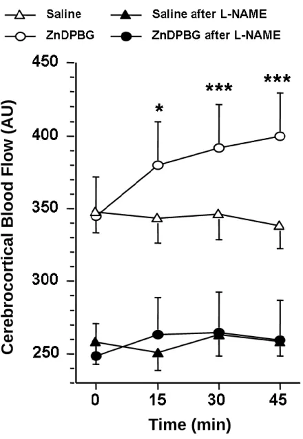
Several HO inhibitors, such as the zinc deuteroporphyrin 2,4-bis glycol (ZnDPBG), are able to cross the blood brain barrier, since given intraperitoneally, they are able to inhibit the HO activity in the brain as well as the peripherial tissues (Vreman et al.
1991; Johnson et al. 1995), while heme does not cross the blood brain barrier (Linden et al. 1987). However, d-aminolevulinic acid synthase, the rate-determining enzyme for new heme synthesis, as well as other enzymes acting in the synthesis of heme, were shown in brain tissue (Moore et al. 1987). The discovery that the brain is not reached by the circulating heme molecules, but rather locally synthesizes its own heme supply via the metabolic pathways needed for the synthesis of heme, shows that the central nervous system is an independent functional HO unit.
2.2.2 The heme oxygenase pathway in the brain and cerebral vasculature
The mechanism of signalling pathways of the HO-CO system under physiological conditions displays various forms. CO activates the soluble guanylyl cyclase (sGC), its rate of activity increasing twofold (Makino et al. 1999 and Vogel et al. 1999). There is a possibility, that CO does not activate sGC directly, because the effect of CO is dramatically increased by the xenobiotic called 3-(5’-hydroxymethyl-2’-furyl)-1- benyilindazol (YC-1) (Friebe et al. 1996; Stone and Marletta 1998). YC-1 and CO together activate sGC as strongly as NO does. There may be a possible endogenous material, which increases the ability of CO to activate sGC.
CO may also bind to heme-containing proteins, and thereby alters their enzymatic activities (White and Marletta 1992; Schmidt 1992; Raff and Janowski 1994; Wada et al. 1985). It follows, that heme proteins are potential signalling messengers for CO.
Enzymes of the cytochrome P450 family are potential mediators of CO actions (White and Marletta 1992). The binding of CO to these enzymes increases during a rise in metabolic need and decreased oxygen tensions. The cytochrome P450 enzymes located on the mitochondrial membrane are involved in the electron transport chain and ATP
28
production, and may be involved in a complex effect of the action of kinases and other ATP-dependent processes.
Under resting conditions, the HO pathway has a dual influence on the hypothalamic circulation: a vasodilation mediated by PGE2, and a simultaneous vasoconstriction due to the reduction of NO synthesis, the two effects being equally potent and neutralizing each other (Horvath et.al. 2008).
The addition of hemin, a HO inducer and substrate of the HO enzyme increased the production of PGE2 in the hypothalamus, an action which was blocked by HO inhibitors, such as zinc protoporphyrin IX (ZnPPIX) and tin mesoporphyrin IX (SnMPIX) (Mancuso et al. 1997). CO may play a basic role in the hypothalamus PGE2 production. Studies done on piglet cerebral microvessels showed that brain parenchymal rather than vascular PGE2 release mediates the vasorelaxant effect of constitutive HO (Kanu et.al. 2006).
In piglets, both exogenous CO and the HO substrate heme-L-lysinate induced pial arterial dilatation and the latter could be inhibited by the HO blocker chromium mesoporphyrin (CrMP). Furthermore, hypoxia-induced vasodilation was inhibited by application of CrMP (Leffler et al. 1999; Leffler et al. 2001; Winestone et al. 2003).
During seizures, the pial arteriolar dilation in piglets and the CBF-increase in adult rats were reported to be attenuated by HO inhibitors (Montecot et al. 1998; Pourcyrous et al.
2002).
As an in vitro paradigm for ischaemia, cultured astrocytes were exposed to hypoxia, which triggered marked increase in the expression of 33 kDa stress protein, identified as HO-1, whose induction was observed within 4 hours of hypoxia and peaked at 12 hours, accompanied by an accelerated transcription of HO-1 mRNA (Imuta et al. 2007).
Consistent with the induction of HO-1, a platelet bioassay revealed the production of CO by reoxygenated astrocytes, and the presence of CO in the medium decelerated the hypoxia-mediated apoptotic type of cell death in cultured cerebral neurons via lowering the activity of caspase-3, a key enzyme regulating apoptotic cell death. This protection against apoptosis was likely mediated by CO-mediated increases in intracellular cGMP, because exposure of hypoxic neurons to CO increased intracellular cGMP levels and addition of cGMP analogue to hypoxic neuronal cultures suppressed caspase-3 activity
29
and promoted neuronal survival, suggesting that a potentially important paracellular pathway may exist through which astrocytes may rescue nearby neurons from ischaemic death (Imuta et al. 2007).
Recently it was demonstrated that endogenous CO dilates cerebral arterioles by augmenting the coupling of Ca2+ sparks to KCa channels in SMCs (Jaggar et al. 2002).
CO elevates the coupling of Ca2+ sparks to the large-conductance Ca2+-activated K+ channels (KCa) in cerebral arterial SMCs and elevates KCa channel Ca2+ sensitivity, leading to more effective KCa channel activation by coupled sparks and KCa channel activation by previously uncoupled Ca2+ sparks (Leffler CW et al. 2011).
It was observed that an astrocytic signal, notably HO2-derived CO, is used by glutamate to stimulate arteriole myocyte KCa channels and dilate cerebral arterioles, as glutamate stimulated CO production by astrocytes with intact HO-2, but did not do that in genetically deficient HO-2 cells. Glutamate activated transient KCa currents and single KCa channels in cerebral arteriole myocytes that were in contact with astrocytes, but did not affect KCa activity in myocytes that were alone (Li et al. 2008).
In vivo, both glutamate and hypoxia dilate newborn pig cerebral arterioles, both dilations are blocked by inhibition of CO production, and both increase cerebrospinal fluid CO concentration (Kanu and Leffler 2007). However, although dilation of newborn pig pial arterioles to glutamate is mediated by activation of KCa channels, consistent with the intermediary signal being CO, neither KCa channel blockers nor the guanylyl cyclase inhibitior, 1H-(1,2,4)oxadiazolo(4,3-a)quinoxalin-1-one (ODQ) blocked dilation to hypoxia, suggesting that the contribution of the HO/CO system to hypoxia-induced dilation is not due to the stimulation of vascular smooth muscle KCa
channels or guanylyl cyclase (Kanu and Leffler 2007). This needs to be investigated further.
2.2.3 The interaction of HO-NO pathways in the brain tissue and cerebral vasculature The physiological roles of NO and CO display notable similarities, as both molecules appear to be neurotransmitters in the brain and the peripheral autonomic nervous system. Furthermore, NO and CO both are endothelial-derived relaxing factors for blood vessels (Baranano and Snyder, 2001). The effect of CO on vascular tone has been studied frequently in the past, and it was found that CO is able to relax smooth muscle-
30
preparations (Graser et al. 1990). At the same time, CO was shown to bind to NOS and to inhibit the production of NO (White and Marletta 1992; Matsuoka et al. 1994;
McMillan and Masters 1995), thereby causing vasoconstriction.
This interaction may also be seen the other way around; that is, NO affects the CO production. For instance, endotoxins and inflammatory cytokines induce HO-1 not directly, but rather via NO release, since their activity was blocked by NOS inhibition (Billar et al. 1992). In further experiments, NO-donors were able to induce HO-1 mRNA expression in aorta endothelium (Motterlini et al. 1996), SMCs (Durante et al.
1997) and rat hepatocytes (Kim et al. 1995).
The participation of CO in the regulation of the cerebral circulation has been poorly studied yet (for review please see: Koehler and Traystman 2002). Freshly isolated cerebral microvessels produce CO from endogenous heme under basal conditions and increased CO production was reported after stimulation of ionotropic glutamate receptors (Parfenova et al. 2003; Leffler et al. 2003a and 2003b). It was observed that in freshly isolated cerebral microvessels from piglets, glutamate activates NOS, producing NO that leads to CO synthesis via a cGMP-dependent elevation of HO-2 catalytic activity, which is consistent with the in vivo findings that either HO or NOS inhibition blocks cerebrovascular dilation to glutamate in piglets (Leffler et al. 2005).
2.3. Endocannabinoids and the cerebral circulation
Endocannabinoids are lipid mediators, isolated from brain and peripheral tissues, which include amides, esters and ethers of long chain polyunsaturated fatty acids (Battista et al. 2004). Endocannabinoids include N-arachidonoylethanolamine (anandamide), 2- arachidonoylglycerol (2-AG), 2-arachidonoylglyceryl ether (noladin ether) and virodhamine, also called inverted anandamide, because the arachidonic acid and ethanolamine is joined together by an ester bond instead of the amide bond of anandamide (Battista et al. 2004).
2.3.1 N-arachidonoylethanolamine (anandamide)
The first endocannabinoid to be isolated was the lipid soluble eicosanoid derivative arachidonoylethanolamide also known as anandamide (ANA) from pig brain (Devane et al. 1992). The sites of ANA synthesis include neurons (Di Marzo et al. 1994 and Cadas