Eur J Pain. 2020;24:383–397. wileyonlinelibrary.com/journal/ejp
|
383O R I G I N A L A R T I C L E
High‐dose phenylephrine increases meningeal blood flow through TRPV1 receptor activation and release of calcitonin gene‐related peptide
Mária Dux
1| Alexandru Babes
2| Jessica Manchen
3| Julika Sertel‐Nakajima
3|
Birgit Vogler
3| Jana Schramm
3| Karl Messlinger
3Abbreviations: AITC, allyl isothiocyanate; BCTC, (4‐(3‐Chloro‐2‐pyridinyl)‐N‐[4‐ (1,1‐dimethylethyl)phenyl]‐1‐piperazinecarboxamide); CGRP, calcitonin gene‐related peptide; DAPI, 4′,6‐diamidino‐2‐phenylindole; EIA, enzyme immunoassay; HC030031, (1,2,3,6‐Tetrahydro‐1,3‐dimethyl‐N‐[4‐(1‐methylethyl) phenyl]‐2,6‐dioxo‐7H‐purine‐7‐acetamide, 2‐(1,3‐Dimethyl‐2,6‐dioxo‐1,2,3,6‐tetrahydro‐7H‐purin‐7‐yl)‐N‐(4‐isopropylphenyl)acetamide); hTRPV1, human transient receptor potential vanilloid 1 channel; IB4, Griffonia simplicifolia isolectin B4; NGF, nerve growth factor; PBS, phosphate‐buffered saline; PU, perfusion unit; SIF, synthetic interstitial fluid; SMA, smooth muscle actin; TG, trigeminal ganglion; TRP, transient receptor potential; TRPA1, transient receptor potential ankyrin 1 channel; TRPM8, transient receptor potential melastatin 8 channel; TRPV1, transient receptor potential vanilloid 1 channel; WT, wild‐type.
1Department of Physiology, University of Szeged, Szeged, Hungary
2Department of Anatomy, Physiology and Biophysics, University of Bucharest, Bucharest, Romania
3Institute of Physiology and
Pathophysiology, Friedrich‐Alexander‐
University Erlangen‐Nürnberg, Nürnberg, Germany
Correspondence
Karl Messlinger, Institute of Physiology and Pathophysiology, Friedrich‐
Alexander‐University Erlangen‐Nürnberg, Universitätsstr. 17, 91054 Erlangen, Germany
Email: karl.messlinger@fau.de Funding information
The study was supported by the European Union (FP7 program EUROHEADPAIN, grant no: 602633), the Hungarian National Research, Development and Innovation Office (K119597) to M.D. and the Alexander von Humboldt Foundation (Research Group Linkage Program between M.D. and K.M.). A. B. acknowledges funding from CNCS/UEFISCDI, grant PN‐
III‐P4‐ID‐PCE‐2016‐0475.
Abstract
Background: The α1‐adrenoceptor agonist, phenylephrine, is used at high concen- trations as a mydriatic agent and for the treatment of nasal congestion. Among its adverse side‐effects transient burning sensations are reported indicating activation of the trigeminal nociceptive system.
Methods: Neuropeptide release, calcium imaging and meningeal blood flow record- ings were applied in rodent models of meningeal nociception to clarify possible re- ceptor mechanisms underlying these pain phenomena.
Results: Phenylephrine above 10 mM dose‐dependently released calcitonin gene‐re- lated peptide (CGRP) from the dura mater and isolated trigeminal ganglia, whereas hyperosmotic mannitol at 90 mM was ineffective. The phenylephrine‐evoked release was blocked by the transient receptor potential vanilloid 1 (TRPV1) antagonist BCTC and did not occur in trigeminal ganglia of TRPV1‐deficient mice. Phenylephrine at 30 mM caused calcium transients in cultured trigeminal ganglion neurons responding to the TRPV1 agonist capsaicin and in HEK293T cells expressing human TRPV1.
Local application of phenylephrine at micromolar concentrations to the exposed rat dura mater reduced meningeal blood flow, whereas concentrations above 10 mM caused increased meningeal blood flow. The flow increase was abolished by pre‐ap- plication of the CGRP receptor antagonist CGRP8‐37 or the TRPV1 antagonist BCTC.
Conclusions: Phenylephrine at high millimolar concentrations activates TRPV1 re- ceptor channels of perivascular afferents and, upon calcium inflow, releases CGRP, which increases meningeal blood flow. Activation of TRPV1 receptors may underlie trigeminal nociception leading to cranial pain such as local burning sensations or headaches caused by administration of high doses of phenylephrine.
This is an open access article under the terms of the Creative Commons Attribution-NonCommercial License, which permits use, distribution and reproduction in any medium, provided the original work is properly cited and is not used for commercial purposes.
© 2019 The Authors. European Journal of Pain published by John Wiley & Sons Ltd on behalf of European Pain Federation - EFIC®
1 | INTRODUCTION
Phenylephrine is a sympathomimetic drug acting as a specific α1‐adrenoceptor agonist. It is clinically used at low doses (50–100 µg) for the treatment of hypotension during spinal, epidural and general anaesthesia (Heesen, Kölhr, Rossaint,
& Straube, 2014; Poterman et al., 2015). The blood pres- sure normalizing effect of phenylephrine is mainly caused by vasoconstriction through the activation of α1‐adrenocep- tors (Reid, 1986). Phenylephrine at high doses (1%–5%, i.e.
60–300 mM) is locally used as a mydriatic agent, for example in the treatment of glaucoma (McAuliffe‐Curtin & Buckley, 1989; Moisseiev et al., 2015). As adverse side‐effects of this treatment increases in blood pressure and pulse rate (Chin, Law, & Chin, 1994) and bronchoconstriction in premature in- fants (Kim, Choi, & Kwak, 2015) have been reported. For the treatment of nasal congestion high local doses (up to 4 mg) of phenylephrine have long been used (Myers & Iazzetta, 1982), and later trials have been published to replace the in- tranasal application by oral phenylephrine (Kollar, Schneider, Waksman, & Krusinska, 2007; Meltzer, Ratner, & McGraw, 2015). Oral doses of up to 30 mg were not different to placebo regarding nasal congestion but a small percentage of patients responded with headaches (Meltzer et al., 2015). Another, so far unexplained, local adverse side‐effect is a painful, burn- ing sensation induced by phenylephrine‐containing eye drops (Moisseiev et al., 2015) or nasal spray as mentioned in med- ical information (https ://medli neplus.gov/drugi nfo/meds/
a6160 49.html), which can hardly be explained by the known α1‐adrenoceptor agonist effects of phenylephrine.
The rationale to investigate the effects of phenylephrine on meningeal blood flow in rodents was derived from ear- lier experiments, in which we used phenylephrine to study the mechanisms underlying increases in blood flow concom- itant with temperature changes in the dura mater (Holom, Messlinger, & Fischer, 2008). We recognized an unexpected effect of phenylephrine, that is it rather potentiated vasodila- tation, which is usually associated with pronociceptive effects in the trigemino‐vascular system. The trigemino‐vascular system is the functional unit of intracranial blood vessels and their trigeminal innervation. In this study we therefore localized α1‐adrenoceptors with immunohistochemistry and probed their possible role using measurement of neuropeptide
release, calcium imaging and recording of vascular responses over a wide range of phenylephrine concentrations in the tri- gemino‐vascular system of rodents. In view of the mentioned temperature experiments (Holom et al., 2008) we addressed the most important transduction channels expressed in no- ciceptive neurons, the family of transient receptor potential (TRP) channels, which may be involved in trigeminal noci- ception activated by high‐dose phenylephrine.
TRP receptor channels of the vanilloid type 1 (TRPV1) have been intensely studied for more than two decades in the spinal and the trigeminal system (Nagy, Sántha, Jancsó, &
Urbán, 2004). They are essential for heat transduction ex- pressed in most of the slowly conducting nociceptive affer- ents (Dux, Sántha, & Jancsó, 2012; Julius, 2013; Numazaki
& Tominaga, 2004). TRP receptor channels of the ankyrin type (TRPA1) have been associated with noxious cold trans- duction, although the matter is not yet entirely clear (Zygmunt
& Högestätt, 2014). TRP receptor channels of the melastatin type 8 (TRPM8) have also been associated with cold trans- duction (Babes, Ciobanu, Neacsu, & Babes, 2011) and are es- pecially interesting for trigeminal nociception, because they show genetic variants in migraine families (Dussor & Cao, 2016). All three TRP receptor channel types are also activated by a variety of endogenous metabolic products and/or de- signed chemical substances determining the chemosensitive properties of polymodal nociceptors (Gerhold & Bautista, 2009; Stucky et al., 2009). Finally, a significant population of TRP expressing afferents are peptidergic containing the va- soactive calcitonin gene‐related peptide (CGRP) or substance P, and all three TRP channels are conductive for calcium and could therefore directly or indirectly be involved in vasomo- tor responses upon their activation (Earley & Brayden, 2015).
Therefore, we focused on these TRP receptor channel types as a possible alternative target for phenylephrine at high doses.
2 | METHODS
All experiments were done in accordance with the ethical guidelines of the International Association for the Study of Pain and in compliance with the guidelines for the welfare of experimental animals of the Federal Republic of Germany and the European Commission (Directive 2010/63/EU). All Significance: Phenylephrine is used at high concentrations as a mydriaticum and for treating nasal congestion. As adverse side‐effects burning sensations and headaches have been described. Phenylephrine at high concentrations causes calcium transients in trigeminal afferents, CGRP release and increased meningeal blood flow upon activation of TRPV1 receptor channels, which is likely underlying the reported pain phenomena.
animals were bred and housed in the animal facility of our Institute at a 12/12 hr day–night cycle. The experimental protocol for in vivo experiments was reviewed by an ethics committee and approved by the local district government of Unterfranken.
2.1 | Immunohistochemistry of trigeminal ganglion and dura mater
Adult male and female Wistar rats (body weight 300–450 g) deeply anaesthetized by inhaling 5% isoflurane and i.p. in- jection of thiopental (Trapanal®, Byk Gulden, Germany, 150–200 mg/kg) were thoracotomized and perfused tran- scardially with physiological saline followed by 4% para- formaldehyde in 0.1 M phosphate buffer (pH 7.4). After removal of the skin, the skull was divided into halves along the midline and the cerebral hemispheres were removed.
Trigeminal ganglia were carefully excised from the Meckel's cave, stored in phosphate‐buffered saline (PBS; 0.01 M, pH 7.4) overnight and transferred into 30% sucrose for another day, then quickly frozen at −20°C. From the frozen blocks sections of 14 µm were cut with a cryotome and mounted on glass slides. For indirect immunofluorescence, the fixed sections were rinsed in PBS, pre‐incubated for 1 or 2 hours at room temperature with a solution of 5% donkey serum (Dianova, Hamburg, Germany), 0.5% Triton X‐100 and 1%
bovine serum albumin in PBS, rinsed in PBS and incubated overnight with polyclonal rabbit antiserum raised against α1‐adrenergic receptors (Sigma A270, 1:100) and goat an- tiserum raised against rat CGRP (Biotrend BT‐17–2090–07, 1:10). To control the specificity of immunostaining, one slide was prepared without first antibodies. Then the slides were washed with PBS, incubated with donkey anti‐rabbit Alexa 488 (Molecular Probes A21 206, 1:100) and donkey anti‐goat Alexa 555 (Molecular Probes A21 432, 1:1,000) secondary antibodies for 1 hour at room temperature, rinsed in PBS and coverslipped in Fluoromount G (Science Services, München, Germany). Some samples were mounted with Roti®‐Mount Fluor Care (Roth, Karlsruhe, Germany) containing 4′,6‐di- amidin‐2‐phenylindol (DAPI) for nuclear DNA staining.
Samples of the dura mater of rats were carefully dis- sected and processed without fixation to preserve optimal immunoreactivity. The whole mounts were immunostained similarly as the ganglion sections while they were kept float- ing in the solutions. They were incubated overnight at 4°C with goat antiserum raised against α1A‐adrenergic recep- tors (Santa Cruz sc‐1477, 1:50) and mouse antiserum raised against smooth muscle actin (SMA; Sigma 2,547, 1:100).
Small parts of dura mater were processed without first an- tibodies for control. Then the whole mounts were washed and incubated with donkey anti‐goat Alexa 488 (Molecular Probes A11055, 1:1,000) and donkey anti‐mouse Alexa 555 (Molecular Probes A31570, 1:1,000) secondary antibodies.
The immunostained samples were analysed using a LSM 780 light and confocal microscope (Carl Zeiss MicroImaging GmbH, Jena, Germany) mounted on an inverted Axio Observer Z1. Two dry objective lenses (10 x and 20 x with numerical apertures of 0.3 and 0.8) were used. Fluorescent structures were observed in the light path mode using red and green filters. Confocal images were taken using filter settings for Alexa 488 and 555. The numbers of image pixels were 1,024 x 1,024 or 512 x 512, and pictures were converted to a 12‐bit RGB tiff‐file using confocal assistant software ZEN 2010.
2.2 | Measurement of CGRP release
Adult Wistar rats (250–420 g) and C57BL/6 mice (21–30 g) of either sex as well as mice with genetically deleted TRPV1 receptor channels (TRPV1‐/‐) bred on the background of C57BL/6 (22–28 g) were killed by CO2 inhalation and decap- itated. After removal of the skin, the skull was divided into halves along the midline and the cerebral hemispheres were removed. Skull halves with intact dura mater were washed at room temperature for 30 min with synthetic interstitial fluid (SIF, see below). The halves were placed in a humid chamber and maintained at 37°C. The cranial fossae were filled with 500 µl (rat) or 200 µl (mice) solution. From skull halves of other mice the trigeminal ganglia were dissected and trans- ferred into wells filled with 200 µl SIF. Vehicle (SIF) was always the first solution, consecutively replaced by solutions of phenylephrine or antagonists in SIF as specified in the results. Solutions were collected at periods of 5 or 10 min by carefully removing the content of the skull halves or the wells with a micropipette. All samples were transferred to Eppendorf cups and 100 µl of each sample were taken, di- luted with 25 µl enzyme immunoassay (EIA) buffer (SPIbio, Paris, France) and immediately frozen at −20°C for subse- quent analysis. CGRP content of the samples was measured using a specific CGRP‐EIA kit (SPIbio, Paris, France). The absorbance of the reaction product representing the CGRP concentration of the sample was determined photometrically using a microplate reader (Opsys MR, Dynex, Denkendorf, Germany). The minimum detection limit of the assay is spec- ified as 5 pg/ml by the manufacturer. The CGRP concentra- tion was calculated in pg/ml.
2.3 | Calcium imaging of trigeminal neurons and HEK293T cells
Adult C57BL/6 wild‐type and TRPV1‐/‐ mice (body weight 21–28 g, both sexes) were used and prepared as for CGRP release. Trigeminal ganglia (TG) were excised, exposed to collagenase and protease (Sigma‐Aldrich, Taufkirchen, Germany) before mechanical dissociation, plated on poly‐D‐
lysine‐coated coverslips and cultured in serum‐free neuronal
medium supplemented with 100 ng/ml mouse nerve growth factor (NGF, Alomone Labs, Israel). After 1‐day incubation at 37°C and 5% CO2, cells were stained by 3 µM Fura‐2‐AM and 0.02% pluronic (Invitrogen, St. Leon Rot, Germany).
HEK293T cells were regularly passaged and controlled for mycoplasma contamination and were transiently transfected with plasmids containing human TRPV1 (hTRPV1) using jetPEI transfection reagent from Polyplus Transfection (Illkirch, France). Cells were plated onto poly‐D‐lysine‐
coated glass coverslips and used for experiments within 24 hr. Coverslips were mounted on an inverse microscope and constantly superfused with extracellular fluid (EF, see below) used as vehicle by a gravity‐driven common‐outlet superfusion. Solutions were applied via different lines running at the same flow rate; this ensured mixing within the common outlet about 1 second before the solution reaches the cells.
Fluorescence evoked by illumination at 340 nm and 380 nm was acquired by a 12‐bit CCD camera (Imago Sensicam QE, Till Photonics) system with a monochromator (Polychrome V, Till Photonics) coupled to an Olympus IX71 microscope, using the TILLvisION 4 (Till Photonics) software package.
After background subtraction the 340/380 nm ratio time series were calculated for individual cells, the mean ± SEM was visualized. The cellular response to phenylephrine and other consecutively applied substances dissolved in vehicle was calculated as the percentage change in fluorescence ratio (i.e. the maximal change in ratio during the stimulus to the baseline ratio under vehicle before the stimulus, ΔR/R0). A value larger than 0.2 (20% increase in ratio) was considered as a response.
2.4 | Meningeal blood flow recordings
Wistar rats of both sexes (body weights of 250–350 g) were anaesthetized with increasing concentrations of isoflurane (up to 5%, Forene, Abbott, Wiesbaden, Germany) inhaled in a closed box, continued by application of 2% isoflurane through a tight mask. Atropine sulphate (Braun Melsungen AG, Melsungen, Germany, 0.5 mg/ml, 1:10 in saline) was injected subcutaneously at 1 ml/kg to prevent salivation.
Femoral artery and vein were cannulated for recording of blood pressure and intravenous (i.v.) infusion of substances.
The animals were tracheotomized to be artificially ventilated with a mixture of oxygen‐enriched room air and 2% isoflu- rane. Depth of anaesthesia was routinely assessed and held at a level in which noxious stimuli (pinching of earlobes) failed to elicit motor reflexes or changes in systemic arterial pressure. The body temperature of the animals was recorded by a thermoprobe inserted into the rectum and was kept at 37–37.5°C with a feedback‐controlled heating pad. Systemic blood pressure was recorded with a pressure transducer con- nected to the catheter inserted into the right femoral artery.
The expiratory CO2 was continuously monitored (Artema
MM 200, Karl Heyer, Bad Ems, Germany) and maintained at 3%–3.5%. The head of the animal was fixed in a stereotaxic frame and held by ear bars and a snout clamp. The eyes were covered with dexpanthenol ointment (Bepanthen®, Bayer Vital GmbH, Leverkusen, Germany) to prevent dehydration of the cornea. A median incision was made along the midline of the scalp, the periosteum was moved aside and, either on one or on both sides, a cranial window of about 8 x 6 mm was drilled into the parietal bone under saline rinsing to expose the cranial dura mater.
The exposed dura mater in the cranial windows was cov- ered by SIF. For chemical stimulation, the SIF was replaced by 40 µl of solutions. Needle type probes of a laser Doppler flowmeter (DRT4, Moor Instruments, Axminster, UK) were positioned over branches of the middle meningeal artery sup- plying the dura mater. Blood flow was recorded at a sampling rate of 10 Hz and expressed in arbitrary perfusion units (PU).
The systemic blood pressure was recorded simultaneously.
Data were stored and processed with the MoorSoft program for Windows. The basal blood flow was the mean flow value measured during a 3 min period prior to the stimulation of the dura mater. Blood flow values during stimulation were as- sessed as mean values measured during the 5 min application period and compared to the respective basal flow measured prior to stimulation. The experiment was terminated by i.v.
application of an overdose of thiopental.
2.5 | Substances
Synthetic interstitial fluid (SIF) contained (in mM) 107.8 NaCl, 26.2 NaHCO3, 9.64 Na‐gluconate, 7.6 sucrose, 5.55 glucose, 3.5 KCl, 1.67 NaH2PO4, 1.53 CaCl2 and 0.69 MgSO4and was buffered to pH 7.4 with carbogen gas (95% O2, 5% CO2).
Mannitol (D‐mannitol, Carl Roth, Karlsruhe, Germany) was freshly dissolved in SIF. Extracellular fluid (EF) for calcium imaging was composed of (in mM): NaCl 140, KCl 4, CaCl2 1.25, MgCl2 1, Glucose 5, Hepes 10). Phenylephrine (Sigma‐
Aldrich, Taufkirchen, Germany) was prepared as a stock solution of 100 mM (pH 7.4) in SIF or in EF (for calcium imaging) and phentolamine (Sigma‐Aldrich, Taufkirchen, Germany) as a stock solution of 10 mM in aqua destillata and further diluted with SIF or EF to the final concentrations (pH 7.4). HC030031 (1,2,3,6‐Tetrahydro‐1,3‐dimethyl‐N‐[4‐
(1‐methylethyl)phenyl]‐2,6‐dioxo‐7H‐purine‐7‐acetamide, 2‐(1,3‐Dimethyl‐2,6‐dioxo‐1,2,3,6‐tetrahydro‐7H‐
purin‐7‐yl)‐N‐(4‐isopropylphenyl)acetamide) and BCTC (4‐(3‐Chloro‐2‐pyridinyl)‐N‐[4‐(1,1‐dimethylethyl) phenyl]‐1‐piperazinecarboxamide) were acquired from Sigma‐Aldrich, Taufkirchen, Germany and prepared as stock solutions of 10 mM in DMSO (HC 030,031) or SIF (BCTC).
CGRP8‐37 (Bachem, Biochemica, Heidelberg, Germany) was dissolved in saline and stored frozen as aliquots of 100 µM. Capsaicin and allyl isothiocyanate (AITC) from
Sigma‐Aldrich, Taufkirchen, Germany were prepared as stock solutions of 10 mM and 1 M, respectively, in ethanol, WS‐12 (Cyclohexanecarboxamide,N‐(4‐methoxyphenyl)‐5‐
methyl‐2‐(1‐methylethyl)) from Santa‐Cruz (Heidelberg, Germany) as a stock solution of 50 mM in DMSO. All stock solutions were kept deep‐frozen, defrosted and diluted with saline or SIF to the respective concentrations immediately before they were used.
2.6 | Biometry and statistics
Prior to main experiments, pilot experiments were performed to calculate the power for statistically evaluable group sizes of CGRP release and blood flow data, as specified in the re- sults. For calcium imaging, group sizes were not designed a priori, rather we tested a maximum of neurons identified in the cultures. Ganglia of two animals each were pooled to dissociate sufficient numbers of neurons distributed to 4–6 culture dishes. For CGRP release experiments phenylephrine and vehicle were tested in parallel using paired hemisected skulls or ganglia, respectively. EIA processing was done by a researcher blinded to the parameters of the release experi- ments. For blood flow measurements multiple substances were applied in a randomized order. Statistical analysis was performed using Statistica software (StatSoft, Tulsa, USA).
For comparing blood flow values before and after application of substances, two‐tailed Student's t‐test was used after test- ing for normal distribution (Shapiro–Wilk's test); if normal- ity failed, the non‐parametric Wilcoxon matched pairs test was used. One‐ and two‐way analysis of variance (ANOVA) extended by Tukey's honest significant test, the Fisher's exact probability test and product‐moment correlations were used as specified in the Results. The level of significance was set at p < .05. Data were normalized to baseline as specified and
are presented as mean values ± standard error of the mean (SEM).
3 | RESULTS
3.1 | Immunohistochemistry of α
1‐ adrenoceptors
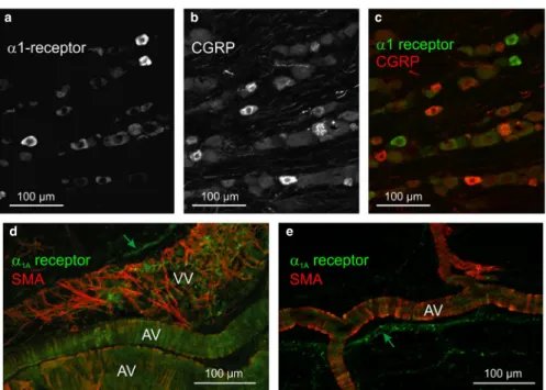
In order to figure out possible binding sites of phenylephrine in the trigeminovascular system, we applied immunohistochemistry using rat trigeminal ganglion sections and the cranial dura mater. In addition to single immunostaining, we combined immunostaining of α1‐adrenoceptor protein with CGRP in the ganglion as a marker for peptidergic neurons, and in the dura mater we combined α1A‐adrenoceptor protein immunostaining with that of SMA as a marker for vascular smooth muscles. In 20 sections of 5 trigeminal ganglia we counted 6,732 neurons.
Among them a small fraction (12.5 ± 1.5%) of mostly small and middle sized neurons was immunopositive for α1‐adrenoceptor protein (Figure 1a‐c). However, combined immunolabelling showed that there was virtually no co‐staining with CGRP‐
immunoreactivity. In the dura mater immunostaining for α1A‐adrenoceptors was mainly seen in the wall of arteries and precapillary arterioles, co‐localized or closely associated with SMA immunoreactivity that outlined circular musculature (Figure 1d‐e). Veins showed network‐like SMA staining but nearly no α1A‐adrenoceptor immunoreactivity. Some perivascular nerve fibres appeared immunopositive for α1A‐ adrenoceptor protein, the most characteristic receptor subtype expressed in primary sensory neurones (Nicholson, Dixon, Spanswick, & Lee, 2005). Trigeminal ganglion sections and dura mater not incubated with primary antisera showed no specific immunostaining indicating that secondary antibodies did not directly bind to any structure in these tissues.
FIGURE 1 Immunohistochemistry in rat trigeminal ganglion (a‐c) and dura mater (d‐e). Some trigeminal ganglion neurons of middle and small sizes show immunoreactivity for α1‐adrenoceptor protein (green), whereas CGRP immunoreactive neurons (red) are mostly small and more frequent. Neurons with both markers were not observed. In the dura mater α1A‐adrenoceptor immunoreactivity is seen in some perivascular nerve fibres (green arrows) apart from large and small arterial (AV) and venous vessels (VV), which show also immunoreactivity for smooth muscle actin (SMA)
a
d e
b c
3.2 | CGRP release from dura mater and trigeminal ganglion
Assuming that phenylephrine may dose‐dependently re- lease CGRP from meningeal afferents, we first made a small series of pilot experiments (n = 4) challenging me- ninges in the rat hemisected preparation with phenylephrine at logarithmic dose steps (10 µM–100 mM) and periods of 5 min. Phenylephrine up to 10 mM did not increase CGRP
release but 100 mM was followed by a more than three- fold increase compared to baseline under superfusion with vehicle (SIF). To control if the high molar concentration of phenylephrine is responsible for the CGRP release, in 6 experiments mannitol at 90 mM was applied followed by SIF, phenylephrine (30 mM) and finally KCl (60 mM).
Mannitol did not cause CGRP release (100.8 ± 10.1% of the baseline), whereas the CGRP release after phenylephrine tended to increase (156.0 ± 28.8%) and was significantly higher after KCl (491.8 ± 91.8%) compared to baseline (re- peated measures ANOVA, F4,20 = 19.1, p < .001; Tukey HSD post hoc test, p < .001) (Figure 2a).
Next we tested meningeal CGRP release with substance application at periods of 10 min, which turned out to be more effective than 5 min. There was again no difference between phenylephrine at 10 mM and vehicle (SIF), whereas applica- tion of phenylephrine at 100 mM was followed by an increase in release to 369.4 ± 38.8% of the SIF baseline (n = 8).
Interestingly, the subsequent application of depolarizing KCl (60 mM) was no longer effective in releasing CGRP (Figure 2b). In parallel control experiments (n = 5), repetitive appli- cation of SIF showed no significant CGRP releasing effect, whereas KCl at the end caused a more than 10‐fold CGRP release compared to baseline. The effects of phenylephrine at 100 mM and KCl are statistically robust (two‐way repeated measures ANOVA, F3,33 = 58.2 for sequence and 92.9 for groups; unequal n HSD post hoc test, p < .001). Finally, application of phenylephrine at 30 mM in a separate set of experiments (n = 6) was followed by an increase in CGRP re- lease to 283.5 ± 57.2% of the SIF baseline (repeated measures ANOVA, F2,8 = 11.4; Tukey HSD post hoc test, p < .05). The response to the subsequent 60 mM KCl application was at- tenuated (184.2 ± 52.2%) and was not significantly different from baseline (p < .06). Thus, there is dose‐dependent CGRP release within a narrow window of efficacy at high doses of phenylephrine, whereas the response to subsequent KCl ap- plication seems to be suppressed depending on the effect of pre‐applied phenylephrine (Figure 2c).
Since the pattern of phenylephrine‐evoked CGRP re- lease was reminiscent of the effect of agonists operating at TRP channels, we next tested if HC030031, an antagonist FIGURE 2 CGRP release from the cranial dura mater in the hemisected rat head ex vivo. (a) CGRP release (mean ± SEM) after synthetic interstitial fluid (SIF), mannitol (Ma) 90 mM, again SIF, phenylephrine (Phe) 30 mM and finally KCl (60 mM) applied successively at 5 min intervals. (b) CGRP release (mean ± SEM) after 10 and 100 mM phenylephrine or repetitive SIF and KCl applied at 10 min intervals. (c) CGRP release normalized to baseline (SIF) after 10, 30 and 100 mM phenylephrine and 60 mM KCl applied for 10 min each. Data were compared using repeated measures ANOVA followed by the Tukey HSD post hoc test (a and c) or two‐way repeated measures ANOVA and unequal n HSD post hoc test (b);* significant difference to baseline (SIF)
at TRPA1 receptors, or BCTC, an antagonist at TRPV1 re- ceptors, modulates CGRP release in the same preparation.
Superfusion of HC030031 (50 µM) itself did not influ- ence CGRP release. After application of phenylephrine at 100 mM together with HC030031, the CGRP release was approximately 5.5‐times over baseline, that is higher than in the previous experiments without HC030031 (n = 6, repeated measures ANOVA and Tukey HSD test, F3,15
= 112.4; Tukey HSD post hoc test, p < .001 for HC030031/
phenylephrine), whereas the subsequent application of KCl (60 mM) was rather ineffective (Figure 3a). Superfusion with BCTC (10 µM) did not cause CGRP release, how- ever, it almost completely abolished the releasing effect of 100 mM phenylephrine (n = 6, repeated measures ANOVA, F3,15 = 62.7; Tukey HSD post hoc test, p = .046 for BCTC/
phenylephrine). Remarkably, the subsequent KCl applica- tion was also ineffective (Figure 3a). This result pointed to TRPV1 but not TRPA1 receptor channels involved in the CGRP releasing effect of phenylephrine.
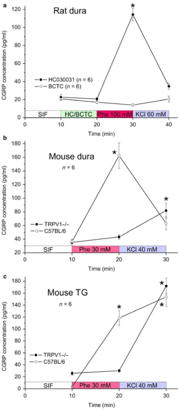
To confirm this hypothesis, we employed the same he- misected cranial preparation in the mouse measuring CGRP release from the dura mater at 10 min intervals (Figure 3b).
Since the mouse dura is more sensitive compared to the rat dura, we used phenylephrine at 30 mM and KCl at 40 mM.
In C57BL/6 wild‐type mice, phenylephrine was followed by a more than fourfold increase in CGRP release com- pared to baseline (SIF), whereas the CGRP releasing effect of the subsequent KCl superfusion was attenuated to 1.6‐
fold the baseline, which was not significantly different from baseline (n = 6, repeated measures ANOVA, F2,10 = 18.5;
Tukey HSD post hoc test, p < .001 for phenylephrine and p = .37 for KCl). In mice lacking functional TRPV1 re- ceptor channels (TRPV1‐/‐) phenylephrine 30 mM was in- effective but KCl caused a more than twofold increase in CGRP release (n = 8, repeated measures ANOVA, F2,14
= 175.1; Tukey HSD post hoc test, p = .46 for phenyleph- rine and p < .001 for KCl).
This result was largely confirmed in isolated mouse trigeminal ganglia (Figure 3c). In ganglia from C57BL/6 mice after phenylephrine at 30 mM the CGRP release was significantly increased but, different to the dura, simi- larly high values were measured after the subsequent KCl (40 mM) application (repeated measures ANOVA, F2,6
= 30.9; Tukey HSD post hoc test, p < .005 for phenyleph- rine and p < .001 for KCl). In TRPV1‐/‐ mice, phenyleph- rine at 30 mM was not followed by a significant CGRP release, whereas KCl was effective (n = 6, repeated mea- sures ANOVA, F2,10 = 88.2; Tukey HSD post hoc test, p = .93 for phenylephrine and p < .001 for KCl).
FIGURE 3 CGRP release from the cranial dura mater of rat (a) and mouse (b) as well as isolated trigeminal ganglia of mouse (c) determined at 10 min intervals, mean values ± SEM. (a) CGRP release at baseline (SIF), after application of the TRPA1 antagonist HC030031 (50 µM) or the TRPV1 antagonist BCTC (10 µM) followed by phenylephrine (100 mM) and KCl (60 mM). (b) CGRP release from C57BL/6 wild‐type and TRPV1‐/‐ mice dura mater after phenylephrine (30 mM) and KCl (40 mM). (c) CGRP release from C57BL/6 wild‐
type and TRPV1‐/‐ mice ganglia after phenylephrine (30 mM) and KCl (40 mM). Data were compared using repeated measures ANOVA followed by the Tukey HSD post hoc test; * significant difference to baseline (SIF)
3.3 | Calcium imaging
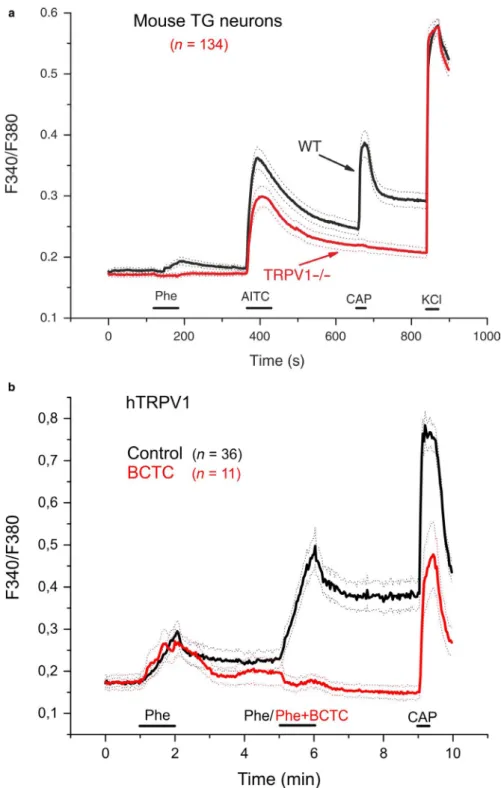
To further test if the CGRP release upon high‐dose phenylephrine is due to calcium inflow through TRP receptor channels, we employed calcium imaging. In the first series of experiments, cultured trigeminal neurons of C57BL/6 mice were challenged with phenylephrine 30 mM for 1 min, followed by agonists of three TRP channels, TRPM8 (in the majority of experiments), TRPA1 and TRPV1. The selective
TRPM8 agonist WS‐12 (5 µM) was applied 90 s after phenylephrine for 30 s, 4 min later the TRPA1 agonist allyl isothiocyanate (AITC) 100 M was applied for 1 min, followed after 3 min by the selective TRPV1 agonist capsaicin 1 M for 20 s (Figure 4a). Finally, a fully depolarizing concentration of KCl (60 mM) was applied for 30 s. Phenylephrine elicited robust calcium transients in 58 of 393 C57BL/6 TG neurons (14.8%). In this whole sample of 393 cells, 202 (51.4%) responded to AITC and 177 (45.0%) responded to capsaicin, which is generally in agreement with published data about the fractions of TRPA1 and TRPV1 expressing cells, respectively (Akopian, Ruparel, Jeske, & Hargreaves, 2007). In a sample of 221 cells also tested with the TRPM8 agonist WS‐12, 25 (11.3%) were responsive (Figure 4a). 37 (63.8%) of the 58 phenylephrine‐sensitive neurons, responded to AITC and 50 (86.2%) to capsaicin. Of 37 phenylephrine‐sensitive neurons, 10 proved to be WS‐12‐sensitive (27%). All neurons responded to KCl (60 mM), which was finally superfused for 30 s as a depolarizing control.
To investigate the possible contribution of the increased osmolarity of the phenylephrine stimulus (30 mM), we car- ried out a control experiment on mouse (C57BL/6) trigem- inal neurons, in which a total of 135 cells were stimulated with mannitol (90 mM; 1 min), followed by phenylephrine (30 mM; 1 min), capsaicin (1 µM; 30 s) and KCl (50 mM;
30 s) (Figure 4b). Of these neurons, only 12 (8.9%) responded to the hyperosmotic stimulus (mannitol), with relatively small calcium increases. In contrast, phenylephrine activated 44 neurons (32.6%), only six of which responded to mannitol.
Of the 44 phenylephrine‐sensitive neurons 34 (77.3%) were also activated by capsaicin, revealing a high co‐expression of TRPV1 among phenylephrine‐responding neurons.
In another series of experiments the effect of the TRPV1 antagonist BCTC (1 M) on the response of trigeminal neurons of two C57BL/6 mice to phenylephrine (30 mM) was tested.
Phenylephrine was first applied for 1 min in the presence of BCTC (BCTC was pre‐applied for 1 min); then after 3 min phenylephrine was applied alone for 1 min. After 4 min, cap- saicin (1 M) was applied for 20 s and finally, after another 3 min, KCl (60 mM) for 30 s. In the presence of BCTC, FIGURE 4 Calcium imaging of dissociated trigeminal
ganglion (TG) neurons from mice. (a) Calcium transients (change in fluorescence ratio) in seven C57BL/6 TG neurons, represented by different colours, as a response to phenylephrine (Phe, 30 mM), the TRPM8 agonist WS‐12 (5 µM), the TRPA1 agonist AITC (100 µM), the TRPV1 agonist capsaicin (CAP, 1 µM) and depolarizing KCl (60 mM). (b) Averaged calcium transient responses of 135 TG neurons stimulated with mannitol (90 mM; 1 min), followed by phenylephrine (30 mM; 1 min), capsaicin (1 µM; 30 s) and KCl (50 mM; 30 s).
(c) Relative responses (maximal change in fluorescence ratio ΔR/
baseline ratio R0) of 22 C57BL/6 neurons to phenylephrine (Phe) in the presence (black circles) and in the absence (open circles) of the TRPV1 antagonist BCTC
the response to phenylephrine was significantly reduced by 71.5% (from 1.13 ± 0.19% to 0.32 ± 0.14%, mean ± SEM, n = 22, two‐tailed Student's paired t test, p < .01, Figure 4c). In this experiment, the fraction of phenylephrine‐sen- sitive cells was 20.8% (22 of 106), whereas 56.6% of cells responded to capsaicin (60 of 106). Sixteen of the 22 phen- ylephrine‐responding cells were capsaicin‐sensitive (72.7%).
Interestingly, in all six phenylephrine‐sensitive neurons not responding to capsaicin, the response to phenylephrine was also inhibited by BCTC.
In TG neurons of two TRPV1‐/‐ mice, the fraction of phen- ylephrine‐sensitive cells was significantly reduced to 5.2% (7 of 134, Fisher exact probability test p < .05 compared to WT;
Figure 5a). In the sample of 134 TRPV1‐/‐ TG neurons, 55 (41%) were activated by AITC and only 2 (1.5%) responded to capsaicin, confirming genetic ablation of the capsaicin re- ceptor, TRPV1. Four (57.1%) of the 7 TRPV1‐/‐ phenyleph- rine‐sensitive neurons were activated by the TRPA1 agonist AITC and none by capsaicin. All neurons responded to KCl (60 mM) superfused for 30 s at the end of the experiment.
FIGURE 5 (a) Averaged calcium transient responses of 172 WT and 134 TRPV1‐/‐ TG neurons to phenylephrine, AITC, capsaicin and KCl. Dotted lines indicate SEM. (b) Calcium imaging of HEK293T cells expressing hTRPV1.
Averaged calcium responses of cells stimulated twice with phenylephrine (Phe, 30 mM) and capsaicin (Cap, 200 nM) under control conditions (black traces) and in the presence of BCTC (5 µM) during the second phenylephrine application (red traces). After wash‐out of phenylephrine/BCTC, cells responded to capsaicin, although at a lower extent compared to the control. Data are presented as mean (continuous lines) ± SEM (dotted lines)
Finally, phenylephrine was tested on recombinant hTRPV1 receptor channels expressed in HEK293T cells. Phenylephrine (30 mM) applied twice for 1 min at an interval of 3 min acti- vated 47 of 157 capsaicin‐sensitive cells (presumably express- ing hTRPV1). Interestingly, the second response appeared higher compared to the first, suggesting sensitization (Figure 5b). Following the removal of phenylephrine the calcium in- crease evoked by the drug did not return completely to baseline, indicating sustained activation of the receptor. In the presence of the TRPV1 antagonist BCTC (5 µM) applied together with the second application, the response to phenylephrine was com- pletely abolished in hTRPV1‐expressing HEK293T cells.
3.4 | Meningeal blood flow
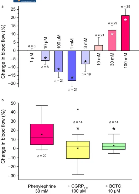
In pilot experiments, phenylephrine was topically applied at concentrations of 1 µM, as well as 30 and 100 mM, and from the variance of blood flow changes a statistically evaluable
group size of n = 8 was calculated. In the main experiments, phenylephrine was applied at increasing concentrations (1 µM‐100 mM) for 5 min each, separated by washout periods of 10 min, during which the meningeal blood flow returned to the pre‐application level. Phenylephrine at 1 µM failed to influence blood flow (0.4 ± 1.1% increase;
n = 8, t‐test), whereas the concentrations of 10 µM‐3 mM significantly reduced the flow compared to baseline. The most powerful decrease in flow was measured at 1 mM concentration (−18.1 ± 3.8%; n = 21, Wilcoxon, p < .001).
In contrast, topical applications of phenylephrine at 10 – 100 mM elicited increases in meningeal blood flow, which were statistically significant at the two highest phenylephrine concentrations, that is at 30 and 100 mM (12.5 ± 2.7%, n = 29 and 21.3 ± 4.1%, n = 25, respectively; t‐test, p < .005 and < 0.001) (Figure 6a).
The systemic blood pressure of the animals was 103.6 ± 3.4 mmHg. Applications of phenylephrine at
FIGURE 6 (a) Changes in meningeal blood flow induced by topical application of different concentrations of phenylephrine onto the exposed dura mater. (b) Changes in meningeal blood flow induced by phenylephrine (30 mM) without pretreatment, after blocking CGRP receptors with CGRP8‐37 (100 µM) and TRPV1 receptors with BCTC (10 µM). The box plot diagram with 25/75% interquartile range shows median (line), mean (square dot) and variation in values. Flow values before and after application of phenylephrine (a) or after phenylephrine with and without pretreatment (b) were compared using the Student’s t‐test;* significant difference
1 µM–3 mM concentrations failed to influence the blood pressure, whereas phenylephrine at 10 – 100 mM induced some increase in pressure. For further blood flow mea- surements the 30 mM concentration of phenylephrine was chosen, because it induced a robust blood flow increase without increasing arterial blood pressure significantly.
After 30 mM phenylephrine the arterial pressure increased on average by 8.7 ± 2.2% (Wilcoxon, p < .001) during the 5 min application period. However, there was no correlation between the changes in arterial pressure and the changes in blood flow (r = −0.18, p > .05; product‐moment correla- tion). In experiments with visible changes in arterial blood pressure, these were limited to the first 3 minutes of the dural phenylephrine application, whereas increases in men- ingeal blood flow were present throughout the whole 5 min application period.
In pilot experiments, the blocking effect on the phenyl- ephrine‐induced (30 mM) blood flow increase was tested, and from the variance of measurements a statistically eval- uable group size of n ≥ 10 was calculated. In the main experiments, blocking the CGRP receptors by pre‐applica- tion of 100 µM CGRP8‐37 to the dura mater significantly inhibited the phenylephrine‐induced blood flow increase (2.3 ± 6.3%; n = 10, t‐test, p < .05). Next we aimed to clarify the role of chemosensitive nociceptors in the dura mater, which are known to release CGRP upon their acti- vation causing arterial vasodilatation and increased blood flow. The majority of these nociceptive afferents express TRPV1 receptor channels. Pretreatment of the dura mater with the TRPV1 receptor antagonist BCTC at 10 µM sig- nificantly inhibited the phenylephrine‐induced increase in meningeal blood flow (4.9 ± 2.0%; n = 10, t‐test, p < .05) (Figure 6b). Together with the data from the CGRP release and the calcium imaging, these results argue for a role of TRPV1 rather than TRPA1 receptors in the phenylephrine‐
induced blood flow increase.
4 | DISCUSSION
The present experiments show that high concentrations (≥ 30 mM) of the α1‐adrenoceptor agonist phenylephrine activate trigeminal neurons in rodent models of trigeminal nociception. This activation is indicated by CGRP release from peptidergic afferents upon calcium inflow and, as neurovascular consequence of the neuropeptide release, increase in meningeal blood flow. It is very unlikely that this activation is due to the high molarity of phenylephrine, since mannitol at 90 mM did neither cause CGRP release nor an effective increase in intracellular calcium. The experiments also showed that the responses to high‐dose phenylephrine are independent of α1‐adrenoceptors, although these adrenoceptors seem to be expressed in some
trigeminal neurons innervating the dura mater, albeit very rarely co‐localized with CGRP. Instead, the responses seem to depend mainly on the activation of TRPV1 receptor channels but not on TRPA1 receptor channels, because phenylephrine‐induced responses present in HEK293T cells transfected with hTRPV1 are largely suppressed by inhibiting TRPV1 and are nearly, though not completely, absent in animals with genetically deleted TRPV1 receptors.
4.1 | Major role for TRPV1 receptors in phenylephrine‐induced trigeminal nociception and pain
TRPV1 receptor channels are expressed in a major pro- portion of nociceptive afferents, constituting the crucial molecular transduction channels for a variety of noxious stimuli associated with painful tissue lesions, inflamma- tion and heat (Holzer, 2008; Rosenbaum & Simon, 2007;
White, Urban, & Nagy, 2011).The unexpected finding that high‐dose phenylephrine is obviously also an agonist at TRPV1 receptors, including human TRPV1, cannot eas- ily be explained by its molecular structure, although it has some similarity with the aromatic end‐structure of capsai- cin. Phenylephrine at high concentrations is routinely used as a mydriatic agent and for the treatment of nasal con- gestions (https ://medli neplus.gov/drugi nfo/meds/a6160 49.html) frequently causing transient local burning or stinging pain sensations (Moisseiev et al., 2015). We now offer a reasonable pathophysiological explanation for these effects, which are reminiscent of mild capsaicin‐evoked pain. There are few other reports suggesting an involve- ment of TRPV1 in pronociceptive effects of phenylephrine.
Subcutaneous injection of phenylephrine (2.5 mg/kg, daily for 2 weeks) in rodents induced abdominal mechanical hy- peralgesia, which was absent in TRPV1‐/‐ mice (Matos et al., 2016); however, pain behaviour was also reversed by a selective α1A‐adrenoceptor antagonist. Injection of phe- nylephrine (0.1–10 µM) in human skin dose‐dependently decreased heat pain threshold but not mechanical pain threshold (Fuchs, Meyer, & Raja, 2001), which would be in line with the sensitization of TRPV1 receptor channels.
Beyond the transient character of TRPV1 opening, an ef- fective activation of TRPV1 receptors followed by massive calcium inflow causes insensitivity to repeated application of the agonists (tachyphylaxis) (Touska, Marsakova, Teisinger,
& Vlachova, 2011) and finally long‐lasting inactivation of the neuron, most likely due to a permanent depolarization block, as it was described for the slowly acting but highly effective TRPV1 agonist resiniferatoxin (Raisinghani, Pabbidi, &
Premkumar, 2005). Phenylephrine at high doses seems to act similarly at TRPV1 receptors and thus may lead to a long‐last- ing depolarization of trigeminal neurons. Thus neuropeptide
release upon depolarizing KCl, which depends primarily on the opening of voltage‐sensitive calcium channels (Kress, Izydorczyk, & Kuhn, 2001), seems to be dose‐dependently blocked after 10 min treatment with high‐dose phenylephrine (see Figure 2b). In our calcium‐imaging experiments neurons responding vigorously to capsaicin showed a long‐lasting calcium signal and responded only weakly to depolarizing KCl (see Figure 4a). Besides, the CGRP secreting sensory fibres in the cranial dura mater seem to be more effectively activated by phenylephrine compared to the dissociated neu- rons and intact ganglia, which after phenylephrine treatment for 10 min still responded to KCl with some calcium signal or neuropeptide release, respectively (compare Figure 3b and 3c). This difference may be due to a higher calcium buffer capacity and larger neuropeptide stores of the cell somata compared to the thin peripheral nerve fibres. However, we cannot explain the observation that, after phenylephrine in the presence of the TRPV1 receptor blocker BCTC, KCl was again ineffective in releasing CGRP from rat dura mater (see Figure 3a). It may be speculated that high‐dose phenyleph- rine has also an inhibitory effect on voltage‐gated calcium channels independent of TRPV1.
Headache is another, yet mechanistically unexplained, side‐effect of phenylephrine noted in instruction leaflets of drugs containing phenylephrine for local (ocular, nasal) application (https ://www.webmd.com/drugs/ 2/drug-6979/
pheny lephr ine-ophth almic-eye/details) (https ://medli neplus.gov/drugi nfo/meds/a6160 49.html). This type of headache is not well defined in the literature and not specif- ically listed in the International Classification of Headache Disorders, third edition (ICHD‐3) (Headache Classification Committee of the International Headache Society (IHS), 2013) , where it could be classified at its best into category 8.1.9 of secondary headaches (‘Headache attributed to oc- casional use of non‐headache medication’). It is generally assumed that headaches involve the activation of intracra- nial afferents of the trigeminal nerve and/or the second order neurons in the spinal trigeminal nucleus, where these afferents are projecting to (Levy, Labastida‐Ramirez, &
MaassenVanDenBrink, 2018; Olesen, Burstein, Ashina, &
Tfelt‐Hansen, 2009; Schueler, Messlinger, Dux, Neuhuber,
& Col, 2013). Headache and ocular pain are frequently as- sociated (Dafer & Jay, 2009), and there is a close associa- tion between nasal/paranasal lesions and headaches (Pinto, Rossi, McQuone, & Sollecito, 2001; Silberstein, 2004). The effectivity of intranasal application of drugs for the treat- ment of headaches is supportive for this view (Rapoport, Bigal, Tepper, & Sheftell, 2004). Possibly intranasal phen- ylephrine reaches directly the meninges by‐passing the blood brain barrier, as it has been discussed for triptans (Rapoport & Winner, 2006). We assume that phenyleph- rine is able to diffuse along the fila of the olfactory neurons through the lamina cribrosa to reach the meninges directly
on this route, as it was shown for other substances. For ex- ample nasal application of irritants such as capsaicin can activate meningeal afferents either directly or via a para- sympathetic reflex, which seem to work at least partially via this nasal‐meningeal pathway (Gottselig & Messlinger, 2004; Kunkler, Ballard, Oxford, & Hurley, 2011). Another hypothesis has very recently been proposed (Zhang, Kunkler, Knopp, Oxford, & Hurley, 2019). The authors have found upregulation of TRPA1 mRNA in trigeminal ganglion neurons specifically innervating the dura mater after chronic exposure of rats to an atmosphere containing the TRPA1 agonist acroleine. They suggest an intragangli- onic transmission from neurons innervating the nasal mu- cosa to neurons innervating the meninges. Irrespective of these proposed mechanism, inhalation of chemical irritants (e.g. acrolein, formaline) can induce headaches in suscepti- ble persons by stimulating TRP receptors of the nasal mu- cosa (Kunkler, Zhang, Johnson, Oxford, & Hurley, 2018).
4.2 | Possible role for other TRP receptor channels and α
1‐adrenoceptors
Another TRP receptor channel responding to a big variety of endogenous and exogenous chemical substances is TRPA1 (Koivisto et al., 2014; Nilius, Appendino, & Owsianik, 2012).
We originally speculated that it may be involved in the phe- nylephrine effects but blockade by the specific TRPA1 antag- onist, HC030031, was ineffective in reducing CGRP release.
In contrast, the specific blockade of TRPV1 with BCTC was very effective in inhibiting neuropeptide release, which was finally confirmed in mice lacking functional TRPV1 recep- tors (see Figure 3a‐c). However, an additional mechanism, by which high‐dose phenylephrine can activate sensory neurons, cannot be excluded, because in the calcium imaging experi- ments the overlap of the responses to capsaicin and phenyle- phrine was not complete: roughly 80% of neurons responding to phenylephrine responded also to capsaicin, whereas some neurons were still activated by phenylephrine in the presence of BCTC (see Figure 4c).
We can also not exclude that phenylephrine causes cal- cium signals in trigeminal neurons without releasing neu- ropeptides, possibly by activation of α1‐adrenoceptors, immunoreactivity of which was found in a minor percentage of trigeminal ganglion neurons and in perivascular nerve fi- bres of the dura mater. The expression of α1‐adrenoceptors has already been described in myelinated and unmyelinated sensory nerve fibres, but not in sympathetic fibres, in rat skin (Dawson, Phillips, Finch, Inglis, & Drummond, 2011).
The expression of α1‐adrenoceptor has been confirmed in dorsal root ganglia, where double labelling showed co‐ex- pression within sub‐populations of CGRP, IB4 and TRPV1 immunoreactive neurons. Following chronic constriction injury, the expression of α1‐adrenoceptors in rat cutaneous
nociceptors was found increased (Drummond, Dawson, Finch, Bennett, & Drummond, 2014). The authors sug- gested that this may contribute to the sensory‐sympathetic coupling increasing the sensitivity to adrenergic agonists after nerve injury.
4.3 | Vascular effects of phenylephrine
In our in vivo experiments blood flow measurements showed decreased meningeal blood flow after the applica- tion of lower phenylephrine concentrations indicating arte- rial vasoconstriction, whereas high concentrations increased meningeal blood flow. Activation of several subtypes of vascular α1‐adrenoceptors constricts blood vessels leading to reduction in blood flow (Civantos Calzada & Aleixandre de Artiñano, 2001). The mechanism is explained by the ac- tivation of the receptor coupled G‐protein Gq/11 followed by phospholipase C and inositol phosphate (IP) turnover and the opening of IP3‐sensitive Ca2+ channels of intracel- lular calcium stores (Docherty, 2010), which is controlled by L‐type voltage‐gated Ca2+ channels of the cell mem- brane (Leloup, Hove, Meyer, Schrijvers, & Fransen, 2015).
In human pial arteries both α1‐ and α2‐adrenoceptors have been found by radioligand‐binding studies (Ferrari‐DiLeo
& Potter, 1985). In isolated feline middle cerebral artery, the α2‐adrenoceptor agonist clonidine was much more po- tent than the α1‐adrenoceptor agonist phenylephrine, sug- gesting the preferential expression of α2‐adrenoceptors (Skärby, Andersson, & Edvinsson, 1983), but this is not clear for dural arteries. The high sensitivity to phenyle- phrine of meningeal blood flow may indicate the preferen- tial action of α1‐adrenoceptors in rat dura mater. According to our immunostainings, α1‐adrenoceptors are expressed in precapillary dural blood vessels, which can regulate blood flow very effectively (see Figure 1b). However, at high concentrations of phenylephrine activation of trigeminal TRPV1 receptors leading to CGRP release and consequent increase in meningeal blood flow is apparent and overrides the α‐adrenoceptor‐induced constriction.
ACKNOWLEDGEMENT
We thank A. Kuhn and S. Haux‐Oertel for excellent technical assistance.
CONFLICT OF INTEREST The authors declare no conflicts of interest.
AUTHOR CONTRIBUTIONS
M.D., A.B. and K.M. designed the study. All authors contributed to perform the experiments. M.D., A.B., J.M.
and K.M. analysed the data. M.D., A.B. and K.M. wrote the paper. All authors discussed the results and commented on the manuscript.
REFERENCES
Akopian, A. N., Ruparel, N. B., Jeske, N. A., & Hargreaves, K. M.
(2007). Transient receptor potential TRPA1 channel desensitization in sensory neurons is agonist dependent and regulated by TRPV1‐
directed internalization. The Journal of Physiology, 583, 175–193.
https ://doi.org/10.1113/jphys iol.2007.133231
Babes, A., Ciobanu, A. C., Neacsu, C., & Babes, R.‐M. (2011). TRPM8, a sensor for mild cooling in mammalian sensory nerve endings.
Current Pharmaceutical Biotechnology, 12, 78–88.
Chin, K. W., Law, N. M., & Chin, M. K. (1994). Phenylephrine eye drops in ophthalmic surgery–a clinical study on cardiovascular ef- fects. Medical Journal of Malaysia, 49, 158–163.
Civantos Calzada, B., & Aleixandre de Artiñano, A. (2001). Alpha‐ad- renoceptor subtypes. Pharmacological Research, 44, 195–208. https ://doi.org/10.1006/phrs.2001.0857
Dafer, R. M., & Jay, W. M. (2009). Headache and the eye. Current Opinion in Ophthalmology, 20, 520–524. https ://doi.org/10.1097/
ICU.0b013 e3283 31270d
Dawson, L. F., Phillips, J. K., Finch, P. M., Inglis, J. J., & Drummond, P. D. (2011). Expression of α1‐adrenoceptors on peripheral nocicep- tive neurons. Neuroscience, 175, 300–314. https ://doi.org/10.1016/j.
neuro scien ce.2010.11.064
Docherty, J. R. (2010). Subtypes of functional alpha1‐adrenoceptor.
Cellular and Molecular Life Sciences, 67, 405–417.
Drummond, E. S., Dawson, L. F., Finch, P. M., Bennett, G. J., &
Drummond, P. D. (2014). Increased expression of cutaneous α1‐ad- renoceptors after chronic constriction injury in rats. The Journal of Pain, 15, 188–196. https ://doi.org/10.1016/j.jpain.2013.10.010 Dussor, G., & Cao, Y.‐Q. (2016). TRPM8 and Migraine. Headache:
the Journal of Head and Face Pain, 56, 1406–1417. https ://doi.
org/10.1111/head.12948
Dux, M., Sántha, P., & Jancsó, G. (2012). The role of chemosensitive afferent nerves and TRP ion channels in the pathomechanism of headaches. Pflügers Archiv ‐ European Journal of Physiology, 464, 239–248. https ://doi.org/10.1007/s00424-012-1142-7
Earley, S., & Brayden, J. E. (2015). Transient receptor potential chan- nels in the vasculature. Physiological Reviews, 95, 645–690. https ://
doi.org/10.1152/physr ev.00026.2014
Ferrari‐DiLeo, G., & Potter, L. T. (1985). Alpha‐adrenoreceptors and muscarine receptors in human pial arteries and microvessels:
A receptor binding study. Journal of Cerebral Blood Flow and Metabolism, 5, 458–464.
Fuchs, P. N., Meyer, R. A., & Raja, S. N. (2001). Heat, but not mechanical hyperalgesia, following adrenergic injections in normal human skin.
Pain, 90, 15–23. https ://doi.org/10.1016/S0304-3959(00)00381-X Gerhold, K. A., & Bautista, D. M. (2009). Molecular and cellu-
lar mechanisms of trigeminal chemosensation. Annals of the New York Academy of Sciences, 1170, 184–189. https ://doi.
org/10.1111/j.1749-6632.2009.03895.x
Gottselig, R., & Messlinger, K. (2004). Noxious chemical stimulation of rat facial mucosa increases intracranial blood flow through a tri- gemino‐parasympathetic reflex–an experimental model for vascular dysfunctions in cluster headache. Cephalalgia, 24, 206–214. https ://
doi.org/10.1111/j.1468-2982.2004.00649.x
. Headache Classification Committee of the International Headache Society (IHS) (2013). The International Classification of Headache Disorders,3rd edition (beta version). Cephalalgia, 33,629–808.
Heesen, M., Kölhr, S., Rossaint, R., & Straube, S. (2014). Prophylactic phenylephrine for caesarean section under spinal anaesthesia:
Systematic review and meta‐analysis. Anaesthesia, 69, 143–165.
https ://doi.org/10.1111/anae.12445
Holom, V. H., Messlinger, K., & Fischer, M. J. M. (2008). Temperature‐
dependent neuronal regulation of arterial blood flow in rat cranial dura mater. Journal of Neuroscience Research, 86, 158–164. https ://
doi.org/10.1002/jnr.21459
Holzer, P. (2008). The pharmacological challenge to tame the tran- sient receptor potential vanilloid‐1 (TRPV1) nocisensor. British Journal of Pharmacology, 155, 1145–1162. https ://doi.org/10.1038/
bjp.2008.351
Julius, D. (2013). TRP channels and pain. Annual Review of Cell and Developmental Biology, 29, 355–384. https ://doi.org/10.1146/annur ev-cellb io-101011-155833
Kim, H. J., Choi, J. G., & Kwak, K.‐H. (2015). Bronchoconstriction following instillation of phenylephrine eye drops in premature in- fants with bronchopulmonary dysplasia: Two cases report. Korean Journal of Anesthesiology, 68, 613–616. https ://doi.org/10.4097/
kjae.2015.68.6.613
Koivisto, A., Chapman, H., Jalava, N., Korjamo, T., Saarnilehto, M., Lindstedt, K., & Pertovaara, A. (2014). TRPA1: A transducer and amplifier of pain and inflammation. Basic & Clinical Pharmacology
& Toxicology, 114, 50–55. https ://doi.org/10.1111/bcpt.12138 Kollar, C., Schneider, H., Waksman, J., & Krusinska, E. (2007). Meta‐
analysis of the efficacy of a single dose of phenylephrine 10 mg compared with placebo in adults with acute nasal congestion due to the common cold. Clinical Therapeutics, 29, 1057–1070. https ://
doi.org/10.1016/j.clint hera.2007.05.021
Kress, M., Izydorczyk, I., & Kuhn, A. (2001). N‐ and L‐ but not P/Q‐type calcium channels contribute to neuropeptide release from rat skin in vitro. NeuroReport, 12, 867–870. https ://doi.org/10.1097/00001 756-20010 3260-00048
Kunkler, P. E., Ballard, C. J., Oxford, G. S., & Hurley, J. H. (2011).
TRPA1 receptors mediate environmental irritant‐induced menin- geal vasodilatation. Pain, 152, 38–44. https ://doi.org/10.1016/j.
pain.2010.08.021
Kunkler, P. E., Zhang, L., Johnson, P. L., Oxford, G. S., & Hurley, J.
H. (2018). Induction of chronic migraine phenotypes in a rat model after environmental irritant exposure. Pain, 159, 540–549.
Leloup, A. J., Van Hove, C. E., De Meyer, G. R. Y., Schrijvers, D. M., &
Fransen, P. (2015). Basal activity of voltage‐gated Ca(2+) channels controls the IP3‐mediated contraction by α(1)‐adrenoceptor stimula- tion of mouse aorta segments. European Journal of Pharmacology, 760, 163–171. https ://doi.org/10.1016/j.ejphar.2015.04.011 Levy, D., Labastida‐Ramirez, A., & MaassenVanDenBrink, A. (2018).
Current understanding of meningeal and cerebral vascular function underlying migraine headache. Cephalalgia, 333102418771350.
https ://doi.org/10.1177/03331 02418 771350
Matos, R., Cordeiro, J. M., Coelho, A., Ferreira, S., Silva, C., Igawa, Y.,
… Charrua, A. (2016). Bladder pain induced by prolonged periph- eral alpha 1A adrenoceptor stimulation involves the enhancement of transient receptor potential vanilloid 1 activity and an increase of urothelial adenosine triphosphate release. Acta Psychologica, 218, 265–275. https ://doi.org/10.1111/apha.12744
McAuliffe‐Curtin, D., & Buckley, C. (1989). Review of alpha adreno- ceptor function in the eye. Eye (Lond), 3(Pt 4), 472–476. https ://doi.
org/10.1038/eye.1989.71
Meltzer, E. O., Ratner, P. H., & McGraw, T. (2015). Oral phenylephrine HCl for nasal congestion in seasonal allergic rhinitis: A random- ized, open‐label, placebo‐controlled study. The Journal of Allergy and Clinical Immunology: in Practice, 3, 702–708. https ://doi.
org/10.1016/j.jaip.2015.05.007
Moisseiev, E., Loberman, D., Zunz, E., Kesler, A., Loewenstein, A., &
Mandelblum, J. (2015). Pupil dilation using drops vs gel: A com- parative study. Eye (Lond), 29, 815–819. https ://doi.org/10.1038/
eye.2015.47
Myers, M. G., & Lazzetta, J. J. (1982). Intranasally administered phenyl- ephrine and blood pressure. Canadian Medical Association Journal, 127, 365–368.
Nagy, I., Sántha, P., Jancsó, G., & Urbán, L. (2004). The role of the vanilloid (capsaicin) receptor (TRPV1) in physiology and pathol- ogy. European Journal of Pharmacology, 500, 351–369. https ://doi.
org/10.1016/j.ejphar.2004.07.037
Nicholson, R., Dixon, A. K., Spanswick, D., & Lee, K. (2005).
Noradrenergic receptor mRNA expression in adult rat superficial dorsal horn and dorsal root ganglion neurons. Neuroscience Letters, 380, 316–321. https ://doi.org/10.1016/j.neulet.2005.01.079 Nilius, B., Appendino, G., & Owsianik, G. (2012). The transient re-
ceptor potential channel TRPA1: From gene to pathophysiology.
Pflugers Archiv. European Journal of Physiology, 464, 425–458.
https ://doi.org/10.1007/s00424-012-1158-z
Numazaki, M., & Tominaga, M. (2004). Nociception and TRP Channels.
Current Drug Targets CNS & Neurological Disorders, 3, 479–485.
Olesen, J., Burstein, R., Ashina, M., & Tfelt‐Hansen, P. (2009).
Origin of pain in migraine: Evidence for peripheral sensitisa- tion. The Lancet Neurology, 8, 679–690. https ://doi.org/10.1016/
S1474-4422(09)70090-0
Pinto, A., De Rossi, S. S., McQuone, S., & Sollecito, T. P. (2001).
Nasal mucosal headache presenting as orofacial pain: A review of the literature and a case report. Oral Surgery, Oral Medicine, Oral Pathology, Oral Radiology and Endodontics, 92, 180–183. https ://
doi.org/10.1067/moe.2001.114006
Poterman, M., Vos, J. J., Vereecke, H. E. M., Struys, M. M. R. F., Vanoverschelde, H., Scheeren, T. W. L., & Kalmar, A. F. (2015).
Differential effects of phenylephrine and norepinephrine on periph- eral tissue oxygenation during general anaesthesia: A randomised controlled trial. European Journal of Anaesthesiology, 32, 571–580.
https ://doi.org/10.1097/EJA.00000 00000 000247
Raisinghani, M., Pabbidi, R. M., & Premkumar, L. S. (2005). Activation of transient receptor potential vanilloid 1 (TRPV1) by resinif- eratoxin. The Journal of Physiology, 567, 771–786. https ://doi.
org/10.1113/jphys iol.2005.087874
Rapoport, A. M., Bigal, M. E., Tepper, S. J., & Sheftell, F. D. (2004).
Intranasal medications for the treatment of migraine and cluster headache. CNS Drugs, 18, 671–685. https ://doi.org/10.2165/00023 210-20041 8100-00004
Rapoport, A., & Winner, P. (2006). Nasal delivery of antimigraine drugs: Clinical rationale and evidence base. Headache, 46(Suppl 4), S192–201. https ://doi.org/10.1111/j.1526-4610.2006.00603.x Reid, J. L. (1986). Alpha‐adrenergic receptors and blood pressure con-
trol. The American Journal of Cardiology, 57, E6–E12. https ://doi.
org/10.1016/0002-9149(86)90716-2