Rapid communication
Cardioprotection by remote ischemic preconditioning of the rat heart is mediated by extracellular vesicles
Zoltán Giricz
a, Zoltán V. Varga
a, Tamás Baranyai
a, Péter Sipos
b, Krisztina Pálóczi
c, Ágnes Kittel
d, Edit I. Buzás
c, Péter Ferdinandy
a,e,⁎
aCardiometabolic Research Group, Department of Pharmacology and Pharmacotherapy, Semmelweis University, Budapest, Hungary
bDepartment of Pharmaceutical Technology, University of Szeged, Szeged, Hungary
cDepartment of Genetics, Cell- and Immunobiology, Semmelweis University, Budapest, Hungary
dDepartment of Pharmacology, Institute of Experimental Medicine, Hungarian Academy of Sciences, Budapest, Hungary
eCardiovascular Research Group, Department of Biochemistry, University of Szeged, Szeged, Hungary
a b s t r a c t a r t i c l e i n f o
Article history:
Received 12 November 2013 Received in revised form 7 January 2014 Accepted 9 January 2014
Available online 16 January 2014 Keywords:
Exosomes Microvesicles Nanoparticles Remote conditioning Cardioprotection Ischemia–reperfusion
Remote ischemic preconditioning (RIPC) of the heart is exerted by brief ischemic insults affected on a remote organ or a remote area of the heart before a sustained cardiac ischemia. To date, little is known about the inter-organ transfer mechanisms of cardioprotection by RIPC. Exosomes and microvesicles/microparticles are vesicles of 30–100 nm and 100–1000 nm in diameter, respectively (collectively termed extracellular vesicles [EVs]). Their content of proteins, mRNAs and microRNAs, renders EV ideal conveyors of inter-organ communica- tion. However, whether EVs are involved in RIPC, is unknown. Therefore, here we investigated whether (1) IPC induces release of EVs from the heart, and (2) EVs are necessary for cardioprotection by RIPC. Hearts of male Wistar rats were isolated and perfused in Langendorff mode. A group of donor hearts was exposed to 3
× 5-5 min global ischemia and reperfusion (IPC) or 30 min aerobic perfusion, while coronary perfusates were col- lected. Coronary perfusates of these hearts were given to another set of recipient isolated hearts. A group of re- cipient hearts received IPC effluent depleted of EVs by differential ultracentrifugation. Infarct size was determined after 30 min global ischemia and 120 min reperfusion. The presence or absence of EVs in perfusates was confirmed by dynamic light scattering, the EV marker HSP60 Western blot, and electron microscopy. IPC markedly increased EV release from the heart as assessed by HSP60. Administration of coronary perfusate from IPC donor hearts attenuated infarct size in non-preconditioned recipient hearts (12.9 ± 1.6% vs. 25.0 ± 2.7%), similarly to cardioprotection afforded by IPC (7.3 ± 2.7% vs. 22.1 ± 2.9%) on the donor hearts. Perfusates of IPC hearts depleted of EVs failed to exert cardioprotection in recipient hearts (22.0 ± 2.3%). This is thefirst demonstration that EVs released from the heart after IPC are necessary for cardioprotection by RIPC, evidencing the importance of vesicular transfer mechanisms in remote cardioprotection.
© 2014 The Authors. Published by Elsevier Ltd.
1. Introduction
Remote ischemic conditioning (RIPC), where a remote area of the heart or another organ is submitted to brief cycles of ischemia– reperfusion, protects the heart against a lethal ischemic insult with efficiency comparable to that of classic in-situ ischemic protocols [1,2]. Although effector pathways of RIPC have been well described, it is currently unclear how cardioprotective signals are propagated between organs [3]. Humoral and neuronal aspects have been
hypothesized, but vesicular transfer mechanisms have not been evi- denced in inter- or intra-organ communication of RIPC signals.
Exosomes and microvesicles/microparticles (collectively termed ex- tracellular vesicles, EVs) are membrane-bound structures secreted by a wide range of mammalian cell types via distinct mechanisms[4,5]. Since EVs contain a high concentration of RNAs and proteins, and since EVs can be secreted and specifically taken up by other cells, they are prime medium for intercellular signal transfer mechanisms[5]. Thus, it is not surprising that EVs have been shown to modulate several essential cel- lular functions, including cell survival mechanisms[6,7]. However, to date, it is not known whether EVs are involved in the transmission of cardioprotective signals in ischemic conditioning maneuvers, particu- larly, their role in the propagation of RIPC has never been studied.
Therefore, here we aimed to investigate whether the release of EVs from the heart is induced by preconditioning stimuli; and to test if EVs are necessary for RIPC-induced cardioprotection by assessing that RIPC can be exerted in the presence and absence of EVs.
Journal of Molecular and Cellular Cardiology 68 (2014) 75–78
⁎ Corresponding author at: Cardiometabolic Research Group, Department of Pharmacology and Pharmacotherapy, Semmelweis University, Nagyvárad tér 4, Budapest H-1089, Hungary. Tel.: +36 1 2104416; fax: +36 1 2104412.
E-mail address:peter.ferdinandy@pharmahungary.com(P. Ferdinandy).
0022-2828 © 2014 The Authors. Published by Elsevier Ltd.
http://dx.doi.org/10.1016/j.yjmcc.2014.01.004
Contents lists available atScienceDirect
Journal of Molecular and Cellular Cardiology
j o u r n a l h o m e p a g e : w w w . e l s e v i e r . c o m / l o c a t e / y j m c c
Open access under CC BY license.
Open access under CC BY license.
2. Materials and methods
This investigation conforms to the Guide for the Care and Use of Lab- oratory Animals published by the US National Institutes of Health (NIH publication No. 85–23, revised 1996) and was approved by the animal ethics committee of the Semmelweis University, Budapest, Hungary.
2.1. Experimental setup, heart perfusion protocol, and assessment of infarct size
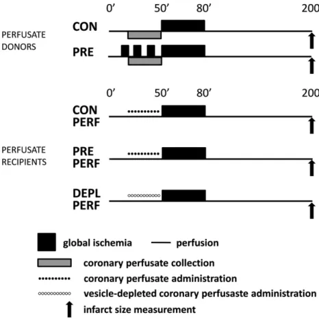
Male Wistar rats (250–350 g) were anesthetized by 85 mg/kg keta- mine and 10 mg/kg xylazine and heparinized. Hearts were isolated and perfused in Langendorff mode with 37 °C Krebs–Henseleit solution for 20 min for stabilization; then hearts were randomized to the following groups. Perfusate donor hearts either received aerobic perfusion for an additional 30 min (CON) or were exposed to 3 × 5-5 min ischemia and reperfusion (PRE). Perfusate recipient hearts were perfused with collected perfusate from either CON or PRE hearts (CON PERF and PRE PERF, respectively). Another group of hearts received perfusate of PRE hearts which had been previously depleted of EVs (DEPL PERF). All hearts were then exposed to a 30 min global ischemia and 2 h reperfu- sion (Fig. 1).
Hearts then were cut into 6–8 slices, slices were weighed, and infarct size was assessed by TTC-staining. Infarct size was expressed as a per- centage of the total heart weight.
2.2. Isolation of EVs and EV depletion
EVs were isolated from collected coronary perfusates byfiltration and differential centrifugation. Briefly, perfusates were dialyzed against 0.45% saline containing 5 mM EDTA for 4 h at room temperature then vacuum-distilled to 40 mL. Concentrated perfusates werefiltered
through 800 nmfilter (Merck, Darmstadt, Germany) and centrifuged at 12,200 ×gfor 20 min at 4 °C. Pellets were saved as microvesicle/
microparticle fraction. Then supernatants werefiltered through 200 nm filter (Merck, Darmstadt, Germany) and centrifuged at 100,000 ×gfor 90 min at 4 °C. Pellets were saved as exosome-rich pellet and the super- natant was saved as EV-depleted perfusate. EV-depleted perfusates were then reconstituted to their original volume with Krebs–Henseleit solution and used in heart perfusion experiments.
2.3. Characterization and assessment of quantity and size distribution of EVs
Isolated vesicles were visualized by transmission electron microsco- py. Vesicle pellets werefixed with 4% formaldehyde, postfixed in 1%
OsO4. EVs were block-stained with 1% uranyl acetate in 50% ethanol, then dehydrated in graded ethanol, and embedded in Taab 812 (Taab Laboratories, Aldermaston, UK). Ultrathin sections were cut and then analyzed with a Hitachi 7100 electron microscope.
Hydrodynamical average particle size of EVs in perfusates was mea- sured by Dynamic Light Scattering (DLS) apparatus Zetasizer Nano ZS (Malvern Instruments, Malvern Hills, UK) (n = 3–4).
The presence and amount of EVs were assessed by HSP60 immuno- blots from vesicular pellets and EV-depleted perfusates.
For a detailed Methods section please see Supplementary data online.
2.4. Statistical analysis
Values are expressed as mean ± SEM. One way analysis of variance (ANOVA) followed by Fisher LSD post-hoc test was used to determine differences in infarct size.
Fig. 1.Experimental protocol of Langendorff-perfused rat hearts. CON: control; PRE: preconditioned; PERF: perfused; DEPL: depleted.
76 Z. Giricz et al. / Journal of Molecular and Cellular Cardiology 68 (2014) 75–78
3. Results
3.1. Ischemic conditioning increases the amount of EVs released into coronary perfusate
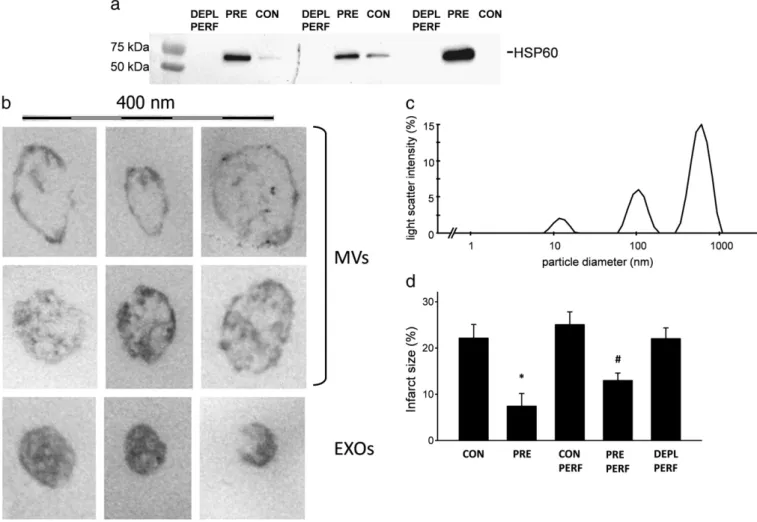
Coronary perfusates from preconditioned hearts (PRE) contained more EVs than perfusates isolated from control (CON) hearts as evi- denced by Western blot against HSP60, a well-accepted marker of EVs (Fig. 2a). Electron micrographs revealed that these EVs can be classified as microvesicle/microparticles as defined by a diameter ofN100 nm and light vesicular structure (Fig. 2b upper panels), and exosomes of b100 nm in diameter (Fig. 2b lower panel).
To further evidence that size of the particulate matter in coronary perfusate conforms to the range of microvesicles and exosomes, we assessed their size distribution by DLS in perfusates from PRE hearts.
As the representative diagram (Fig. 2c) shows, three populations of par- ticles could be distinguished in our samples. Beside a small fraction of particles with hypothetical diameter of approximately 10 nm, the main particulate constituents fell into the size range of exosomes (b100 nm) and microvesicles (100–1000 nm).
3.2. Perfusates depleted of EVs did not decrease infarct size
Isolated hearts that received 3 × 5-5 min ischemia and reperfusion (PRE) before 30 min global ischemia demonstrated a significantly re- duced infarct size as compared to aerobically perfused hearts (CON).
In hearts perfused with coronary perfusates collected from PRE hearts (PRE PERF) infarct size was significantly lower than in hearts perfused
with coronary perfusate from CON hearts (CON PERF). Perfusates of PERF hearts which had been depleted of EVs were also given to recipient hearts (DEPL PERF). Infarct size after 30 min ischemia and 2 h reperfu- sion in DEPL PERF hearts did not differ significantly from the extent of infarction observed in CON PERF hearts (Fig. 2d).
4. Discussion
We have shown here for thefirst time in the literature that the re- lease of EVs from the heart after preconditioning stimuli is increased and that EVs are responsible for the transmission of remote condition- ing signals for cardioprotection.
Previously several humoral and neuronal transmitter mechanisms have been hypothesized to play a role in the propagation of remote ischemic conditioning, however, to date none of them is generally accepted. Dickson et al. have proposedfirst the involvement of hu- moral transmission pathways showing that transfusion of blood from preconditioned rabbits confers cardioprotection in a naïve non- preconditioned animal against ischemia/reperfusion injury[8]. Later, Breivik et al. showed that the soluble factor is likely to be hydrophobic [9]. The role of neuronal pathways has also been studied, but results are also still controversial[10,11].
Here we evidence a novel, vesicular mechanism for the transmission of cardioprotective signals from a preconditioned heart to another heart subjected to coronary occlusion and reperfusion, which might explain how the suspected humoral and/or released neuronal factors of remote conditioning are transmitted. Ischemia-induced release of EVs from cul- tured cardiomyocytes was reported by Malik et al.[12]recently, which
Fig. 2.a: HSP60 immunoblots of depleted perfusates (DEPL PERF), EV pellets of preconditioned (PRE) and control perfusates (CON). b: Representative electron micrographs of EVs in the size ranges of microvesicles and exosomes isolated from coronary perfusate of preconditioned hearts. Scale bar represents 400 nm. c: DLS analysis of perfusates showing distribution of different vesicle populations. d: Infarct size indicated as percent of the total heart volume. Results are expressed as mean ± SEM; n = 5–8. *pb0.05 vs. CON, #pb0.05 vs. CON PERF.
Z. Giricz et al. / Journal of Molecular and Cellular Cardiology 68 (2014) 75–78 77
is in agreement with our currentfindings that EV-release of isolated hearts increases after brief ischemic episodes. Elsewhere, exosomes de- rived from mesenchymal stem cell cultures have been shown to exert cardioprotection in mice[13], and microvesicles isolated from cell culture medium of GATA-4-overexpressing bone marrow stem cells protected neonatal cardiomyocytes from ischemic injuries[14]. Since in the latter two reports EVs from untreated cells induced pro-survival signals, based on our currentfindings, we cannot exclude the possibility that EVs released from the heart under basal conditions might be also cardioprotective, would their amount be as high as after precondition- ing stimuli. Seemingly controversial to ourfindings, microvesicles derived from blood of animals underwent hind limb ischemia/reperfu- sion failed to decrease infarct size in rats[15], which might suggest that exosomes rather than microvesicles are responsible for the propa- gation of cardioprotective signals. Nonetheless, despite our present findings clearly show that an increased release of EVs is indispensable for RIPC-induced cardioprotection ex-vivo, further experiments are warranted to investigate this phenomenon using a clinically relevant in-vivo model for remote conditioning by hindlimb ischemia.
In summary, this is thefirst demonstration that EVs are the carrier mechanism of the cardioprotective effect of RIPC of the heart, although, further molecular and in-vivo experimentation is warranted to decipher the nature of actual effector factors carried by these vesicles.
Sources of funding
This work was supported by the following grants: Hungarian Scien- tific Research Fund (OTKA PD 109051, OTKA ANN 107803 and OTKA NK 84043) and FP7-PEOPLE-2011-ITN-PITN-GA-2011-289033“DYNANO”. ZG holds a“János Bolyai Fellowship”from the Hungarian Academy of Sciences and ZVV was supported by the National Program of Excellence (TAMOP 4.2.4.A/1-11-1-2012-0001). PF is a Szentágothai Fellow of the National Program of Excellence (TAMOP 4.2.4.A/2-11-1-2012-0001).
Conflict of interest statement None.
Acknowledgment
We are indebted to Dorottya Gódor, Krisztina Dobosi and Ibolya Benkes Jenőné (Semmelweis University) for their skillful technical assistance.
Appendix A. Supplementary data
Supplementary data to this article can be found online athttp://dx.
doi.org/10.1016/j.yjmcc.2014.01.004.
References
[1]Przyklenk K, Bauer B, Ovize M, Kloner RA, Whittaker P. Regional ischemic‘precondi- tioning’protects remote virgin myocardium from subsequent sustained coronary occlusion. Circulation 1993;87:893–9.
[2]Kharbanda RK, Mortensen UM, White PA, Kristiansen SB, Schmidt MR, Hoschtitzky JA, et al. Transient limb ischemia induces remote ischemic preconditioning in vivo.
Circulation 2002;106:2881–3.
[3]Hirsch E, Hilfiker-Kleiner D, Balligand JL, Tarone G, De Windt L, Bauersachs J, et al.
Interaction of the heart and its close and distant neighbours: report of the Meeting of the ESC Working Groups Myocardial Function and Cellular Biology. Cardiovasc Res 2013;99:595–9.
[4]Keller S, Sanderson MP, Stoeck A, Altevogt P. Exosomes: from biogenesis and secre- tion to biological function. Immunol Lett 2006;107:102–8.
[5]Gyorgy B, Szabo TG, Pasztoi M, Pal Z, Misjak P, Aradi B, et al. Membrane vesicles, cur- rent state-of-the-art: emerging role of extracellular vesicles. Cell Mol Life Sci 2011;68:2667–88.
[6]Waldenstrom A, Genneback N, Hellman U, Ronquist G. Cardiomyocyte microvesicles contain DNA/RNA and convey biological messages to target cells. PLoS One 2012;7:
e34653.
[7]Qu JL, Qu XJ, Zhao MF, Teng YE, Zhang Y, Hou KZ, et al. Gastric cancer exosomes pro- mote tumour cell proliferation through PI3K/Akt and MAPK/ERK activation. Dig Liver Dis 2009;41:875–80.
[8]Dickson EW, Reinhardt CP, Renzi FP, Becker RC, Porcaro WA, Heard SO. Ischemic pre- conditioning may be transferable via whole blood transfusion: preliminary evidence.
J Thromb Thrombolysis 1999;8:123–9.
[9]Breivik L, Helgeland E, Aarnes EK, Mrdalj J, Jonassen AK. Remote postconditioning by humoral factors in effluent from ischemic preconditioned rat hearts is mediated via PI3K/Akt-dependent cell-survival signaling at reperfusion. Basic Res Cardiol 2011;106:135–45.
[10]Gho BC, Schoemaker RG, van den Doel MA, Duncker DJ, Verdouw PD. Myocardial protection by brief ischemia in noncardiac tissue. Circulation 1996;94:2193–200.
[11]Weinbrenner C, Nelles M, Herzog N, Sarvary L, Strasser RH. Remote precondi- tioning by infrarenal occlusion of the aorta protects the heart from infarction:
a newly identified non-neuronal but PKC-dependent pathway. Cardiovasc Res 2002;55:590–601.
[12]Malik ZA, Kott KS, Poe AJ, Kuo T, Chen L, Ferrara KW, et al. Cardiac myocyte exosomes: stability, HSP60, and proteomics. Am J Physiol Heart Circ Physiol 2013;304:H954–65.
[13]Lai RC, Arslan F, Lee MM, Sze NS, Choo A, Chen TS, et al. Exosome secreted by MSC reduces myocardial ischemia/reperfusion injury. Stem Cell Res 2010;4:214–22.
[14]Yu B, Gong M, Wang Y, Millard RW, Pasha Z, Yang Y, et al. Cardiomyocyte protection by GATA-4 gene engineered mesenchymal stem cells is partially mediated by translocation of miR-221 in microvesicles. PLoS One 2013;8:
e73304.
[15]Jeanneteau J, Hibert P, Martinez MC, Tual-Chalot S, Tamareille S, Furber A, et al. Mi- croparticle release in remote ischemic conditioning mechanism. Am J Physiol Heart Circ Physiol 2012;303:H871–7.
78 Z. Giricz et al. / Journal of Molecular and Cellular Cardiology 68 (2014) 75–78