ANATOMICAL AND HISTOLOGICAL CHARACTERISTICS OF THE LUNGS IN THE GROUND SQUIRREL
(SPERMOPHILUS CITELLUS)
Miloš BLAGOJEVIĆ1*, Ivana BOŽIČKOVIĆ2, Gordana UŠĆEBRKA3, Olivera LOZANČE1, Milena ĐORĐEVIĆ1, Zoran ZORIĆ1 and Ivana NEŠIĆ1
1Department of Anatomy, Faculty of Veterinary Medicine, University of Belgrade, Bulevar oslobođenja 18, Belgrade, Serbia; 2Department of Animal Science, Faculty of Agriculture, University of Belgrade, Belgrade, Serbia; 3Department of Veterinary Medicine, Faculty of Agriculture, University of Novi Sad, Novi Sad, Serbia
(Received 30 January 2018; accepted 16 April 2018)
The aim of this work was to study the topography, morphology, vasculari- sation, histology and innervation of the lungs in the ground squirrel (Spermophi- lus citellus) and compare these data with those concerning the rat, mole rat, rabbit and mouse. The research was carried out on 15 animals. It was revealed that the right lung has four lobes (cranial, middle, caudal and accessory lobes), while the left lung is not divided into segments. The functional vessels are a. pulmonalis dextra et sinistra and vv. pulmonales (5–6), while the nutritive vessels of the lungs are a. bronchoesophagea dextra and v. bronchoesophagea dextra. Histological tissue sections of the lungs revealed that the wall of terminal bronchioles contains no cartilage and the mucosal epithelium is pseudostratified, cubic and ciliated.
Clara cells (club cells, bronchiolar exocrine cells) are present but have no cilia.
The lung alveolar diameter is 37 μm on average, and the thickness of the alveolar wall and the interalveolar septa is 1.38 μm. Destruction of the alveolar walls, ac- cumulation of erythrocytes in the capillaries of alveolar septa and destruction of the cytolemma of the capillary endothelium were detected. In addition, connective tissue fibres and peripheral nerves were detected by silver impregnation.
Key words: Ground squirrel, lungs, morphology, vascularisation, histo- logical structure
The ground squirrel (Spermophilus citellus) belongs to the class Mamma- lia, order Rodentia, and family Sciuridae. It is a real hibernator and remains con- tinuously underground from around August or September to March (Millesi et al., 1998). It is known that significant immunological alterations (Bohr et al., 2014) as well as changes in several aspects of pulmonary mechanics occur in the lungs of hibernating ground squirrels (Milsom and Reid, 1995).
Numerous authors have studied the anatomy and histology of the lungs in several species of experimental animals, such as the rabbit (McLaughlin and
*Corresponding author; E-mail: mblagojevic@vet.bg.ac.rs; Phone: 00381 (63) 248-786
Chiasson, 1990; Ramchandi et al., 2001; Autifi et al., 2015), the rat (Hislop and Reid, 1978; Correa et al., 2001; Hyde et al., 2009; Koptyev et al., 2014), the mole rat (Blagojević, 2010; İlgun et al., 2014) and the mouse (Popesko et al., 1990b; Chaturvedi and Lee, 2005; Thiesse et al., 2010; Zhang et al., 2011).
However, there are no data in the available literature on the topography, morphology, vascularisation and innervation of the lungs of the ground squirrel except for a brief description of organs in its pectoral cavity (Blagojević, 2010) and of the a. intercostalis suprema, a branch of the a. subclavia dextra (Nikolić et al., 2004). For this reason, we undertook a detailed study on the topography, morphology, vascularisation, histological structure and innervation of the lungs in the ground squirrel.
Materials and methods
The investigation was carried out on 15 ground squirrels (7 captured ani- mals and 8 preserved preparations) of both sexes, which weighed between 200 and 300 g. The ground squirrels were trapped in the area of south Banat (Deliblato Sands). All animals were found to be clinically healthy.
Being an endangered species, the ground squirrel is protected by law in the Republic of Serbia. For this reason, the Ministry of Environmental Protection of the Republic of Serbia issued an approval (No. 353-01-752/2008-03) for ob- taining the animals from their natural habitats and the Ethics Committee of the Faculty of Veterinary Medicine in Belgrade (No. 01-218, 21. 04. 2008) issued an approval for conducting this research.
The investigations were performed by anatomical and histological methods.
Anatomical methods
The animals were premedicated with xylazine (Rompun, Bayer, Canada) in a dose of 0.1 ml/kg i.m., deeply anaesthetised by an intramuscular injection of ketamine (Ketamidor 10%, Richter Pharma, Austria) in a dose of 0.1 ml/kg i.m., and sacrificed by exsanguination.
The topographical position and morphology of lungs were studied on fresh specimens and preparations preserved in 3% formaldehyde solution.
In order to obtain preparations with arterial and venous vascularisation of the lungs, after bleeding out, gelatin stained with painting tempera was injected into the thoracic aorta with increased pressure. The blood vessels were tied up, forcing the contrast medium from the arteries to enter the veins through the ca- pillary anastomoses.
In addition to this procedure, the pulmonary arteries were injected through the pulmonary trunk with stained gelatin. After 24 h the blood vessels were pre- pared and photographed with a digital camera (Olympus X-760, AF 3x optical zoom, 10.0 megapixels).
Histological methods
Immediately after bleeding out, fragments of the lungs were preserved in a 10% solution of neutral formalin. After dehydration in a series of alcohols they were embedded in paraffin, cut into 5- to 6-μm-thick sections, and stained with haematoxylin and eosin.
For the impregnation of connective tissue fibres and peripheral nerves with silver, tissue sections were stained using the special histochemical method according to Gomori and Gordon-Sweet (Bancroft, 1967). The histological prep- arations were examined with a light microscope (Leica DM/LS with digital cam- era Leica DC 300).
The Latin terminology used in this work is adopted from the list issued by the International Committee on Veterinary Gross Anatomical Nomenclature (Nomina Anatomica Veterinaria, 2012).
Results
The lungs of the ground squirrel are located within the thorax, in the pleu- ral cavity. The thoracic cavity has three compartments: the pericardial cavity en- closing the heart, the right pleural cavity with the right lung, and the left pleural cavity with the left lung.
The lungs are connected to the trachea by the bronchial tree, and also con- nected to the surrounding organs by the aorta, the v. cava cranialis sinistra et dextra, the v. cava caudalis and the v. azygos dextra. The lungs are connected to the right ventricle of the heart by the pulmonary trunk and its branches, and to the left atrium of the heart by the pulmonary veins.
The lungs dorsally reach the dorsal and ventrally the ventral chest wall.
The apex of the right lung extends further forward than the apex of the left lung.
Both have a slightly concave base which lies adjacent to the diaphragm.
The lungs have a spongy, soft and elastic consistency. The lungs of young animals are bright red, while those of older ones are dark red in colour. Except at the hilum of the lungs, the pulmonary pleura tightly adheres to the surface of the lungs and follows all their fissures. The a. pulmonalis, the ramus bronchalis from the a. bronchoesophagea dextra and the nerves enter each lung half through their hilum, which is where the vv. pulmonales, the v. bronchoesophagea dextra and the lymph vessels leave each lung half.
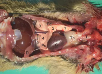
The inner, medial or mediastinal surface of the lungs lies on the lateral side of the vertebral column and the mediastinum. Its caudal part (pars verte- bralis) is convex, and the cranial part (pars mediastinalis) is uneven. The heart, the blood vessels and the oesophagus produce impressions on the mediastinal surface of the lungs. The medial surface of the apical portion of the lungs leans on the thymus, when it is present (Fig. 1 Th).
Fig. 1. Topographical position of the organs in the pectoral cavity of the ground squirrel (Spermophilus citellus). Ventral view. 1 – lobus cranialis dexter, 2 – lobus medius dexter, 3 –lobus caudalis dexter, 4 – lobus accessorius dexter, 5 – v. cava caudalis, Pd – pulmo dexter,
Ps – pulmo sinister, T – trachea, Th – thymus, C – cor, D – diaphragm
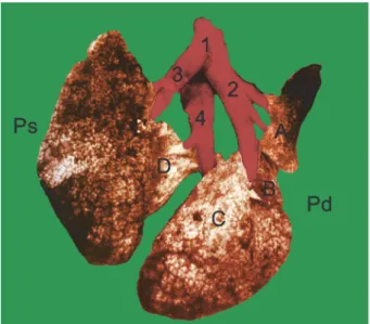
Fig. 2. Lungs of the ground squirrel (Spermophilus citellus). Dorsal view (a) and ventral view (b).
1 – lobus cranialis dexter, 2 – lobus medius dexter, 3 – lobus caudalis dexter, 4 – lobus accessorius dexter, T – trachea, Bd – bronchus principalis dexter, Bs – bronchus principalis sinister,
Pd – pulmo dexter, Ps – pulmo sinister
The lungs consist of the right (pulmo dexter) (Fig. 1 Pd, Fig. 2 Pd, Fig. 3 Pd) and the left lung (pulmo sinister) (Fig. 1 Ps, Fig. 2 Ps, Fig. 3 Ps), intercon- nected by the pulmonary and bronchial trunk. Both lungs are positioned asym- metrically, the left one more cranial than the right, because the right lung is not separated from the left lung in the median plane but only in the sagittal plane.
The bifurcation of the trachea into right and left principal bronchi is also asym- metric. Therefore, the right bronchus is positioned more cranially than the left bronchus, and it is shorter than the latter (Fig. 2 Bd).
b a
Fig. 3. Vascularisation of the lungs of the ground squirrel (Spermophilus citellus). Dorsal view.
1 – truncus pulmonalis, 2 – a. pulmonalis dextra, 3 – a. pulmonalis sinistra, 4 – v. pulmonalis, A – lobus cranialis dexter, B – lobus medius dexter, C – lobus caudalis dexter,
D – lobus accessorius dexter, Pd – pulmo dexter, Ps – pulmo sinister
The right lung
The right lung is divided into four lobes by deep fissures. The lobes are named according to the ramification of the mainstem bronchi within the right lung. The right principal bronchus enters the lung at its root and branches into lobar bronchi which enter each lobe of the lung.
In accordance with the ramification of the bronchial tree, the right lung (pulmo dexter) (Fig. 1 Pd, Fig. 2 Pd, Fig. 3 Pd) consists of four lobes: the cranial (lobus cranialis dexter) (Fig. 1 – 1, Fig. 2 – 1, Fig. 3 A), the middle (lobus medi- us dexter) (Fig. 1 – 2, Fig. 2 – 2, Fig. 3 – B), the caudal (lobus caudalis dexter) (Fig. 1 – 3, Fig. 2 – 3, Fig. 3 – C) and the accessory lobe (lobus accessorius dex- ter) (Fig. 1 – 4, Fig. 2 – 4, Fig. 3 – D). The right cranial lobe is irregular pyramid shaped. The right middle lobe is elongated, resembling an irregular pyramid. The right caudal and accessory lobes are of pyramid shape. In the animals observed in the present study, the right caudal lobe was found to be the largest lobe of the lungs, and in its cranial portion the presence of the thymus was noticed. The v.
cava caudalis is supported by the plica venae cavae and positioned between the accessory and the caudal lobes of the right lung (Fig. 1 – 5).
Fig. 4. Lungs of the ground squirrel (Spermophilus citellus). Terminal bronchiole (TB), respiratory bronchiole (RB), alveolar ductus (AD), alveoli (A), pneumocyte I (PI), pneumocyte II (PII), alveolar macrophage (M). (a) Haematoxylin and eosin (HE) ×10, bar = 100 μm; (b) HE ×40,
bar = 100 μm
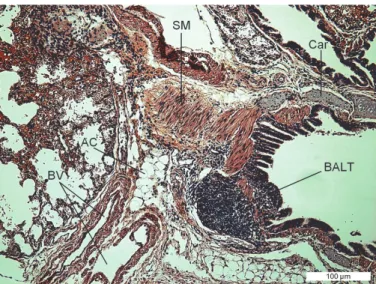
Fig. 5. Lungs of the ground squirrel (Spermophilus citellus). Bronchus-associated lymphoid tissue (BALT), adipocyte (AC), hyaline cartilage (Car), smooth muscle (SM), blood vessel (BV).
HE ×10, bar = 100 μm
The left lung
Being smaller than the right one, the left lung (Fig. 1 – Ps, Fig. 2 – Ps, Fig. 3 – Ps) is not divided into lobes. Its shape is elongated, vaguely resembling a coni- cal frustum. The left bronchus bifurcates into two branches (Fig. 2b – Bs), one entering the cranial, and the other the caudal portion of the left lung.
The average weight of both lungs is 0.77 g, the right lung being heavier than the left one. The weight of the right lung is 0.46 g, the cranial lobe weighs 0.08 g, the middle lobe 0.08 g, the caudal lobe 0.21 g and the accessory lobe 0.09 g.
The weight of the left lung is 0.31 g.
b a
Vascularisation of the lungs
Vascularisation of the lungs of the ground squirrel consists of functional and nutritive blood vessels.
The functional blood vessels of the lungs are the pulmonary trunk (truncus pulmonalis) and the pulmonary veins (vv. pulmonales).
The truncus pulmonalis (Fig. 3 – 1) is a short blood vessel, with a thinner wall than that of the aorta. The trunk arises from the right ventricle of the heart, on the ostium trunci pulmonalis at the conus arteriosus. Its initial part is located within the pericardium, between the auricles of the heart, cranially and to the left from the beginning of the aorta. It runs caudodorsally to the root of lungs and leaves the pericardium dorsal to the base of the left ventricle. After a course of 5 to 6 mm, the pulmonary trunk divides into the right and left pulmonary arteries (a. pulmonalis dextra et a. pulmonalis sinistra). Each of them enters the hilum of the corresponding lung and the branches of the pulmonary artery accompany the bronchial tree.
The a. pulmonalis dextra (Fig. 3 – 2) passes across the dorsal wall of the left atrium of the heart, and runs to the right and caudodorsally to the base of the heart, between the orifice of the right and left cranial caval veins (v. cava crani- alis dextra et sinistra). It accompanies the right principal bronchus and, before entering the right lung, gives off branches: the ramus lobi cranialis to the cranial lobe, the ramus lobi medii to the middle lobe, the ramus lobi accessorii to the accessory lobe, and the ramus lobi caudalis to the caudal lobe of the right lung.
The a. pulmonalis sinistra (Fig. 3 – 3) runs caudolaterally and to the left, towards the left lung, enters it and accompanies the left principal bronchus and bronchial tree into the left lung. Along its course to the left lung it crosses the left cranial caval vein (v. cava cranialis sinistra).
The vv. pulmonales (Fig. 3 – 4), which return arterial blood from the lungs to the left atrium of the heart, are 5 to 6 in number, and differ in size and thick- ness. In four animals the pulmonary veins from each lung tended to fuse together to form a wide common vein. The branches of the pulmonary veins in the lungs do not accompany the branches of the pulmonary arteries and the bronchial tree, but cross them.
The nutritive blood vessels of the lungs are the a. bronchoesophagea dex- tra and the v. bronchoesophagea dextra.
The a. bronchoesophagea dextra, a branch of the a. intercostalis suprema dextra, is divided into the ramus bronchalis and the ramus esophageus. Blood is conveyed to the lungs by the ramus bronchalis dexter and ramus bronchalis sin- ister, which are branches of the ramus bronchalis.
The v. bronchoesophagea dextra takes blood from the lungs and enters the v. azygos dextra.
Histological structure of the lungs
The lungs consist of parenchyma, interstitium, blood vessels, and nerves.
The parenchyma is composed of the conductive pathways (bronchi and bronchi- oles) and the alveoli.
After bifurcation of the trachea into asymmetrical right and left principal bronchi, each of the latter enters the lungs through their root and divides into secondary or lobar, and tertiary or segmental bronchi up to terminal bronchioles.
The lobar bronchi and several of their branches have cartilages embedded in their walls. In the lobar, intralobular and interlobular blood vessels, the thickness of the tunica media was expressed (Fig. 5 – SM and Fig. 6 – SM).
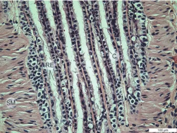
The mucosal epithelium of larger and terminal bronchioles is a pseudo- stratified prismatic ciliated epithelium, but the epithelial cells of the mucous membrane of terminal bronchioles are cubic in shape (Fig. 6). In the terminal bronchial epithelium, Clara cells are present, but they have no cilia (Fig. 6 – C).
Cartilages are absent from the wall of terminal bronchioles.
Fig. 6. Lungs of the ground squirrel (Spermophilus citellus). Respiratory epithelium (RE), smooth muscle (SM), Clara cells (C). HE ×40, bar = 100 μm
Each terminal bronchiole branches into two (and rarely more than two) respiratory bronchioles (Fig. 4 – RB). Along the walls of the respiratory bronchi- oles, numerous openings (alveolar channels, ductus alveolaris) and alveoli can be observed. In the distal parts of the alveolar channels the smooth muscles dis- appear. The alveoli open into alveolar bags, and their shape is correct oval to very ellipsoidal. The alveoli are separated from each other by interalveolar septa.
Destruction of alveolar walls, accumulation of erythrocytes in the capillaries of interalveolar septa, as well as destruction of the cytolemma of the capillary endo- thelium have been detected (Fig. 5).
The alveoli are small and variable in size, and the thickness of the alveolar epithelium also varies. The diameter of the alveoli is 37 μm on average, and the thickness of the alveolar wall and the interalveolar septa is 1.38 μm. The areas of epithelium on the alveolar wall and also in the interalveolar septa are small, in some places barely noticeable and discontinuous. The alveolar epithelium con- sists of a large number of squamous cells – type I pneumocytes and a lower number of type II pneumocytes, which are parallelepiped shaped. Individual macrophages can be found in the lumen of the alveoli (Fig. 4).
The lung parenchyma is characterised by the presence of bronchus- associated lymphoid tissue (BALT) (Fig. 5), which is a component of the diffuse mucosa-associated lymphoid tissue (MALT). There are also areas containing numerous adipocytes around the bronchi and blood vessels (Fig. 5 – AC).
The lung parenchyma of ground squirrels is rich in connective tissue fibres and peripheral nerves. Connective tissue fibres impregnated with silver are no- ticeable from the tunica submucosa continuously to the basal membrane of the lamina epithelialis of bronchioles and terminal bronchioles (Fig. 7), as well as around the alveoli.
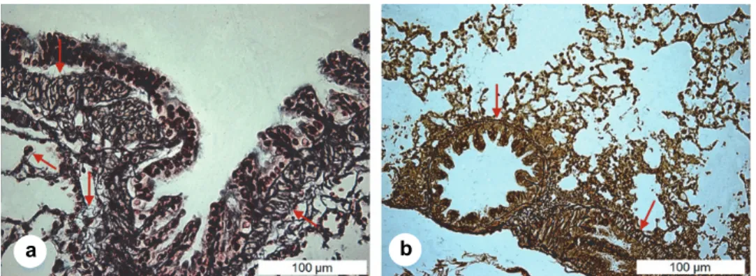
Fig. 7. Lungs of the ground squirrel (Spermophilus citellus). Fibres of connective tissue in bron- chioles and terminal bronchioles ↓ (a), Gomori ×40, bar = 100 μm and ↓ (b), Gordon-Sweet ×20,
bar = 100 μm
Discussion
The data obtained about the lungs of the ground squirrel were compared to those about the rat (Rattus norvegicus), mouse (Mus musculus), mole rat (Spalax leucodon), rabbit (Oryctolagus cuniculus) and a non-human primate (the rhesus monkey, Macaca mulatta) with the intention of demonstrating similarities and differences in topography, morphology, vascularisation and innervation.
a b
The lungs of the ground squirrel as well as those of rats (Hebel and Strom- berg, 1976), rabbits (McLaughlin and Chiasson, 1990) and mole rats (Blagojević, 2010) are located in the pleural cavity.
The right lung of the ground squirrel, like that of the mole rat (Blagojević, 2010; İlgun et al., 2014), mouse (Thiesse et al., 2010) and rabbit (McLaughlin and Chiasson, 1990; Popesko et al., 1990a; Ramchandi et al., 2001; Autifi et al., 2015), is segmented into four (cranial, middle, caudal and accessory) lobes. The right cranial lobe of the lung in the rat is divided into a cranial and a caudal part (Hebel and Stromberg, 1976; Hislop and Reid, 1978).
The left lung in the ground squirrel, mole rat (Blagojević, 2010; İlgun et al., 2014), rat (Hislop and Reid, 1978) and mouse (Popesko et al., 1990b; Thiesse et al., 2010) is not divided into lobes. The left lung of the rabbit consists of two lobes: the cranial and the caudal (Ramchandi et al., 2001; Autifi et al., 2015).
The left cranial lobe is partially divided into cranial and caudal portions by an in- complete fissure (McLaughlin and Chiasson, 1990).
The lungs of the ground squirrel have no visible subpleural pulmonary lobules unlike those of domestic animals, for example the pig and large rumi- nants (Gledić, 2012). This finding is in accordance with that reported by Autifi et al. (2015) on the rabbit and by Nakakuki (1980), who conducted studies on the lungs of rabbits and some other mammalian species.
The functional blood vessels of the lungs in the ground squirrel and rabbit are the truncus pulmonalis and the vv. pulmonales (Popesko et al., 19900a; Auti- fi et al., 2015) while the truncus pulmonalis and a single v. pulmonalis in rats (Hebel and Stromberg, 1976; Popesko et al., 1990b) and mole rats (Blagojević, 2010). There are most frequently 5 to 6 pulmonary veins in ground squirrels, but four pulmonary veins and two trunks from each lung in rabbits (Autifi et al., 2015) and a single pulmonary vein in rats (Hebel and Stromberg, 1976). All of these veins drain into the left atrium of the heart.
The a. bronchoesophagea dextra, a branch of the a. intercostalis suprema dextra, is a nutritive arterial vessel of the lungs in the ground squirrel. The ramus bronchalis of the a. bronchoesophagea dextra branches into the ramus bronchalis dexter et sinister, which supply the corresponding lung with blood. The a. bron- choesophagea dextra, a branch of the a. intercostalis suprema dextra and the a.
bronchoesophagea sinistra, a branch of the aorta thoracica in the mole rat (Blagojević, 2010) and the a. bronchialis dextra, a branch of the truncus costocervicalis, and the a. bronchialis sinistra, a branch of the a. thoracica interna in the rat (Hebel and Stromberg, 1976) are the nutritive arterial vessels of the lungs.
The nutritive vein vessels of the lungs are the v. bronchoesophagea dextra in the ground squirrel and the v. bronchoesophagea dextra et sinistra in the mole rat (Blagojević, 2010) and the rat (Hebel and Stromberg, 1976). The histological structure of the lung parenchyma in the ground squirrel is similar to that of the rat (Correa et al., 2001; Koptyev et al., 2014) and some other mammals (Junguei-
ra and Carneiro, 2003; Chaturvedi and Lee, 2005; Gledić, 2012). There is a suc- cessive branching of the lobar bronchi within the parenchyma in the ground squirrel and other experimental animals, giving rise to terminal bronchioles, the most distal parts of the conducting portion of the respiratory system. Each termi- nal bronchiole branches further into two, and rarely more than two, respiratory bronchioles.
Cartilages are found at the ends of the lobar bronchi in the ground squirrel, rat (Koptyev et al., 2014) and mouse (Thiesse et al., 2010; Chaturvedi and Lee, 2005). By contrast, they are not present in the distal bronchioles of the rhesus monkey (Hyde et al., 2009).
The mucous membrane of larger bronchioles in the ground squirrel is cov- ered by a pseudostratified prismatic ciliated epithelium, while in the rat there is a monolayer plural-row ciliated epithelium (Koptyev et al., 2014). As in the rat (Hyde et al., 2009), in the terminal bronchiole epithelium of the ground squirrel Clara cells are present, but they have no cilia.
The presence of BALT is evident in the interstitium of bronchioles and blood vessels in the ground squirrel. Two out of nine groups of inspected rats had little lymphoid tissue, which the author considered normal (Lamb, 1975).
The diameter of the pulmonary alveoli, as well as the thickness of the al- veolar epithelium, are small and variable. The areas of epithelium in the alveolar wall and interalveolar septa are small, at some sites barely noticeable and inter- rupted. The average diameter of the pulmonary alveoli was 37 μm in the ground squirrel, 70.2 μm in the rat (Hebel and Stromberg, 1976), and 38 to 80 µm in adult mouse (Zhang et al., 2011).
The alveolar epithelium of the ground squirrel consists of two types of cells: numerous squamous cells (type I pneumocytes) and less numerous type II pneumocytes. The latter are cubic cells in the rat (Koptyev et al., 2014) and cy- lindrical in the ground squirrel.
Destruction of the alveolar walls, accumulation of erythrocytes in the ca- pillaries of the alveolar septa, as well as local damage and destruction of the cy- tolemma of the capillary endothelium are characteristic features of the lungs in ground squirrels, hibernating squirrels (Kozlova and Yurchenko, 1996) and rats (Lamb, 1975; Koptyev et al., 2014).
References
Autifi, M. A. H., El-Banna, A. K. and Ebaid, A. E.-S. (2015): Morphological study of rabbit lung, bronchial tree and pulmonary vessels using corrosion cast technique. Al-Azhar Assiut Med. J. 13, 41–51.
Bancroft, J. D. (1967): An Introduction to Histochemical Technique. Butterworths, London.
Blagojević, M. (2010): Morphology, topography, vascularization and innervation of the organs in the thoracic cavity of experimental animals [in Serbian]. PhD Thesis, Faculty of Veterinary Medicine, University of Belgrade, Belgrade, Serbia.
Bohr, M., Brooks, R. A. and Kurtz, C. C. (2014): Hibernation induces immune changes in the lung of 13-lined ground squirrels (Ictidomys tridecemlineatus). Develop. Comp. Immunol. 47, 178–184.
Chaturvedi, A. and Lee, Z. (2005): Three-dimensional segmentation and skeletonisation to build an airway tree data structure for small animals. Phys. Med. Biol. 50, 1405–1419.
Correa, F. C. F., Ciminelli, B. P., Falcao, H., Alcantara, C. J. B., Contador, S. R., Medeiros, S. A., Zin, A. W. and Rocco, R. M. P. (2001): Respiratory mechanics and lung histology in nor- mal rats anesthetized with sevoflurane. J. Appl. Physiol. 91, 803–810.
Gledić, D. (2012): Veterinary Histology [in Serbian]. Naučna KMD, Beograd.
Hebel, R. and Stromberg, M. W. (1976): Anatomy of the Laboratory Rat. The Williams-Wilkins Company, Baltimore, USA.
Hislop, A. and Reid, L. (1978): Normal structure and dimensions of the pulmonary arteries in the rat. J Anat. 125, 71–83.
Hyde, D. M., Hamid, Q., Irvin, G. C. and Dallas, M. (2009): Anatomy, pathology and physiology of the tracheobronchial tree: emphasis on the distal airways. J. Allergy Clin. Immunol. 124, S72–77.
İlgun, R., Yoldas, A., Kuru, N. and Özkan, Z. E. (2014): Macroscopic anatomy of the lower respir- atory system in mole rats (Spalax leucodon). Anat. Histol. Embryol. 43, 474–481.
Jungueira, C. L. and Carneiro, J. (2003): Basic Histology. Editora Guanabara Koogan S. A., Rio de Janeiro, Brazil.
Koptyev, M. M., Pronina, O. M., Danylchenko, S. I., Avetikov, D. S. and Stavitsky, S. O. (2014):
Histological features of rat’s normal lung tissue. Eur. Int. J. Sci. Technol. 3, 33–38.
Kozlova, V. F. and Yurchenko, T. N. (1996): Structural aspects hibernating animal adaptation.
Probl. Cryobiol. 3, 43–50.
Lamb, D. (1975): Rat lung pathology and quality of laboratory animals. Lab. Anim. 9, 1–8.
McLaughlin, A. C. and Chiasson, B. R. (1990): Laboratory Anatomy of the Rabbit. W. C. Brown Co. Publishers, USA.
Millesi, E., Huber, S., Dittami, J., Hoffmann, I. and Daan, S. (1998): Parameters of mating effort and success in male European ground squirrels (Spermophilus citellus). Ethology 104, 298–313.
Milsom, K. W. and Reid, W. D. (1995): Pulmonary mechanics of hibernating squirrels (Sper- mophilus lateralis). Respir. Physiol. 101, 311–320.
Nakakuki, S. (1980): Comparative anatomical studies on the mammalian lung. Bull. Fac. Agric.
21, 1–73.
Nikolić, Z., Đelić, D., Blagojević, Z., Mrvić-Jovičič, V., Drekić, D. and Zorić, Z. (2004): The sub- clavian artery and its branches in the ground squirrel (Citellus citellus). Acta Vet. Beograd 54, 227–237.
Nomina Anatomica Veterinaria, fifth edition (2012): Editorial Committee, Hannover (Germany), Columbia, MO (USA), Ghent (Belgium), Sapporo (Japan).
Popesko, P., Rajtová, V. and Horák, J. (1990a): A Colour Atlas of Anatomy of Small Laboratory Animals. Volume One: Rabbit, Guinea Pig. Priroda Publishing House, Bratislava.
Popesko, P., Rajtová, V. and Horák, J. (1990b): A Colour Atlas of Anatomy of Small Laboratory Animals. Volume Two: Rat, Mouse, Golden Hamster. Priroda Publishing House, Bratislava.
Ramchandi, R., Bates, J. H., Shen, B., Suki, B. and Tepper, R. S. (2001): Airway branching mor- phology of mature and immature rabbit lungs. J. Appl. Physiol. 90, 1584–1592.
Thiesse, J., Namati, E., Sieren, C. J., Smith, R. A., Reinhardt, M. J., Hoffman, A. E. and McLen- nan, G. (2010): Lung structure phenotype variation in inbred mouse strains revealed through in vivo micro-CT imaging. J. Appl. Physiol. 109, 1960–1968.
Zhang, L., Li, D. and Luo, S. (2011): Non-invasive microstructure and morphology investigation of the mouse lung: Qualitative description and quantitative measurement. PLoS One 6, e17400.