Revised manuscript,
dedicated to the memory of Professor Parimal Sen
This version was published in Science and Culture 84, 229-238 (2018)
Peptide bioconjugates for intracellular targeting Ferenc Hudecz 1,2*
1 Research Group of Peptide Chemistry, Eötvös L. University (ELTE), Hungarian Academy of Sciences,
P.O.Box 32, H-1518 Budapest 112, 2Department of Organic Chemistry, ELTE, Budapest, Hungary
E-mail: fhudecz@elte.hu
Abstract: The increasing number of papers in which bioconjugates of oligo- and/or polypeptides are present indicates the importance and potential of these constructs. In this contribution there is an attempt to provide an overview of our results concerning the preparation, functional characterization and mainly biomedicinal application of conjugates with chemotherapeutic agents against tumour, infectious diseases, with T-cell epitope peptides or with “reporter” entities to be used also for mechanistic studies prepared in our laboratories last decades.
Introduction
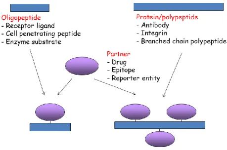
During the last two decades cellular targeting by peptide-based bioconjugates resulted in novel approaches for which various drugs, reporter entities or even epitopes were attached to appropriate structures1-4. These two- or three-component constructs are widely utilized for biomedical research studying their potential to eliminate selectively tumor cells, intracellular microbes or to study uptake mechanisms or changes in protein expression profiles. The partner molecules are attached by covalent bond and preserve relevant functional properties (like biological activity or „reporter properties”) after conjugation (Figure 1.)
Figure 1: Peptide/polypeptide/protein based bioconjugates.
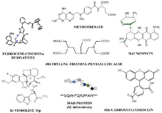
With selected examples here we outline our findings derived from novel two- or three- component conjugates of antitumour/antimicrobial compound (e.g. daunomycin [Dau] 5-12, methotrexate [MTX] 13-20, vinblastine21, ferrocene derivatives22 or antitubelculars23-25), enzyme (e.g. calpain) activator/substrate 26, 27 linear T-cell epitope peptides 28- 32 or “reporter”
molecules (e. g. radioligand 33-37, fluorophore 38-41)(Figure 2).
Figure 2: Structure of examples of drugs (daunomycin, methotrexate, vindoline and ferrocene derivatives), linear T-cell epitope peptide from M. tuberculosis and of “reporter” entities [5(6)-carboxyfluorescein (Cf) and diethylene-triamine pentaacetic acid (DTPA) chelator] used as partner for conjugation.
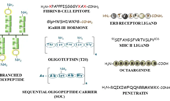
As peptide-partner component of the bioconjugates (a) novel branched chain polymeric polypeptides 5-8, 11, 13-15, 17, 20, 28-31, 33-37, 40, 41
with different side chain structure, charge properties, and solution conformation were designed, (b) well-established or newly discovered oligopeptide with specific recognition unit, like cell surface-receptor binding ligand (e.g. Erb-B212, MHC for T-cell epitope 23-25, or (c) cell penetrating peptides [CPP] (e.g.
oligoarginine, penetratin) 9, 10, 18, 21, 22, 26, 27
were chosen for construction of targeting bioconjugates (Figure 3)
Figure 3: Peptide/polypeptide component of the bioconjugates for targeting: branched chain polymeric polypeptides, oligotuftsin (T20), sequential oligopeptide carrier (SOC) (a), cell surface-receptor binding ligand (e.g. Erb-B2), linear T-cell (MHC-II), B-cell epitope (b) or cell penetrating peptides (e.g. oligoarginine, penetratin) (c).
.
MTX-conjugates with new, potential chemotherapeutic properties against cancer cells and intracellular parasite Leishmania donovani
Pentaglutamylated methotrexate conjugates with cell penetrating oligopeptides
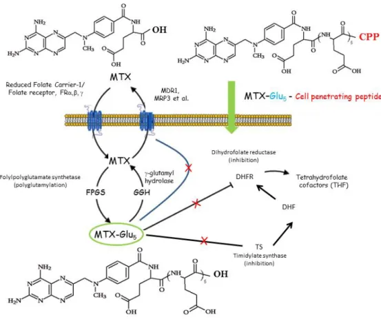
A folate antagonist (MTX) or its pentaglutamylated (MTX-Glu5) derivative was conjugated with cell-penetrating oligopeptides (penetratin or octaarginine) with or without tetrapeptide spacer unit (GFLG). 5(6)-carboxyfluorescein (Cf) labelled derivatives were also prepared18. The in vitro cytostatic activity and cellular-uptake of conjugates were studied on breast carcinoma cells (on MDA-MB-231 as resistant and on MCF-7 as sensitive cells). Although the presence of Glu5 moiety significantly decreased the level of internalisation, but all conjugates were able to penetrate - to some extent - into both cell types. However, only the conjugates of penetratin showed in vitro cytostatic activity. The most effective conjugates include MTX-Glu5-Penetratin(desMet) and MTX-Glu5-GFLG-Penetratin(desMet) containing a tetrapeptide spacer unit. The latter one was effective on both cell cultures, while the former compound was active only on the resistant tumour cells.
Figure 4: Schematic route of cellular uptake and mechanism of action of methotrexate (MTX), pentaglutamylated MTX (MTX-Glu5) and pentaglutamylated MTX – cell penetrating peptide conjugate (MTX-Glu5-CPP) 18, 42, 43
Our results suggest that the translocation of polyglutamylated MTX could be dependent on the structure of the CPP (penetratin desMet vs Arg8) as well as on the spacer between the partners. In conclusion the intracellular delivery of MTX-Glu5-may be a new avenue to treat resistant tumors19.
Methotrexate conjugates with branched chain polymeric polypeptides
Considering the inhibitory potential of MTX against a group of intracellular parasites (Leishmania) of macrophages causing leishmaniasis in man44,45 we have prepared MTX conjugates with branched chain polymeric polypeptides 13-15.
We have demonstrated with a set of structurally related branched polypeptides (Figure 5) that the side chain composition and amino acid sequence could markedly determine the physico- chemical characteristics including solution conformation, as well as the cytotoxicity, blood clearance, biodistribution and even the interaction with phospholipid mono- or bilayers of the compound 46-49
Figure 5: Schematic structure of XAK type branched chain polymeric polypeptide with poly[L-Lys] backbone. K = Lys, A = Ala, X = E (Glu), Ac-E (Ac-Glu), Suc-E (Suc-Glu), L (Leu), S (Ser), or O (Orn).
Similarly, in conjugates the functional characteristics of the covalently attached moiety are highly influenced by the properties of the macromolecular carrier component (Figure 5).
Conjugates with various chemotherapeutic drugs (e.g. Dau5,6, MTX13, GnRH antagonist50,51, amiloride52 or with several radionuclides34-37 were prepared and tested for antitumour effect or imaging efficacy.
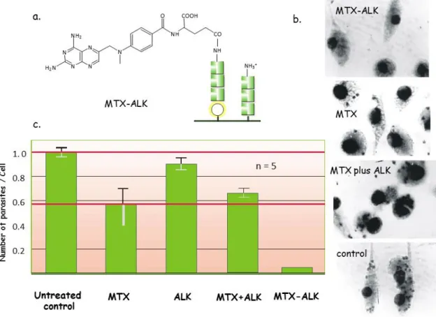
In order to investigate the potential of branched chain polymeric polypeptides for MTX targeting to Leishmania donovani infected macrophages proliferating intracellularly, a new set of conjugates with amphoteric (EAK) or polycationic (AK, SAK, ALK) (Figures 5 and 6a) polypeptide was prepared14,15 and the leishmanicid activity was tested in collaboration with A.C. Ghose et al. at the Bose Institute in Kolkata.
The effect of these conjugates has been studied and compared with that of the free drug/carrier and the non-covalent mixture of the two partners in mouse macrophages in vitro as well as in vivo in L. donovani infected in BALB/c mice. Among conjugates studied, MTX attached to polycationic ALK (MTX-ALK) exhibited pronounced anti-parasite activity both in vitro (Figure 6b) and in vivo (Figure 6c).
Figure 6: Schematic structure of branched chain polymeric polypeptide ALK and MTX (a), the in vitro (b) and in vivo effect of the conjugate (MTX-ALK), free MTX, free polypeptide (ALK) and of the mixture of ALK+MTX treatment of L. donovani infected macrophages (b) or BALB/c mice (c).
Chemotactic responses could play a significant role during the course of parasite–host–cell interaction, and also in Leishmania differentiation. Therefore MTX attached to XAK type polypeptides (X=Ser, SAK or X= Glu, EAK) (Figure 5) by thioether linkage with/without GFLGC oligopeptide spacer were investigated for anti-Leishmania braziliensis parasite effect, cytotoxicity/cytostatic as well as chemotactic properties under in vitro conditions. Data suggest that chemotaxis produced by the MTX-XAK polypeptide conjugates depends on specific chemical characteristics. For example, the identity of the N-terminal amino acid (X=
Ser vs. Glu) in the branch and also the attachment site of MTX in the conjugates (- vs. - carboxylic groups) significantly influences the elicited chemotaxis. Specific cytotoxic/cytostatic effects of the conjugates on parasites/murine and human cells of the macrophage/monocyte system respectively, suggest that these MTX-conjugates can be considered as selective compounds for Leishmania17.
Recently, a new group of XAK type branched polypeptides with side chain terminal X = Ile, Nle, Leu, Val, or Arg amino acid53 and their MTX-conjugates with amide linkage were synthesized20 using optimized procedures. All XAK polypeptides with hydrophobic amino acid (Ile, Nle, Leu or Val) were essentially non-toxic on murine bone marrow-derived macrophages from Balb/c mice, whereas the polypeptide with terminal Arg (RAK) proved to be markedly cytotoxic20.
Oligopeptide conjugates with new, potential chemotherapeutic properties against cancer cells
In addition to the biologically active pentaglutamylated MTX – CPP conjugates (see above) our research group reported on the synthesis of new, small or medium size oligopeptide- antitumour compound conjugates54 with targeting of specific cell surface structures (e.g. Erb- B2 receptor12, GnRH-III 55-58 or selectin59) and/or cell penetrating properties (e.g.
oligoarginine 9,10,18,21,22,59
, penetratin 18,26). In case of the most active conjugates promising first studies were performed to understand the mechanism of action (e.g. effect on the tubulin system 21, analysis of the conjugate induced protein expression profile. 12,57
Recently, a new set of vindoline derivatives, amino acid conjugates were prepared60. and selected derivatives, Br-vindoline-(L/D)-Trp, were coupled with octaarginine cell penetrating peptide at the N-terminal with amide bond.61 In vitro cytotoxic/cytostatic properties of vindoline derivatives studied on human leukemia cells (HL-60) suggested that in contrast to free vindoline different modifications of the ring system and/or the conjugation with L- or D- Trp result in active compounds (IC50 = 25-80 µM)58. The conjugation of the Br-vindoline derivatives increased significantly the in vitro antitumor activity on HL-60, MCF-7 and MDA-MB-231 cells. However, in vivo the Br-vindoline-(L)-Trp-Arg8 conjugate could inhibit the tumour growth but none of the conjugates increased the survival of mouse leukaemia (P388) and murine colon carcinoma (C26) bearing mice in vivo61.
In order to understand the differences between the efficacy of antitumour effect of free and oligopeptide conjugated drug at cellular level, we have investigated the protein expression profiles of an appropriate cell line after optimized treatment with the free or conjugated drug.
For these studies Dau, a DNA-binding antineoplastic agent applied in the treatment of various types of cancer, like osteosarcomas and acute myeloid leukemia was selected (Figure 7).
As demonstrated earlier the covalent coupling of Dau with branched polypeptide or oligopeptide carrier could improve its selectivity, decrease the side effects as well as alter the drug uptake and intracellular fate. In a novel pairs of conjugate Dau was attached to ErbB2 receptor peptide ligand (LTVSPWY) for targeting cancer cells expressing ErbB2 receptor12 or to oligoarginine9,10.
First, in vitro cytostatic effect and cellular uptake of Dau=Aoa-LTVSPWY-NH2 conjugate were studied on human cancer cell lines expressing different levels of ErbB2 receptor. The conjugate had the highest cytostatic effect as well as the most marked cellular uptake on HL- 60 cells
Figure 7: Structure of free Dau and of Dau-conjugate with ERbB2 receptor ligand peptide (LTVSPWY) (a), the protein expression profile of free Dau-treated (b) and of Dau=Aoa- LTVSPWY-NH2 conjugate-treated (c) HL-60 cells. Numbers show the significantly different spots on the gel after treatment for 24 h.
We compared the protein expression profile of treated and non-treated HL-60 human leukemia cells and also of conjugate vs. free Dau treated cells. We have observed that the expression levels of several proteins were altered due to the treatment with the conjugate or with the free drug (Dau) in comparison with proteins from untreated cells (Figure 7).
Proteomic analysis suggests that the expression of cytoskeletal, metabolic proteins and proteins involved in signalling processes were altered12: three proteins, whose expression was lower (tubulin beta chain) or markedly higher (proliferating cell nuclear antigen and protein kinase C inhibitor protein 1) after administration of cells with Dau-conjugate vs. free drug.
Comparison of the protein expression profile of HL-60 cells was found to be dependent not only on the conditions (e.g. concentration selected for treatment: IC25, IC 37.5 and IC50 values), but also on the peptide component (Erb2 receptor ligand vs. cell penetrating oligoarginine [data not shown]).
It should be mentioned that similar studies were performed with another conjugate composed of GnRH-III oligopeptide, as targeting ligand and Dau 57.The results of proteomic analysis reported could be utilized not only for clarification of mechanism of action of bioconjugates, but also for target identification and/or validation.
Conjugates with “reporter” function including intracellular enzyme substrate or established/newly designed fluorophore for the analysis of cellular uptake and intracellular activities
Cellular uptake mechanism and intracellular localization of conjugates as well as the cellular response activities of the attached „cargo” entities could be analysed by using labelled compounds. Here we summarise our results related to branched chain polymeric polypeptides and also cell penetrating peptide - enzyme substrate/activator conjugates.
Analysis of the cellular uptake mechanism of branched chain polypeptides labelled by fluorophore
Selective delivery of chemotherapeutic drugs (e.g. MTX, Dau) or radionucleide36,37, or epitope peptide29 into infected macrophages, tumour or antigen presenting cells, respectively could be a promising approach for improved therapies.
MTX conjugate with branched chain polypeptides exhibited pronounced anti-Leishmania activity in vitro and in vivo as mentioned above14,15. Polypeptide conjugated Dau treatment of tumour bearing mice proved to be markedly pronounced6. However, these effects were very much dependent of the structural characteristics of the branched chain polypeptide partner of the bioconjugates used.
In order to identify structural requirements for efficient transcellular delivery of branched polypeptides, we have studied murine bone marrow culture-derived macrophages (BMM) from 129/ICR mice after incubation with amphoteric (EAK) (Figure 5), and two polyanionic polypeptides differing in negative charge density: Ac-EAK has one negative charge per unit, while Suc-EAK bears two negative charges per unit under physiological conditions. In order to characterize the translocation, polypeptides with closely related topography were labelled with Cf 40 (Figure 8a).
We found that this internalization process is dependent on experimental conditions (e.g.
polypeptide concentration, (Figure 8b), temperature, (Figure 8c) and incubation time (Figure 8d) applied during the treatment. The polyanionic polypeptide with high charge density (Suc- EAK) translocated more efficiently under identical circumstances than AcEAK, which had a lower negitive charge density, while the amphoteric compound (EAK) were ingested only at limited level. Using macrophages from wild-type (SR-A) and class-A scavenger receptor knockout (SR-A-/-) mice, as well as specific inhibitors we demonstrated that SR-A is involved in the endocytosis of synthetic polyanionic polypeptides, especially Suc-EAK.
Similar findings were reported on the uptake properties of these polyanionic polypeptides by murine macrophage-like cell line J77441. Thus, polypeptide with multiple negative charges seems to be appropriate as a macromolecular carrier for specific drug delivery into macrophages and these compounds could be considered as basis for development of further carriers and bioconjugates.
Figure 8: Schematic structure of carboxyfluorescein labelled XAK type branched chain polypeptides with poly[L-Lys] backbone (a), and the cellular uptake as a function of concentration (b), time (c) and temperature (d). K = Lys, A = Ala, X = E (Glu), Ac-E (Ac- Glu) or Suc-E (Suc-Glu).
For comparative analysis three polycationic branched chain polypeptides with different side chain structure XAK (X = Ser, SAK) (Figure 5) and XiK (X = Pro) were also labeled by Cf and analysed under the same conditions. Data indicated that the uptake characteristics of murine J774 macrophage cells were dependent of the side chain length and composition.
Polypeptide with a single amino acid in the branches (PiK) was internalized mostly, but the uptake of SAK with longer side chain terminated by Ser residue, was significant only at the highest concentration studied in these in vitro experiments41.
Considering that branched chain polypeptides with chemotherapeutic drugs (e.g. Dau, MTX) exerted significant antitumour activities in vitro5, 13, internalization studies of polypeptides were also conducted by tumour cell lines as well.
The uptake and cellular localization studies were performed on tumour cell lines (HT-29, human colon carcinoma, HepG2, human hepatocellular carcinoma) and an extended group of polycationic branched chain polypeptides with Cf reporter moiety.62 This includes XAK (X = Ser, Arg, Ser, Thr), (Figure 5), AXK (X = Leu), XiK (X = Pro, Leu, His) types of architecture and poly[Lys], as control.
In the case of HT-29 cells, the efficacy of the uptake, based on calculated fluorescence mean data normalized to Cf content was the following: Cf-HiK > Cf-LiK > Cf-RAK ∼ Cf-SAK >
Cf-poly[Lys] > Cf-ALK at 50 μg/mL. The internalization profile of HepG2 cells was slightly
different as follows: Cf-LiK > Cf-RAK > Cf-HiK > Cf-poly[Lys] > Cf-ALK. The comparison of the uptake by the two tumour cell lines indicates some similarities. Namely, the most engulfed three polypeptides (Cf-LiK, Cf-HiK, and Cf-RAK) and the least ingested polypeptides (Cf-PiK, Cf-TAK) were identical. (Essentially no (Cf-TAK) or negligible (Cf- PiK) uptake was observed on either of the cell cultures.) 62
In conclusion, the translocation is very much dependent on the side chain length, identity of amino acid X and on the distance between the poly[Lys] backbone and the terminal positive charge. It is important to emphasize that the engulfment by the cell lines is not necessarily the same. For example polypeptide PiK was transported into J774 macrophage cell efficiently, but it was only taken up at low level by the two tumour cell lines. Our findings using a wide set of relevant inhibitors also suggest that macropinocytosis and caveole/lipid raft mediated endocytosis are typically involved in the uptake of the cationic polymeric polypeptides.
Cellular uptake and intracellular cleavage of the cell penetrating conjugate of calpain substate peptide labelled by fluorophor(s)
For the analysis of the intracellular activity of calpain in vivo a novel substrate 62 of the enzyme was designed, prepared and covalently attached to cell penetrating peptide (oligoarginine) and conjugated with two fluorophores acting as FRET partners (Figure 9a.) 27 It should be noted that the in both constructs (free and conjugated substrates) the necessary distance between the two FRET partners was preserved.
First, the substrate properties of the new constructs were verified by using isolated, cell-free calpain B enzyme. Not only the labelled free substrate, but also the conjugated derivative maintained the substrate properties. In order to study the translocation properties of the free and conjugated peptides, the differential internalization was demonstrated in COST-7 cells by fluorescent microscopy experiments. In contrast to the free, labelled peptide (Dabcyl- TPLKSPPPSPR-EDANS), its heptaarginine conjugate, substrate conjugate (Dabcyl- TPLKSPPPSPRE(EDANS)RRRRRRR-NH2) exhibited high and homogeneous fluorescence in cells27 (Figure 9b.) Thus with the introduction of fluorescent labels it was possible to demonstrate that the - enzyme substrate-conjugate retained the properties of the attached partners, namely the enzyme substrate related activity (specific cleavability by calpain) and the capability to enter the cell.
For the verification of substrate to be cleaved by intracellular calpain, the substrate conjugate was added to the lysate of Drosophila melanogaster S2 cells and the calpain activity was determined. It was observed that the fluorescence intensity was changed indicating that substrate conjugate was functioning in the cell lysate, which could be inhibited by Calpain inhibitor I (Figure 9c.) 27
Results derived also from similar studies using cell penetrating peptide – calpain enzyme activator/inhibitor26 conjugates strongly indicate that this new cell-penetrating conjugates could be useful for determining as well as up or down-regulating intracellular calpain activity in cell lysate or in intact living cells of various origins.
Figure 9: Structure of calpain substrate – heptaarginine (CPP) conjugate labelled with two FRET-partner fluorophores [N-terminal Dabcyl and EDANS attached to the side chain of Glu (E)] (a), internalization of the free and CPP-conjugated substrates by COST cells (b), cleavage of the labelled substrate-CPP conjugate by calpain in S2 cell lysate with/without Ca2+ and specific enzyme inhibitor (c).
Conclusions
Bioconjugation chemistry could represent a special and highly multidisciplinary field of organic chemistry. For the design of the chemical reaction strategy one has to consider the basic requirement: the partners must preserve their functional properties after the completion of the synthesis and purification. In peptide/polypeptide based bioconjugates the functional properties could mean bioactivity (e.g. recognition properties, cellular uptake, intracellular trafficking, biodistribution) as well as “reporter” properties (e.g. fluorescence).
We found that the chemical structure of the targeting peptide partner, the topology of the attached entities as well as the nature of the linkage inserted have marked effects on binding (recognition) properties, intracellular targeting and subcellular distribution as well as cellular responses (e.g. cytostasis/cytotoxicity, protein expression profile) detected.
Acknowledgements: To grants from Hungarian Research Fund (OTKA CK 80689, NK 104864, K 104385, K 104928, PD-83923, TÉT_09-1-2010-0010 and GVOP-3.2.1-2004-04- 0352/3.0.
References:
1. N. Mihala and F. Hudecz, F, Amino Acids, Peptides and Proteins (Eds. E. Farkas, M.
Ryadnov), Royal Society of Chemistry, London, 2012, Vol. 37, 1-39.
2. K. Uray and F. Hudecz, F, Amino Acids, Peptides and Proteins (Eds. E. Farkas, M.
Ryadnov), Royal Society of Chemistry, London, 2014, Vol. 39, 68-113.
3. Sz. Bősze and F. Hudecz, F, Amino Acids, Peptides and Proteins (Eds. M. Ryadnov, F.
Hudecz), Royal Society of Chemistry, London, 2016, Vol. 41, 146-198.
4. Z. Bánóczi, Z., and F. Hudecz, Amino Acids, Peptides and Proteins (Eds. M. Ryadnov, F.
Hudecz), Royal Society of Chemistry, Cambridge, 2018, Vol. 42, 85-145.
5. F. Hudecz, J.A. Clegg, J. Kajtár, M. J. Embleton, M. Szekerke and R.W. Baldwin, Bioconjugate Chemistry, 3, 49-57 (1992).
6. D. Gaál and F. Hudecz, Eur.J.Cancer, 34, 155-161 (1998).
7. J. Reményi, B. Balázs, S. Tóth, A. Falus, G. Tóth and F. Hudecz, Biochem. Biophys. Res.
Commun. 303, 556-561 (2003).
8. J. Reményi, G. Csík, P. Kovács, F. Reig and F. Hudecz, Biochim.Biophys. Acta, 1758, 280-289 (2006).
9. Z. Bánóczi, B. Peregi, E. Orbán, R. Szabó and F. Hudecz, ARKIVOC, 140-153 (2008).
10. Zs. Miklán, E. Orbán, G. Csík, G. Schlosser, A. Magyar and F. Hudecz, Biopolymers (Peptide Science), 92, 489-501 (2009).
11. R. Szabó, Z. Bánóczi, G. Mező, O. Láng, L. Kőhidai and F. Hudecz, F.: Biochim.
Biophys. Acta, 1798, 2209-2216 (2010).
12. E. Orbán, M. Manea, A. Marquadt, Z. Bánóczi, G. Csík, E. Fellinger, Sz. Bősze and F.
Hudecz, Bioconjugate Chemistry, 22, 2154-2165 (2011).
13. F. Hudecz, J.A. Clegg, J. Kajtár, M.J. Embleton, M.V. Pimm, M. Szekerke and R.W.Baldwin, Bioconjugate Chemistry, 4, 25-33 (1993).
14. Gy. Kóczán, A.K. Ghosh, A. Mookherjee, A.C. Ghose, and F. Hudecz, The Immunologist, S1, 610 (1998).
15. Gy. Kóczán, A.C. Ghose, A. Mookerjee and F. Hudecz, Bioconjugate Chemistry, 13, 518-524 (2002).
16. K.B. Bai, O. Láng, E. Orbán, R. Szabó, L. Kőhidai, F. Hudecz and G. Mező, Bioconjugate Chemistry, 19, 2260-2269 (2008).
17. E. Díaz, L. Kőhidai, A. Ríos, A.Silva, O. Vanegas, R. Szabó, G. Mező, F. Hudecz and A.
Ponte Sucre, Experimental Parasitology 135,134–141 (2013).
18. I. Szabó, E. Orbán, G. Schlosser, F. Hudecz and Z. Bánóczi, Eur. J. Med. Chemistry, 115, 361-368 (2016).
19. M. Sebestyén, Gy. Kóczán and F. Hudecz, Amino Acids, 48, 2599-2604 (2016).
20. R. Szabó, M. Sebestyén, Gy. Kóczán and F. Hudecz, Proceedings of 34th European Peptide Symposium, (Eds.: Beck-Sickinger, A., Mörl, K., Bellmann-Sickert, K., Els-Heindl, S., Diederichsen, U.), European Peptide Society, Leipzig, Germany, 2017, p. 98.
21. Z. Bánóczi, Á. Gorka-Kereskényi, J. Reményi, E. Orbán, L. Hazai, N. Tőkési, J. Oláh, J, Ovádi, Z. Béni, V. Háda, Cs. Szántay Jr., F. Hudecz, Gy. Kalaus and Cs. Szántay, Bioconjugate Chemistry, 21, 1948-1955 (2010).
22. Zs. Miklán, R. Szabó, V. Zsoldos-Mady, Z. Bánóczi and F. Hudecz, Biopolymers, 88, 108-114 (2007).
23. K. Horváti, G. Mező, E. Szabó, F. Hudecz and Sz. Bősze, J. Peptide Sci. 15,385-91 (2009).
24. K.Horváti K, Bacsa B, Szabó N, Dávid S, G. Mező G, Grolmusz V, Vértessy B, F.
Hudecz F, and Sz.Bősze S.: Bioconjugate Chemistry 23, 900-907 (2012.)
25. K.Horváti, K., Bacsa, B., Szabó, N., K. Fodor, K., Balka, Gy., M. Rusvai, M., Kiss, É., G.
Mező, G., Grolmusz, V., Vértessy, B., F. Hudecz, F., and Sz. Bősze, Sz.: Tuberculosis (Edinb). 95, 207-211(2015).
26. Z. Bánóczi, Á. Tantos, A. Farkas, P. Tompa, P. Friedrich and F. Hudecz, Bioconjugate Chemistry 18, 130-137 (2007).
27. Z. Bánóczi, A. Alexa, A. Farkas, P. Friedrich and F. Hudecz, Bioconjugate Chemistry, 19, 1378-1381 (2008).
28. K .A.Wilkinson, M.H. Vordermeier, R. Wilkinson, J. Iványi and F. Hudecz, Bioconjugate Chemistry, 9, 539-547 (1998).
29. K.A. Wilkinson, F. Hudecz, H.M. Vordermeier, J. Ivanyi, and R.J. Wilkinson, Eur. J.
Immunol. 29, 2788-2796 (1999).
30. F. Hudecz, F.: Biologicals, 29,197-207 (2001).
31. M. Manea, G. Mező., F. Hudecz and M. Przybylski, Biopolymers, 76, 503-11 (2004).
32. M. Manea, F. Hudecz, M. Przybylski and G. Mező, Bioconjugate Chemistry, 16, 921-928 (2005).
33. J. A. Clegg, F. Hudecz, G. Mező, M.V. Pimm, M. Szekerke and R.W. Baldwin, Bioconjugate Chemistry, 1, 425-430 (1990).
34. M.V. Pimm, S. J. Gribben, K. Bogdán and F. Hudecz, J. Controlled Release, 37,161-172 (1995).
35. M.V. Pimm and F. Hudecz, J. Cancer Res. Clin. Oncol. 122, 45-54 (1996).
36. A.C. Perkins, M. Frier, M.V. Pimm and F. Hudecz, J. Labelled Compounds 41, 631-638 (1998).
37. M.V. Pimm, A.C. Perkins, S.J. Gribben, G. Mező, D. Gaál and F. Hudecz, Ann. Nuclear Medicine, 9, 247-251 (1995).
38. I.B. Nagy, I.Varga and F. Hudecz, Anal. Biochemistry, 287,17-24 (2000).
39. Sz. Bősze, G. Csík, Gy. Kóczán and F. Hudecz, Biopolymers, 81, 81-91 (2006).
40. R. Szabó, L. Peiser, A. Plüddemann, Sz. Bősze, S.Heinsbroek, S. Gordon and F. Hudecz, Bioconjugate Chemistry, 16, 1442-1450 (2005).
41. R. Szabó, G. Mező, É. Pállinger, P. Kovács, L. Kőhidai, Sz., Bősze and F. Hudecz, Bioconjugate Chemistry, 19, 1078-1088 (2008).
42. J. Walling, Invest New Drugs 24, 37-77 (2006).
43. C. Chen, J. Ke, X. E. Zhou, W. Yi, J. S. Brunzelle, J. Li, E. L.Yong, H. E. Xu and K.
Melcher, Nature 500, 486-489 (2013).
44. S. Majumdar and T.J. Siahaan, Med Res Rev. 32, 637-658 (2012).
45. A. Mukhopadhyay, G. Chaudhuri, S.K. Arora, S. Sehgal and S. K. Basu, Science, 244, 705-707 (1989).
46. F. Hudecz, Anti-Cancer Drugs, 6, 171-193(1995).
47. F. Hudecz, Self-Assembling peptide systems in biology, medicine and engineering. (Eds.:
Agelli, A., Boden, N., Zhang, S.) Kluwer Academic Publisher, The Netherlands, 2001, p. 139.
48. I. B. Nagy, Zs. Majer and F. Hudecz, Biopolymers, 58, 152-164 (2001).
49. I. B. Nagy, F. Hudecz, M.A. Alsina and F. Reig, Biopolymers, 70, 323-335 (2003).
50. B. Vincze, I. Pályi, D. Daubner, A. Kálnay, G. Mező, F. Hudecz, M. Szekerke, I. Teplán, and I. Mező, J. Cancer Res. Clin.Oncol. 120, 578-584 (1994).
51. G. Mező, I., Mező, M.V. Pimm, J. Kajtár, A. Seprődi, I. Teplán, M. Kovács, B. Vincze, I.
Pályi, M. Idei, M., Szekerke and F. Hudecz, Bioconjugate Chemistry, 7, 642-650 (1996).
52. J. Pató, K. Ulbrich, P. Baker, G. Mező and F. Hudecz, J. Bioactive and Compatible Polymers 14, 99-121 (1999).
53. M. Sebestyén, R. Szabó, L. Kőhidai, É. Pállinger, G. Mező, Gy. Kóczán and F. Hudecz, Structural Chemistry, 28, 527-536 (2017).
54. F. Hudecz, Z. Bánóczi and G. Csík, Medicinal Research Reviews, 25, 679-786 (2005).
55. G. Mező, M.Manea, I. Szabó, B. Vincze and M. Kovács, Curr. Med. Chem. 15, 2366- 2379 (2008).
56. M. Manea, U. Leurs, E. Orbán, Zs. Baranyai, P. Ohlschlager, A. Marquardt, Á. Schulcz, M. Tejeda, B. Kapuvári, J. Tóvári and G. Mező, Bioconjugate Chemistry, 22, 1320-1329 (2011).
57. V.N. Schreier, L. Pethő, E. Orbán, A. Marquardt, B.A. Petre, G. Mező and M. Manea, PLoS One. 9, 9(4):e94041 (2014).
58. R. Hegedüs, A. Pauschert, E. Orbán, I. Szabó, D. Andreu, A. Marquardt, G. Mező and M.
Manea, Biopolymers, 104,167-177 (2015).
59. Zs. Miklán, E. Orbán, Z. Bánóczi and F. Hudecz, J. Peptide Science, 17, 805-811 (2011).
60. P. Keglevich, L.Hazai, Á. Gorka-Kereskényi, L. Péter, J. Gyenese, Zs. Lengyel, Gy.
Kalaus, Zs. Dubrovay, M. Dékány, E. Orbán, Z. Bánóczi, Cs. Szántay, Jr., and Cs. Szántay, Heterocycles, 87, 2299-2317 (2013).
61. Z. Bánóczi, Z., Tóvári, J., Szabó, I., Regenbach, F.L., Randelovic, I., Keglevich, A., Keglevich P., Lengyel, Zs., Gorka-Kereskényi, Á., Szántay, Jr. Cs., Hazai, L. and F. Hudecz, F.: Proceedings, 34th European Peptide Symposium, (Eds.: Beck-Sickinger, A., Mörl, K., Bellmann-Sickert, K., Els-Heindl, S., Diederichsen, U.), Leipzig, Germany, 2017, p. 91.
62. R. Szabó, M. Sebestyén, Gy. Kóczán, Orosz, Á, Mező, G., and F. Hudecz, ACS Combinatorial Science, 19, 246−254 (2017).
63. P. Tompa, P. Buzder-Lantos, Á. Tantos, A. Farkas, A. Szilágyi, Z. Bánóczi, F. Hudecz and P. Friedrich, J. Biol. Chem. 279, 20775–20785 (2004).
Figure legends
Figure 1: Peptide/polypeptide/protein based bioconjugates
Figure 2: Structure of examples of drugs (daunomycin, methotrexate, vindoline and ferrocene derivatives), linear T-cell epitope peptide from M. tuberculosis and of “reporter” entities [5(6)-carboxyfluorescein (Cf) and diethylene-triamine pentaacetic acid (DTPA) chelator] used as partner for conjugation.
Figure 3: Peptide/polypeptide component of the bioconjugates for targeting: (a) branched chain polymeric polypeptides, oligotuftsin (T20), sequential oligopeptide carrier (SOC), (b) cell surface-receptor binding ligand (e.g. Erb-B2), linear T-cell (MHC-II), B-
epitope or (c) cell penetrating peptides (e.g. oligoarginine, penetratin).
Figure 4: Schematic route of cellular uptake and mechanism of action of methotrexate (MTX), pentaglutamylated MTX (MTX-Glu5) and pentaglutamylated MTX – cell penetrating peptide conjugate (MTX-Glu5-CPP) 18, 42, 43
Figure 5: Schematic structure of XAK type branched chain polymeric polypeptide with poly[L-Lys] backbone. K = Lys, A = Ala, X = E (Glu), Ac-E (Ac-Glu), Suc-E (Suc-Glu), L (Leu), S (Ser), or O (Orn).
Figure 6: Schematic structure of branched chain polymeric polypeptide ALK and methotrexate (MTX) (a), the in vitro (b) and in vivo effect of the conjugate (MTX-ALK), free MTX, free polypeptide (ALK) and of the mixture of ALK+MTX treatment of L. donovani infected macrophages (b) or BALB/c mice (c).
Figure 7: Structure of free Dau and of Dau-conjugate with ERbB2 receptor ligand peptide (LTVSPWY) (a), the protein expression profile of free Dau-treated (b) and of Dau=Aoa- LTVSPWY-NH2 conjugate-treated (c) HL-60 cells. Numbers show the significantly different spots on the gel after treatment for 24 h
Figure 8: Schematic structure of 5(6)-carboxyfluorescein labelled XAK type branched chain polypeptides with poly[L-Lys] backbone (a), and the cellular uptake as a function of concentration (b), time (c) and temperature (d). K = Lys, A = Ala, X = E (Glu), Ac-E (Ac- Glu) or Suc-E (Suc-Glu).
Figure 9: Structure of calpain substrate – heptaarginine (CPP) conjugate labelled with two FRET-partner fluorophores [N-terminal Dabcyl and EDANS attached to the side chain of Glu (E)] (a), internalization of the free and CPP-conjugated substrates by COST cells (b), cleavage of the labelled substrate-CPP conjugate by calpain in S2 cell lysate with/without Ca2+ and specific enzyme inhibitor (c).
![Figure 5: Schematic structure of XAK type branched chain polymeric polypeptide with poly[L-Lys] backbone](https://thumb-eu.123doks.com/thumbv2/9dokorg/1399724.117153/6.892.134.757.137.557/figure-schematic-structure-branched-chain-polymeric-polypeptide-backbone.webp)
![Figure 8: Schematic structure of carboxyfluorescein labelled XAK type branched chain polypeptides with poly[L-Lys] backbone (a), and the cellular uptake as a function of concentration (b), time (c) and temperature (d)](https://thumb-eu.123doks.com/thumbv2/9dokorg/1399724.117153/11.892.145.762.147.579/schematic-structure-carboxyfluorescein-labelled-branched-polypeptides-concentration-temperature.webp)
![Figure 9: Structure of calpain substrate – heptaarginine (CPP) conjugate labelled with two FRET-partner fluorophores [N-terminal Dabcyl and EDANS attached to the side chain of Glu (E)] (a), internalization of the free and CPP-conjugated subst](https://thumb-eu.123doks.com/thumbv2/9dokorg/1399724.117153/13.892.169.733.145.559/structure-substrate-heptaarginine-conjugate-labelled-fluorophores-internalization-conjugated.webp)
